Embed Size (px)
Citation preview
EHRA Accreditation Exam
- Sample MCQs –
Dear EHRA Member, Dear Colleague,
As you know, the EHRA Accreditation Process is becoming increasingly recognised as an important step for clinical practices within Europe.
The following slides contain examples of questions from past EHRA Accreditation Exam in EP.
Correct answers are on the last slide so that you may test yourself.
Invasive cardiac electrophysiology
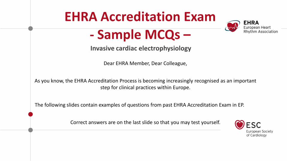
1. ECG, bipolar and distal electrode unipolar recordings from the mapping catheterduring atrial pacing are shown.
Please tick the best statement about the site where the recordings are obtained.
EHRA AP Exam Samples
a. Not appropriate for RF application since AV continuity is not present
b. Not appropriate for RF application because AV relation >1
c. Appropriate for RF application because early ventricular activation
d. Appropriate for RF application because a negative monophasic unipolar ventriculogram
e. None of the other statements is correct
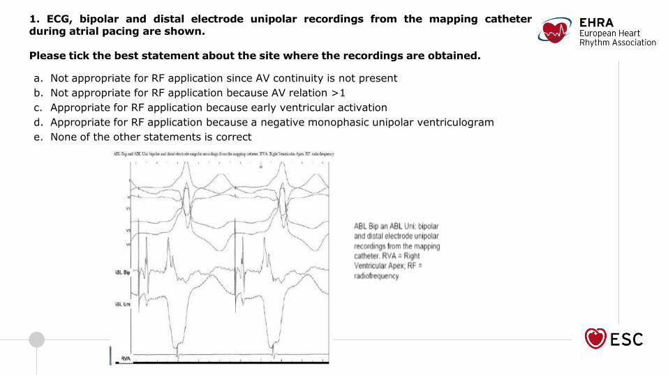
2. A 24 year-old male presented with a narrow QRS-complex tachycardia andunderwent electrophysiological study. During recording he presented the followingECG.
What is the most likely mechanism of the tachycardia ?
a. Ectopic right atrial tachycardia
b. Orthodromic AV reentrant tachycardia
c. Slow-fast AV nodal reentrant tachycardia
d. Left-sided accessory pathway
e. None of the other mentioned mechanisms is likely
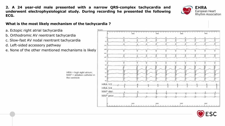
3. ECG and bipolar recordings during the beginning of a train of stimulation from the coronary sinus (A1A1) are shown. The recording is most probably :
a. Suggestive of two accessory pathways b. Suggestive of one accessory pathway and AV nodal reentrant tachycardia c. Suggestive of one accessory pathway and one orthodromic echo beat d. Suggestive of one accessory pathway and intraatrial reentry e. Suggestive of one accessory pathway and nodal automaticity
4. Which of the following criteria is not typical of the Mahaim-type syndrome ?
a. Wide-QRS-complex tachycardia
b. Pre-excitation during incremental atrial pacing
c. Decremental antegrade conduction properties
d. Retrograde conduction via atriofascicular pathways
5. Which diagnosis is associated with one or more right accessory pathways ?
a. Coronary sinus diverticulumb. Atrial septal defectc. Ventricular septal defectd. Ebstein’s anomalye. Pulmonary stenosis
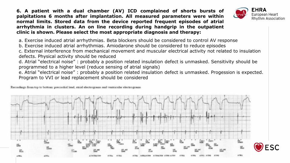
6. A patient with a dual chamber (AV) ICD complained of shorts bursts ofpalpitations 6 months after implantation. All measured parameters were withinnormal limits. Stored data from the device reported frequent episodes of atrialarrhythmia in clusters. An on line recording during handgrip in the outpatientclinic is shown. Please select the most appropriate diagnosis and therapy:
a. Exercise induced atrial arrhythmias. Beta blockers should be considered to control AV responseb. Exercise induced atrial arrhythmias. Amiodarone should be considered to reduce episodesc. External interference from mechanical movement and muscular electrical activity not related to insulation defects. Physical activity should be reducedd. Atrial “electrical noise” : probably a position related insulation defect is unmasked. Sensitivity should be programmed to a higher level (reduce sensing of atrial signals)e. Atrial “electrical noise” : probably a position related insulation defect is unmasked. Progession is expected. Program to VVI or lead replacement should be considered
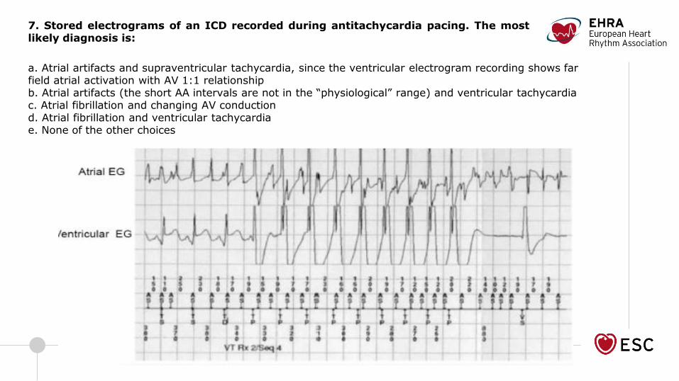
7. Stored electrograms of an ICD recorded during antitachycardia pacing. The mostlikely diagnosis is:
a. Atrial artifacts and supraventricular tachycardia, since the ventricular electrogram recording shows far field atrial activation with AV 1:1 relationship b. Atrial artifacts (the short AA intervals are not in the “physiological” range) and ventricular tachycardiac. Atrial fibrillation and changing AV conduction d. Atrial fibrillation and ventricular tachycardia e. None of the other choices
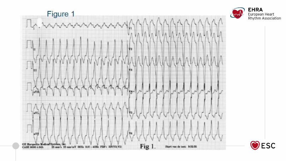
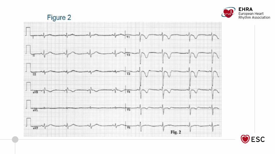
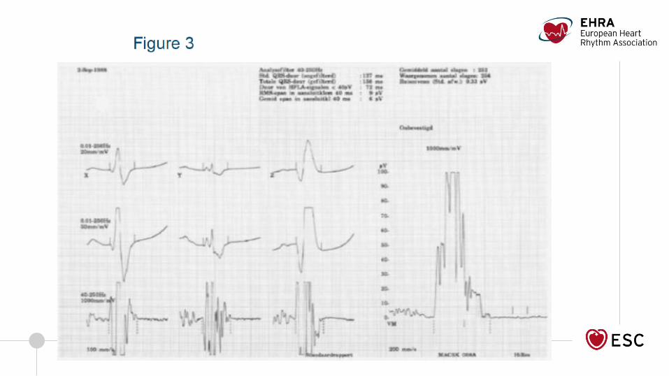
8. A 16-year-old athlete presented with an episode of exercise induced wide QRScomplex tachycardia (Figure 1). Figure 2 shows the 12-lead ECG in sinus rhythm.Figure 3 shows the result of signal-averaged ECG examination. Following infusion of0.7mg/kg ajmaline, the ST segment did not show any change in the precordial leads.
What is the most likely diagnosis ?
a. Idiopathic right ventricular tachycardia b. Brugada syndrome c. Arrhythmogenic right ventricular dysplasia d. Catecholaminergic ventricular tachycardia e. Supraventricular tachycardia with aberrancy or pre-excitation
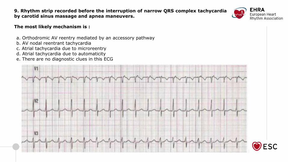
9. Rhythm strip recorded before the interruption of narrow QRS complex tachycardiaby carotid sinus massage and apnea maneuvers.
The most likely mechanism is :
a. Orthodromic AV reentry mediated by an accessory pathway b. AV nodal reentrant tachycardia c. Atrial tachycardia due to microreentryd. Atrial tachycardia due to automaticity e. There are no diagnostic clues in this ECG
10. Some supraventricular tachycardias are difficult to classify due to early retrograde activation during tachycardia (short VA-interval). Differential diagnosis is atrial tachycardia or paraseptal orthodromic AV reentrant tachycardia.
Which maneuver is of little value to differentiate between AV nodal reentrant tachycardia and AV reentrant tachycardia ?
a. Para-hisian pacing b. Pacing the right ventricle at the tachycardia cycle length and compare the VA-interval to the VA-interval recorded during tachycardia c. Adenosine during tachycardia d. Adenosine during ventricular pacing e. Stimulation to test for tachycardia reset
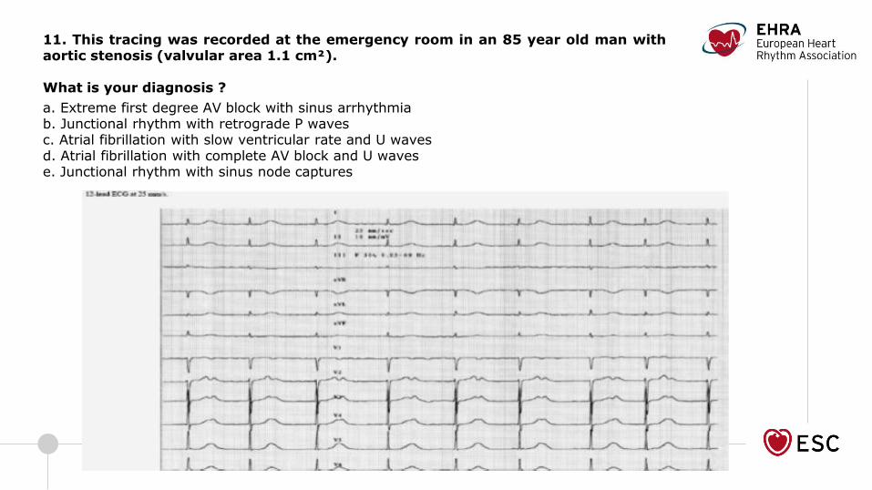
11. This tracing was recorded at the emergency room in an 85 year old man withaortic stenosis (valvular area 1.1 cm²).
What is your diagnosis ?
a. Extreme first degree AV block with sinus arrhythmia b. Junctional rhythm with retrograde P waves c. Atrial fibrillation with slow ventricular rate and U waves d. Atrial fibrillation with complete AV block and U waves e. Junctional rhythm with sinus node captures
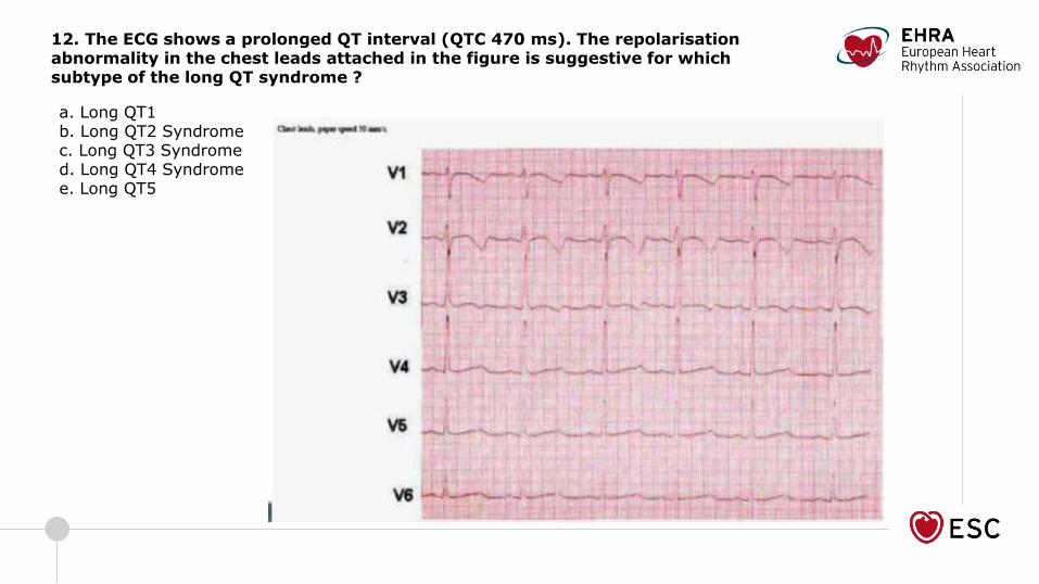
12. The ECG shows a prolonged QT interval (QTC 470 ms). The repolarisationabnormality in the chest leads attached in the figure is suggestive for which subtype of the long QT syndrome ?
a. Long QT1 b. Long QT2 Syndrome c. Long QT3 Syndrome d. Long QT4 Syndrome e. Long QT5
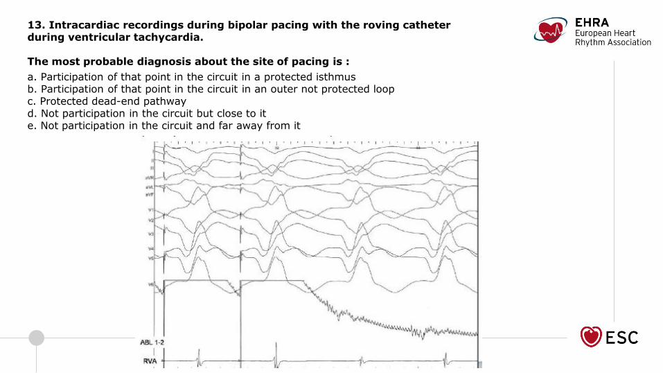
13. Intracardiac recordings during bipolar pacing with the roving catheter during ventricular tachycardia.
The most probable diagnosis about the site of pacing is :
a. Participation of that point in the circuit in a protected isthmus b. Participation of that point in the circuit in an outer not protected loop c. Protected dead-end pathway d. Not participation in the circuit but close to it e. Not participation in the circuit and far away from it
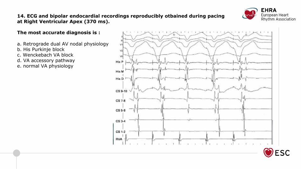
14. ECG and bipolar endocardial recordings reproducibly otbained during pacing at Right Ventricular Apex (370 ms).
The most accurate diagnosis is :
a. Retrograde dual AV nodal physiology b. His Purkinje block c. Wenckebach VA block d. VA accessory pathway e. normal VA physiology
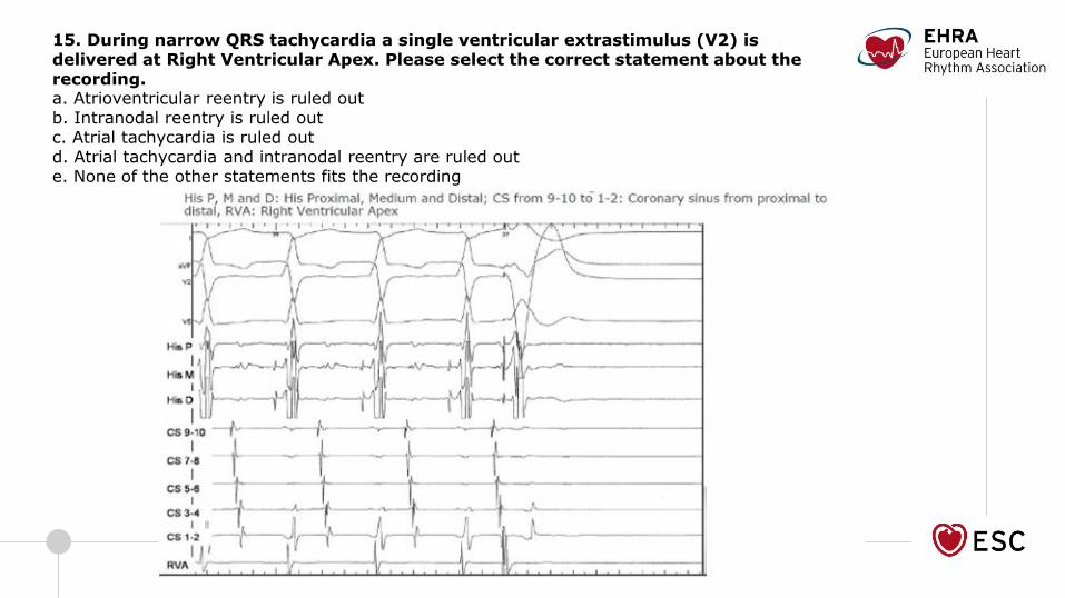
15. During narrow QRS tachycardia a single ventricular extrastimulus (V2) is delivered at Right Ventricular Apex. Please select the correct statement about the recording. a. Atrioventricular reentry is ruled out b. Intranodal reentry is ruled out c. Atrial tachycardia is ruled out d. Atrial tachycardia and intranodal reentry are ruled out e. None of the other statements fits the recording

15. During narrow QRS tachycardia a single ventricular extrastimulus (V2) is delivered at Right Ventricular Apex. Please select the correct statement about the recording. a. Atrioventricular reentry is ruled out b. Intranodal reentry is ruled out c. Atrial tachycardia is ruled out d. Atrial tachycardia and intranodal reentry are ruled out e. None of the other statements fits the recording
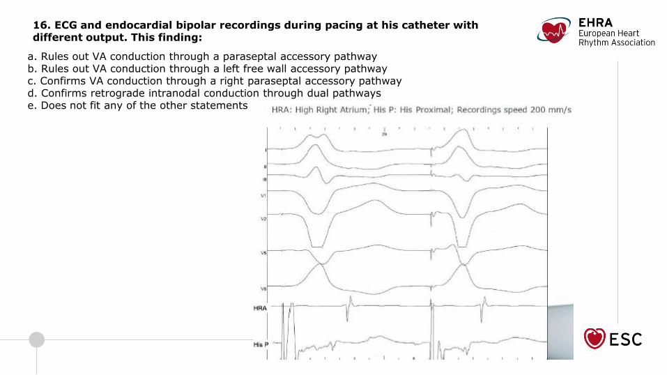
16. ECG and endocardial bipolar recordings during pacing at his catheter with different output. This finding:
a. Rules out VA conduction through a paraseptal accessory pathway b. Rules out VA conduction through a left free wall accessory pathway c. Confirms VA conduction through a right paraseptal accessory pathway d. Confirms retrograde intranodal conduction through dual pathwayse. Does not fit any of the other statements
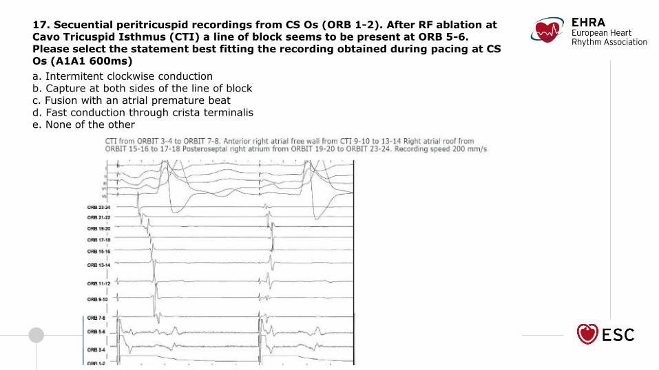
17. Secuential peritricuspid recordings from CS Os (ORB 1-2). After RF ablation at Cavo Tricuspid Isthmus (CTI) a line of block seems to be present at ORB 5-6. Please select the statement best fitting the recording obtained during pacing at CS Os (A1A1 600ms)
a. Intermitent clockwise conduction b. Capture at both sides of the line of block c. Fusion with an atrial premature beat d. Fast conduction through crista terminalise. None of the other
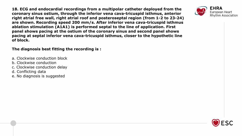
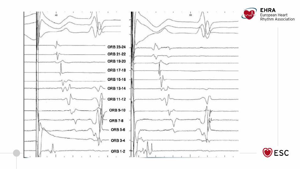
18. ECG and endocardial recordings from a multipolar catheter deployed from the coronary sinus ostium, through the inferior vena cava-tricuspid isthmus, anterior right atrial free wall, right atrial roof and posteroseptal region (from 1-2 to 23-24) are shown. Recording speed 200 mm/s. After inferior vena cava-tricuspid isthmus ablation stimulation (A1A1) is performed septal to the line of application. First panel shows pacing at the ostium of the coronary sinus and second panel shows pacing at septal inferior vena cava-tricuspid isthmus, closer to the hypothetic line of block.
The diagnosis best fitting the recording is :
a. Clockwise conduction block b. Clockwise conduction c. Clockwise conduction delay d. Conflicting data e. No diagnosis is suggested
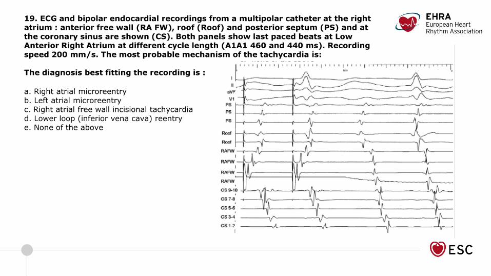
19. ECG and bipolar endocardial recordings from a multipolar catheter at the right atrium : anterior free wall (RA FW), roof (Roof) and posterior septum (PS) and at the coronary sinus are shown (CS). Both panels show last paced beats at Low Anterior Right Atrium at different cycle length (A1A1 460 and 440 ms). Recording speed 200 mm/s. The most probable mechanism of the tachycardia is:
The diagnosis best fitting the recording is :
a. Right atrial microreentryb. Left atrial microreentryc. Right atrial free wall incisional tachycardia d. Lower loop (inferior vena cava) reentry e. None of the above
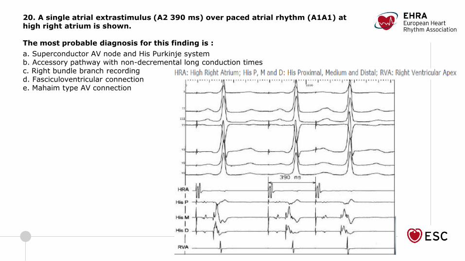
20. A single atrial extrastimulus (A2 390 ms) over paced atrial rhythm (A1A1) at high right atrium is shown.
The most probable diagnosis for this finding is :
a. Superconductor AV node and His Purkinje system b. Accessory pathway with non-decremental long conduction times c. Right bundle branch recording d. Fasciculoventricular connection e. Mahaim type AV connection
ANSWERSQ. # Answer Q. # Answer
1 C 11 C
2 C 12 B
3 C 13 A
4 D 14 D
5 D 15 C
6 E 16 A
7 D 17 C
8 C 18 A
9 B 19 E
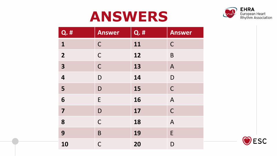
10 C 20 D