Embed Size (px)
Citation preview
PERSPECTIVEpublished: 16 September 2015
doi: 10.3389/fgene.2015.00293
Edited by:Tiago Collares,
Federal University of Pelotas, Brazil
Reviewed by:Thomas John,
Ludwig Institute for Cancer Research,Australia
Howard Donninger,University of Louisville, USA
*Correspondence:Vilceu Bordignon,
Department of Animal Science, McGillUniversity, Sainte-Anne-de-Bellevue,
QC H9X 3V9, [email protected];
Luis B. Agellon,School of Dietetics and Human
Nutrition, McGill University,Sainte-Anne-de-Bellevue, QC H9X
3V9, [email protected]
Specialty section:This article was submitted to
Cancer Genetics,a section of the journal
Frontiers in Genetics
Received: 30 June 2015Accepted: 04 September 2015Published: 16 September 2015
Citation:Gutierrez K, Dicks N, Glanzner WG,Agellon LB and Bordignon V (2015)
Efficacy of the porcine speciesin biomedical research.
Front. Genet. 6:293.doi: 10.3389/fgene.2015.00293
Efficacy of the porcine species inbiomedical researchKarina Gutierrez1, Naomi Dicks1, Werner G. Glanzner1, Luis B. Agellon2* andVilceu Bordignon1*
1 Department of Animal Science, McGill University, Sainte-Anne-de-Bellevue, QC, Canada, 2 School of Dietetics and HumanNutrition, McGill University, Sainte-Anne-de-Bellevue, QC, Canada
Since domestication, pigs have been used extensively in agriculture and kept ascompanion animals. More recently they have been used in biomedical research,given they share many physiological and anatomical similarities with humans. Recenttechnological advances in assisted reproduction, somatic cell cloning, stem cell culture,genome editing, and transgenesis now enable the creation of unique porcine models ofhuman diseases. Here, we highlight the potential applications and advantages of usingpigs, particularly minipigs, as indispensable large animal models in fundamental andclinical research, including the development of therapeutics for inherited and chronicdisorders, and cancers.
Keywords: Large animal models, biomedical research, swine, pigs, minipigs, clones, transgenics
Introduction
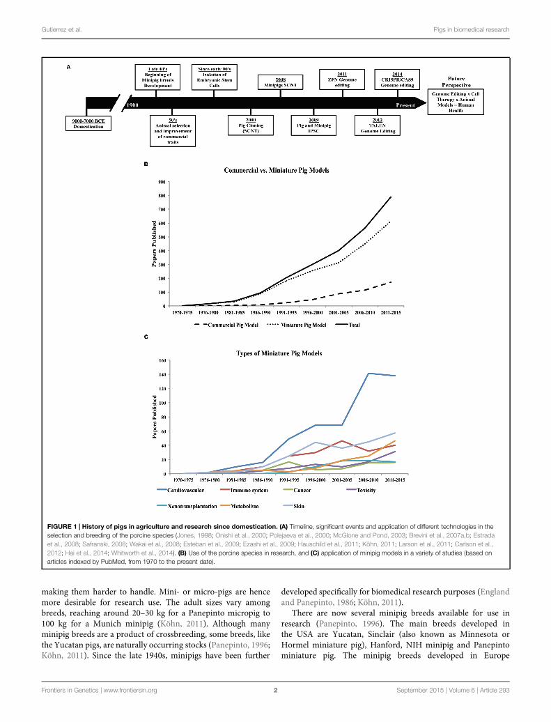
The first evidence of swine domestication dates back to approximately 7000–9000 years ago (Jones,1998; McGlone and Pond, 2003; Köhn, 2011; Larson et al., 2011; Figure 1A). China and Europehave been, since domestication, the pig-breeding centers dictating the profile of the pig breeds(Jones, 1998; Amills et al., 2001). The reason for domestication was to provide meat as a sourceof food protein, which stimulated pig selection and farming (Jones, 1998; Köhn, 2011). Studieshave been conducted using genome-wide genotyping and genetic variability to trace the migration,selection, and improvement from ancient wild species to modern swine (Giuffra et al., 2000; Bosseet al., 2014a,b). It is generally accepted that the majority of all modern breeds are derived from theEurasian wild boar (European and Asian wild boars; Porter, 1993; Bosse et al., 2014b). Althoughpig selection started just after domestication, it has only been since the mid-20th century thatperformance has been used as the main tool in the animal selection process (Safranski, 2008). Morerecently, molecular biology technologies, genome-wide association studies, and next-generationsequencing have been applied to enhance the selection process of domesticated pig breeds (e.g.,Duroc, Landrace, Pietrain, Yorkshire, etc.) to further improve traits of high economic value suchas feed conversion, meat quality, growth, precocious puberty, and prolificity (Sahana et al., 2013;Tart et al., 2013; Jiang et al., 2014; Sanchez et al., 2014).
The variety of modern pig breeds available today (Buchanan and Stalder, 2011), are a productof human intervention since domestication, but especially during the last century (Figure 1A).Besides breeds specialized for food production, smaller sized breeds (miniature- and micro-pigs)with certain characteristics such as obedience, friendly nature, and cognitive ability have also beenselected for the purpose of companion animals. In addition, their use in biomedical research hasbeen increasing considerably in the last years (Figure 1B).
Compared with other animals used in research (e.g., mice, rats, rabbits, and dogs), domesticfarm pigs are much larger (>300 kg adult size), therefore, requiring more space and feed, and
Frontiers in Genetics | www.frontiersin.org 1 September 2015 | Volume 6 | Article 293
Gutierrez et al. Pigs in biomedical research
FIGURE 1 | History of pigs in agriculture and research since domestication. (A) Timeline, significant events and application of different technologies in theselection and breeding of the porcine species (Jones, 1998; Onishi et al., 2000; Polejaeva et al., 2000; McGlone and Pond, 2003; Brevini et al., 2007a,b; Estradaet al., 2008; Safranski, 2008; Wakai et al., 2008; Esteban et al., 2009; Ezashi et al., 2009; Hauschild et al., 2011; Köhn, 2011; Larson et al., 2011; Carlson et al.,2012; Hai et al., 2014; Whitworth et al., 2014). (B) Use of the porcine species in research, and (C) application of minipig models in a variety of studies (based onarticles indexed by PubMed, from 1970 to the present date).
making them harder to handle. Mini- or micro-pigs are hencemore desirable for research use. The adult sizes vary amongbreeds, reaching around 20–30 kg for a Panepinto micropig to100 kg for a Munich minipig (Köhn, 2011). Although manyminipig breeds are a product of crossbreeding, some breeds, likethe Yucatan pigs, are naturally occurring stocks (Panepinto, 1996;Köhn, 2011). Since the late 1940s, minipigs have been further
developed specifically for biomedical research purposes (Englandand Panepinto, 1986; Köhn, 2011).
There are now several minipig breeds available for use inresearch (Panepinto, 1996). The main breeds developed inthe USA are Yucatan, Sinclair (also known as Minnesota orHormel miniature pig), Hanford, NIH minipig and Panepintominiature pig. The minipig breeds developed in Europe
Frontiers in Genetics | www.frontiersin.org 2 September 2015 | Volume 6 | Article 293
Gutierrez et al. Pigs in biomedical research
are Göttingen, Munich, Berlin, Mini-Lewe, Czech-Republic,Vietnamese potbellied and Mini-Sib. In Asia, the breeds includeOhmini, Clawn, Lee Sung, and Chinese minipigs. The Göttingenand Yucatan breeds are the most commonly used minipigsin research, although there is no apparent clear reason forpreference. Unlike the Yucatan, a natural breed, the Göttingenminipig was developed specifically for research use. Other breedsare used only by specific research groups, thus limiting theirwidespread availability in research. Nevertheless, the interest inthe use of pigs in biomedical research has been rising over the last40–45 years (Figure 1B).
Use of Pigs in Biomedical Research
Biomedical research is broad, spanning studies on underlyingdisease mechanisms to the evaluation of safety and effectivenessof preventative measures, diagnostic tests, and therapies. Mostanimal studies in recent times have used the murine speciesdue to their small size, fast reproductive cycles and shortlifespan. In addition, the availability of murine embryonic stemcells, fully annotated genome, and facile tools for targetedgenetic manipulation have all contributed to the elucidation ofgene functions and disease pathophysiology. However, in manycases, mouse models do not adequately represent features ofhuman disorders (Seok et al., 2013). In this regard, animalsthat better represent human pathophysiology are required. Pigsand humans share many similarities such as size, physiology,anatomy, metabolic profile, and longer lifespan (Panepinto,1996; Spurlock and Gabler, 2008; Kuzmuk and Schook, 2011;Swindle et al., 2012). For example, pig skin is structurally similarto human skin regarding thickness and spacing between hairfollicles, making it useful for studies on wound healing andburn lesions (Sullivan et al., 2001). Pigs also share anatomicaland physiological similarities with respect to the renal system,making them valuable for pharmacological studies (Dalgaard,2014; Huppertz et al., 2015). Pigs can also be useful in the studyof nutrient absorption and intestinal transport, as well as thepathogenesis of gastrointestinal diseases (Sangild et al., 2014). Allthese characteristics contribute to the development of superiormodels of human conditions (Kuzmuk and Schook, 2011).
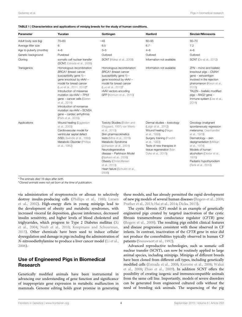
The choice between outbred or inbred strains can havea significant impact on research outcomes (Festing, 2014).While, outbred strains may be better suited for quantitativetrait loci studies, experiments addressing mechanistic aspectswould benefit from the use of inbred strains (Chia et al.,2005). Some minipig breeds are already established for specificapplications due to their unique characteristics (Table 1). Pigshave also been used for testing new therapies, devices, andefficacy and safety of new drugs prior to human trials. Forinstance, a novel endovascular chemotherapy filter, designed toreduce circulatory drug excess in vitro, was successfully testedin pigs (Patel et al., 2014). As well, a new method for pediatricliver transplantation was validated using pigs (Leal et al., 2015).Regarding pharmacokinetic and cytotoxic tests, pigs have beenused for testing topical skin formulations (Mitra et al., 2015),and are considered a better choice compared to dogs for the
study of drugs that are metabolized by the aldehyde oxidase(AOX1), N-acetyltransferase (NAT1 or NAT2) or cytochrome(CYP2C9-like) enzymes (Dalgaard, 2014).
In general, there is low incidence of naturally occurringpathologies described in pigs. The reason for this is twofold. First,human intervention by way of selective breeding has eliminatedgenes that increased disease susceptibility. Second, the majorityof the domestic farm pigs are slaughtered at a young age (<6months old), precluding the detection of late onset diseases suchas cancer. On the other hand, Vietnamese potbellied minipigsraised as companion animals do reach old ages. Indeed, aretrospective study found a variety of neoplasms with widespreadmetastases in these pigs of advanced age (∼11 years; Newmanand Rohrbach, 2012). The most common malignances foundincluded hepatic and intestinal carcinomas, and uterine andovarian smooth muscle tumors (Newman and Rohrbach, 2012).
Occurrence of malignant spontaneously regressingmelanomas has been described in Sinclair minipigs (Millikanet al., 1974; Oxenhandler et al., 1979). Selective interbreeding,by removing animals with red coat color that do not developthe lesions, increased the frequency of tumor formation inthese selected minipigs (Millikan et al., 1974). The tumorsappear from birth and culminate in skin depigmentation aftertumor regression showing a phenotype similar to human vitiligo(Millikan et al., 1974). Studies conducted in these minipigshave shown decreased telomerase activity during melanomaregression (Pathak et al., 2000), which has also been observedby inhibiting telomerase activity in human melanoma cells(Burchett et al., 2014). Therefore, these minipigs may representa useful model to study malignant melanomas because thetumors appear spontaneously and then either regress or growprogressively and metastasize similarly to human melanomas(Oxenhandler et al., 1979).
Another example of a naturally occurring condition in pigs isthe dwarf phenotype, caused by a single amino acid change in theα1 chain of type X collagen (Nielsen et al., 2000). The COL10A1gene, which encodes type X collagen, is expressed in hypertrophicchondrocytes during endochondral ossification. In humans, anamino acid variation in the same position of the type X collagenprotein has been shown to be the cause of Schmid metaphysealchondrodysplasia (SMCD), a mild skeletal disorder associatedwith dwarfism (Warman et al., 1993). Since mice lacking type Xcollagen do not develop abnormalities in long bone development(Rosati et al., 1994), pigs represent a better animal model ofhuman SMCD.
Another naturally occurring disease observed in Yucatanminipigs mimics human ventricular septal defect (VSD; Swindleet al., 1990). The VSD in pigs can be observed in fetal stagessimilar to the congenital anomaly in humans, and can be usedfor the study of new methods of diagnosis or therapies (Swindleet al., 1990; Amin et al., 2006).
Despite a number of natural occurring pig phenotypes thatresemble human diseases, for most of human pathologies itis difficult to find representative animal models in nature.Thus, manipulation of diet, use of drugs and/or surgerieshas been necessary to generate appropriate models. Forexample, minipig models for Type I diabetes were induced
Frontiers in Genetics | www.frontiersin.org 3 September 2015 | Volume 6 | Article 293
Gutierrez et al. Pigs in biomedical research
TABLE 1 | Characteristics and applications of minipig breeds for the study of human conditions.
Parameter Yucatan Gottingen Hanford Sinclair/Minnesota
Adult body size (kg) 70–83 ∼45 80–95 55–70
Average litter size 6 6.5 6.7 7.2
Age to puberty (months) 4–6 3–5 4–6 4–6
Genetic background Purebred Outbred Outbred Outbred
Cloning somatic cell nuclear transfer(SCNT; Estrada et al., 2008)
SCNT (Wakai et al., 2008) Information not available SCNT (Do et al., 2012)
Transgenics Homologous recombinationBRCA1 (breast cancersusceptibility gene 1) –gene knockout by rAAV –model for breast cancer(Luo et al., 2011, 2012)∗Introduction of missensemutation via rAAV – TP53gene – cancer cells (Sierenet al., 2014)Introduction of nonsensemutation via rAAV – SCN5Agene – cardiac arrhythmia(Park et al., 2015)
Homologous recombinationBRCA1 (breast cancersusceptibility gene 1) –gene knockout by rAAV –model for breast cancer(Luo et al., 2012)†
rAAV vectors encodingGFP (Kornum et al., 2010)
Information not available ZFN – mono and biallelicknockout pigs – CMAHgene – xenoantigeninvolved in the rejectionphenomenon (Kwon et al.,2013)TALEN – biallelic modifiedpigs – RAG2 gene –immune system (Lee et al.,2014)
Applications Wound healing (Egglestonet al., 2000)Cardiovascular model forventricular septal defect(VSD; Swindle et al., 1990)Metabolic Disorder (Phillipset al., 1982)
Toxicity Studies (Bollen andEllegaard, 1997; van Mierloet al., 2013)Skin pharmacokineticstests (Mitra et al., 2015)Metabolic Syndrome(Johansen et al., 2001)Neurodegenerativedisease – Parkinson Model(Bjarkam et al., 2008)Obesity (Christoffersenet al., 2013)Heart failure (Schuleri et al.,2008)
Dermal studies – toxicology(Leigh et al., 2012)Wound healing (Regeret al., 1999)Surgery training (Purohitet al., 1993)Tests of new therapies intissue regeneration (VanDyke et al., 2015)
Oncology (malignantspontaneously regressionmelanoma; Oxenhandleret al., 1979)Dermatology – skindepigmentation (Millikanet al., 1974)Models of humanalcoholism (Dexter et al.,1976)Pediatric hypothyroidism(Tank et al., 2013)
∗The animals died 18 days after birth.†Cloned animals were not yet born at the time of publication.
via administration of streptozotocin or alloxan to selectivelydestroy insulin-producing cells (Phillips et al., 1980; Larsenet al., 2002). High-energy diets in young minipigs lead tothe development of obesity and metabolic syndromes, withincreased visceral fat deposition, glucose intolerance, decreasedinsulin sensitivity, and higher levels of blood cholesterol andtriglycerides, which progress to Type 2 Diabetes mellitus (Xiet al., 2004; Neeb et al., 2010; Koopmans and Schuurman,2015). Other chemicals have been used to induce cellulardysregulation and damage in pigs including the administration ofN-nitrosodiethylamine to produce a liver cancer model (Li et al.,2006).
Use of Engineered Pigs in BiomedicalResearch
Genetically modified animals have been instrumental inadvancing our understanding of gene function and significanceof inappropriate gene expression in metabolic malfunction inmammals. Genome editing holds great promise in generating
these models, and has already permitted the rapid developmentof new pig models of several human diseases (Rogers et al., 2008;Prather et al., 2013; Hai et al., 2014; Dicks, 2015).
The cystic fibrosis (CF) model is an example of geneticallyengineered pigs created by targeted inactivation of the cysticfibrosis transmembrane conductance regulator (CFTR) gene(Rogers et al., 2008). The resulting pigs exhibit clinical featuresand disease progression consistent with those observed in CFinfants. In contrast, inactivation of the CFTR gene in mice didnot produce the comorbidities typically observed in human CFpatients (Snouwaert et al., 1992).
Advanced reproductive technologies, such as somatic cellnuclear transfer (SCNT), can now be routinely applied to largeanimal species, including minipigs. Minipigs of different breedshave been cloned from different cell types, including geneticallymodified cells (Estrada et al., 2008; Kurome et al., 2008; Wakaiet al., 2008; Zhao et al., 2009). In addition SCNT offers thepossibility of creating isogenic and immunocompatible animalsfrom the same cell line. Importantly, models of severe disorderscan be generated from engineered cultured cells without theneed of breeding sick animals. The sequencing of the pig
Frontiers in Genetics | www.frontiersin.org 4 September 2015 | Volume 6 | Article 293
Gutierrez et al. Pigs in biomedical research
genome is another key development in the production of gene-modified pigs in the post-genomic era (Schook et al., 2015a).Genome editing techniques, including zinc finger nucleases(ZFN), transcription activator-like effector nucleases (TALEN),and clustered, regularly interspaced, short palindromic repeats(CRISPR) together with CRISPR associated (Cas) nucleases(CRISPR/Cas), now allow the precise manipulation of genesequences in germ, embryonic and somatic cells (Hauschild et al.,2011; Carlson et al., 2012; Cong et al., 2013; Hai et al., 2014;Whitworth et al., 2014; Dicks, 2015). Among these methods,the CRISPR/Cas9 system is emerging as the method of choicebecause it permits gene editing to be accomplished in only onestep by injecting both the specific guide RNAs and endonucleaseinto zygotes (Hai et al., 2014; Whitworth et al., 2014).
Another example of human disease that has the potential tobe studied in genetically engineered pigs is heart arrhythmias(Park et al., 2015). Mutations in the SCN5A gene, whichencodes a subunit of the cardiac sodium channel Nav1.5required for excitability and conduction in the myocardium,were found in patients with Bruguda syndrome (Hedley et al.,2009). SCN5AE558X/+ engineered Yucatan minipigs with reducedexpression of the sodium channel Nav1.5 have been created andthese animals exhibit conduction abnormalities and susceptibilityto ventricular arrhythmias (Park et al., 2015). There has also beenconsiderable interest in genetically modified pig strains suitablefor xenotransplantation. Most research into the development ofappropriate xenotransplantation strains focused on addressinghyperacute rejection, which is initiated rapidly and involvespreformed natural human antibodies and the complement system(Cooper et al., 2002). This has been possible by targeting cellsurface antigens such as α-1,3 galactosyltransferase (Miyagawaet al., 2001; Lai et al., 2002; Phelps et al., 2003; Takahagi et al.,2005) or complement regulatory proteins such as human decayaccelerating factor (Murakami et al., 2002). The pigs madedeficient of α-1,3 galactosyltransferase have contributed to thereduction of immunogenicity of donor tissue/organs (Phelpset al., 2003). Transgenic pigs expressing antibodies againstcytotoxic T-cell lymphocyte antigen receptor, a cell-mediatedimmune response suppressor, were also developed (Phelps et al.,2009).
A pig model for the human familial adenomatous polyposiswas generated by inactivation of the adenomatous polyposis coli(APC) gene (Flisikowska et al., 2012). Mice lacking the APC geneexhibit non-metastatic neoplasias only in the small intestine (Suet al., 1992). However, the pig model of colon and rectal cancerreproduces the human features of the disease, which includes thedevelopment of polyps spread along the whole large bowel inyoung animals. A candidate gene for the development of breastand ovarian cancer models is the breast cancer-associated gene1 (BRCA1), which has been manipulated in both Yucatan andGöttingen cells, but lines of modified minipigs remain to beproduced (Luo et al., 2011, 2012). The TP53 gene, which encodesthe tumor suppressor protein p53 and is the most commonlyobserved suppressed gene in human tumors, was found to bemutated in Li-Fraumeni patients having increased risk to developmultiple types of cancers (Gonzalez et al., 2009). Suppression ofp53 in mesenchymal stem cells derived from pig bone marrow
exhibits chemoresistance in vitro (Leuchs et al., 2012). Mutationof TP53 gene in Yucatan minipigs resulted in development oflymphomas and osteogenic tumors (Sieren et al., 2014). Morerecently, a new engineered pig strain termed “oncopig” wasdeveloped, which promises inducible formation of a wide varietyof cancers that are potentially novel platforms for research andtherapeutics development (Schook et al., 2015b). These examplesillustrate the potential of genetically engineered pigs as robustmodels for the study of human pathologies that are not wellrepresented in small laboratory animal species.
Improving the Usefulness of Pigs inBiomedical Research
Rodents have been the choice animal model for basic research,but are not always suitable for translational research dueto marked differences in size, lifespan as well as metabolic,anatomical, and physiological discrepancies. On the other hand,the pig is more closely related to humans in terms of theseparameters (Swindle et al., 2012) and, therefore, is better suitedfor recapitulation of human diseases. Indeed, the use of thepig in translational research is increasingly gaining acceptance(Figure 1C). Dogs and non-human primates have traditionallybeen used for this purpose, but rising ethical concerns havereduced their favor and increased demand for alternatives(Swindle et al., 2012). The number of peer-reviewed papersdescribing the use of pigs as biomedical models has riseneightfold over the past 30 years (Figure 1B). Already, the pig hasbecome well established in many areas of research and training.For instance, in the past 20 years the pig has replaced the dog asa model for surgical training and has also gained FDA approvalfor the testing of surgical implantation devices intended forhuman use (Swindle et al., 2012; Schook et al., 2015a). Minipigmodels, which aremuch smaller in size compared to the domesticfarm breeds, offer lower operating costs compared to other largeanimal models and also reduce the concern of ethical acceptancegiven the already widespread use of pigs in agriculture (Bollenand Ellegaard, 1997; Swindle et al., 2012).
Pigs offer many exciting applications, including stem cellresearch, tissue engineering and xenotransplantation. Althoughincredible advances in transgenic pigs harboring variousengineered alterations designed to minimize graft versus hostrejection (Lai et al., 2002; Phelps et al., 2003, 2009; Klose et al.,2005; Takahagi et al., 2005; Hauschild et al., 2011; Petersenet al., 2011; Jeong et al., 2013), much work remains to beaccomplished since multiple genes need to be manipulatedgiven the various types of tissue rejection reactions (Takahagiet al., 2005; Whyte and Prather, 2011; Jeong et al., 2013).Porcine induced pluripotent stem cells (iPSCs) have beenproduced (Esteban et al., 2009) and chimeric pigs weregenerated using iPSC (West et al., 2010, 2011). This is highlyrelevant since study of porcine iPSCs have eventual humanapplications (Esteban et al., 2009), such as cell-based therapies.However, the mechanisms of cellular reprogramming, directedcell differentiation and species-specific cell culture requirementsnecessitate further investigation (Ezashi et al., 2012). The
Frontiers in Genetics | www.frontiersin.org 5 September 2015 | Volume 6 | Article 293
Gutierrez et al. Pigs in biomedical research
International Society for Stem Cell Research has indicated intheir guidelines for translational use that validationmust occur inboth small and large animal models (Aigner et al., 2010). Tissuerepair is another potential application of engineered pig models.Cartilage tissue grafts have been created using chondrocytesisolated from infant minipigs (Deponti et al., 2014), andmandibular condyle grafts have been generated from Yucatanminipig adipose-derivedmesenchymal stem cells (Abukawa et al.,2003). There has also been successful regeneration of bone defectsusing engineered bone graft tissues in minipig models (Grögeret al., 2003). If custom donor transgenic minipig strains can becreated, this could open the doors to other engineered tissuereplacements for human uses. For example, the use of blastocystcomplementation and pluripotent stem cells has been appliedto direct the development of otherwise missing organs in pigs(Matsunari et al., 2013). This has increased the hope that it mayone day be possible to create non-immunogenic donor organs inpigs using human iPSCs (Matsunari et al., 2013; Feng et al., 2015).Finally, similarities in the porcine and human immune systemhave sparked interest in vaccine development and efficacy testingin pigs (Meurens et al., 2012).
The completion of the porcine genome project in 2012 hasfurther facilitated the use of pigs in research. Data from thisproject has enabled the comparative analysis of genetic sequencesand development of the necessary tools to create and validatetargeted genetic alterations in the porcine genome (Gun andKues, 2014; Schook et al., 2015a). In addition, the development ofRNASeq technology has facilitated transcriptome analysis, whichfurther improves our ability to identify important targets relatedto certain phenotypic traits (Ropka-Molik et al., 2014). Otherrecent achievements in the pig include the use of inducible orconditional systems to control transgene expression (Kues et al.,2006; Klymiuk et al., 2012), and tissue-specific expression of theCre recombinase (Li et al., 2009; Luo et al., 2014). These advanceswill ensure the continued development of various pig strains forresearch, similar to what has already been accomplished in mice.
Summary
It is clear that the use of the pig as a biomedical modelis increasingly gaining approval due to physiopathologicalsimilarities with humans. However, some obstacles remainto be overcome in order to realize the full potential of theporcine species in developing new diagnostic and therapeuticapproaches. Despite the sequencing of the porcine genome,full annotation has yet to be completed. This is essential tofacilitate interrogation of the pig genome and investigationof less characterized genes. Efforts to develop a completeporcine proteome map as well as epigenome map arecurrently underway (Meurens et al., 2012; Schook et al.,2015a). These databases are necessary to understand diseasepathogenesis (Meurens et al., 2012; Schook et al., 2015a).Moreover, the availability of both inbred and outbred breedsof minipigs extends the utility of these species as a viablelarge animal model. Continuing refinements and adaptationof technologies for genome editing, cell/tissue-specific genetargeting strategies, stem cells and somatic cell cloning willfurther facilitate the creation of specialized pig strains forbiomedical research.
Acknowledgments
Research in our laboratories is funded by grants from theNatural Sciences and Engineering Research Council of Canada(to VB and to LA). KG is supported by a scholarship from theScience without Borders Program of the Brazilian Coordinationfor the Improvement of Higher Education Personnel (CAPES).WG is supported by a scholarship from the Brazilian NationalCouncil for Scientific and Technological Development (CNPq).ND is supported by an Alexander Graham Bell Canada GraduateScholarship from the Natural Sciences and Engineering ResearchCouncil of Canada.
References
Abukawa, H., Terai, H., Hannouche, D., Vacanti, J. P., Kaban, L. B., andTroulis, M. J. (2003). Formation of a mandibular condyle in vitro by tissueengineering. J. Oral Maxillofac. Surg. 61, 94–100. doi: 10.1053/joms.2003.50015
Aigner, B., Renner, S., Kessler, B., Klymiuk, N., Kurome, M., Wunsch, A., et al.(2010). Transgenic pigs as models for translational biomedical research. J. Mol.Med. (Berl.) 88, 653–664. doi: 10.1007/s00109-010-0610-9
Amills, M., Clop, A., Ramírez, O., and Pérez-Enciso, M. (2010). “Origin and geneticdiversity of pig breeds,” in Encyclopedia of Life Sciences (eLS) (Chichester: JohnWiley & Sons Ltd). doi: 10.1002/9780470015902.a0022884
Amin, Z., Woo, R., Danford, D. A., Froemming, S. E., Reddy, V. M., Lof, J.,et al. (2006). Robotically assisted perventricular closure of perimembranousventricular septal defects: preliminary results in Yucatan pigs. J. Thorac.Cardiovasc. Surg. 131, 427–432. doi: 10.1016/j.jtcvs.2005.10.034
Bjarkam, C. R., Nielsen, M. S., Glud, A. N., Rosendal, F., Mogensen, P., Bender, D.,et al. (2008). Neuromodulation in a minipig MPTPmodel of Parkinson disease.Br. J. Neurosurg. 22(Suppl. 1), S9–S12. doi: 10.1080/02688690802448285
Bollen, P., and Ellegaard, L. (1997). The Gottingen minipig in pharmacologyand toxicology. Pharmacol. Toxicol. 80(Suppl. 2), 3–4. doi: 10.1111/j.1600-0773.1997.tb01980.x
Bosse, M., Madsen, O., Megens, H. J., Frantz, L. A., Paudel, Y., Crooijmans,R. P., et al. (2014a). Hybrid origin of European commercial pigs examinedby an in-depth haplotype analysis on chromosome 1. Front. Genet. 5:442. doi:10.3389/fgene.2014.00442
Bosse, M., Megens, H.-J., Frantz, L. A. F., Madsen, O., Larson, G., Paudel, Y.,et al. (2014b). Genomic analysis reveals selection for Asian genes in Europeanpigs following human-mediated introgression. Nat. Commun. 5:4392. doi:10.1038/ncomms5392
Brevini, T. A., Antonini, S., Cillo, F., Crestan, M., and Gandolfi, F.(2007a). Porcine embryonic stem cells: facts, challenges and hopes.Theriogenology 68(Suppl. 1), S206–S213. doi: 10.1016/j.theriogenology.2007.05.043
Brevini, T. A., Tosetti, V., Crestan, M., Antonini, S., and Gandolfi, F.(2007b). Derivation and characterization of pluripotent cell linesfrom pig embryos of different origins. Theriogenology 67, 54–63. doi:10.1016/j.theriogenology.2006.09.019
Buchanan, D. S., and Stalder, K. (2011). “Breeds of pigs,” in The Genetics of the Pig,eds M. F. Rothschild and A. Ruvinsky (Cambridge, MA: CAB International).
Burchett, K. M., Yan, Y., and Ouellette, M. M. (2014). Telomeraseinhibitor Imetelstat (GRN163L) limits the lifespan of human pancreaticcancer cells. PLoS ONE 9:e85155. doi: 10.1371/journal.pone.0085155
Frontiers in Genetics | www.frontiersin.org 6 September 2015 | Volume 6 | Article 293
Gutierrez et al. Pigs in biomedical research
Carlson, D. F., Tan, W., Lillico, S. G., Stverakova, D., Proudfoot, C., Christian, M.,et al. (2012). Efficient TALEN-mediated gene knockout in livestock. Proc. Natl.Acad. Sci. U.S.A. 109, 17382–17387. doi: 10.1073/pnas.1211446109
Chia, R., Achilli, F., Festing, M. F., and Fisher, E. M. (2005). The origins and usesof mouse outbred stocks. Nat. Genet. 37, 1181–1186. doi: 10.1038/ng1665
Christoffersen, B., Golozoubova, V., Pacini, G., Svendsen, O., and Raun, K.(2013). The young Gottingen minipig as a model of childhood and adolescentobesity: influence of diet and gender. Obesity (Silver Spring) 21, 149–158. doi:10.1002/oby.20249
Cong, L., Ran, F. A., Cox, D., Lin, S., Barretto, R., Habib, N., et al. (2013). Multiplexgenome engineering using CRISPR/Cas systems. Science 339, 819–823. doi:10.1126/science.1231143
Cooper, D. K., Gollackner, B., and Sachs, D. H. (2002). Will the pigsolve the transplantation backlog? Annu. Rev. Med. 53, 133–147. doi:10.1146/annurev.med.53.082901.103900
Dalgaard, L. (2014). Comparison of minipig, dog, monkey and human drugmetabolism and disposition. J. Pharmacol. Toxicol. Methods 74, 80–92. doi:10.1016/j.vascn.2014.12.005
Deponti, D., Di Giancamillo, A., Gervaso, F., Domenicucci, M., Domeneghini, C.,Sannino, A., et al. (2014). Collagen scaffold for cartilage tissue engineering: thebenefit of fibrin glue and the proper culture time in an infant cartilage model.Tissue Eng. Part A 20, 1113–1126. doi: 10.1089/ten.TEA.2013.0171
Dexter, J. D., Tumbleson, M. E., Hutcheson, D. P., and Middleton, C. C. (1976).Sinclair(S-1) miniature swine as a model for the study of human alcoholism.Ann. N. Y. Acad. Sci. 273, 188–193. doi: 10.1111/j.1749-6632.1976.tb52881.x
Dicks, N. (2015). “Somatic cell nuclear transfer and the creation of transgeniclarge animal models,” in Somatic GenomeManipulation: Advances, Methods andApplications, edsD. J. Donnelly, X.-Q. Li and T.G. Jensen (NewYork: Springer),123–143.
Do, M., Jang, W. G., Hwang, J. H., Jang, H., Kim, E. J., Jeong, E. J., et al. (2012).Inheritance of mitochondrial DNA in serially recloned pigs by somatic cellnuclear transfer (SCNT). Biochem. Biophys. Res. Commun. 424, 765–770. doi:10.1016/j.bbrc.2012.07.031
Eggleston, T. A., Roach, W. P., Mitchell, M. A., Smith, K., Oler, D., and Johnson,T. E. (2000). Comparison of two porcine (Sus scrofa domestica) skin models forin vivo near-infrared laser exposure. Comp. Med. 50, 391–397.
England, D. C., and Panepinto, L. M. (1986). “Conceptual and operational historyof the development of miniature swine,” in Swine in Biomedical Research, ed.M. E. Tumbleson (New York, NY: Plenum Press).
Esteban, M. A., Xu, J., Yang, J., Peng, M., Qin, D., Li, W., et al. (2009). Generationof induced pluripotent stem cell lines from Tibetan miniature pig. J. Biol. Chem.284, 17634–17640. doi: 10.1074/jbc.M109.008938
Estrada, J. L., Collins, B., York, A., Bischoff, S., Sommer, J., Tsai, S., et al. (2008).Successful cloning of the Yucatan minipig using commercial/occidental breedsas oocyte donors and embryo recipients. Cloning Stem Cells 10, 287–296. doi:10.1089/clo.2008.0005
Ezashi, T., Telugu, B. P., Alexenko, A. P., Sachdev, S., Sinha, S., and Roberts, R. M.(2009). Derivation of induced pluripotent stem cells from pig somatic cells.Proc. Natl. Acad. Sci. U.S.A. 106, 10993–10998. doi: 10.1073/pnas.0905284106
Ezashi, T., Telugu, B. P., and Roberts, R. M. (2012). Induced pluripotent stemcells from pigs and other ungulate species: an alternative to embryonicstem cells? Reprod. Domest. Anim. 47(Suppl. 4), 92–97. doi: 10.1111/j.1439-0531.2012.02061.x
Feng, W., Dai, Y., Mou, L., Cooper, D. K., Shi, D., and Cai, Z. (2015). The Potentialof the Combination of CRISPR/Cas9 and Pluripotent Stem Cells to ProvideHuman Organs from Chimaeric Pigs. Int. J. Mol. Sci. 16, 6545–6556. doi:10.3390/ijms16036545
Festing, M. F. (2014). Evidence should trump intuition by preferring inbredstrains to outbred stocks in preclinical research. ILAR J. 55, 399–404. doi:10.1093/ilar/ilu036
Flisikowska, T., Merkl, C., Landmann, M., Eser, S., Rezaei, N., Cui, X., et al.(2012). A porcine model of familial adenomatous polyposis. Gastroenterology143, e1171–e1177. doi: 10.1053/j.gastro.2012.07.110
Giuffra, E., Kijas, J. M., Amarger, V., Carlborg, O., Jeon, J. T., and Andersson, L.(2000). The origin of the domestic pig: independent domestication andsubsequent introgression. Genetics 154, 1785–1791.
Gonzalez, K. D., Noltner, K. A., Buzin, C. H., Gu, D., Wen-Fong, C. Y., Nguyen,V. Q., et al. (2009). Beyond Li Fraumeni Syndrome: clinical characteristics
of families with p53 germline mutations. J. Clin. Oncol. 27, 1250–1256. doi:10.1200/JCO.2008.16.6959
Gröger, A., Kläring, S., Merten, H.-A., Holste, J., Kaps, C., and Sittinger, M. (2003).Tissue engineering of bone formandibular augmentation in immunocompetentminipigs: preliminary study. Scand. J. Plastic Reconstruct. Surg. Hand Surg. 37,129–133. doi: 10.1080/02844310310007728
Gun, G., and Kues, W. A. (2014). Current progress of genetically engineeredpig models for biomedical research. Biores. Open. Access. 3, 255–264. doi:10.1089/biores.2014.0039
Hai, T., Teng, F., Guo, R., Li, W., and Zhou, Q. (2014). One-step generation ofknockout pigs by zygote injection of CRISPR/Cas system. Cell Res. 24, 372–375.doi: 10.1038/cr.2014.11
Hauschild, J., Petersen, B., Santiago, Y., Queisser, A. L., Carnwath, J. W., Lucas-Hahn, A., et al. (2011). Efficient generation of a biallelic knockout in pigsusing zinc-finger nucleases. Proc. Natl. Acad. Sci. U.S.A. 108, 12013–12017. doi:10.1073/pnas.1106422108
Hedley, P. L., Jorgensen, P., Schlamowitz, S., Moolman-Smook, J., Kanters, J. K.,Corfield, V. A., et al. (2009). The genetic basis of Brugada syndrome: a mutationupdate.Hum. Mutat. 30, 1256–1266. doi: 10.1002/humu.21066
Huppertz, N., Tolba, R., and Grosse, J. (2015). Micturition in Gottingen minipigs:first reference in vivo data for urological research and review of literature. Lab.Anim. doi: 10.1177/0023677215570993 [Epub ahead of print].
Jeong, Y. H., Park, C. H., Jang, G. H., Jeong, Y. I., Hwang, I. S., Jeong, Y. W., et al.(2013). Production of multiple transgenic Yucatan miniature pigs expressinghuman complement regulatory factors, human CD55. CD59, and H-transferasegenes. PLoS ONE 8:e63241. doi: 10.1371/journal.pone.0063241
Jiang, J., Wang, J., Wang, H., Zhang, Y., Kang, H., Feng, X., et al. (2014). Globalcopy number analyses by next generation sequencing provide insight into piggenome variation. BMC Genomics 15:593. doi: 10.1186/1471-2164-15-593
Johansen, T., Hansen, H. S., Richelsen, B., and Malmlof, R. (2001). The obeseGottingen minipig as a model of the metabolic syndrome: dietary effectson obesity, insulin sensitivity, and growth hormone profile. Comp. Med. 51,150–155.
Jones, G. F. (1998). “Genetic aspects of domestication, common breeds and theirorigin,” in The Genetics of the Pig, eds M. F. Rothschild and A. Ruvinsky(New York: CAB International).
Klose, R., Kemter, E., Bedke, T., Bittmann, I., Keler, B., Endres, R., et al.(2005). Expression of biologically active human TRAIL in transgenic pigs.Transplantation 80, 222–230. doi: 10.1097/01.tp.0000164817.59006.c2
Klymiuk, N., Bocker, W., Schonitzer, V., Bahr, A., Radic, T., Frohlich, T., et al.(2012). First inducible transgene expression in porcine large animal models.FASEB J. 26, 1086–1099. doi: 10.1096/fj.11-185041
Köhn, F. (2011). “History and development of miniature, micro- and minipigs,”in The Minipig in Biomedical Research, eds P. A. Mcanulty, A. D. Dayan, N. C.Ganderup and K. L. Hastings (Boca Raton, FL: CRC Press).
Koopmans, S. J., and Schuurman, T. (2015). Considerations on pig modelsfor appetite, metabolic syndrome and obese type 2 diabetes: fromfood intake to metabolic disease. Eur. J. Pharmacol. 759, 231–239. doi:10.1016/j.ejphar.2015.03.044
Kornum, B. R., Stott, S. R., Mattsson, B., Wisman, L., Ettrup, A., Hermening, S.,et al. (2010). Adeno-associated viral vector serotypes 1 and 5 targeted tothe neonatal rat and pig striatum induce widespread transgene expressionin the forebrain. Exp. Neurol. 222, 70–85. doi: 10.1016/j.expneurol.2009.12.009
Kues, W. A., Schwinzer, R., Wirth, D., Verhoeyen, E., Lemme, E., Herrmann, D.,et al. (2006). Epigenetic silencing and tissue independent expression of anovel tetracycline inducible system in double-transgenic pigs. FASEB J. 20,1200–1202. doi: 10.1096/fj.05-5415fje
Kurome, M., Ishikawa, T., Tomii, R., Ueno, S., Shimada, A., Yazawa, H., et al.(2008). Production of transgenic and non-transgenic clones in miniaturepigs by somatic cell nuclear transfer. J. Reprod. Dev. 54, 156–163. doi:10.1262/jrd.20038
Kuzmuk, K. N., and Schook, L. B. (2011). “Pig as a model for biomedical sciences,”in The Genetics of the Pig, eds M. F. Rothschild and A. Ruvinsky (Cambridge,MA: CAB International).
Kwon, D. N., Lee, K., Kang, M. J., Choi, Y. J., Park, C., Whyte, J. J., et al. (2013).Production of biallelic CMP-Neu5Ac hydroxylase knock-out pigs. Sci. Rep. 3,1981. doi: 10.1038/srep01981
Frontiers in Genetics | www.frontiersin.org 7 September 2015 | Volume 6 | Article 293
Gutierrez et al. Pigs in biomedical research
Lai, L., Kolber-Simonds, D., Park, K. W., Cheong, H. T., Greenstein, J. L., Im, G. S.,et al. (2002). Production of alpha-1,3-galactosyltransferase knockout pigs bynuclear transfer cloning. Science 295, 1089–1092. doi: 10.1126/science.1068228
Larsen, M. O., Wilken, M., Gotfredsen, C. F., Carr, R. D., Svendsen, O., andRolin, B. (2002). Mild streptozotocin diabetes in the Gottingen minipig.A novel model of moderate insulin deficiency and diabetes. Am. J.Physiol. Endocrinol. Metab. 282, E1342–E1351. doi: 10.1152/ajpendo.00564.2001
Larson, G., Cucchi, T., and Dobney, K. (2011). “Genetic aspects of pigdomestication,” in The Genetics of the Pig, edsM. F. Rothschild and A. Ruvinsky(Cambridge, MA: CAB International).
Leal, A. J., Tannuri, A. C., Belon, A. R., Guimaraes, R. R., Coelho, M. C., GoncalvesJde, O., et al. (2015). Effects of ischemic preconditioning in a pig modelof large-for-size liver transplantation. Clinics (Sao Paulo) 70, 126–135. doi:10.6061/clinics/2015(02)10
Lee, K., Kwon, D. N., Ezashi, T., Choi, Y. J., Park, C., Ericsson, A. C., et al.(2014). Engraftment of human iPS cells and allogeneic porcine cells into pigswith inactivated RAG2 and accompanying severe combined immunodeficiency.Proc. Natl. Acad. Sci. U.S.A. 111, 7260–7265. doi: 10.1073/pnas.1406376111
Leigh, H., Forbes, P. D., Lawson, C., Kim, D. Y., White, D., Brown, L. D.,et al. (2012). Miniature swine model of phototoxicity testing. Photodermatol.Photoimmunol. Photomed. 28, 34–41. doi: 10.1111/j.1600-0781.2011.00633.x
Leuchs, S., Saalfrank, A., Merkl, C., Flisikowska, T., Edlinger, M., Durkovic, M.,et al. (2012). Inactivation and inducible oncogenic mutation of p53 in genetargeted pigs. PLoS ONE 7:e43323. doi: 10.1371/journal.pone.0043323
Li, L., Pang, D., Wang, T., Li, Z., Chen, L., Zhang, M., et al. (2009). Productionof a reporter transgenic pig for monitoring Cre recombinase activity. Biochem.Biophys. Res. Commun. 382, 232–235. doi: 10.1016/j.bbrc.2009.02.146
Li, X., Zhou, X., Guan, Y., Wang, Y. X., Scutt, D., and Gong, Q. Y. (2006).N-nitrosodiethylamine-induced pig liver hepatocellular carcinoma model:radiological and histopathological studies. Cardiovasc. Intervent. Radiol. 29,420–428. doi: 10.1007/s00270-005-0099-8
Luo, W., Li, Z., Huang, Y., Han, Y., Yao, C., Duan, X., et al. (2014). Generationof AQP2-Cre transgenic mini-pigs specifically expressing Cre recombinase inkidney collecting duct cells. Transg. Res. 23, 365–375. doi: 10.1007/s11248-013-9774-8
Luo, Y., Bolund, L., and Sorensen, C. B. (2012). Pig gene knockout byrAAV-mediated homologous recombination: comparison of BRCA1 geneknockout efficiency in Yucatan and Gottingen fibroblasts with slightlydifferent target sequences. Transg. Res. 21, 671–676. doi: 10.1007/s11248-011-9563-1
Luo, Y., Li, J., Liu, Y., Lin, L., Du, Y., Li, S., et al. (2011). High efficiency of BRCA1knockout using rAAV-mediated gene targeting: developing a pig model forbreast cancer. Transg. Res. 20, 975–988. doi: 10.1007/s11248-010-9472-8
Matsunari, H., Nagashima, H., Watanabe, M., Umeyama, K., Nakano, K.,Nagaya, M., et al. (2013). Blastocyst complementation generates exogenicpancreas in vivo in apancreatic cloned pigs. Proc. Natl. Acad. Sci. U.S.A. 110,4557–4562. doi: 10.1073/pnas.1222902110
McGlone, J., and Pond, W. (2003). Pig Production: Biological Principles andApplications. Florence: Thomson - Delmar Learning.
Meurens, F., Summerfield, A., Nauwynck, H., Saif, L., and Gerdts, V. (2012). Thepig: a model for human infectious diseases. Trends Microbiol. 20, 50–57. doi:10.1016/j.tim.2011.11.002
Millikan, L. E., Boylon, J. L., Hook, R. R., and Manning, P. J. (1974). Melanomain Sinclair swine: a new animal model. J. Invest. Dermatol. 62, 20–30. doi:10.1111/1523-1747.ep12676714
Mitra, A., Leyes, A., Manser, K., Roadcap, B., Mestre, C., Tatosian, D., et al.(2015). Use of minipig skin biopsymodel as an innovative tool to design topicalformulation to achieve desired pharmacokinetics in humans. J. Pharm. Sci. 104,1701–1708. doi: 10.1002/jps.24383
Miyagawa, S., Murakami, H., Takahagi, Y., Nakai, R., Yamada, M.,Murase, A., et al. (2001). Remodeling of the major pig xenoantigen byN-acetylglucosaminyltransferase III in transgenic pig. J. Biol. Chem. 276,39310–39319. doi: 10.1074/jbc.M104359200
Murakami, H., Nagashima, H., Takahagi, Y., Miyagawa, S., Fujimura, T.,Toyomura, K., et al. (2002). Transgenic pigs expressing human decay-accelerating factor regulated by porcineMCP gene promoter.Mol. Reprod. Dev.61, 302–311. doi: 10.1002/mrd.10043
Neeb, Z. P., Edwards, J. M., Alloosh, M., Long, X., Mokelke, E. A., and Sturek, M.(2010). Metabolic syndrome and coronary artery disease in Ossabaw comparedwith Yucatan swine. Comp. Med. 60, 300–315.
Newman, S. J., and Rohrbach, B. (2012). Pot-bellied pig neoplasia: aretrospective case series (2004-2011). J. Vet. Diagn. Invest. 24, 1008–1013. doi:10.1177/1040638712452725
Nielsen, V. H., Bendixen, C., Arnbjerg, J., Sorensen, C. M., Jensen, H. E., Shukri,N. M., et al. (2000). Abnormal growth plate function in pigs carrying adominant mutation in type X collagen. Mamm. Genome 11, 1087–1092. doi:10.1007/s003350010212
Onishi, A., Iwamoto, M., Akita, T., Mikawa, S., Takeda, K., Awata, T., et al. (2000).Pig cloning by microinjection of fetal fibroblast nuclei. Science 289, 1188–1190.doi: 10.1126/science.289.5482.1188
Oxenhandler, R. W., Adelstein, E. H., Haigh, J. P., Hook, R. R. Jr., and Clark, W.H. Jr. (1979). Malignant melanoma in the Sinclair miniature swine: an autopsystudy of 60 cases. Am. J. Pathol. 96, 707–720.
Panepinto, L. M. (1996). “Miniature swine breeds used worldwide in research,”in Advances in Swine in Biomedical Research, eds M. E. Tumbleson and L. B.Schook (New York, NY: Plenum Press).
Park, D. S., Cerrone, M., Morley, G., Vasquez, C., Fowler, S., Liu, N., et al.(2015). Genetically engineered SCN5A mutant pig hearts exhibit conductiondefects and arrhythmias. J. Clin. Invest. 125, 403–412. doi: 10.1172/JCI76919
Patel, A. S., Saeed, M., Yee, E. J., Yang, J., Lam, G. J., Losey, A. D., et al. (2014).Development and validation of endovascular chemotherapy filter device forremoving high-dose doxorubicin: preclinical study. J. Med. Device. 8, 0410081–0410088. doi: 10.1115/1.4027444
Pathak, S., Multani, A. S., McConkey, D. J., Imam, A. S., and Amoss, M. S. Jr.(2000). Spontaneous regression of cutaneous melanoma in sinclair swine isassociated with defective telomerase activity and extensive telomere erosion.Int. J. Oncol. 17, 1219–1224.
Petersen, B., Ramackers, W., Lucas-Hahn, A., Lemme, E., Hassel, P., Queisser,A. L., et al. (2011). Transgenic expression of human heme oxygenase-1 inpigs confers resistance against xenograft rejection during ex vivo perfusionof porcine kidneys. Xenotransplantation 18, 355–368. doi: 10.1111/j.1399-3089.2011.00674.x
Phelps, C. J., Ball, S. F., Vaught, T. D., Vance, A. M., Mendicino, M., Monahan,J. A., et al. (2009). Production and characterization of transgenic pigs expressingporcine CTLA4-Ig. Xenotransplantation 16, 477–485. doi: 10.1111/j.1399-3089.2009.00533.x
Phelps, C. J., Koike, C., Vaught, T. D., Boone, J., Wells, K. D., Chen, S. H., et al.(2003). Production of alpha 1,3-galactosyltransferase-deficient pigs. Science 299,411–414. doi: 10.1126/science.1078942
Phillips, R. W., Panepinto, L. M., Spangler, R., and Westmoreland, N. (1982).Yucatan miniature swine as a model for the study of human diabetes mellitus.Diabetes Metab. Res. Rev. 31, 30–36.
Phillips, R. W., Panepinto, L. M., Will, D. H., and Case, G. L. (1980). The effects ofalloxan diabetes on Yucatan miniature swine and their progeny.Metabolism 29,40–45. doi: 10.1016/0026-0495(80)90096-7
Polejaeva, I. A., Chen, S.-H., Vaught, T. D., Page, R. L., Mullins, J., Ball, S., et al.(2000). Cloned pigs produced by nuclear transfer from adult somatic cells.Nature 407, 86–90. doi: 10.1038/35024082
Porter, V. (1993). Pigs: A Handbook to the Breeds of the World. Ithaca, NY: CornellUniversity Press.
Prather, R. S., Lorson, M., Ross, J. W., Whyte, J. J., and Walters, E. (2013).Genetically engineered pig models for human diseases.Annu. Rev. Anim. Biosci.1, 203–219.
Purohit, D. M., Swindle, M. M., Smith, C. D., Othersen, H. B. Jr., and Kazanovicz,J. M. (1993). Hanford miniature swine model for extracorporeal membraneoxygenation. J. Invest. Surg. 6, 503–508. doi: 10.3109/08941939309141640
Reger, S. I., Hyodo, A., Negami, S., Kambic, H. E., and Sahgal, V. (1999).Experimental wound healing with electrical stimulation. Artif. Organs. 23,460–462. doi: 10.1046/j.1525-1594.1999.06365.x
Rogers, C. S., Stoltz, D. A., Meyerholz, D. K., Ostedgaard, L. S., Rokhlina, T., Taft,P. J., et al. (2008). Disruption of the CFTR gene produces a model of cysticfibrosis in newborn pigs. Science 321, 1837–1841. doi: 10.1126/science.1163600
Ropka-Molik, K., Zukowski, K., Eckert, R., Gurgul, A., Piorkowska, K., andOczkowicz, M. (2014). Comprehensive analysis of the whole transcriptomes
Frontiers in Genetics | www.frontiersin.org 8 September 2015 | Volume 6 | Article 293
Gutierrez et al. Pigs in biomedical research
from two different pig breeds using RNA-Seq method. Anim. Genet. 45, 674–684. doi: 10.1111/age.12184
Rosati, R., Horan, G. S., Pinero, G. J., Garofalo, S., Keene, D. R., Horton, W. A.,et al. (1994). Normal long bone growth and development in type X collagen-nullmice. Nat. Genet. 8, 129–135. doi: 10.1038/ng1094-129
Safranski, T. J. (2008). Genetic selection of boars. Theriogenology 70, 1310–1316.doi: 10.1016/j.theriogenology.2008.06.020
Sahana, G., Kadlecova, V., Hornshoj, H., Nielsen, B., and Christensen, O. F. (2013).A genome-wide association scan in pig identifies novel regions associated withfeed efficiency trait. J. Anim. Sci. 91, 1041–1050. doi: 10.2527/jas.2012-5643
Sanchez, M. P., Tribout, T., Iannuccelli, N., Bouffaud, M., Servin, B., Tenghe, A.,et al. (2014). A genome-wide association study of production traits in acommercial population of Large White pigs: evidence of haplotypes affectingmeat quality. Genet. Sel. Evol. 46, 12. doi: 10.1186/1297-9686-46-12
Sangild, P. T., Ney, D. M., Sigalet, D. L., Vegge, A., and Burrin, D. (2014).Animal models of gastrointestinal and liver diseases. Animal models of infantshort bowel syndrome: translational relevance and challenges. Am. J. Physiol.Gastrointest. Liver Physiol. 307, G1147–G1168. doi: 10.1152/ajpgi.00088.2014
Schook, L. B., Collares, T. V., Darfour-Oduro, K. A., De, A. K., Rund, L. A.,Schachtschneider, K. M., et al. (2015a). Unraveling the Swine genome:implications for human health. Annu. Rev. Anim. Biosci. 3, 219–244. doi:10.1146/annurev-animal-022114-110815
Schook, L. B., Collares, T. V., Hu, W., Liang, Y., Rodrigues, F. M., Rund, L. A.,et al. (2015b). A genetic porcine model of cancer. PLoS ONE 10:e0128864. doi:10.1371/journal.pone.0128864
Schuleri, K. H., Boyle, A. J., Centola, M., Amado, L. C., Evers, R., Zimmet, J. M.,et al. (2008). The adult Gottingen minipig as a model for chronic heart failureafter myocardial infarction: focus on cardiovascular imaging and regenerativetherapies. Comp. Med. 58, 568–579.
Seok, J., Warren, H. S., Cuenca, A. G., Mindrinos, M. N., Baker, H. V., Xu, W.,et al. (2013). Genomic responses in mouse models poorly mimic humaninflammatory diseases. Proc. Natl. Acad. Sci. U.S.A. 110, 3507–3512. doi:10.1073/pnas.1222878110
Sieren, J. C., Meyerholz, D. K., Wang, X. J., Davis, B. T., Newell, J. D. Jr.,Hammond, E., et al. (2014). Development and translational imaging of aTP53 porcine tumorigenesis model. J. Clin. Invest. 124, 4052–4066. doi:10.1172/JCI75447
Snouwaert, J. N., Brigman, K. K., Latour, A. M., Malouf, N. N., Boucher, R. C.,Smithies, O., et al. (1992). An animal model for cystic fibrosis made by genetargeting. Science 257, 1083–1088. doi: 10.1126/science.257.5073.1083
Spurlock, M. E., and Gabler, N. K. (2008). The development of porcine models ofobesity and the metabolic syndrome. J. Nutr. 138, 397–402.
Su, L. K., Kinzler, K. W., Vogelstein, B., Preisinger, A. C., Moser, A. R.,Luongo, C., et al. (1992). Multiple intestinal neoplasia caused by a mutationin the murine homolog of the APC gene. Science 256, 668–670. doi:10.1126/science.256.5060.1114-c
Sullivan, T. P., Eaglstein, W. H., Davis, S. C., and Mertz, P. (2001). The pigas a model for human wound healing. Wound Repair. Regen. 9, 66–76. doi:10.1046/j.1524-475x.2001.00066.x
Swindle, M. M., Makin, A., Herron, A. J., Clubb, F. J. Jr., and Frazier, K. S. (2012).Swine as models in biomedical research and toxicology testing. Vet. Pathol. 49,344–356. doi: 10.1177/0300985811402846
Swindle, M. M., Thompson, R. P., Carabello, B. A., Smith, A. C., Hepburn, B. J.,Bodison, D. R., et al. (1990). Heritable ventricular septal defect in Yucatanminiature swine. Lab. Anim. Sci. 40, 155–161.
Takahagi, Y., Fujimura, T., Miyagawa, S., Nagashima, H., Shigehisa, T.,Shirakura, R., et al. (2005). Production of alpha 1,3-galactosyltransferase
gene knockout pigs expressing both human decay-accelerating factor andN-acetylglucosaminyltransferase III. Mol. Reprod. Dev. 71, 331–338. doi:10.1002/mrd.20305
Tank, J. C., Weiner, D. S., Jacquet, R., Childs, D., Ritzman, T. F., Horne, W. I.,et al. (2013). The effects of hypothyroidism on the proximal femoral physis inminiature swine. J. Orthop. Res. 31, 1986–1991. doi: 10.1002/jor.22467
Tart, J. K., Johnson, R. K., Bundy, J. W., Ferdinand, N. N., Mcknite, A. M., Wood,J. R., et al. (2013). Genome-wide prediction of age at puberty and reproductivelongevity in sows. Anim. Genet. 44, 387–397. doi: 10.1111/age.12028
Van Dyke, T. E., Hasturk, H., Kantarci, A., Freire, M. O., Nguyen, D., Dalli, J.,et al. (2015). Proresolving nanomedicines activate bone regeneration inperiodontitis. J. Dent. Res. 94, 148–156. doi: 10.1177/0022034514557331
van Mierlo, G. J., Cnubben, N. H., Kuper, C. F., Wolthoorn, J., Van Meeteren-Kreikamp, A. P., Nagtegaal, M. M., et al. (2013). The Gottingen minipig(R) asan alternative non-rodent species for immunogenicity testing: a demonstratorstudy using the IL-1 receptor antagonist anakinra. J. Immunotoxicol. 10, 96–105.doi: 10.3109/1547691X.2012.735274
Wakai, T., Sugimura, S., Yamanaka, K., Kawahara, M., Sasada, H., Tanaka, H.,et al. (2008). Production of viable cloned miniature pig embryos using oocytesderived from domestic pig ovaries. Cloning Stem Cells 10, 249–262. doi:10.1089/clo.2007.0045
Warman, M. L., Abbott, M., Apte, S. S., Hefferon, T., Mcintosh, I., Cohn,D. H., et al. (1993). A type X collagen mutation causes Schmid metaphysealchondrodysplasia. Nat. Genet. 5, 79–82. doi: 10.1038/ng0993-79
West, F. D., Terlouw, S. L., Kwon, D. J., Mumaw, J. L., Dhara, S. K., Hasneen, K.,et al. (2010). Porcine induced pluripotent stem cells produce chimeric offspring.Stem Cells Dev. 19, 1211–1220. doi: 10.1089/scd.2009.0458
West, F. D., Uhl, E. W., Liu, Y., Stowe, H., Lu, Y., Yu, P., et al. (2011). Brief report:chimeric pigs produced from induced pluripotent stem cells demonstrategermline transmission and no evidence of tumor formation in young pigs. StemCells 29, 1640–1643. doi: 10.1002/stem.713
Whitworth, K. M., Lee, K., Benne, J. A., Beaton, B. P., Spate, L. D., Murphy, S. L.,et al. (2014). Use of the CRISPR/Cas9 system to produce genetically engineeredpigs from in vitro-derived oocytes and embryos. Biol. Reprod. 91, 78. doi:10.1095/biolreprod.114.121723
Whyte, J. J., and Prather, R. S. (2011). Genetic modifications of pigs for medicineand agriculture.Mol. Reprod. Dev. 78, 879–891. doi: 10.1002/mrd.21333
Xi, S., Yin, W., Wang, Z., Kusunoki, M., Lian, X., Koike, T., et al.(2004). A minipig model of high-fat/high-sucrose diet-induced diabetes andatherosclerosis. Int. J. Exp. Pathol. 85, 223–231. doi: 10.1111/j.0959-9673.2004.00394.x
Zhao, J., Ross, J.W., Hao, Y., Spate, L. D.,Walters, E. M., Samuel,M. S., et al. (2009).Significant improvement in cloning efficiency of an inbred miniature pig byhistone deacetylase inhibitor treatment after somatic cell nuclear transfer. Biol.Reprod. 81, 525–530. doi: 10.1095/biolreprod.109.077016
Conflict of Interest Statement: The authors declare that the research wasconducted in the absence of any commercial or financial relationships that couldbe construed as a potential conflict of interest.
Copyright © 2015 Gutierrez, Dicks, Glanzner, Agellon and Bordignon. This is anopen-access article distributed under the terms of the Creative Commons AttributionLicense (CC BY). The use, distribution or reproduction in other forums is permitted,provided the original author(s) or licensor are credited and that the originalpublication in this journal is cited, in accordance with accepted academic practice.No use, distribution or reproduction is permitted which does not comply with theseterms.
Frontiers in Genetics | www.frontiersin.org 9 September 2015 | Volume 6 | Article 293


![Porcine Epidemic Diarrhea [Autosaved]](https://img.pdfslide.us/doc/110x75/577c808c1a28abe054a92a69/porcine-epidemic-diarrhea-autosaved.jpg)


