Embed Size (px)
Citation preview
1
Effects of mobile phone radiation on heart rate: a radiation-detector controlled pilot study 1
2
ABSTRACT 3
Objectives: To investigate to what degree radiofrequency electromagnetic radiation, induced by 4
a mobile phone placed on the chest, impacts cardiac rhythm. 5
Design: n=1, single blinded pilot study 6
Setting: Academic hospital, Maastricht, the Netherlands 7
Participants: One healthy female 24 years old participant. 8
Interventions: The participant underwent four experimental sessions, spread over four days. A 9
session consisted of four consecutive 15 minute conditions, three with a sham phone and one 10
with a dialling mobile phone. The participant was blind for the condition. During each condition, 11
per-millisecond electrocardiac activity (lead V4) and radiofrequency radiation was recorded 12
jointly. 13
Primary outcome measures: Heart rate. The association with radiation was analysed at two 14
levels, (i) at macrolevel, based on averaged condition effects, and (ii) at microlevel, focusing on 15
radiation peak-related effects within the exposure condition. 16
Results: The macrolevel analysis clearly indicated that heart rate was lowered during the 17
radiation exposure condition. The heart rate during the preceding and subsequent sham phone 18
condition was respectively 1.014 beats/minute (p < 0.001) and 1.009 beats/minute (p < 0.001) 19
higher compared to the radiation exposure condition. In order to conduct radiation-detector 20
controlled microlevel analyses, 142 critical segments were identified, in which a radiation-free 21
period was followed by a radiation peak. The heart rate during the radiation-free period showed a 22
mean increase, whereas the radiation peak period was associated with a mean decrease in heart 23
rate (time*period interaction: p=0.001). Thus, the macrolevel finding was confirmed at 24
microlevel. 25
Conclusions: Mobile phone radiation may impact heart rate, suggesting urgent further study to 26
assess physiological safety parameters. 27
28
Corresponding author: 29
Suzanne Roggeveen 30
P.O. Box 616 (location Vijverdal- SN2) 31
6200 MD Maastricht, the Netherlands 32
e-mail: [email protected] 33
T +31 43 38 84 111 34
Suzanne Roggeveen1, Jim van Os
1,2, Johan Gielissen
3, Ron Mengelers
1, Klaus Golombeck
1 and 35
Richel Lousberg1 36
1Department of Psychiatry and Psychology, Maastricht University, The Netherlands. 37
PeerJ PrePrints | http://dx.doi.org/10.7287/peerj.preprints.485v1 | CC-BY 3.0 Open Access | rec: 4 Sep 2014, publ: 4 Sep 2014
PrePrin
ts
2
2King’s College London, King’s Health Partners, Department of Psychosis Studies, Institute of 38
Psychiatry, London, United Kingdom 39
3 Department of Instrumentation, Faculty of Psychology and Neuroscience, Maastricht 40
University, The Netherlands. 41
Running title: Effects of mobile phone radiation on heart rate 42
Keywords: cellular phone, electromagnetic radiation, electrocardiography, heart rate 43
PeerJ PrePrints | http://dx.doi.org/10.7287/peerj.preprints.485v1 | CC-BY 3.0 Open Access | rec: 4 Sep 2014, publ: 4 Sep 2014
PrePrin
ts
3
INTRODUCTION 44
The number of mobile phones and the amount of mobile phone usage has expanded massively in 45
the last decennium. In 2011 there were 5.9 billion mobile phone subscriptions, whilst about 16.7 46
billion text messages were sent each day in 2010(InternationalCommunicationUnion, 2011). In 47
addition to the basic phone function, many other functions have been developed, transforming 48
mobile phones into multimedia devices. For young people, mobile phones have become an 49
integrated part of everyday behavioural interactions. A less conspicuous statistic, showing a 50
parallel increase, is the level of exposure to radiofrequency electromagnetic fields (RF-EMF), 51
necessary for mobile phone connections, particularly in densely populated countries. 52
The electromagnetic field is classified according to wavelengths. It contains the following 53
varieties of radiation: ionizing radiation, ultraviolet, visible light, infrared, radiofrequency (10 54
kHz-300 GHz, mobile phones are within this range) and extremely low frequencies. For ionizing 55
radiation, the photon energy is large enough to knock out electrons from atoms and molecules. It 56
is acknowledged that ionizing radiation leads to cellular damage in biological tissue. Lower 57
frequencies of the electromagnetic spectrum are called ‘non-ionizing radiation’. There is debate 58
to what degree non-ionizing radiation may also induce biological changes. It has been 59
demonstrated that electromagnetic fields from 100 kHz and higher cause a thermal, heating 60
effect(Adair & Black, 2003). Apart from thermal effects, mobile phone use may also induce non-61
thermal effects. Non-thermal effects refer to the possible direct and indirect effects of absorbed 62
energy inside biological tissue. However, how these may be mediated at cellular level remains 63
unclear(Gaestel, 2010). 64
For thermal effects, the rate at which energy is absorbed per unit of biological tissue is known as 65
the ‘specific absorption rate’ (SAR), expressed in watts per kilogram (W/kg). The International 66
Commission on Non-Ionizing Radiation Protection has recommended a SAR-limit of 0.08 W/kg 67
average for the entire body and a SAR-limit of 2W/kg average for the head. For electromagnetic 68
radiation up to 10 GHz, the localized SAR averaging mass is any 10 grams of contiguous tissue, 69
averaged over a 6 minute period(ICNIRP, 1998). These values are maintained by several 70
countries, but substantial differences exist between countries in public recommendations in 71
relation to mobile phone use. 72
As opposed to RF-EMF producing devices such as televisions and microwaves, the radiation 73
caused by a mobile phone may be more invasive, as direct body contact with a mobile phone is 74
the norm and the radiation the device emits is practically inescapable. In recent years, intense use 75
of mobile phones has raised concerns about possible adverse health effects. The number of 76
studies on this topic is increasing rapidly, with a primary focus on pathological effects, such as 77
hypothesized carcinogenesis and infertility. However, despite the large body of work, results 78
remain largely inconclusive due to contradictory findings. A possible explanation is that mobile 79
phone radiation may not produce harmful health effects in the short term. However, possible 80
adverse health effects, particularly in the long-term, cannot be entirely discarded. In addition, the 81
PeerJ PrePrints | http://dx.doi.org/10.7287/peerj.preprints.485v1 | CC-BY 3.0 Open Access | rec: 4 Sep 2014, publ: 4 Sep 2014
PrePrin
ts
4
field is rife with possible conflicting interests(Huss, Egger, Hug, Huwiler-Müntener, & Röösli, 82
2006). 83
Within the broad spectrum of mobile phone radiation research, effects on electrophysiological 84
functioning have also been examined. Although a large number of studies exist on the effects on 85
electroencephalography, relatively few studies have been performed to investigate the possible 86
cardiovascular effects of RF-EMF. Given that the heart is a vital organ, the functions of which 87
are subserved by electrically excitable tissue, more research is required to unequivocally assess 88
its susceptibility to RF-EMF, particularly given the fact that phones often are carried in 89
proximity to the heart. The hypothesis that RF-EMF has systemic effects on the autonomic 90
nervous system has been voiced frequently(Ahamed, Karthick, & Joseph, 2008; Andrzejak et al., 91
2008; Bortkiewicz, Zmyślony, Gadzicka, & Szymczak, 2006; Kwon, Choi, Kim, Yoo, & Kim, 92
2012; Parazzini et al., 2007), however the majority of studies investigating RF-EMF effects on 93
heart rate (HR) showed non-significant results(Andrzejak, et al., 2008; Atlasz et al., 2006; 94
Barutcu et al., 2011; Braune, Riedel, Schulte-Monting, & J., 2002; Huber et al., 2003; Kwon, et 95
al., 2012; Nam et al., 2009; Oftedal, Straume, Johnsson, & Stovner, 2007; Parazzini, et al., 2007; 96
Tahvanainen et al., 2004; Tamer, Gunduz, & Ozyildirim, 2009; Wilen, Johansson, Kalezic, 97
Lyskov, & Sandstrom, 2006). Nevertheless, an animal study as well as experiments on human 98
subjects show a strong tendency towards lowering of the HR under RF-EMF exposure(Augner, 99
Gnambs, Winker, & Barth, 2012; Colak et al., 2012; Hietanen, Hamalainen, & Husman, 2002). 100
An important source of variability between studies concerns the use of different exposure 101
methods. Whereas some studies use a continuously radiating module or a computer-controlled 102
cellular phone(Atlasz, et al., 2006; Hietanen, et al., 2002; Kwon, et al., 2012; Nam, et al., 2009; 103
Oftedal, et al., 2007; Parazzini, et al., 2007; Tahvanainen, et al., 2004; Wilen, et al., 2006), other 104
studies use a regular mobile phone. Although a module may be preferred to exactly control the 105
radiation exposure, it may not represent an accurate simulation of reality. 106
To our knowledge, no studies have investigated a direct relationship between a radiation peak 107
and the immediate subsequent change in cardiac function. In addition, effect analyses are 108
regularly based on averaged radiation (condition) effects, whereas it may be argued that a more 109
fundamental and important question is to what degree radiation peaks (caused by a mobile 110
phone) impact on subsequent electrocardiac activity. A further issue is that consecutive heart rate 111
values are strongly interdependent. In the analysis, this interdependency should be taken into 112
account. 113
A pilot study was set up to test the hypothesis whether radiation, induced by a mobile phone, 114
causes a decrease in heart rate. In addition to the standard ‘macrolevel’ analysis (based on 115
averaged condition effects), analyses were also carried out at ‘microlevel’ (i.e. radiation peak-116
related effects), taking into account interdependency among observations. This study was also 117
intended to trial a new procedure for a larger programme of research. 118
MATERIALS AND METHODS 119
PeerJ PrePrints | http://dx.doi.org/10.7287/peerj.preprints.485v1 | CC-BY 3.0 Open Access | rec: 4 Sep 2014, publ: 4 Sep 2014
PrePrin
ts
5
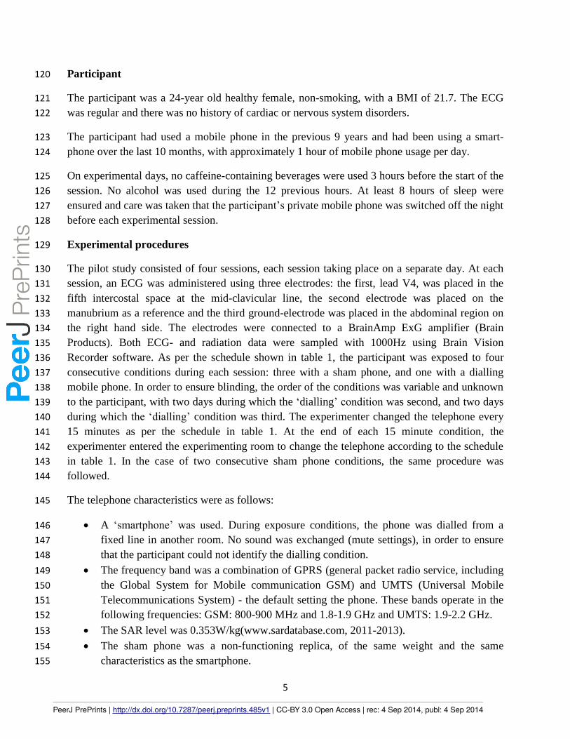
Participant 120
The participant was a 24-year old healthy female, non-smoking, with a BMI of 21.7. The ECG 121
was regular and there was no history of cardiac or nervous system disorders. 122
The participant had used a mobile phone in the previous 9 years and had been using a smart-123
phone over the last 10 months, with approximately 1 hour of mobile phone usage per day. 124
On experimental days, no caffeine-containing beverages were used 3 hours before the start of the 125
session. No alcohol was used during the 12 previous hours. At least 8 hours of sleep were 126
ensured and care was taken that the participant’s private mobile phone was switched off the night 127
before each experimental session. 128
Experimental procedures 129
The pilot study consisted of four sessions, each session taking place on a separate day. At each 130
session, an ECG was administered using three electrodes: the first, lead V4, was placed in the 131
fifth intercostal space at the mid-clavicular line, the second electrode was placed on the 132
manubrium as a reference and the third ground-electrode was placed in the abdominal region on 133
the right hand side. The electrodes were connected to a BrainAmp ExG amplifier (Brain 134
Products). Both ECG- and radiation data were sampled with 1000Hz using Brain Vision 135
Recorder software. As per the schedule shown in table 1, the participant was exposed to four 136
consecutive conditions during each session: three with a sham phone, and one with a dialling 137
mobile phone. In order to ensure blinding, the order of the conditions was variable and unknown 138
to the participant, with two days during which the ‘dialling’ condition was second, and two days 139
during which the ‘dialling’ condition was third. The experimenter changed the telephone every 140
15 minutes as per the schedule in table 1. At the end of each 15 minute condition, the 141
experimenter entered the experimenting room to change the telephone according to the schedule 142
in table 1. In the case of two consecutive sham phone conditions, the same procedure was 143
followed. 144
The telephone characteristics were as follows: 145
A ‘smartphone’ was used. During exposure conditions, the phone was dialled from a 146
fixed line in another room. No sound was exchanged (mute settings), in order to ensure 147
that the participant could not identify the dialling condition. 148
The frequency band was a combination of GPRS (general packet radio service, including 149
the Global System for Mobile communication GSM) and UMTS (Universal Mobile 150
Telecommunications System) - the default setting the phone. These bands operate in the 151
following frequencies: GSM: 800-900 MHz and 1.8-1.9 GHz and UMTS: 1.9-2.2 GHz. 152
The SAR level was 0.353W/kg(www.sardatabase.com, 2011-2013). 153
The sham phone was a non-functioning replica, of the same weight and the same 154
characteristics as the smartphone. 155
PeerJ PrePrints | http://dx.doi.org/10.7287/peerj.preprints.485v1 | CC-BY 3.0 Open Access | rec: 4 Sep 2014, publ: 4 Sep 2014
PrePrin
ts
6
As described in the introduction, a real mobile phone was used as inductor of RF-EMF. The 156
timing of radiation-peaks was detected with a radiation detector (type: HF59B, Gigahertz 157
Solutions), connected to an omnidirectional antenna. This detector was connected from the DC 158
output with an auxiliary plug to the ExG-amplifier. The detector was placed in the upright 159
position, 30 cm above the table (at which the participant was sitting) and 20 cm left from the 160
participant. The phone was placed adjacent to the left side of the sternum, bordering the 161
sternoclavicular joint at the caudal side, thus ensuring that there was no contact between the 162
phone and the V4 lead. Previous testing experiments showed that there was no direct disturbing 163
interference of the mobile phone impacting on either the V4 electrode or the amplifier (tested 164
with a shielded and non-shielded electrode). The backside of the phone was placed on the skin. 165
The phone was fixed using an elastic band. In order to maintain the participant’s alertness and to 166
guarantee a relatively stable mood, she read affectively neutral sections of a book during the 167
experiment. All experimental sessions were carried out in the afternoon. 168
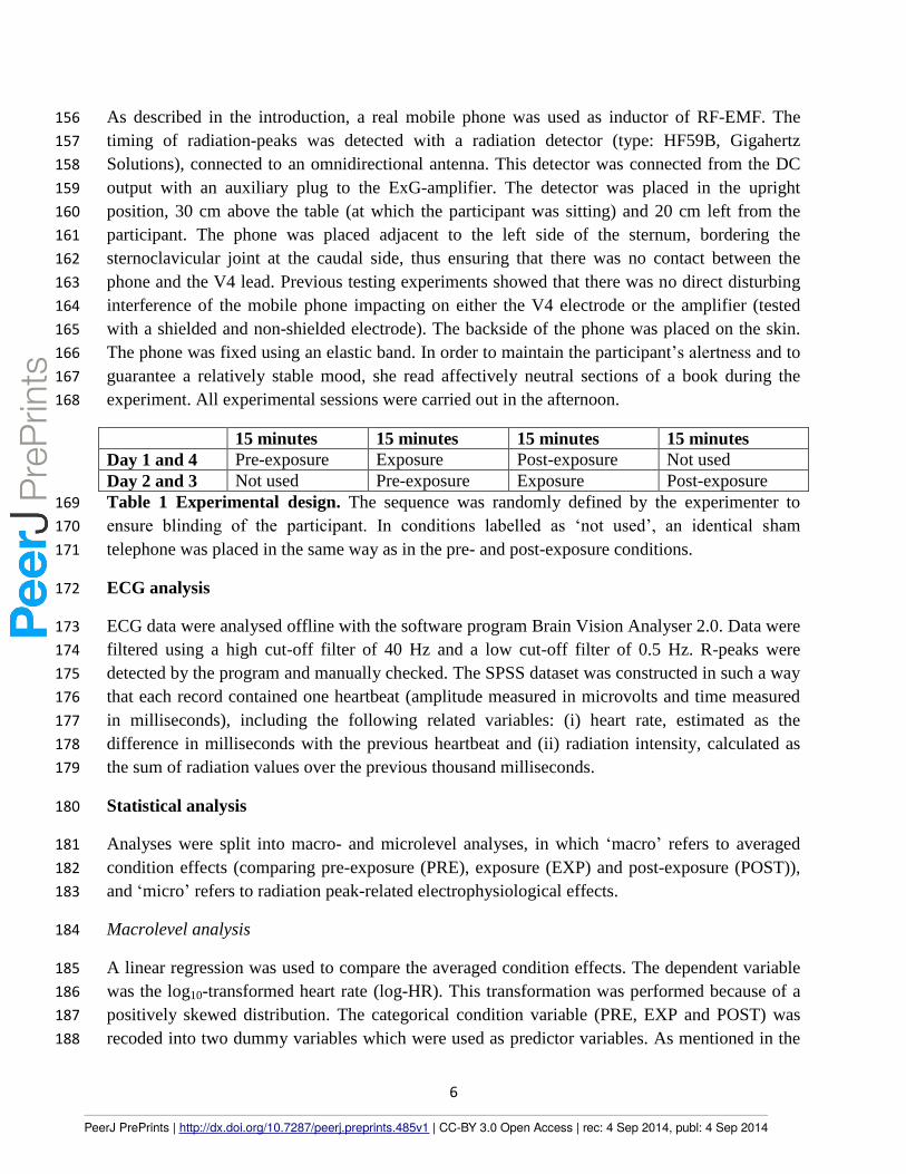
15 minutes 15 minutes 15 minutes 15 minutes
Day 1 and 4 Pre-exposure Exposure Post-exposure Not used
Day 2 and 3 Not used Pre-exposure Exposure Post-exposure
Table 1 Experimental design. The sequence was randomly defined by the experimenter to 169
ensure blinding of the participant. In conditions labelled as ‘not used’, an identical sham 170
telephone was placed in the same way as in the pre- and post-exposure conditions. 171
ECG analysis 172
ECG data were analysed offline with the software program Brain Vision Analyser 2.0. Data were 173
filtered using a high cut-off filter of 40 Hz and a low cut-off filter of 0.5 Hz. R-peaks were 174
detected by the program and manually checked. The SPSS dataset was constructed in such a way 175
that each record contained one heartbeat (amplitude measured in microvolts and time measured 176
in milliseconds), including the following related variables: (i) heart rate, estimated as the 177
difference in milliseconds with the previous heartbeat and (ii) radiation intensity, calculated as 178
the sum of radiation values over the previous thousand milliseconds. 179
Statistical analysis 180
Analyses were split into macro- and microlevel analyses, in which ‘macro’ refers to averaged 181
condition effects (comparing pre-exposure (PRE), exposure (EXP) and post-exposure (POST)), 182
and ‘micro’ refers to radiation peak-related electrophysiological effects. 183
Macrolevel analysis 184
A linear regression was used to compare the averaged condition effects. The dependent variable 185
was the log10-transformed heart rate (log-HR). This transformation was performed because of a 186
positively skewed distribution. The categorical condition variable (PRE, EXP and POST) was 187
recoded into two dummy variables which were used as predictor variables. As mentioned in the 188
PeerJ PrePrints | http://dx.doi.org/10.7287/peerj.preprints.485v1 | CC-BY 3.0 Open Access | rec: 4 Sep 2014, publ: 4 Sep 2014
PrePrin
ts
7
introduction, consecutive heart rate values are strongly interdependent. A time series analysis 189
was performed to adjust for autocorrelation and to check whether the macrolevel effect of the 190
linear regression would remain significant. Thus, a time series model, using an ARIMA structure 191
with 10 lags of autocorrelation, with the two condition predictor variables, was carried out. 192
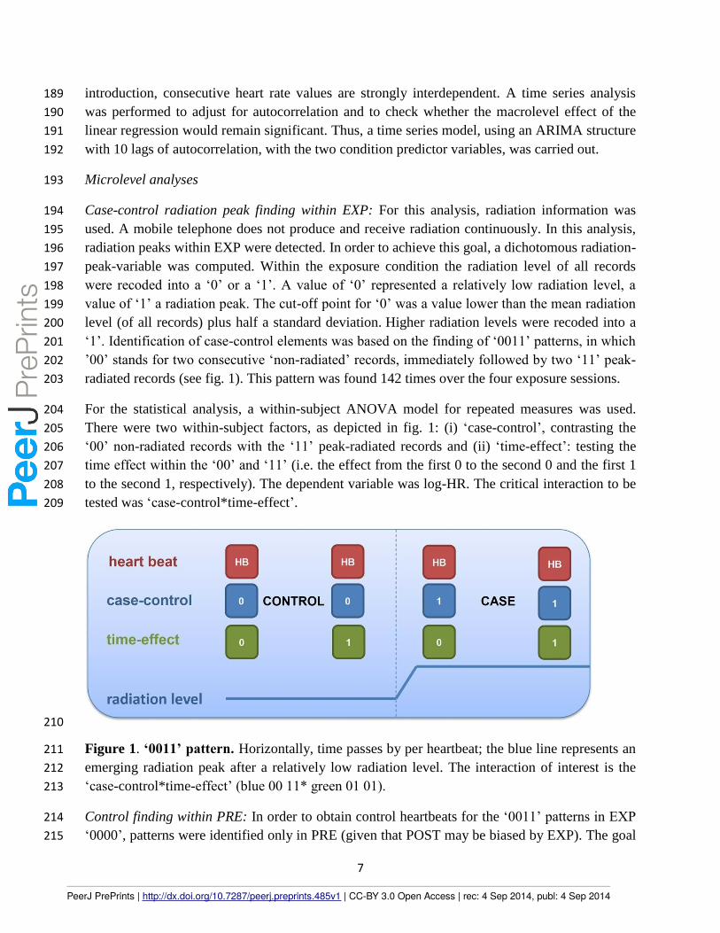
Microlevel analyses 193
Case-control radiation peak finding within EXP: For this analysis, radiation information was 194
used. A mobile telephone does not produce and receive radiation continuously. In this analysis, 195
radiation peaks within EXP were detected. In order to achieve this goal, a dichotomous radiation-196
peak-variable was computed. Within the exposure condition the radiation level of all records 197
were recoded into a ‘0’ or a ‘1’. A value of ‘0’ represented a relatively low radiation level, a 198
value of ‘1’ a radiation peak. The cut-off point for ‘0’ was a value lower than the mean radiation 199
level (of all records) plus half a standard deviation. Higher radiation levels were recoded into a 200
‘1’. Identification of case-control elements was based on the finding of ‘0011’ patterns, in which 201
’00’ stands for two consecutive ‘non-radiated’ records, immediately followed by two ‘11’ peak-202
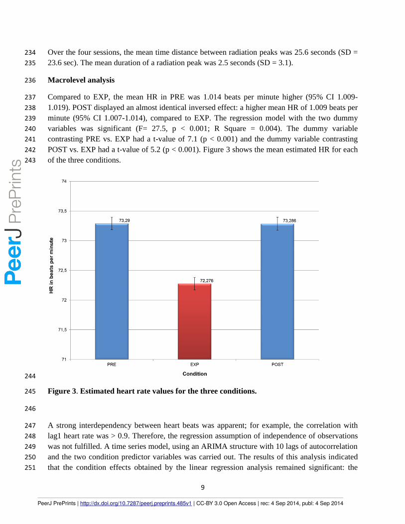
radiated records (see fig. 1). This pattern was found 142 times over the four exposure sessions. 203
For the statistical analysis, a within-subject ANOVA model for repeated measures was used. 204
There were two within-subject factors, as depicted in fig. 1: (i) ‘case-control’, contrasting the 205
‘00’ non-radiated records with the ‘11’ peak-radiated records and (ii) ‘time-effect’: testing the 206
time effect within the ‘00’ and ‘11’ (i.e. the effect from the first 0 to the second 0 and the first 1 207
to the second 1, respectively). The dependent variable was log-HR. The critical interaction to be 208
tested was ‘case-control*time-effect’. 209
210
Figure 1. ‘0011’ pattern. Horizontally, time passes by per heartbeat; the blue line represents an 211
emerging radiation peak after a relatively low radiation level. The interaction of interest is the 212
‘case-control*time-effect’ (blue 00 11* green 01 01). 213
Control finding within PRE: In order to obtain control heartbeats for the ‘0011’ patterns in EXP 214
‘0000’, patterns were identified only in PRE (given that POST may be biased by EXP). The goal 215
PeerJ PrePrints | http://dx.doi.org/10.7287/peerj.preprints.485v1 | CC-BY 3.0 Open Access | rec: 4 Sep 2014, publ: 4 Sep 2014
PrePrin
ts
8
was to compare the ‘0011’ patterns from EXP with the radiation free ‘0000’ pattern, derived 216
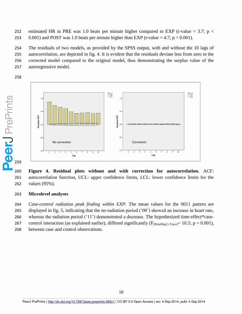
from PRE. A ‘0000’ pattern represents a radiation level lower than the median radiation value in 217
four consecutive records in the PRE condition. In order to balance the amount of controls with 218
the cases (defined as ‘0011’ found in EXP), a random sample of 142 ‘0000’ patterns were used 219
for the analysis. A comparable within-subject ANOVA model was used, but with the inclusion of 220
a between-subjects-factor (‘condition’), contrasting EXP with PRE. The critical interaction to be 221
tested was the ‘case-control*time-effect*condition’. 222
RESULTS 223
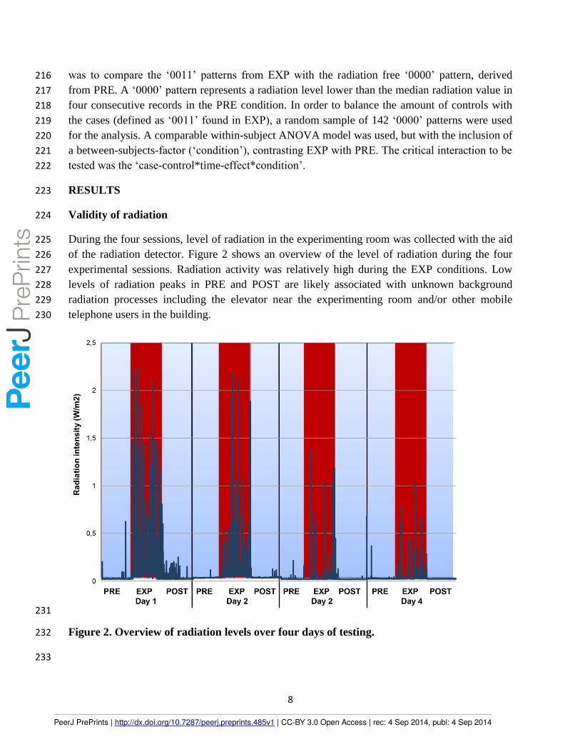
Validity of radiation 224
During the four sessions, level of radiation in the experimenting room was collected with the aid 225
of the radiation detector. Figure 2 shows an overview of the level of radiation during the four 226
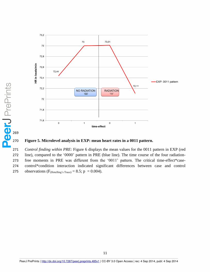
experimental sessions. Radiation activity was relatively high during the EXP conditions. Low 227
levels of radiation peaks in PRE and POST are likely associated with unknown background 228
radiation processes including the elevator near the experimenting room and/or other mobile 229
telephone users in the building. 230
231
Figure 2. Overview of radiation levels over four days of testing. 232
233
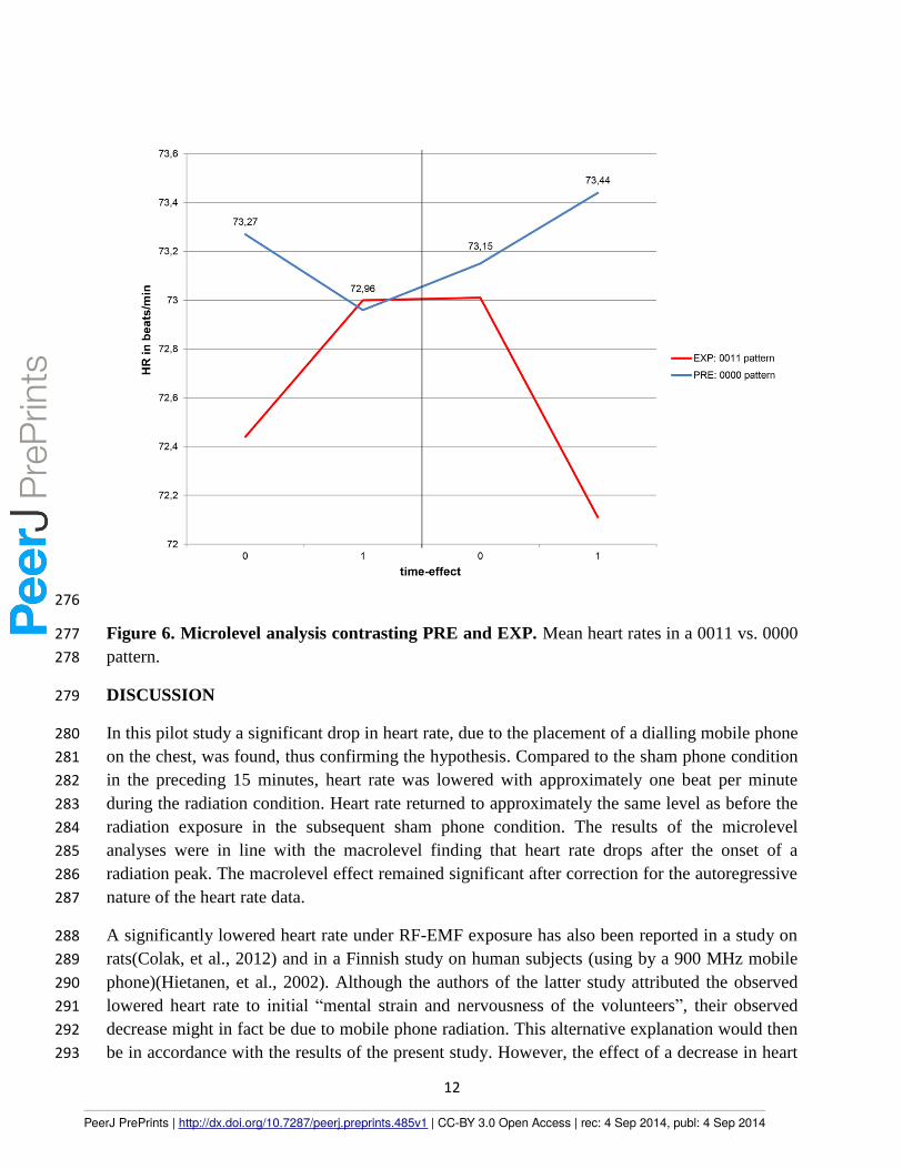
PeerJ PrePrints | http://dx.doi.org/10.7287/peerj.preprints.485v1 | CC-BY 3.0 Open Access | rec: 4 Sep 2014, publ: 4 Sep 2014
PrePrin
ts
9
Over the four sessions, the mean time distance between radiation peaks was 25.6 seconds (SD = 234
23.6 sec). The mean duration of a radiation peak was 2.5 seconds (SD = 3.1). 235
Macrolevel analysis 236
Compared to EXP, the mean HR in PRE was 1.014 beats per minute higher (95% CI 1.009-237
1.019). POST displayed an almost identical inversed effect: a higher mean HR of 1.009 beats per 238
minute (95% CI 1.007-1.014), compared to EXP. The regression model with the two dummy 239
variables was significant (F= 27.5, p < 0.001; R Square = 0.004). The dummy variable 240
contrasting PRE vs. EXP had a t-value of 7.1 (p < 0.001) and the dummy variable contrasting 241
POST vs. EXP had a t-value of 5.2 (p < 0.001). Figure 3 shows the mean estimated HR for each 242
of the three conditions. 243
244
Figure 3. Estimated heart rate values for the three conditions. 245
246
A strong interdependency between heart beats was apparent; for example, the correlation with 247
lag1 heart rate was > 0.9. Therefore, the regression assumption of independence of observations 248
was not fulfilled. A time series model, using an ARIMA structure with 10 lags of autocorrelation 249
and the two condition predictor variables was carried out. The results of this analysis indicated 250
that the condition effects obtained by the linear regression analysis remained significant: the 251
PeerJ PrePrints | http://dx.doi.org/10.7287/peerj.preprints.485v1 | CC-BY 3.0 Open Access | rec: 4 Sep 2014, publ: 4 Sep 2014
PrePrin
ts
10
estimated HR in PRE was 1.0 beats per minute higher compared to EXP (t-value = 3.7; p < 252
0.001) and POST was 1.0 beats per minute higher than EXP (t-value = 4.7; p < 0.001). 253
The residuals of two models, as provided by the SPSS output, with and without the 10 lags of 254
autocorrelation, are depicted in fig. 4. It is evident that the residuals deviate less from zero in the 255
corrected model compared to the original model, thus demonstrating the surplus value of the 256
autoregressive model. 257
258
259
Figure 4. Residual plots without and with correction for autocorrelation. ACF: 260
autocorrelation function, UCL: upper confidence limits, LCL: lower confidence limits for the 261
values (95%). 262
Microlevel analyses 263
Case-control radiation peak finding within EXP: The mean values for the 0011 pattern are 264
displayed in fig. 5, indicating that the no-radiation period (‘00’) showed an increase in heart rate, 265
whereas the radiation period (‘11’) demonstrated a decrease. The hypothesized time-effect*case-266
control interaction (as explained earlier), differed significantly (F(Hotelling’s Trace)= 10.5; p = 0.001), 267
between case and control observations. 268
PeerJ PrePrints | http://dx.doi.org/10.7287/peerj.preprints.485v1 | CC-BY 3.0 Open Access | rec: 4 Sep 2014, publ: 4 Sep 2014
PrePrin
ts
11
269
Figure 5. Microlevel analysis in EXP: mean heart rates in a 0011 pattern. 270
Control finding within PRE: Figure 6 displays the mean values for the 0011 pattern in EXP (red 271
line), compared to the ‘0000’ pattern in PRE (blue line). The time course of the four radiation-272
free moments in PRE was different from the ‘0011’ pattern. The critical time-effect*case-273
control*condition interaction indicated significant differences between case and control 274
observations (F(Hotelling’s Trace) = 8.5; p = 0.004). 275
PeerJ PrePrints | http://dx.doi.org/10.7287/peerj.preprints.485v1 | CC-BY 3.0 Open Access | rec: 4 Sep 2014, publ: 4 Sep 2014
PrePrin
ts
12
276
Figure 6. Microlevel analysis contrasting PRE and EXP. Mean heart rates in a 0011 vs. 0000 277
pattern. 278
DISCUSSION 279
In this pilot study a significant drop in heart rate, due to the placement of a dialling mobile phone 280
on the chest, was found, thus confirming the hypothesis. Compared to the sham phone condition 281
in the preceding 15 minutes, heart rate was lowered with approximately one beat per minute 282
during the radiation condition. Heart rate returned to approximately the same level as before the 283
radiation exposure in the subsequent sham phone condition. The results of the microlevel 284
analyses were in line with the macrolevel finding that heart rate drops after the onset of a 285
radiation peak. The macrolevel effect remained significant after correction for the autoregressive 286
nature of the heart rate data. 287
A significantly lowered heart rate under RF-EMF exposure has also been reported in a study on 288
rats(Colak, et al., 2012) and in a Finnish study on human subjects (using by a 900 MHz mobile 289
phone)(Hietanen, et al., 2002). Although the authors of the latter study attributed the observed 290
lowered heart rate to initial “mental strain and nervousness of the volunteers”, their observed 291
decrease might in fact be due to mobile phone radiation. This alternative explanation would then 292
be in accordance with the results of the present study. However, the effect of a decrease in heart 293
PeerJ PrePrints | http://dx.doi.org/10.7287/peerj.preprints.485v1 | CC-BY 3.0 Open Access | rec: 4 Sep 2014, publ: 4 Sep 2014
PrePrin
ts
13
rate in this n=1 study, due to mobile phone radiation, needs to be placed into perspective since 294
the majority of studies(Andrzejak, et al., 2008; Atlasz, et al., 2006; Barutcu, et al., 2011; Braune, 295
et al., 2002; Huber, et al., 2003; Kwon, et al., 2012; Nam, et al., 2009; Oftedal, et al., 2007; 296
Parazzini, et al., 2007; Tahvanainen, et al., 2004; Tamer, et al., 2009; Wilen, et al., 2006) did not 297
report large or significant effects. Nevertheless, in a recent meta-analysis on this topic, the 298
direction of the effect, albeit statistically inconclusive, is towards lower heart rate in association 299
with mobile phone radiation(Augner, et al., 2012). 300
In order to compare the results of the present study with those from others, it should be noted 301
that the methodology of the present study differs in several aspects. First, in the current study, 302
the mobile phone was placed on the chest instead of the ear. This placement in the proximity to 303
the heart may have increased the effect size (and thus the significance level). Second, in contrast 304
to all other studies, this study was based on the data of a single participant. Although each of the 305
four experimental days contained one exposure and two sham conditions, there was no between-306
subject factor. Third, the RF-EMF exposure condition in this study was always directly preceded 307
and followed by a sham condition, whereas in other studies the sham-condition was regularly 308
performed on a separate day. Fourth, with respect to data analysis, the current study also focused 309
on immediate radiation peak (microlevel) effects, which may represent a more sensitive way to 310
detect radiation-related impact on heart rate. 311
A possible mechanism for the observed drop in heart rate has been proposed in the literature, 312
suggesting that RF-EMF may activate the autonomic nerve system resulting in a slow diastolic 313
depolarization(Ahamed, et al., 2008; Andrzejak, et al., 2008; Inc. & Cardiology, 1996; Kwon, et 314
al., 2012; Parazzini, et al., 2007). The microlevel finding of an increase in heart rate directly 315
preceded by radiation peaks (see fig. 5) could be explained as a ‘compensatory’ effect for the 316
decrease in heart rate caused by earlier radiation peaks. Both the direct radiation peak effect as 317
well as the compensating effect were absent (fig. 6 even suggests a trend in the opposite 318
direction) in the PRE phase, thus providing more support for the notion of a genuine radiation 319
peak effect in the EXP phase. 320
The observed heart rate effect needs to be interpreted in a clinical perspective. Although the 321
effect is significant, the short reversible change of one beat per minute unlikely represents a large 322
or clinically relevant change in healthy individuals in the short term. The goal of this pilot study 323
was not to demonstrate risk, but to investigate whether there are electrocardiac changes at all. 324
The results indicate that more research is required to examine long-term effects. It is also 325
recommended to investigate the effects of mobile phone radiation in specific (vulnerable) 326
subgroups, such as children(Feychting, 2011). 327
Limitations 328
Some critical limitations require consideration. First, the fact that the reported results are based 329
on a n=1 sample restricts the generalisability of the findings. A replication study with a larger 330
sample size is necessary. Significant findings may be due to a (relatively) high susceptibility to 331
PeerJ PrePrints | http://dx.doi.org/10.7287/peerj.preprints.485v1 | CC-BY 3.0 Open Access | rec: 4 Sep 2014, publ: 4 Sep 2014
PrePrin
ts
14
RF-EMF of the participant(Bergqvist & Vogel, 1997; Hietanen, et al., 2002; Nam, et al., 2009; 332
Oftedal, et al., 2007; Wilen, et al., 2006). Although the participant in the current study did not 333
report RF-EMF sensitivity (headaches or other complaints) it cannot be ruled out that individual 334
susceptibility played a role. Another critical issue is the fact that this study was single blinded. It 335
is, however, unlikely that the non-blindness of the experimenter influenced the results. Third, as 336
can be seen in fig. 2, radiation level decreased over the four days of testing. A post-hoc 337
explanation may be that this effect was due to randomly different occupation of the telecom 338
network over the four days. A final critical aspect pertains to the chosen setting of the frequency 339
band GPRS/UMTS. This setting was chosen because it mimics real phone usage. As a 340
consequence, however, the exact frequency in which radiation took place was unknown 341
(although the exact timing of radiation was). 342
Future studies are required to further explore the interference of RF-EMF with human 343
physiology. Within the field of electrophysiology, heart rate variability, EEG and respiratory rate 344
would be interesting parameters to explore RF-EMF influence on the autonomic nervous system. 345
Another aspect to elaborate further is the microlevel technique of analysis. In order to unravel 346
microlevel peak-related radiation effects, a radiation detector is indispensable. Finally, because 347
of its potentially global relevance, it is important to set up longitudinal studies to examine 348
whether there are also long-term effects of RF-EMF. 349
PeerJ PrePrints | http://dx.doi.org/10.7287/peerj.preprints.485v1 | CC-BY 3.0 Open Access | rec: 4 Sep 2014, publ: 4 Sep 2014
PrePrin
ts
15
REFERENCES 350
Adair, E. R., & Black, D. R. (2003). Thermoregulatory responses to RF energy absorption. [Review]. 351 Bioelectromagnetics, Suppl 6, S17-38. doi: 10.1002/bem.10133 352
Ahamed, V. I., Karthick, N. G., & Joseph, P. K. (2008). Effect of mobile phone radiation on heart rate 353 variability. Computers in biology and medicine, 38(6), 709-712. doi: 354 10.1016/j.compbiomed.2008.03.004 355
Andrzejak, R., Poreba, R., Poreba, M., Derkacz, A., Skalik, R., Gac, P., . . . Pilecki, W. (2008). The Influence 356 of the Call with a Mobile Phone on Heart Rate Variability Parameters in Healthy Volunteers. 357 [Field Report]. Industrial Health, 46, 409-417. 358
Atlasz, T., Kellenyi, L., Kovacs, P., Babai, N., Thuroczy, G., Hejjel, L., & Hernadi, I. (2006). The application 359 of surface plethysmography for heart rate variability analysis after GSM radiofrequency 360 exposure. [Research Support, Non-U.S. Gov't]. Journal of biochemical and biophysical methods, 361 69(1-2), 233-236. doi: 10.1016/j.jbbm.2006.03.017 362
Augner, C., Gnambs, T., Winker, R., & Barth, A. (2012). Acute effects of electromagnetic fields emitted by 363 GSM mobile phones on subjective well-being and physiological reactions: a meta-analysis. 364 [Meta-Analysis]. The Science of the total environment, 424, 11-15. doi: 365 10.1016/j.scitotenv.2012.02.034 366
Barutcu, I., Esen, A. M., Kaya, D., Turkmen, M., Karakaya, O., Saglam, M., . . . Kirma, C. (2011). Do mobile 367 phones pose a potential risk to autonomic modulation of the heart? Pacing and clinical 368 electrophysiology : PACE, 34(11), 1511-1514. doi: 10.1111/j.1540-8159.2011.03162.x 369
Bergqvist, U., & Vogel, E. (1997). Possible health implications of subjective symptoms and 370 electromagnetic fields. A report prepared by a European group of experts for the European 371 Commission DG V. (Vol. 19): Arbetslivsinstitutet, National Institute for Working Life. 372
Bortkiewicz, A., Zmyślony, M., Gadzicka, E., & Szymczak, W. (2006). Neurovegetative Disturbances in 373 Workers Exposed to 50 Hz Electromagnetic Fields. International Journal of Occupational 374 Medicine and Environmental Health, 19(1), 53-60. doi: 10.2478/v10001-006-0001-1 375
Braune, S., Riedel, A., Schulte-Monting, J., & J., R. (2002). Influence of a Radiofrequency Electromagnetic 376 Field on Cardiovascular and Hormonal Parameters of the Autonomic Nervous System in Healthy 377 Individuals. Radiation Research, 158(3), 352-356. 378
Colak, C., Parlakpinar, H., Ermis, N., Tagluk, M. E., Sarihan, E., Dilek, O. F., . . . Acet, A. (2012). Effects of 379 electromagnetic radiation from 3G mobile phone on heart rate, blood pressure and ECG 380 parameters in rats. [Research Support, Non-U.S. Gov't]. Toxicology and industrial health, 28(7), 381 629-638. doi: 10.1177/0748233711420468 382
Feychting, M. (2011). Mobile phones, radiofrequency fields, and health effects in children--383 epidemiological studies. [Review]. Progress in biophysics and molecular biology, 107(3), 343-384 348. doi: 10.1016/j.pbiomolbio.2011.09.016 385
Gaestel, M. (2010). Biological monitoring of non-thermal effects of mobile phone radiation: recent 386 approaches and challenges. [Research Support, Non-U.S. Gov't 387
Review]. Biological reviews of the Cambridge Philosophical Society, 85(3), 489-500. doi: 10.1111/j.1469-388 185X.2009.00112.x 389
Hietanen, M., Hamalainen, A. M., & Husman, T. (2002). Hypersensitivity symptoms associated with 390 exposure to cellular telephones: no causal link. [Research Support, Non-U.S. Gov't]. 391 Bioelectromagnetics, 23(4), 264-270. 392
Huber, R., Schuderer, J., Graf, T., Jutz, K., Borbely, A. A., Kuster, N., & Achermann, P. (2003). Radio 393 frequency electromagnetic field exposure in humans: Estimation of SAR distribution in the brain, 394
PeerJ PrePrints | http://dx.doi.org/10.7287/peerj.preprints.485v1 | CC-BY 3.0 Open Access | rec: 4 Sep 2014, publ: 4 Sep 2014
PrePrin
ts
16
effects on sleep and heart rate. [Clinical Trial Comparative Study Controlled Clinical Trial 395 Research Support, Non-U.S. Gov't Validation Studies]. Bioelectromagnetics, 24(4), 262-276. doi: 396 10.1002/bem.10103 397
Huss, A., Egger, M., Hug, K., Huwiler-Müntener, K., & Röösli, M. (2006). Source of Funding and Results of 398 Studies of Health Effects of Mobile Phone Use: Systematic Review of Experimental Studies. 399 Environmental Health Perspectives, 115(1), 1-4. doi: 10.1289/ehp.9149 400
ICNIRP. (1998). Guidelines for limiting exposure to time-varying electric, magnetic, and electromagnetic 401 fields (up to 300 GHz). Health Physics, 74(4), 494-522. 402
Inc., A., & Cardiology, E. S. o. (1996). Heart rate variability Standards of measurement, physiological 403 interpretation, and clinical use. European Heart Journal, 17, 354-381. 404
InternationalCommunicationUnion. (2011). ICT facts and figures 405 Kwon, M. K., Choi, J. Y., Kim, S. K., Yoo, T. K., & Kim, D. W. (2012). Effects of radiation emitted by 406
WCDMA mobile phones on electromagnetic hypersensitive subjects. Environmental Health, 407 69(11), 1-8. 408
Nam, K. C., Lee, J. H., Noh, H. W., Cha, E. J., Kim, N. H., & Kim, D. W. (2009). Hypersensitivity to RF fields 409 emitted from CDMA cellular phones: a provocation study. [Research Support, Non-U.S. Gov't]. 410 Bioelectromagnetics, 30(8), 641-650. doi: 10.1002/bem.20518 411
Oftedal, G., Straume, A., Johnsson, A., & Stovner, L. J. (2007). Mobile phone headache: a double blind, 412 sham-controlled provocation study. [Randomized Controlled Trial 413
Research Support, Non-U.S. Gov't]. Cephalalgia : an international journal of headache, 27(5), 447-455. 414 doi: 10.1111/j.1468-2982.2007.01336.x 415
Parazzini, M., Ravazzani, P., Tognola, G., Thuroczy, G., Molnar, F. B., Sacchettini, A., . . . Mainardi, L. T. 416 (2007). Electromagnetic fields produced by GSM cellular phones and heart rate variability. 417 Bioelectromagnetics, 28(2), 122-129. doi: 10.1002/bem.20275 418
Tahvanainen, K., Nino, J., Halonen, P., Kuusela, T., Laitinen, T., Lansimies, E., . . . Lindholm, H. (2004). 419 Cellular phone use does not acutely affect blood pressure or heart rate of humans. [Clinical Trial 420 Comparative Study Randomized Controlled Trial Research Support, Non-U.S. Gov't]. 421 Bioelectromagnetics, 25(2), 73-83. doi: 10.1002/bem.10165 422
Tamer, A., Gunduz, H., & Ozyildirim, S. (2009). The cardiac effects of a mobile phone positioned closest 423 to the heart. Anadolu Kardiyol Derg, 9, 380-384. 424
Wilen, J., Johansson, A., Kalezic, N., Lyskov, E., & Sandstrom, M. (2006). Psychophysiological tests and 425 provocation of subjects with mobile phone related symptoms. [Randomized Controlled Trial 426
Research Support, Non-U.S. Gov't]. Bioelectromagnetics, 27(3), 204-214. doi: 10.1002/bem.20195 427 . www.sardatabase.com. (2011-2013) 428
PeerJ PrePrints | http://dx.doi.org/10.7287/peerj.preprints.485v1 | CC-BY 3.0 Open Access | rec: 4 Sep 2014, publ: 4 Sep 2014
PrePrin
ts