Embed Size (px)
Citation preview
Effects of Linomide on AdvancedProstate-Seminal Vesicle Cancers in
Lobund-Wistar RatsMorris Pollard*
Lobund Laboratory, University of Notre Dame, Notre Dame, Indiana
BACKGROUND. Angiogenesis and antiangiogenesis, as applied to oncology, are phenom-ena in which (1) tumors acquire a new blood vascular system from the host that is needed fortheir growth progression and metastasis; and (2) factors are produced that interfere withneovascularization, thereby inhibiting growth and metastasis of the tumor. Linomide, achemical antiangiogenesis agent, inhibited the growth of transplanted tumors in mice and ratsand inhibited the early development of metastasizing tumors induced in the prostate-seminalvesicle (P-SV) complex of genetically susceptible Lobund-Wistar (L-W) rats.METHODS. L-W rats with small induced P-SV tumors were treated with a recommendeddosage of linomide (100 mg/kg BW/day) by the intraperitoneal and oral routes. The rats weremonitored for the next 1–2 months, and the primary and metastatic tumors were comparedwith related data in drug-free tumor-bearing control rats.RESULTS. P-SV tumors in linomide-treated and untreated control rats continued to grow,except that in the former (1) the tumors were marginally smaller, (2) the centers of the primaryP-SV tumors had failed to grow, (3) the peripheral areas of the tumors contained activelyproliferating tumor cells, and (4) metastatic P-SV tumors in the lungs were disrupted withfocal areas of necrosis, but areas of intact tumor cells survived. Spread of tumor cells into theperitoneal cavity was not inhibited. Rats on orally administered linomide lived significantlylonger than rats inoculated by the intraperitoneal route and untreated control rats. The dosageof linomide used showed evidence of toxicity.CONCLUSIONS. Although primary and metastatic P-SV tumors were damaged in L-W ratstreated with linomide, this antiangiogenic drug was of minimal therapeutic benefit to rats inwhich a palpable P-SV tumor had developed before onset of treatments. Prostate 35:43–49,1998. © 1998 Wiley-Liss, Inc.
KEY WORDS: antiangiogenesis; linomide; prostate-seminal vesicle cancer
INTRODUCTION
To the oncologist, angiogenesis is a complex phe-nomenon in which a small avascular aggregate of tu-mor cells receives a blood supply from the host thatcontributes to the growth and spread of the tumor.However, angiogenesis has important physiologicalfunctions: a broad range of actions, which include pla-centa formation [1], wound healing [2], the develop-ment of collateral circulation, and the sequences offetal development [3]. This is in contrast to the harm-ful effects of tumorigenesis, where tumor growth be-yond microscopic size is angiogenesis dependent [4].
Lists of angiogenic peptides have been identified asfactors that contribute to tumor growth, and of anti-angiogenic agents that suppress or reverse the tumor-
igenic process [5–7]. Vascular endothelial growth fac-tor (VEGF) was noted as an important mitogen [8]whose in vivo growth-promoting effect on a propa-gating rhabdomyosarcoma was significantly sup-pressed by a specific monoclonal antibody (MAb) [9].Linomide (Roquinimex), a quinoline-3-carboxamide,is a unique antiangiogenic agent with low toxicity [10].Linomide interfered with tumor growth and metasta-sis when administered to mice with replicating trans-
Contract grant sponsor: University of Notre Dame; Contract grantsponsor: The Coleman Foundation; Contract grant sponsor: TheScully Trust.*Correspondence to: Morris Pollard, Lobund Laboratory, Universityof Notre Dame, Notre Dame, IN 46556.Received 5 May 1997; Accepted 5 August 1997
The Prostate 35:43–49 (1998)
© 1998 Wiley-Liss, Inc.
planted cells of B16 melanoma [11] and Lewis lungcarcinoma [12], to rats with Dunning tumor [10,13],and to rats treated early with metastasizing prostate(PA-III) cancer cells (M. Pollard, unpublished obser-vations, 1996) (Table I). Model systems of neoplasticdiseases have moved from in vitro-propagated and invivo-transplanted tumor cells to a higher level of rel-evance to the counterpart disease in humans; inducedautochthonous neoplastic diseases with which one caninvestigate the complete tumorigenic spectrum frominitiation to promotion, to progression and metastasis.It was exemplary of this trend that early administra-tions of linomide suppressed the development of ad-enocarcinomas that were induced in (1) the prostate-seminal vesicle (P-SV) complex of Lobund-Wistar (L-W) rats, and (2) in the breasts of Sprague-Dawley rats[14]. In inhibiting angiogenesis, linomide was found tobe cytostatic, not cytotoxic, to tumor cells; its tumor-inhibiting effect was attributed to the curtailed supplyof new blood vessels required for the expandingpopulation of tumor cells.
L-W rats are genetically predisposed to the sponta-neous and induced development of metastasizing ad-enocarcinomas in testosterone-sensitive cells in the ac-cessory sex glands: the anterior and dorsolateral lobesof the prostate and the seminal vesicles (P-SV) [15–19].P-SV tumors developed (1) spontaneously in 26% ofaging L-W rats an average 26 months [16]; and (2) byinduction in 70–90% of L-W rats an average 10.5months after administration of methylnitrosourea(MNU) and testosterone propionate (TP) [16,19]. P-SV
tumors induced by MNU/TP were actually palpableat earlier stages of development an average 7.5 monthswhen 0.5 cm in diameter; within the next 1–2 months,the tumors attained large size (3- to 4-cm diameter).Metastatic tumors were rarely observed in rats withsmall tumors, but metastases to the lungs or into theperitoneal cavity, or both, were frequent in rats withlarge tumors (>10 g in weight).
In the report presented here, linomide was admin-istered to L-W rats with small palpable induced P-SVtumors to determine whether this antiangiogenic drugwould interfere or modify (1) continued growth ofestablished small tumors, (2) metastatic spread of thetumor cells to the lungs and/or into the peritonealcavity, and (3) extension of life span.
MATERIALS AND METHODS
Rats
Specific pathogen-free L-W rats were propagated atrandom in isolated air conditioned rooms with 12-hrlight/dark cycles. They were fed, ad libitum, naturalingredient diet L-485, which was formulated with ad-ditional minerals and vitamins (TekLad, Madison,WI). The rats were maintained in plastic cages on abedding of wood shavings. Although L-W rats werenot intentionally inbred, they did not reject reciprocalskin grafts. In aging L-W rats, carcinomas developedspontaneously in the P-SV, in the liver, and in thelungs [16,20]; however, spontaneous tumors havebeen observed rarely in rats under age 18 months.
All experimental protocols involving animals weremonitored by the University Animal Care Committeein accordance with the guidelines in the Public HealthService Guide for the Care and Use of Laboratory Ani-mals.
Chemical Agents
Linomide (N-phenylmethyl-1,2-dihydro-4-hydroxy-1-methyl 2-oxo-quinoline-3-carboxamide) isa water-soluble, low-molecular-weight (Mr = 308,000)drug, of relatively low toxicity. It is the registeredtrademark for Roquinimex (Pharmacia–Upjohn, Lund,Sweden). MNU was purchased from Ash Stevens (De-troit, MI). Testosterone propionate was purchasedfrom Sigma Chemical Co., St. Louis, MO. The optimaldosage recommended for linomide was 100 mg/kgBW/day by intraperitoneal inoculation [10]. Linomidewas dissolved in sterile distilled water for intraperito-neal inoculation (100 mg/kg BW/rat/day), or it wasadded to the drinking water (1 mg linomide/1 ml wa-ter). The dosage was based on consumption of 8–11 ml
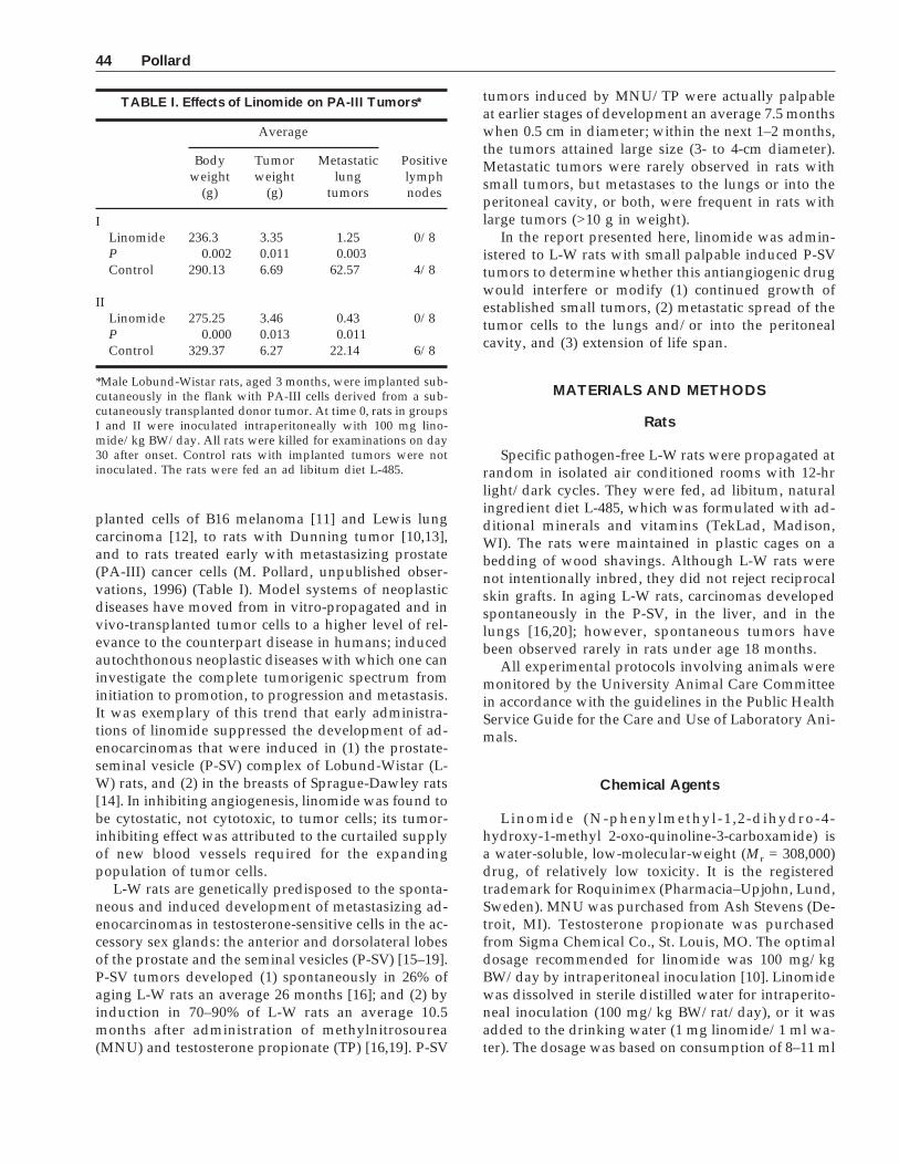
TABLE I. Effects of Linomide on PA-III Tumors*
Average
Positivelymphnodes
Bodyweight
(g)
Tumorweight
(g)
Metastaticlung
tumors
ILinomide 236.3 3.35 1.25 0/8P 0.002 0.011 0.003Control 290.13 6.69 62.57 4/8
IILinomide 275.25 3.46 0.43 0/8P 0.000 0.013 0.011Control 329.37 6.27 22.14 6/8
*Male Lobund-Wistar rats, aged 3 months, were implanted sub-cutaneously in the flank with PA-III cells derived from a sub-cutaneously transplanted donor tumor. At time 0, rats in groupsI and II were inoculated intraperitoneally with 100 mg lino-mide/kg BW/day. All rats were killed for examinations on day30 after onset. Control rats with implanted tumors were notinoculated. The rats were fed an ad libitum diet L-485.
44 Pollard
water/100 g body weight [21]. Therefore, rats weigh-ing 300 g consumed ∼30 mg linomide, and 400 g ratsconsumed ∼40 mg/kg BW/day.
Experimental Design
The procedure for induction of P-SV tumors in L-Wrats, and characteristics of the resulting tumors, havebeen described [17]. Briefly, L-W rats aged 3 monthswere inoculated intravenously with acidified MNU(30 mg/kg BW); and then, at an interval of 2 months,given two subcutaneous implants of TP (25 mg each)enclosed in a Silastic membrane. When small palpableP-SV tumors (∼0.5-cm diameter) were detected, eachrat was administered linomide either by intraperito-neal inoculation or in the drinking water. At frequentintervals, each rat was monitored for physical condi-tion and by palpation for changes in tumor size duringthe next 1–2 months. When the palpable tumors in thelinomide-treated and in the untreated controlsreached 3–4 cm in diameter, each rat was killed byinhaled halothane and exsanguination from the heartand then examined for gross changes at autopsy. Be-cause the large prostate-related tumors had spread toall lobes, weights at all stages of tumor development(early and late) were determined from the weight ofthe entire prostate complex. Body weight and weightand appearance of liver, the prostate complex, testes,and tumors in the peritoneal cavity were recorded.Survival times of linomide-treated rats were com-pared with survival of untreated tumor-bearing con-trol rats. Cross sections of the P-SV tumor and slices of
other involved organs were fixed in 10% buffered for-malin. Lungs were inflated through the trachea withBouin’s solution, then immersed in Bouin’s for 18 hr,transferred to 70% ethanol, and examined at 5× mag-nification for numbers of focal subpleural tumors. Thetissues were processed for histological examinationsand stained by hematoxylin and eosin (H&E). Resultswere assessed for statistical significance by Student’st-test.
RESULTS
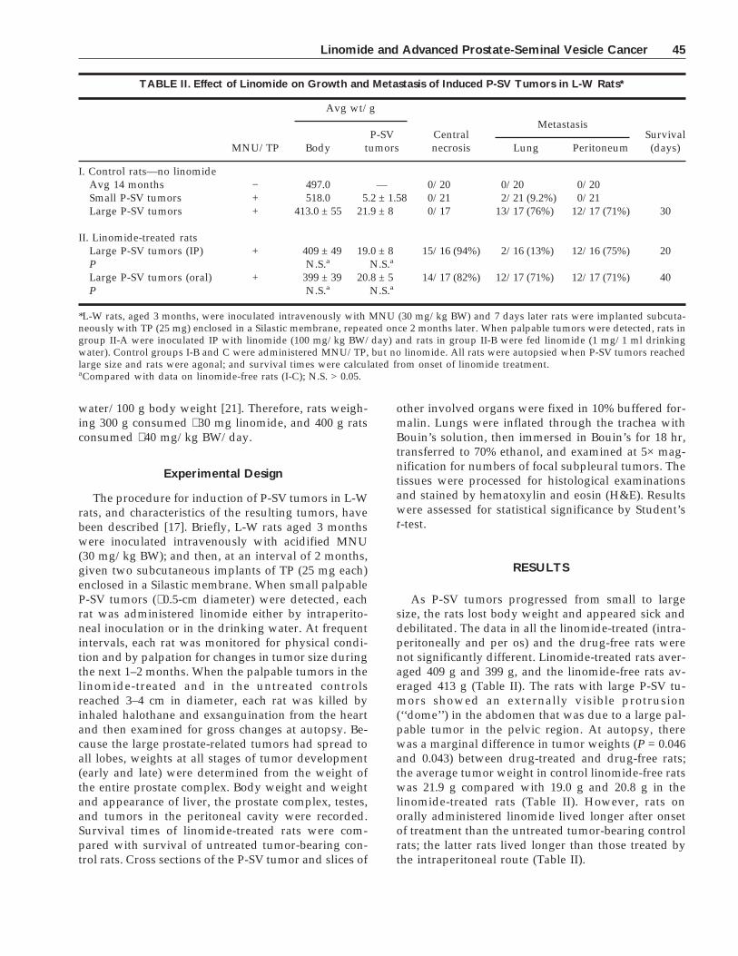
As P-SV tumors progressed from small to largesize, the rats lost body weight and appeared sick anddebilitated. The data in all the linomide-treated (intra-peritoneally and per os) and the drug-free rats werenot significantly different. Linomide-treated rats aver-aged 409 g and 399 g, and the linomide-free rats av-eraged 413 g (Table II). The rats with large P-SV tu-mors showed an externally visible protrusion(‘‘dome’’) in the abdomen that was due to a large pal-pable tumor in the pelvic region. At autopsy, therewas a marginal difference in tumor weights (P = 0.046and 0.043) between drug-treated and drug-free rats;the average tumor weight in control linomide-free ratswas 21.9 g compared with 19.0 g and 20.8 g in thelinomide-treated rats (Table II). However, rats onorally administered linomide lived longer after onsetof treatment than the untreated tumor-bearing controlrats; the latter rats lived longer than those treated bythe intraperitoneal route (Table II).
TABLE II. Effect of Linomide on Growth and Metastasis of Induced P-SV Tumors in L-W Rats*
MNU/TP
Avg wt/g
Centralnecrosis
MetastasisSurvival(days)Body
P-SVtumors Lung Peritoneum
I. Control rats—no linomideAvg 14 months − 497.0 — 0/20 0/20 0/20Small P-SV tumors + 518.0 5.2 ± 1.58 0/21 2/21 (9.2%) 0/21Large P-SV tumors + 413.0 ± 55 21.9 ± 8 0/17 13/17 (76%) 12/17 (71%) 30
II. Linomide-treated ratsLarge P-SV tumors (IP) + 409 ± 49 19.0 ± 8 15/16 (94%) 2/16 (13%) 12/16 (75%) 20P N.S.a N.S.a
Large P-SV tumors (oral) + 399 ± 39 20.8 ± 5 14/17 (82%) 12/17 (71%) 12/17 (71%) 40P N.S.a N.S.a
*L-W rats, aged 3 months, were inoculated intravenously with MNU (30 mg/kg BW) and 7 days later rats were implanted subcuta-neously with TP (25 mg) enclosed in a Silastic membrane, repeated once 2 months later. When palpable tumors were detected, rats ingroup II-A were inoculated IP with linomide (100 mg/kg BW/day) and rats in group II-B were fed linomide (1 mg/1 ml drinkingwater). Control groups I-B and C were administered MNU/TP, but no linomide. All rats were autopsied when P-SV tumors reachedlarge size and rats were agonal; and survival times were calculated from onset of linomide treatment.aCompared with data on linomide-free rats (I-C); N.S. > 0.05.
Linomide and Advanced Prostate-Seminal Vesicle Cancer 45
Drug-Free Control Rats
The average weights of the prostate complex in 20drug-free, tumor-free L-W rats, age 14 months, was3.98 ± 0.63 g. Their average body weight was 497 ± 20g (Table II). Among 21 control rats with small inducedpalpable P-SV tumors, the average body weight was518 ± 15 g; and the average weight of the prostatecomplex was 5.2 ± 1.58 g. Note that the inductionprocedure of P-SV tumors was started when the ratswere 3 months old. In 12 rats, their tumors had devel-oped in the seminal vesicles; in 6 rats, the tumors haddeveloped in the dorsolateral and anterior lobes; andin 3 rats, the tumors had developed in both areas. Thetumor cells had penetrated the basement membrane ofa duct or an acinus and were dispersed into an exten-sive stroma of connective tissue that had developed inthe surrounding area. In two rats, small lung metas-tases had developed.
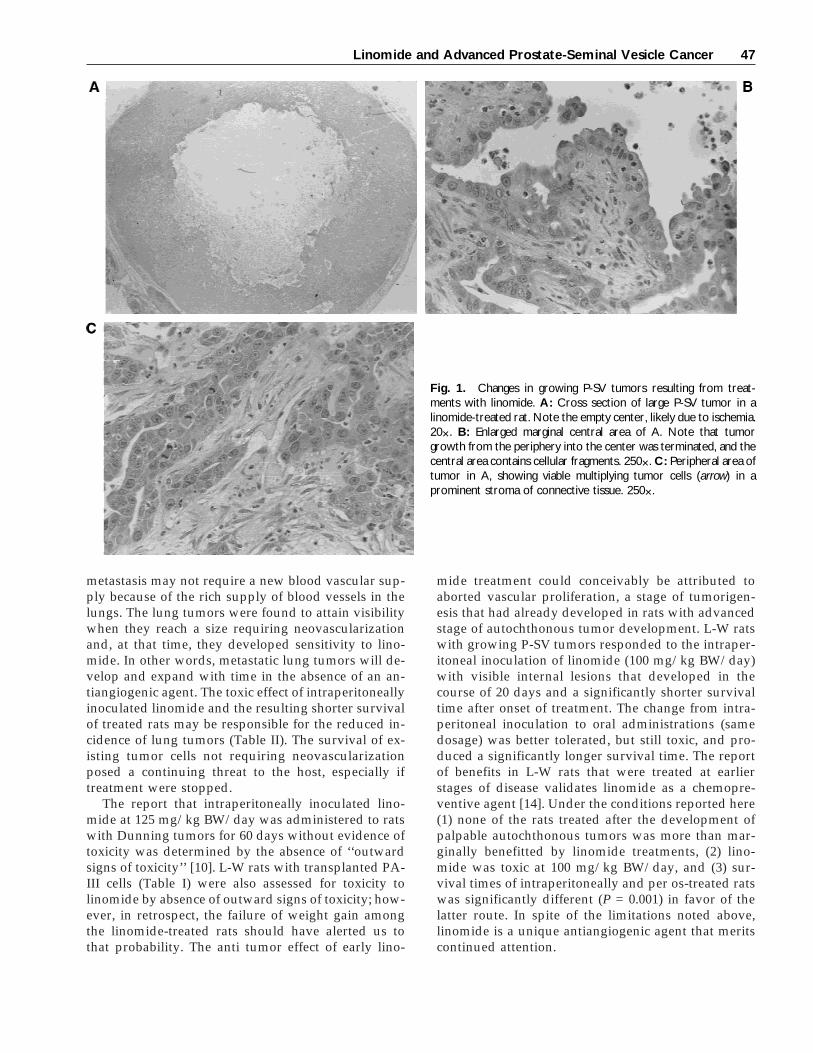
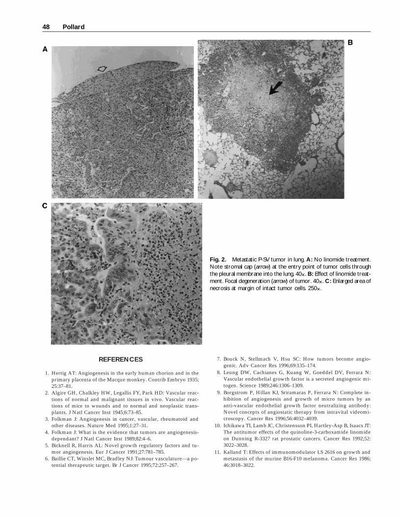
Among 17 control rats in which large tumors haddeveloped, the average body weight was 413 ± 55 gand their P-SV tumors weighed avg 21.9 ± 8 g (TableII). Their P-SV tumors were hard, with knobby sur-faces, and cirrhotic when incised. The anatomical sitesfrom which the large tumors had developed were ob-scured. The P-SV tumors were moderately differenti-ated adenocarcinomas with occasional mitotic figuresand numerous myelocytes. The centers of the largeP-SV tumors were relatively cell-free and consisted offibrin strands with active stromal cells and small is-lands of tumor cells. The peripheral (marginal) areasof the P-SV tumors contained numerous single tumorcells and cells that had developed into moderately dif-ferentiated adenocarcinomas in a prominent stroma.Thirteen (76%) of the rats had developed metastaticsubpleural moderately differentiated adenocarcino-mas in the lungs which were actively expanding intothe lung parenchyma (see Fig. 2A). The metastatic tu-mor cells frequently developed a stromal ‘‘cap’’ at thepoint of entry of tumor cells into the lung, where theydeveloped adenomatous structures in random pat-terns with occasional mitotic figures, a prominentstroma, and numerous myelocytes. In 12 (71%) of therats, the tumor cells had penetrated the prostate cap-sule into the peritoneal cavity, where they developedinto discrete pearl-like tumors attached to the perito-neal lining; occasionally, they had attached to, andpenetrated, the liver.
Linomide-Treated Rats
Among 16 rats that had been inoculated intraperi-toneally with linomide when their tumors were small,the average body weight at autopsy was 409 ± 49 gand the average weight of their large tumors was 19.0
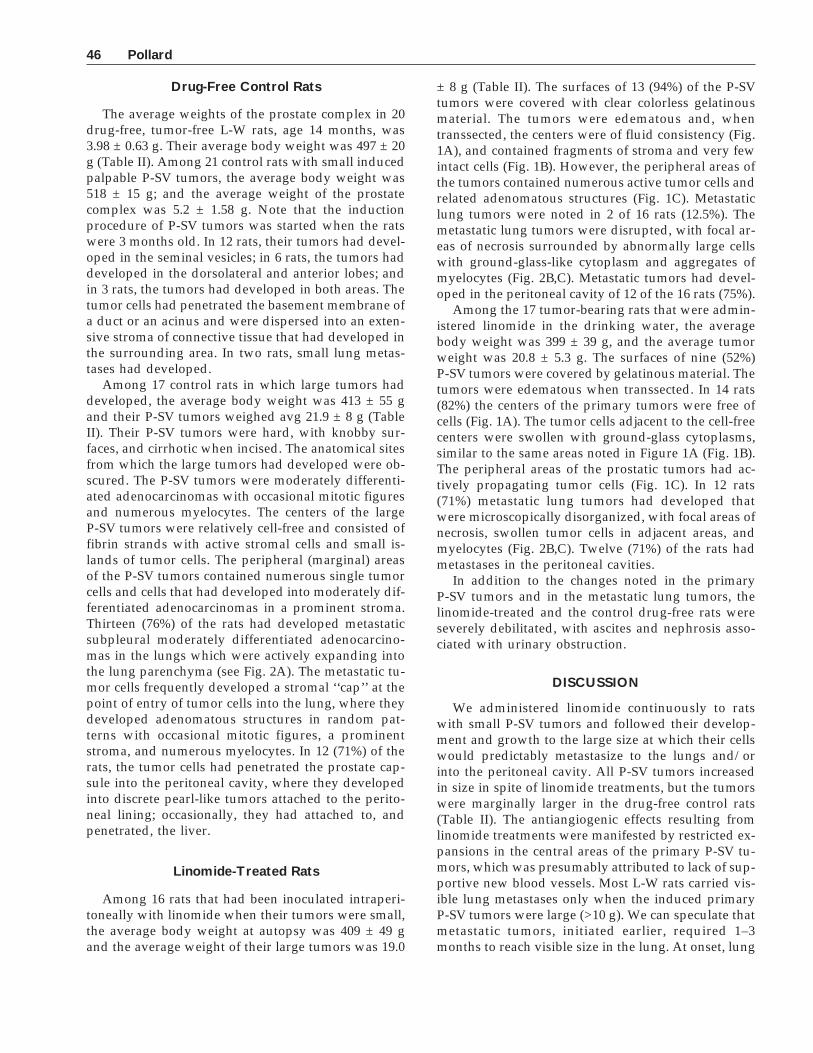
± 8 g (Table II). The surfaces of 13 (94%) of the P-SVtumors were covered with clear colorless gelatinousmaterial. The tumors were edematous and, whentranssected, the centers were of fluid consistency (Fig.1A), and contained fragments of stroma and very fewintact cells (Fig. 1B). However, the peripheral areas ofthe tumors contained numerous active tumor cells andrelated adenomatous structures (Fig. 1C). Metastaticlung tumors were noted in 2 of 16 rats (12.5%). Themetastatic lung tumors were disrupted, with focal ar-eas of necrosis surrounded by abnormally large cellswith ground-glass-like cytoplasm and aggregates ofmyelocytes (Fig. 2B,C). Metastatic tumors had devel-oped in the peritoneal cavity of 12 of the 16 rats (75%).
Among the 17 tumor-bearing rats that were admin-istered linomide in the drinking water, the averagebody weight was 399 ± 39 g, and the average tumorweight was 20.8 ± 5.3 g. The surfaces of nine (52%)P-SV tumors were covered by gelatinous material. Thetumors were edematous when transsected. In 14 rats(82%) the centers of the primary tumors were free ofcells (Fig. 1A). The tumor cells adjacent to the cell-freecenters were swollen with ground-glass cytoplasms,similar to the same areas noted in Figure 1A (Fig. 1B).The peripheral areas of the prostatic tumors had ac-tively propagating tumor cells (Fig. 1C). In 12 rats(71%) metastatic lung tumors had developed thatwere microscopically disorganized, with focal areas ofnecrosis, swollen tumor cells in adjacent areas, andmyelocytes (Fig. 2B,C). Twelve (71%) of the rats hadmetastases in the peritoneal cavities.
In addition to the changes noted in the primaryP-SV tumors and in the metastatic lung tumors, thelinomide-treated and the control drug-free rats wereseverely debilitated, with ascites and nephrosis asso-ciated with urinary obstruction.
DISCUSSION
We administered linomide continuously to ratswith small P-SV tumors and followed their develop-ment and growth to the large size at which their cellswould predictably metastasize to the lungs and/orinto the peritoneal cavity. All P-SV tumors increasedin size in spite of linomide treatments, but the tumorswere marginally larger in the drug-free control rats(Table II). The antiangiogenic effects resulting fromlinomide treatments were manifested by restricted ex-pansions in the central areas of the primary P-SV tu-mors, which was presumably attributed to lack of sup-portive new blood vessels. Most L-W rats carried vis-ible lung metastases only when the induced primaryP-SV tumors were large (>10 g). We can speculate thatmetastatic tumors, initiated earlier, required 1–3months to reach visible size in the lung. At onset, lung
46 Pollard
metastasis may not require a new blood vascular sup-ply because of the rich supply of blood vessels in thelungs. The lung tumors were found to attain visibilitywhen they reach a size requiring neovascularizationand, at that time, they developed sensitivity to lino-mide. In other words, metastatic lung tumors will de-velop and expand with time in the absence of an an-tiangiogenic agent. The toxic effect of intraperitoneallyinoculated linomide and the resulting shorter survivalof treated rats may be responsible for the reduced in-cidence of lung tumors (Table II). The survival of ex-isting tumor cells not requiring neovascularizationposed a continuing threat to the host, especially iftreatment were stopped.
The report that intraperitoneally inoculated lino-mide at 125 mg/kg BW/day was administered to ratswith Dunning tumors for 60 days without evidence oftoxicity was determined by the absence of ‘‘outwardsigns of toxicity’’ [10]. L-W rats with transplanted PA-III cells (Table I) were also assessed for toxicity tolinomide by absence of outward signs of toxicity; how-ever, in retrospect, the failure of weight gain amongthe linomide-treated rats should have alerted us tothat probability. The anti tumor effect of early lino-
mide treatment could conceivably be attributed toaborted vascular proliferation, a stage of tumorigen-esis that had already developed in rats with advancedstage of autochthonous tumor development. L-W ratswith growing P-SV tumors responded to the intraper-itoneal inoculation of linomide (100 mg/kg BW/day)with visible internal lesions that developed in thecourse of 20 days and a significantly shorter survivaltime after onset of treatment. The change from intra-peritoneal inoculation to oral administrations (samedosage) was better tolerated, but still toxic, and pro-duced a significantly longer survival time. The reportof benefits in L-W rats that were treated at earlierstages of disease validates linomide as a chemopre-ventive agent [14]. Under the conditions reported here(1) none of the rats treated after the development ofpalpable autochthonous tumors was more than mar-ginally benefitted by linomide treatments, (2) lino-mide was toxic at 100 mg/kg BW/day, and (3) sur-vival times of intraperitoneally and per os-treated ratswas significantly different (P = 0.001) in favor of thelatter route. In spite of the limitations noted above,linomide is a unique antiangiogenic agent that meritscontinued attention.
Fig. 1. Changes in growing P-SV tumors resulting from treat-ments with linomide. A: Cross section of large P-SV tumor in alinomide-treated rat. Note the empty center, likely due to ischemia.20×. B: Enlarged marginal central area of A. Note that tumorgrowth from the periphery into the center was terminated, and thecentral area contains cellular fragments. 250×. C: Peripheral area oftumor in A, showing viable multiplying tumor cells (arrow) in aprominent stroma of connective tissue. 250×.
Linomide and Advanced Prostate-Seminal Vesicle Cancer 47
REFERENCES
1. Hertig AT: Angiogenesis in the early human chorion and in theprimary placenta of the Macque monkey. Contrib Embryo 1935;25:37–81.
2. Algire GH, Chalkley HW, Legallis FY, Park HD: Vascular reac-tions of normal and malignant tissues in vivo. Vascular reac-tions of mice to wounds and to normal and neoplastic trans-plants. J Natl Cancer Inst 1945;6:73–85.
3. Folkman J: Angiogenesis in cancer, vascular, rheumatoid andother diseases. Nature Med 1995;1:27–31.
4. Folkman J: What is the evidence that tumors are angiogenesis-dependant? J Natl Cancer Inst 1989;82:4–6.
5. Bicknell R, Harris AL: Novel growth regulatory factors and tu-mor angiogenesis. Eur J Cancer 1991;27:781–785.
6. Baillie CT, Winslet MC, Bradley NJ: Tumour vasculature—a po-tential therapeutic target. Br J Cancer 1995;72:257–267.
7. Bouck N, Stellmach V, Hsu SC: How tumors become angio-genic. Adv Cancer Res 1996;69:135–174.
8. Leung DW, Cachianes G, Kuang W, Goeddel DV, Ferrara N:Vascular endothelial growth factor is a secreted angiogenic mi-togen. Science 1989;246:1306–1309.
9. Borgstrom P, Hillan KJ, Sriramaras P, Ferrara N: Complete in-hibition of angiogenesis and growth of micro tumors by ananti-vascular endothelial growth factor neutralizing antibody:Novel concepts of angiostatic therapy from intravital videomi-croscopy. Cancer Res 1996;56:4032–4039.
10. Ichikawa TI, Lamb JC, Christensson PI, Hartley-Asp B, Isaacs JT:The antitumor effects of the quinoline-3-carboxamide linomideon Dunning R-3327 rat prostatic cancers. Cancer Res 1992;52:3022–3028.
11. Kalland T: Effects of immunomodulator LS 2616 on growth andmetastasis of the murine B16-F10 melanoma. Cancer Res 1986;46:3018–3022.
Fig. 2. Metastatic P-SV tumor in lung. A: No linomide treatment.Note stromal cap (arrow) at the entry point of tumor cells throughthe pleural membrane into the lung. 40×. B: Effect of linomide treat-ment. Focal degeneration (arrow) of tumor. 40×. C: Enlarged area ofnecrosis at margin of intact tumor cells. 250×.
48 Pollard
12. Borgstrom P, Torres Filho IP, Hartley-Asp B: Inhibition of an-giogenesis and metastasis of the Lewis lung cells carcinoma bythe quinoline-3-carboxamide, linomide. Anticancer Res 1995;15:719–728.
13. Vukanovic J, Passaniti A, Hirata T, Traystman RJ, Hartley-AspB, Isaacs JT: Antiangiogenic effects of the quinoline-3-carboxamide linomide. Cancer Res 1993;53:1833–1837.
14. Joseph IBJK, Vukanovic J, Isaacs JT: Antiangiogenic treatmentwith linomide as chemoprevention for prostate, seminal vesicle,and breast carcinogenesis in rodents. Cancer Res 1996;56:3404–3408.
15. Pollard M: Spontaneous prostate adenocarcinomas in agedgerm free Wistar rats. J Natl Cancer Inst 1973;51:1235–1241.
16. Pollard M: The Lobund-Wistar rat model of prostate cancer. JCell Biochem 1992;16H(suppl):84–88.
17. Pollard M, Luckert PH: Autochthonous prostate adenocarcino-mas in Lobund-Wistar rats: A model system. Prostate 1987;11:219–227.
18. Hoover DM, Best KL, McKenny BK, Tamura RN, Neubauer BL:Experimental induction of neoplasia in the accessory sex organsof male Lobund-Wistar rats. Cancer Res 1990;50:142–146.
19. Pollard M, Luckert PH: Early manifestations of induced prostatetumors in Lobund-Wistar rats. Cancer Lett 1992;67:113–116.
20. Pollard M, Luckert PH: Phenobarbital promotes multistage pul-monary carcinogenesis in MNU-inoculated L-W rats. In Vivo1997;11:55–60.
21. Kohn DF, Barthold SW: Biology and diseases of rats. In Fox JW,Cohen BJ, Loew FM (eds): ‘‘Laboratory Animal Medicine.’’ SanDiego: Academic Press, 1984:95–120.
Linomide and Advanced Prostate-Seminal Vesicle Cancer 49