Embed Size (px)
Citation preview

Panje et al. Radiation Oncology (2015) 10:47 DOI 10.1186/s13014-015-0338-3
source: https://doi.org/10.7892/boris.75350 | downloaded: 13.3.2017
RESEARCH Open Access
Guidance of treatment decisions in risk-adaptedprimary radiotherapy for prostate cancer usingmultiparametric magnetic resonance imaging: asingle center experienceCedric Panje1, Thierry Panje1, Paul Martin Putora1, Suk-kyum Kim2, Sarah Haile3, Daniel M Aebersold4
and Ludwig Plasswilm1*
Abstract
Background: Magnetic resonance imaging (MRI) of the prostate is considered to be the most precise noninvasivestaging modality for localized prostate cancer. Multiparametric MRI (mpMRI) dynamic sequences have recently beenshown to further increase the accuracy of staging relative to morphological imaging alone. Correct radiologicalstaging, particularly the detection of extraprostatic disease extension, is of paramount importance for targetvolume definition and dose prescription in highly-conformal curative radiotherapy (RT); in addition, it may affectthe risk-adapted duration of additional antihormonal therapy. The purpose of our study was to analyze theimpact of mpMRI-based tumor staging in patients undergoing primary RT for prostate cancer.
Methods: A total of 122 patients admitted for primary RT for prostate cancer were retrospectively analyzedregarding initial clinical and computed tomography-based staging in comparison with mpMRI staging. Bothtumor stage shifts and overall risk group shifts, including prostate-specific antigen (PSA) level and the Gleasonscore, were assessed. Potential risk factors for upstaging were tested in a multivariate analysis. Finally, the impactof mpMRI-based staging shift on prostate RT and antihormonal therapy was evaluated.
Results: Overall, tumor stage shift occurred in 55.7% of patients after mpMRI. Upstaging was most prominent inpatients showing high-risk serum PSA levels (73%), but was also substantial in patients presenting with low-riskPSA levels (50%) and low-risk Gleason scores (45.2%). Risk group changes occurred in 28.7% of the patients withconsequent treatment adaptations regarding target volume delineation and duration of androgen deprivationtherapy. High PSA levels were found to be a significant risk factor for tumor upstaging and newly diagnosedseminal vesicle infiltration assessed using mpMRI.
Conclusions: Our findings suggest that mpMRI of the prostate leads to substantial tumor upstaging, and canconsiderably affect treatment decisions in all patient groups undergoing risk-adapted curative RT for prostate cancer.
Keywords: MRI, Multiparametric, Prostate cancer, Radiotherapy
BackgroundExternal beam radiotherapy (RT) of the prostate hasbeen established as an effective therapeutic option forlocalized prostate cancer as a single treatment modalityor in conjunction with systemic androgen deprivationtherapy (ADT); it has achieved excellent rates of
* Correspondence: [email protected] of Radiation Oncology, Kantonsspital St. Gallen, St. Gallen, SwitzerlandFull list of author information is available at the end of the article
© 2015 Panje et al.; licensee BioMed Central. TCommons Attribution License (http://creativecreproduction in any medium, provided the orDedication waiver (http://creativecommons.orunless otherwise stated.
locoregional and biochemical control [1-3]. Recent phaseIII studies have demonstrated an additional improve-ment in oncological outcome for patients presentingwith adverse risk factors including advanced T-stage,markedly elevated serum prostate-specific antigen (PSA)and high-grade disease by means of treatment intensifi-cation such as radiation dose escalation and the additionof ADT [4-6]. Current consensus guidelines recommendextension of the target volume beyond the prostatic
his is an Open Access article distributed under the terms of the Creativeommons.org/licenses/by/4.0), which permits unrestricted use, distribution, andiginal work is properly credited. The Creative Commons Public Domaing/publicdomain/zero/1.0/) applies to the data made available in this article,
Table 1 Risk group classification for localized prostatecancer according to the National Comprehensive CancerNetwork guidelines
T Stage PSA (ng/ml) Gleason score
(Very) low risk T1–T2a < 10 2–6
Intermediate risk T2b-c 10–20 7
(Very) high risk T3a-b >20 8–10
Panje et al. Radiation Oncology (2015) 10:47 Page 2 of 9
capsule for locally advanced disease (T3 stage) to ac-count for extracapsular extension (ECE) and seminalvesicle invasion (SVI) [7]. Such an accurate target vol-ume definition is particularly important when highlyconformal techniques such as image-guided intensity-modulated radiotherapy (IMRT) are used [8]. However,as a definite pathological specimen is not available likeafter radical prostatectomy, curative RT for prostate can-cer relies primarily on accurate clinical and radiologicaltumor staging for risk group-adapted treatment intensifi-cation as well as for target volume delineation [7,9].To date, magnetic resonance imaging (MRI) of the
prostate is considered to be the most accurate imagingmodality available for the noninvasive determination ofthe local extent of prostate cancer [10,11]. More recently,prostate morphological imaging involving T1- and T2-weighted MRI has been routinely complemented by multi-parametric dynamic sequences such as diffusion-weightedimaging (DWI) and dynamic contrast-enhanced imaging,which have been shown to further increase specificity andsensitivity [12-14]. MRI of prostate cancer allows for thereliable detection of adverse pathological features such asECE and SVI [15-18]. It has also shown a superior congru-ence with the final surgical-pathological staging relative todigital rectal examination, transrectal ultrasound-guidedbiopsy and computed tomography (CT) [19,20], as well ascompared with prediction tools such as the Partin tables[21,22] and the Kattan nomogram [23].A small number of previous studies have demon-
strated that morphological MRIs of localized prostatecancer resulted in a significant tumor stage shift withconsequent implications regarding target volume defin-ition [24] and more accurate prediction of treatmentoutcome [25]. However, to our knowledge, the impact ofstate-of-the-art multiparametric MRI (mpMRI) of theprostate on curative RT for prostate cancer and add-itional ADT has not yet been specifically investigated.Consequently, the purpose of our study was to retro-spectively analyze the value of mpMRI for prostate can-cer staging before curative RT and its impact ontreatment decisions.
MethodsPatient selectionAfter review and the approval of the institutional ethicscommittee (Ethics Committee St. Gallen, Switzerland),160 patients with clinically localized prostate cancerwere identified. These patients had been referred to theCantonal Hospital St. Gallen between January 2010 andDecember 2013 for primary RT, based on patient prefer-ence or medical inoperability; they had all received ampMRI of the prostate before RT. For further analysispatients who met the following staging criteria beforeundergoing a pelvic MRI scan (n = 122) were included:
histopathological confirmation of prostate cancer usingtransrectal ultrasound-guided biopsy evaluated accord-ing to the Gleason grading system [26]; a complete med-ical history and physical examination; a serum PSAmeasurement; and a CT scan of the abdomen and pelvis.Clinical stage before the MRI scan was determined usingthe 2010 International Union Against Cancer (UICC),7th edition staging criteria [27].Based on the pre-treatment serum PSA, histopatho-
logical Gleason Score and clinical staging patients wereassigned to low-risk, intermediate-risk and high-riskgroups according to the 2014 National ComprehensiveCancer Network (NCCN) guidelines on prostate can-cer (Table 1) [28].
mpMRI protocolmpMRI of the prostate was typically performed on thesame day as the planning CT at 1 week prior to the be-ginning of RT. MRI examinations were performed usinga 3 Tesla (38%) or 1.5 Tesla MRI (59%) scanner (Verio,Avanto and Symphony: Siemens Healthcare, Forchheim,Germany), and the signals were acquired using a 32-channel-phased-array-bodycoil (Siemens Healthcare,Forchheim, Germany). Morphological imaging includedT2-weighted turbo spin-echo (TSE) sequences in axialand sagittal planes, as well as precontrast T1-weightedTSE sequences in coronal planes covering the prostateand the seminal vesicles. DWIs were acquired usingsingle-shot spin-echo-echo planar imaging with differ-ent b-values. Dynamic contrast-enhanced MRI (DCE-MRI) was acquired using 3D T1-weighted spoiledgradient echo sequence and contrast agent was injectedusing a motorized power injector. The DCE-MRI data-sets were transferred to a dedicated radiological work-station (Multimodality Workplace: Siemens Healthcare,Forchheim, Germany) and analyzed by a board-certifiedradiologist. Three patients (2.5%) underwent an equiva-lent MRI study in other institutions prior to RT.
Risk-adapted institutional treatment stratification andtarget volume definitionPlanning CT and mpMRI were usually scheduled on thesame day to allow for image fusion and improved MR-based target volume delineation [29,30]. Target volumedefinition was performed according to current guidelines
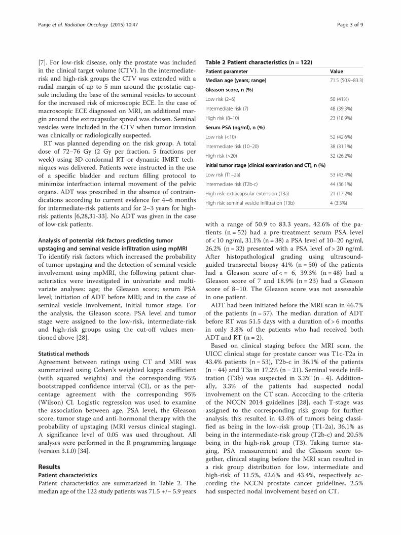
Table 2 Patient characteristics (n = 122)
Patient parameter Value
Median age (years; range) 71.5 (50.9–83.3)
Gleason score, n (%)
Low risk (2–6) 50 (41%)
Intermediate risk (7) 48 (39.3%)
High risk (8–10) 23 (18.9%)
Serum PSA (ng/ml), n (%)
Low risk (<10) 52 (42.6%)
Intermediate risk (10–20) 38 (31.1%)
High risk (>20) 32 (26.2%)
Initial tumor stage (clinical examination and CT), n (%)
Low risk (T1–2a) 53 (43.4%)
Intermediate risk (T2b-c) 44 (36.1%)
High risk: extracapsular extension (T3a) 21 (17.2%)
High risk: seminal vesicle infiltration (T3b) 4 (3.3%)
Panje et al. Radiation Oncology (2015) 10:47 Page 3 of 9
[7]. For low-risk disease, only the prostate was includedin the clinical target volume (CTV). In the intermediate-risk and high-risk groups the CTV was extended with aradial margin of up to 5 mm around the prostatic cap-sule including the base of the seminal vesicles to accountfor the increased risk of microscopic ECE. In the case ofmacroscopic ECE diagnosed on MRI, an additional mar-gin around the extracapsular spread was chosen. Seminalvesicles were included in the CTV when tumor invasionwas clinically or radiologically suspected.RT was planned depending on the risk group. A total
dose of 72–76 Gy (2 Gy per fraction, 5 fractions perweek) using 3D-conformal RT or dynamic IMRT tech-niques was delivered. Patients were instructed in the useof a specific bladder and rectum filling protocol tominimize interfraction internal movement of the pelvicorgans. ADT was prescribed in the absence of contrain-dications according to current evidence for 4–6 monthsfor intermediate-risk patients and for 2–3 years for high-risk patients [6,28,31-33]. No ADT was given in the caseof low-risk patients.
Analysis of potential risk factors predicting tumorupstaging and seminal vesicle infiltration using mpMRITo identify risk factors which increased the probabilityof tumor upstaging and the detection of seminal vesicleinvolvement using mpMRI, the following patient char-acteristics were investigated in univariate and multi-variate analyses: age; the Gleason score; serum PSAlevel; initiation of ADT before MRI; and in the case ofseminal vesicle involvement, initial tumor stage. Forthe analysis, the Gleason score, PSA level and tumorstage were assigned to the low-risk, intermediate-riskand high-risk groups using the cut-off values men-tioned above [28].
Statistical methodsAgreement between ratings using CT and MRI wassummarized using Cohen’s weighted kappa coefficient(with squared weights) and the corresponding 95%bootstrapped confidence interval (CI), or as the per-centage agreement with the corresponding 95%(Wilson) CI. Logistic regression was used to examinethe association between age, PSA level, the Gleasonscore, tumor stage and anti-hormonal therapy with theprobability of upstaging (MRI versus clinical staging).A significance level of 0.05 was used throughout. Allanalyses were performed in the R programming language(version 3.1.0) [34].
ResultsPatient characteristicsPatient characteristics are summarized in Table 2. Themedian age of the 122 study patients was 71.5 +/− 5.9 years
with a range of 50.9 to 83.3 years. 42.6% of the pa-tients (n = 52) had a pre-treatment serum PSA levelof < 10 ng/ml, 31.1% (n = 38) a PSA level of 10–20 ng/ml,26.2% (n = 32) presented with a PSA level of > 20 ng/ml.After histopathological grading using ultrasound-guided transrectal biopsy 41% (n = 50) of the patientshad a Gleason score of < = 6, 39.3% (n = 48) had aGleason score of 7 and 18.9% (n = 23) had a Gleasonscore of 8–10. The Gleason score was not assessablein one patient.ADT had been initiated before the MRI scan in 46.7%
of the patients (n = 57). The median duration of ADTbefore RT was 51.5 days with a duration of > 6 monthsin only 3.8% of the patients who had received bothADT and RT (n = 2).Based on clinical staging before the MRI scan, the
UICC clinical stage for prostate cancer was T1c-T2a in43.4% patients (n = 53), T2b-c in 36.1% of the patients(n = 44) and T3a in 17.2% (n = 21). Seminal vesicle infil-tration (T3b) was suspected in 3.3% (n = 4). Addition-ally, 3.3% of the patients had suspected nodalinvolvement on the CT scan. According to the criteriaof the NCCN 2014 guidelines [28], each T-stage wasassigned to the corresponding risk group for furtheranalysis; this resulted in 43.4% of tumors being classi-fied as being in the low-risk group (T1-2a), 36.1% asbeing in the intermediate-risk group (T2b-c) and 20.5%being in the high-risk group (T3). Taking tumor sta-ging, PSA measurement and the Gleason score to-gether, clinical staging before the MRI scan resulted ina risk group distribution for low, intermediate andhigh-risk of 11.5%, 42.6% and 43.4%, respectively ac-cording the NCCN prostate cancer guidelines. 2.5%had suspected nodal involvement based on CT.
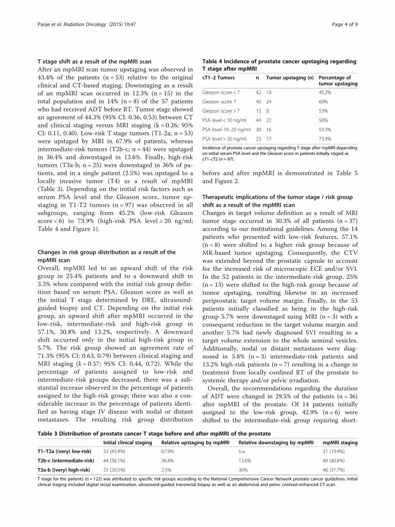
Table 4 Incidence of prostate cancer upstaging regardingT stage after mpMRI
cT1–2 Tumors n Tumor upstaging (n) Percentage oftumor upstaging
Gleason score < 7 42 19 45.2%
Gleason score 7 40 24 60%
Gleason score > 7 15 8 53%
PSA level < 10 ng/ml 44 22 50%
PSA level 10–20 ng/ml 30 16 53.3%
PSA level > 20 ng/ml 23 17 73.9%
Incidence of prostate cancer upstaging regarding T stage after mpMRI dependingon initial serum PSA level and the Gleason score in patients initially staged ascT1–cT2 (n = 97).
Panje et al. Radiation Oncology (2015) 10:47 Page 4 of 9
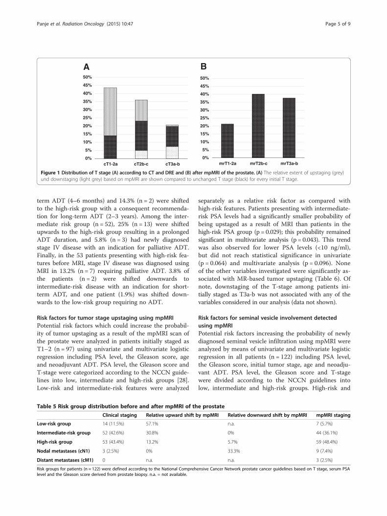
T stage shift as a result of the mpMRI scanAfter an mpMRI scan tumor upstaging was observed in43.4% of the patients (n = 53) relative to the originalclinical and CT-based staging. Downstaging as a resultof an mpMRI scan occurred in 12.3% (n = 15) in thetotal population and in 14% (n = 8) of the 57 patientswho had received ADT before RT. Tumor stage showedan agreement of 44.3% (95% CI: 0.36, 0.53) between CTand clinical staging versus MRI staging (k = 0.26; 95%CI: 0.11, 0.40). Low-risk T stage tumors (T1-2a; n = 53)were upstaged by MRI in 67.9% of patients, whereasintermediate-risk tumors (T2b-c; n = 44) were upstagedin 36.4% and downstaged in 13.6%. Finally, high-risktumors (T3a-b; n = 25) were downstaged in 36% of pa-tients, and in a single patient (2.5%) was upstaged to alocally invasive tumor (T4) as a result of mpMRI(Table 3). Depending on the initial risk factors such asserum PSA level and the Gleason score, tumor up-staging in T1-T2 tumors (n = 97) was observed in allsubgroups, ranging from 45.2% (low-risk Gleasonscore < 6) to 73.9% (high-risk PSA level > 20 ng/ml;Table 4 and Figure 1).
Changes in risk group distribution as a result of thempMRI scanOverall, mpMRI led to an upward shift of the riskgroup in 25.4% patients and to a downward shift in3.3% when compared with the initial risk group defin-ition based on serum PSA, Gleason score as well asthe initial T stage determined by DRE, ultrasound-guided biopsy and CT. Depending on the initial riskgroup, an upward shift after mpMRI occurred in thelow-risk, intermediate-risk and high-risk group in57.1%, 30.8% and 13.2%, respectively. A downwardshift occurred only in the initial high-risk group in5.7%. The risk group showed an agreement rate of71.3% (95% CI: 0.63, 0.79) between clinical staging andMRI staging (k = 0.57; 95% CI: 0.44, 0.72). While thepercentage of patients assigned to low-risk andintermediate-risk groups decreased, there was a sub-stantial increase observed in the percentage of patientsassigned to the high-risk group; there was also a con-siderable increase in the percentage of patients identi-fied as having stage IV disease with nodal or distantmetastases. The resulting risk group distribution
Table 3 Distribution of prostate cancer T stage before and af
Initial clinical staging Relative upstagin
T1–T2a ((very) low-risk) 53 (43.4%) 67.9%
T2b-c (intermediate-risk) 44 (36.1%) 36.4%
T3a-b ((very) high-risk) 25 (20.5%) 2.5%
T stage for the patients (n = 122) was attributed to specific risk groups according toclinical staging included digital rectal examination, ultrasound-guided transrectal bi
before and after mpMRI is demonstrated in Table 5and Figure 2.
Therapeutic implications of the tumor stage / risk groupshift as a result of the mpMRI scanChanges in target volume definition as a result of MRItumor stage occurred in 30.3% of all patients (n = 37)according to our institutional guidelines. Among the 14patients who presented with low-risk features, 57.1%(n = 8) were shifted to a higher risk group because ofMR-based tumor upstaging. Consequently, the CTVwas extended beyond the prostatic capsule to accountfor the increased risk of microscopic ECE and/or SVI.In the 52 patients in the intermediate-risk group, 25%(n = 13) were shifted to the high-risk group because oftumor upstaging, resulting likewise in an increasedperiprostatic target volume margin. Finally, in the 53patients initially classified as being in the high-riskgroup 5.7% were downstaged using MRI (n = 3) with aconsequent reduction in the target volume margin andanother 5.7% had newly diagnosed SVI resulting in atarget volume extension to the whole seminal vesicles.Additionally, nodal or distant metastases were diag-nosed in 5.8% (n = 3) intermediate-risk patients and13.2% high-risk patients (n = 7) resulting in a change intreatment from locally confined RT of the prostate tosystemic therapy and/or pelvic irradiation.Overall, the recommendations regarding the duration
of ADT were changed in 29.5% of the patients (n = 36)after mpMRI of the prostate. Of 14 patients initiallyassigned to the low-risk group, 42.9% (n = 6) wereshifted to the intermediate-risk group requiring short-
ter mpMRI of the prostate
g by mpMRI Relative downstaging by mpMRI mpMRI staging
n.a. 31 (19.4%)
13.6% 49 (40.6%)
36% 46 (37.7%)
the National Comprehensive Cancer Network prostate cancer guidelines. Initialopsy as well as an abdominal and pelvic contrast-enhanced CT scan.
A B
0%
5%
10%
15%
20%
25%
30%
35%
40%
45%
50%
cT1-2a cT2b-c cT3a-b0%
5%
10%
15%
20%
25%
30%
35%
40%
45%
50%
mrT1-2a mrT2b-c mrT3a-b
Figure 1 Distribution of T stage (A) according to CT and DRE and (B) after mpMRI of the prostate. (A) The relative extent of upstaging (grey)und downstaging (light grey) based on mpMRI are shown compared to unchanged T stage (black) for every initial T stage.
Panje et al. Radiation Oncology (2015) 10:47 Page 5 of 9
term ADT (4–6 months) and 14.3% (n = 2) were shiftedto the high-risk group with a consequent recommenda-tion for long-term ADT (2–3 years). Among the inter-mediate risk group (n = 52), 25% (n = 13) were shiftedupwards to the high-risk group resulting in a prolongedADT duration, and 5.8% (n = 3) had newly diagnosedstage IV disease with an indication for palliative ADT.Finally, in the 53 patients presenting with high-risk fea-tures before MRI, stage IV disease was diagnosed usingMRI in 13.2% (n = 7) requiring palliative ADT. 3.8% ofthe patients (n = 2) were shifted downwards tointermediate-risk disease with an indication for short-term ADT, and one patient (1.9%) was shifted down-wards to the low-risk group requiring no ADT.
Risk factors for tumor stage upstaging using mpMRIPotential risk factors which could increase the probabil-ity of tumor upstaging as a result of the mpMRI scan ofthe prostate were analyzed in patients initially staged asT1–2 (n = 97) using univariate and multivariate logisticregression including PSA level, the Gleason score, ageand neoadjuvant ADT. PSA level, the Gleason score andT-stage were categorized according to the NCCN guide-lines into low, intermediate and high-risk groups [28].Low-risk and intermediate-risk features were analyzed
Table 5 Risk group distribution before and after mpMRI of th
Clinical staging Relative upward shift b
Low-risk group 14 (11.5%) 57.1%
Intermediate-risk group 52 (42.6%) 30.8%
High-risk group 53 (43.4%) 13.2%
Nodal metastases (cN1) 3 (2.5%) 0%
Distant metastases (cM1) 0 n.a.
Risk groups for patients (n = 122) were defined according to the National Comprehlevel and the Gleason score derived from prostate biopsy. n.a. = not available.
separately as a relative risk factor as compared withhigh-risk features. Patients presenting with intermediate-risk PSA levels had a significantly smaller probability ofbeing upstaged as a result of MRI than patients in thehigh-risk PSA group (p = 0.029); this probability remainedsignificant in multivariate analysis (p = 0.043). This trendwas also observed for lower PSA levels (<10 ng/ml),but did not reach statistical significance in univariate(p = 0.064) and multivariate analysis (p = 0.096). Noneof the other variables investigated were significantly as-sociated with MR-based tumor upstaging (Table 6). Ofnote, downstaging of the T-stage among patients ini-tially staged as T3a-b was not associated with any of thevariables considered in our analysis (data not shown).
Risk factors for seminal vesicle involvement detectedusing mpMRIPotential risk factors increasing the probability of newlydiagnosed seminal vesicle infiltration using mpMRI wereanalyzed by means of univariate and multivariate logisticregression in all patients (n = 122) including PSA level,the Gleason score, initial tumor stage, age and neoadju-vant ADT. PSA level, the Gleason score and T-stagewere divided according to the NCCN guidelines intolow, intermediate and high-risk groups. High-risk and
e prostate
y mpMRI Relative downward shift by mpMRI mpMRI staging
n.a. 7 (5.7%)
0% 44 (36.1%)
5.7% 59 (48.4%)
33.3% 9 (7.4%)
n.a. 3 (2.5%)
ensive Cancer Network prostate cancer guidelines based on T stage, serum PSA

A B
0%
5%
10%
15%
20%
25%
30%
35%
40%
45%
50%
low-risk intermediate-risk high-risk stage IV0%
5%
10%
15%
20%
25%
30%
35%
40%
45%
50%
low-risk intermediate-risk high-risk stage IV
Figure 2 Distribution of overall risk groups based on PSA, Gleason score and (A) T stage determined by CT and DRE compared to (B) Tstage assessed by mpMRI. (A) The relative extent of risk group upshift (grey) und downshift (light grey) due to mpMRI-based T stage changesare shown for every initial risk group compared to patients without risk group shift (black).
Panje et al. Radiation Oncology (2015) 10:47 Page 6 of 9
intermediate-risk features were analyzed separately as arelative risk factor as compared with low-risk features.PSA levels above the low-risk range were associatedwith an increased risk which reached statistical signifi-cance in univariate analysis for the intermediate-riskPSA range (p = 0.041), but not for the high-risk PSArange (p = 0.11). The same trend was found in multi-variate analysis which, however, was not quite statisti-cally significant (p = 0.058). In contrast, none of theother factors that were evaluated appeared to play asignificant role in this investigation.
DiscussionMRI of the prostate has already been established as avaluable tool for radiation oncologists in improving tar-get volume delineation [35-37] and has also been investi-gated for treatment planning [38]. It has been shownthat morphologic MRI enables a more reproducible def-inition of the prostate gland [39-41], particularly when itcomes to the definition of the prostate apex [42,43]; itleads to a smaller CTV volume and the sparing of morenormal tissue relative to CT-based target volume defin-ition [44]. Consequently, in prostate cancer it has beensuggested that MRI-based treatment may reducetreatment-associated toxicity [30].
Table 6 Multivariate logistic regression analysis of potential r
OR
Low-risk PSA (<10 ng/ml) 0.37
Intermediate-risk PSA (10–20 ng/ml) 0.29
Low-risk Gleason score (<7) 0.99
Intermediate-risk Gleason score (7) 1.88
Age 1.02
ADT pre-mpMRI 1.34
Multivariate logistic regression analysis was used to demonstrate the relative probastaged as cT1-cT2 (n = 97) depending on specific patient parameters. Patients in thedue to mpMRI than patients in the high-risk PSA group (p = 0.043). OR = Odds ratio
Apart from precise organ delineation, the accurateradiological tumor staging of prostate cancer is playingan increasingly important role in the era of IMRT [8].Using highly-conformal RT techniques and small plan-ning target volume margins, the detection and inclusionof extracapsular disease and seminal vesicle infiltrationinto the treatment volume is of paramount importancein avoiding a geographic miss and consequent tumorunder-dosage [45].MRI of the prostate can offer additional benefit in RT
planning because it is regarded as the most reliable im-aging modality regarding the determination of the localextension of prostate cancer, and it exhibits the highestcongruence with the “gold standard” of post-operativepathological staging [10,11].Surgical series have demonstrated that both the digital
rectal examination, as well as other radiological tech-niques such as CT scans of the abdomen and transrectalultrasound, only exhibit low correlations with definitivepathological staging after radical prostatectomy; indeed,they only accurately predict the extent of the local tumor61–70% of the time [19,20]. In contrast, morphologicalT1- and T2-weighted MR imaging alone has alreadyshown an accuracy of 84% for the detection of extrapro-static disease in clinically staged T2 prostate cancer [46].
isk factors for T upstaging as a result of mpMRI
95% confidence interval p-value
(0.11, 1.15) 0.096
(0.08, 0.93) 0.043
(0.27, 3.65) 0.99
(0.51, 7.11) 0.34
(0.95, 1.10) 0.54
(0.55, 3.25) 0.52
bility of tumor upstaging after mpMRI of the prostate in patients initiallyintermediate-risk PSA group had a significantly lower probability of upstaging
. ADT = Androgen deprivation therapy.

Panje et al. Radiation Oncology (2015) 10:47 Page 7 of 9
In recent years, mpMRI sequences have been introducedinto prostate cancer imaging in order to further enhancetumor staging [14]. As a result of the early rapid en-hancement and early washout of prostate cancer tumors,DCE-MRI provides a means of determining tumorextension more accurately than morphological imaging[10], and has demonstrated an improved overall stagingaccuracy of 95% in a single-institution series [17]. Inter-estingly, this improvement in prostate cancer tumor sta-ging due to DCE-MRI has been found to be particularlyimportant for less experienced readers [47]. Likewise,recent literature reviews have confirmed that additionalDWI can detect prostate cancer tumor foci more accur-ately than conventional anatomic imaging alone [12,48].For these reasons, mpMRI of the prostate has beenestablished in our institution as a standard imaging pro-cedure in all patients before primary RT for prostatecancer. Although this approach is not uncommonamong RT units, it cannot at the moment be consideredas a modality with interdisciplinary consensus. Currentguidelines recommend the use of mpMRI for local sta-ging of prostate cancer ahead of curative therapy mainlyfor high-risk patients [28,31], whereas its value is ques-tioned in patients presenting with low-risk features [49].The purpose of the present study was therefore toanalyze retrospectively the extent of the shift in tumorstage as a result of the use of mpMRI, and the conse-quent impact on risk group distribution and treatmentdecisions in primary RT for prostate cancer.In our patient series, a considerable impact on pros-
tate tumor staging as a consequence of the use ofmpMRI was observed, with tumor upstaging in 43.4%and downstaging in 12.3% of patients. These findingsare consistent with the findings from a previously pub-lished series of 199 patients with localized prostate can-cer; in this series morphological MRI scans of theprostate, without DCE imaging or DWI, led to tumorupstaging in 52% and to tumor downstaging in 3% ofpatients before RT [25].Although our study showed a trend towards a signifi-
cantly lower probability of MRI-based tumor upstagingin patients with an intermediate or low-risk PSA level,there was no subgroup identifiable where it appearedreasonable to omit mpMRI before RT. Even in the low-risk subgroups (Gleason score < 7; PSA level < 10 ng/ml),a substantial upstaging of 45–50% could be observed.However, taking all of the established risk factors, suchas the Gleason score, PSA level and tumor stage to-gether, mpMRI of the prostate only led to a change inrisk group in 25.4% of the patients in our series. Thismight be explained by the fact that patients with a lo-cally advanced tumor stage detected by MRI may oftenpresent with other adverse features which had alreadybeen assessed before MRI.
Tumor stage shift and change in risk group taken to-gether resulted in a change in the RT dose and targetvolume, as well as in the recommendations for add-itional ADT in 30% of patients. These findings are inconcordance with recent data from a study by Changet al. [24] who reported a significant upstaging of 29%before primary RT for prostate cancer in a comparablepatient series, using morphological MRIs without multi-parametric dynamic sequences. Interestingly, Clure et al.reported a similar extent of treatment adaptation of 27%as a result of MRI staging in a series of 104 patientswho underwent robotic-assisted laparoscopic prosta-tectomy [50]. Similarly, Hricak et al. reported a changein surgical plan based on MRI scans in 39% of patientswho underwent radical prostatectomy for localizedprostate cancer [51].Whereas it seems to be legitimate to increase treatment
intensity for patients with mpMRI based upstaging,caution should be exerted for treatment de-intensificationin cases where mpMRI led to downstaging: in our series, aconsiderable number of downstaging was found inpatients who received ADT before RT. It is, however,mainly not determinable whether MR-based downstagingoccured due to previous diagnostic inaccuracy of the pre-MRI staging or if it was an ADT effect. If the latter can’tbe definitely ruled out, downstaging should not subse-quently lead to de-intensification of radiotherapy. In orderto avoid this uncertainty of ADT interference, mpMRIshould be conducted as part of the initial staging proced-ure before start of any neoadjuvant treatment.Our study had several limitations. First, all data were
reviewed retrospectively and staging information wasobtained from patient histories. In addition, the report-ing radiologists had not been blinded to previously ob-tained clinical staging. However, these circumstancesmay represent the daily practice in a major cancer centermore accurately than would be the case in a dedicatedprospective trial. Second, clinical and MRI staging werenot correlated to oncological outcome as has beenreported in other studies [25,52]; this was because thefollow-up time was limited as we evaluated a very recentpatient series. The main focus of our study was on theimpact of MRI staging on treatment decisions, not onits predictive value. mpMRI sequences were notreported separately from morphological MRI, so theiradditional value for treatment decisions could not beindependently assessed. Additionally, in contrast tostudies regarding the surgical treatment of prostatecancer, MRI staging could not be validated using defini-tive pathological staging. Finally, the impact of MRI ontarget volume delineation and treatment volume rela-tive to CT-based planning was not analyzed in thecurrent study, because it has been investigated exten-sively by others [29,30,53].

Panje et al. Radiation Oncology (2015) 10:47 Page 8 of 9
ConclusionsThe use of mpMRI in patients admitted for primary RTfor prostate cancer resulted in a substantial shift intumor stage in > 50%, with consequent treatment adapta-tions in nearly one third of the patients. In our singlecenter series, we could not identify any subgroup of pa-tients where mpMRI did not lead to considerable up-staging. In conclusion, our findings suggest that patientsfrom all risk groups may benefit from mpMRI of the pros-tate regarding the choice of the optimal risk-adapted RT.
Competing interestsThe authors declare that they have no competing interests.
Authors’ contributionsCP participated in the study design, managed the database and drafted themanuscript. TP carried out the review of medical records, participated indatabase management and helped to draft the manuscript. PMP dealt withthe ethics approval and participated in the study design and revised themanuscript. SK gave advice regarding radiological aspects of the study andhelped to draft the manuscript. SH performed the statistical analysis. DÄgave advice concerning scientific aspects of the study and helped to draftthe manuscript. LP conceived of the study, supervised the development ofthe study design and data interpretation, and revised the manuscript. Allauthors read and approved the final version of the manuscript.
Author details1Department of Radiation Oncology, Kantonsspital St. Gallen, St. Gallen,Switzerland. 2Department of Radiology and Nuclear Medicine, Kantonsspital St.Gallen, St. Gallen, Switzerland. 3Clinical Trials Unit, Kantonsspital St. Gallen, St. Gallen,Switzerland. 4Department of Radiation Oncology, Bern University Hospital, Bern,Switzerland.
Received: 26 November 2014 Accepted: 21 January 2015
References1. Dearnaley DP, Jovic G, Syndikus I, Khoo V, Cowan RA, Graham JD, et al.
Escalated-dose versus control-dose conformal radiotherapy for prostatecancer: long-term results from the MRC RT01 randomised controlled trial.Lancet Oncol. 2014;15:464–73.
2. Al-Mamgani A, Heemsbergen WD, Levendag PC, Lebesque JV. Subgroupanalysis of patients with localized prostate cancer treated within theDutch-randomized dose escalation trial. Radiother Oncol. 2010;96:13–8.
3. Welz S, Nyazi M, Belka C, Ganswindt U. Surgery vs. radiotherapy in localizedprostate cancer. Which is best? Radiat Oncol. 2008;3:23.
4. Nguyen QN, Levy LB, Lee AK, Choi SS, Frank SJ, Pugh TJ, et al. Long-termoutcomes for men with high-risk prostate cancer treated definitively withexternal beam radiotherapy with or without androgen deprivation. Cancer.2013;119:3265–71.
5. Zietman AL, Bae K, Slater JD, Shipley WU, Efstathiou JA, Coen JJ, et al.Randomized trial comparing conventional-dose with high-dose conformalradiation therapy in early-stage adenocarcinoma of the prostate: long-termresults from proton radiation oncology group/american college of radiology95–09. J Clin Oncol. 2010;28:1106–11.
6. Bolla M, Van Tienhoven G, Warde P, Dubois JB, Mirimanoff RO, Storme G,et al. External irradiation with or without long-term androgen suppressionfor prostate cancer with high metastatic risk: 10-year results of an EORTCrandomised study. Lancet Oncol. 2010;11:1066–73.
7. Boehmer D, Maingon P, Poortmans P, Baron MH, Miralbell R, RemouchampsV, et al. Guidelines for primary radiotherapy of patients with prostate cancer.Radiother Oncol. 2006;79:259–69.
8. Teh BS, Bastasch MD, Wheeler TM, Mai WY, Frolov A, Uhl BM, et al. IMRT forprostate cancer: defining target volume based on correlated pathologicvolume of disease. Int J Radiat Oncol Biol Phys. 2003;56:184–91.
9. McLaughlin PW, Troyer S, Berri S, Narayana V, Meirowitz A, Roberson PL,et al. Functional anatomy of the prostate: implications for treatmentplanning. Int J Radiat Oncol Biol Phys. 2005;63:479–91.
10. Pinto F, Totaro A, Palermo G, Calarco A, Sacco E, D’Addessi A, et al. Imagingin prostate cancer staging: present role and future perspectives. Urol Int.2012;88:125–36.
11. Thompson J, Lawrentschuk N, Frydenberg M, Thompson L, Stricker P. Therole of magnetic resonance imaging in the diagnosis and management ofprostate cancer. BJU Int. 2013;112 Suppl 2:6–20.
12. Tan CH, Wei W, Johnson V, Kundra V. Diffusion-weighted MRI in thedetection of prostate cancer: meta-analysis. AJR Am J Roentgenol.2012;199:822–9.
13. Wu LM, Xu JR, Ye YQ, Lu Q, Hu JN. The clinical value of diffusion-weightedimaging in combination with T2-weighted imaging in diagnosing prostatecarcinoma: a systematic review and meta-analysis. AJR Am J Roentgenol.2012;199:103–10.
14. Verma S, Turkbey B, Muradyan N, Rajesh A, Cornud F, Haider MA, et al.Overview of dynamic contrast-enhanced MRI in prostate cancer diagnosisand management. AJR Am J Roentgenol. 2012;198:1277–88.
15. Sala E, Akin O, Moskowitz CS, Eisenberg HF, Kuroiwa K, Ishill NM, et al.Endorectal MR imaging in the evaluation of seminal vesicle invasion:diagnostic accuracy and multivariate feature analysis. Radiology.2006;238:929–37.
16. Yu KK, Hricak H, Alagappan R, Chernoff DM, Bacchetti P, Zaloudek CJ.Detection of extracapsular extension of prostate carcinoma with endorectaland phased-array coil MR imaging: multivariate feature analysis. Radiology.1997;202:697–702.
17. Bloch BN, Furman-Haran E, Helbich TH, Lenkinski RE, Degani H, Kratzik C,et al. Prostate cancer: accurate determination of extracapsular extensionwith high-spatial-resolution dynamic contrast-enhanced and T2-weightedMR imaging–initial results. Radiology. 2007;245:176–85.
18. Ren J, Huan Y, Wang H, Ge Y, Chang Y, Yin H, et al. Seminal vesicleinvasion in prostate cancer: prediction with combined T2-weighted anddiffusion-weighted MR imaging. Eur Radiol. 2009;19:2481–6.
19. Hricak H, Dooms GC, Jeffrey RB, Avallone A, Jacobs D, Benton WK, et al.Prostatic carcinoma: staging by clinical assessment, CT, and MR imaging.Radiology. 1987;162:331–6.
20. Mullerad M, Hricak H, Kuroiwa K, Pucar D, Chen HN, Kattan MW, et al.Comparison of endorectal magnetic resonance imaging, guided prostatebiopsy and digital rectal examination in the preoperative anatomicallocalization of prostate cancer. J Urol. 2005;174:2158–63.
21. Augustin H, Fritz GA, Ehammer T, Auprich M, Pummer K. Accuracy of 3-Teslamagnetic resonance imaging for the staging of prostate cancer in comparisonto the Partin tables. Acta Radiol. 2009;50:562–9.
22. Wang L, Hricak H, Kattan MW, Chen HN, Scardino PT, Kuroiwa K.Prediction of organ-confined prostate cancer: incremental value of MRimaging and MR spectroscopic imaging to staging nomograms.Radiology. 2006;238:597–603.
23. Wang L, Hricak H, Kattan MW, Chen HN, Kuroiwa K, Eisenberg HF, et al.Prediction of seminal vesicle invasion in prostate cancer: incremental valueof adding endorectal MR imaging to the Kattan nomogram. Radiology.2007;242:182–8.
24. Chang JH, Lim Joon D, Nguyen BT, Hiew CY, Esler S, Angus D, et al. MRIscans significantly change target coverage decisions in radical radiotherapyfor prostate cancer. J Med Imaging Radiat Oncol. 2014;58:237–43.
25. Jackson AS, Parker CC, Norman AR, Padhani AR, Huddart RA, Horwich A,et al. Tumour staging using magnetic resonance imaging in clinically localisedprostate cancer: relationship to biochemical outcome after neo-adjuvantandrogen deprivation and radical radiotherapy. Clin Oncol (R Coll Radiol).2005;17:167–71.
26. Epstein JI, Allsbrook Jr WC, Amin MB, Egevad LL. The 2005 InternationalSociety of Urological Pathology (ISUP) Consensus Conference on GleasonGrading of Prostatic Carcinoma. Am J Surg Pathol. 2005;29:1228–42.
27. Sobin LH, Gospodarowicz MK, Wittekind C. TNM classification of malignanttumours. 7th ed. John Wiley & Sons; 2011.
28. Mohler JL, Kantoff PW, Armstrong AJ, Bahnson RR, Cohen M, D’Amico AV,et al. Prostate cancer, version 2.2014. J Natl Compr Canc Netw.2014;12:686–718.
29. Rasch C, Barillot I, Remeijer P, Touw A, van Herk M, Lebesque JV. Definitionof the prostate in CT and MRI: a multi-observer study. Int J Radiat Oncol BiolPhys. 1999;43:57–66.
30. Sander L, Langkilde NC, Holmberg M, Carl J. MRI target delineation mayreduce long-term toxicity after prostate radiotherapy. Acta Oncol.2014;53:809–14.

Panje et al. Radiation Oncology (2015) 10:47 Page 9 of 9
31. Heidenreich A, Bastian PJ, Bellmunt J, Bolla M, Joniau S, van der Kwast T,et al. EAU guidelines on prostate cancer. part 1: screening, diagnosis, andlocal treatment with curative intent-update 2013. Eur Urol. 2014;65:124–37.
32. Horwitz EM, Bae K, Hanks GE, Porter A, Grignon DJ, Brereton HD, et al.Ten-year follow-up of radiation therapy oncology group protocol 92–02: aphase III trial of the duration of elective androgen deprivation in locallyadvanced prostate cancer. J Clin Oncol. 2008;26:2497–504.
33. D’Amico AV, Manola J, Loffredo M, Renshaw AA, DellaCroce A, Kantoff PW.6-month androgen suppression plus radiation therapy vs radiation therapyalone for patients with clinically localized prostate cancer: a randomizedcontrolled trial. JAMA. 2004;292:821–7.
34. R Core Team. R: A Language and Environment for Statistical Computing.http://www.R-project.org. Vienna, Austria; 2014.
35. Khoo VS, Dearnaley DP, Finnigan DJ, Padhani A, Tanner SF, Leach MO.Magnetic resonance imaging (MRI): considerations and applications inradiotherapy treatment planning. Radiother Oncol. 1997;42:1–15.
36. Khoo VS. MRI–“magic radiotherapy imaging” for treatment planning? Br JRadiol. 2000;73:229–33.
37. Rischke HC, Nestle U, Fechter T, Doll C, Volegova-Neher N, Henne K, et al. 3Tesla multiparametric MRI for GTV-definition of Dominant IntraprostaticLesions in patients with Prostate Cancer–an interobserver variability study.Radiat Oncol. 2013;8:183.
38. Korsholm ME, Waring LW, Edmund JM. A criterion for the reliable use ofMRI-only radiotherapy. Radiat Oncol. 2014;9:16.
39. Debois M, Oyen R, Maes F, Verswijvel G, Gatti G, Bosmans H, et al. Thecontribution of magnetic resonance imaging to the three-dimensionaltreatment planning of localized prostate cancer. Int J Radiat Oncol Biol Phys.1999;45:857–65.
40. Parker CC, Damyanovich A, Haycocks T, Haider M, Bayley A, Catton CN.Magnetic resonance imaging in the radiation treatment planning oflocalized prostate cancer using intra-prostatic fiducial markers for computedtomography co-registration. Radiother Oncol. 2003;66:217–24.
41. Khoo EL, Schick K, Plank AW, Poulsen M, Wong WW, Middleton M, et al.Prostate contouring variation: can it be fixed? Int J Radiat Oncol Biol Phys.2012;82:1923–9.
42. Kagawa K, Lee WR, Schultheiss TE, Hunt MA, Shaer AH, Hanks GE. Initialclinical assessment of CT-MRI image fusion software in localization of theprostate for 3D conformal radiation therapy. Int J Radiat Oncol Biol Phys.1997;38:319–25.
43. Sannazzari GL, Ragona R, Ruo Redda MG, Giglioli FR, Isolato G, Guarneri A.CT-MRI image fusion for delineation of volumes in three-dimensionalconformal radiation therapy in the treatment of localized prostate cancer. BrJ Radiol. 2002;75:603–7.
44. Hentschel B, Oehler W, Strauss D, Ulrich A, Malich A. Definition of the CTVprostate in CT and MRI by using CT-MRI image fusion in IMRT planning forprostate cancer. Strahlenther Onkol. 2011;187:183–90.
45. Heemsbergen WD, Al-Mamgani A, Witte MG, van Herk M, Lebesque JV.Radiotherapy with rectangular fields is associated with fewer clinical failuresthan conformal fields in the high-risk prostate cancer subgroup: results froma randomized trial. Radiother Oncol. 2013;107:134–9.
46. D’Amico AV, Schnall M, Whittington R, Malkowicz SB, Schultz D,Tomaszewski JE, et al. Endorectal coil magnetic resonance imagingidentifies locally advanced prostate cancer in select patients with clinicallylocalized disease. Urology. 1998;51:449–54.
47. Futterer JJ, Engelbrecht MR, Huisman HJ, Jager GJ, Hulsbergen-van De Kaa CA,Witjes JA, et al. Staging prostate cancer with dynamic contrast-enhancedendorectal MR imaging prior to radical prostatectomy: experienced versus lessexperienced readers. Radiology. 2005;237:541–9.
48. Jin G, Su DK, Luo NB, Liu LD, Zhu X, Huang XY. Meta-analysis ofdiffusion-weighted magnetic resonance imaging in detecting prostatecancer. J Comput Assist Tomogr. 2013;37:195–202.
49. Lavery HJ, Brajtbord JS, Levinson AW, Nabizada-Pace F, Pollard ME, SamadiDB. Unnecessary imaging for the staging of low-risk prostate cancer iscommon. Urology. 2011;77:274–8.
50. McClure TD, Margolis DJ, Reiter RE, Sayre JW, Thomas MA, Nagarajan R, et al.Use of MR imaging to determine preservation of the neurovascular bundlesat robotic-assisted laparoscopic prostatectomy. Radiology. 2012;262:874–83.
51. Hricak H, Wang L, Wei DC, Coakley FV, Akin O, Reuter VE, et al. The role ofpreoperative endorectal magnetic resonance imaging in the decisionregarding whether to preserve or resect neurovascular bundles duringradical retropubic prostatectomy. Cancer. 2004;100:2655–63.
52. Riaz N, Afaq A, Akin O, Pei X, Kollmeier MA, Cox B, et al. Pretreatmentendorectal coil magnetic resonance imaging findings predict biochemicaltumor control in prostate cancer patients treated with combinationbrachytherapy and external-beam radiotherapy. Int J Radiat Oncol Biol Phys.2012;84:707–11.
53. Villeirs GM, L Verstraete K, De Neve WJ, De Meerleer GO. Magneticresonance imaging anatomy of the prostate and periprostatic area: a guidefor radiotherapists. Radiother Oncol. 2005;76:99–106.
Submit your next manuscript to BioMed Centraland take full advantage of:
• Convenient online submission
• Thorough peer review
• No space constraints or color figure charges
• Immediate publication on acceptance
• Inclusion in PubMed, CAS, Scopus and Google Scholar
• Research which is freely available for redistribution
Submit your manuscript at www.biomedcentral.com/submit