Embed Size (px)
Citation preview

Kidney International, Vol. 57 (2000), pp. 183–190
Effects of ICAM-1 antisense oligonucleotide on thetubulointerstitium in mice with unilateral ureteral obstruction
QING-LI CHENG, XIANG-MEI CHEN, FENG LI, HONG-LI LIN, YI-ZHOU YE, and BO FU
Division of Nephrology, General Hospital of Chinese PLA, Beijing, China
Effects of ICAM-1 antisense oligonucleotide on the renal tubu- molecule fibrinogen, and the matrix factor hyaluronanlointerstitium in mice with unilateral ureteral obstruction. [1–5]. ICAM-1 plays an important role in inflammatory
Background. To extend our previous study of the therapy processes and in the T-cell–mediated immune responseof the renal lesions of unilateral ureteral obstruction (UUO)system. In normal conditions, ICAM-1 is constitutivelyin mice by an inhibitor of intercellular adhesion molecule-1expressed by capillary endothelial cells of kidney, and(ICAM-1), we investigated the blocking effects of ICAM-1
antisense oligonucleotides (ASONs) on the ICAM-1 expres- ICAM-1 expresses at basal or low levels on the surfacesion in mouse kidney. of glomerular mesangial cells and renal tubular epithelial
Methods. First, ICAM-1 ASON was transducted into mouse cells (TECs) [6]. However, in response to stimuli fromrenal tubular epithelial cells to investigate the effects ofinflammatory mediators such as cytokines, the expres-ICAM-1 ASON in vitro. Second, fluorescein isothiocyanatesion of ICAM-1 can be markedly up-regulated in mesan-(FITC)-labeled ICAM-1 ASON was injected intravenously to
determine the distribution of the ASON in vivo. Third, the gial and epithelial cells of the kidney [7, 8]. Althoughexpression of ICAM-1 in kidney and the changes of renal ICAM-1 by itself probably does not elicit an inflamma-morphology were observed to investigate the therapeutic ef-
tory response and more likely functions as an inflamma-fects of ICAM-1 ASON on the UUO mice in vivo.tion-enhancing molecule, increased ICAM-1 expressionResults. The expressions of ICAM-1 in the epithelial cells
induced by interleukin-1b were inhibited by ICAM-1 ASON of the cells has been implicated in a variety of disordersat the dosages of 100 and 200 nmol/L. Twenty-four hours after characterized by local inflammation in kidney [9].an introvenous injection with FITC-labeled ICAM-1 ASON, Many studies have demonstrated that antibodies againstthe highest level of fluorescein was detected within the proximal
ICAM-1 have beneficial effects in animal models of in-tubules in mouse kidney. Results of immunohistology andflammatory kidney diseases, and preliminary clinical dataNorthern blot showed that the ICAM-1 expression was mark-
edly reduced in the obstructed kidney after treatment with have also shown that the ICAM-1 monoclonal antibodyICAM-1 ASON. The ASON also alleviated the infiltration of has good therapeutic effects in renal allografts [10–12].inflammatory cells and accumulation of the extracellular matrix In our previous study, we observed that the expressionin the tubulointerstitium of UUO mice without apparent side
of ICAM-1 and the infiltration of inflammatory cells ineffects.the renal tubulointerstitium decreased after the adminis-Conclusion. Our data demonstrate that ICAM-1 ASON is
taken up primarily by the proximal tubular cells of mouse tration of an ICAM-1 monoclonal antibody in a mousekidney. ICAM-1 ASON can selectively inhibit the ICAM-1 model of unilateral ureteral obstruction (UUO) [13].expression of the renal tubular cells both in vitro and in vivo. Because antisense oligonucleotides (ASONs) provide
a novel strategy to inhibit RNA transcription and therebythe synthesis of the protein, ASONs as therapeutic agents
The intercellular adhesion molecule-1 (ICAM-1) is a have been developed during the past years. Recently,transmembrane glycoprotein that belongs to the Ig super- ASONs that inhibit the expression of ICAM-1 on eithergene family. Its ligands are the membrane-bound integ- human or murine endothelial cells were identified [14, 15].rin receptor lymphocyte function-associated antigen-1 The murine-specific ASON was shown to selectively in-(LFA-1) and Mac-1 on leukocytes, CD43, the soluble hibit the expression of ICAM-1 and infiltration of leuko-
cytes in lesions of a colitis model or a transplantationmodel in a sequence-specific manner [16, 17].Key words: intercellular adhesion molecule, inflammation, obstructed
kidney, proximal tubule cells, extracellular matrix. To extend our previous study of the therapy of therenal lesions of UUO mice by inhibitors of ICAM-1, weReceived for publication December 1, 1998investigated the blocking effects of ICAM-1 ASONs onand in revised form June 14, 1999
Accepted for publication September 1, 1999 the ICAM-1 expression of the cultured renal TECs invitro, the distribution of ICAM-1 ASONs in the kidney 2000 by the International Society of Nephrology
183

Cheng et al: Effects of ICAM-1 ASON on tubulointerstitial lesions184
after intravenous injection, and the therapeutic effects controls for immunostaining consisted of equivalent con-centration of an irrelevant murine monoclonal antibodyof ICAM-1 ASONs on the renal injury of UUO mice.and the cells without stimulation of IL-1b.
METHODS Distribution of oligonucleotide in mouse in vivoOligonucleotides synthesis Studies were performed in female NIH mice (mean
weight 31 6 3.6 g) purchased from the animal centerPhosphorothioate oligonucleotides were synthesizedby Cybersyn Inc. (Lenni, PA, USA). The sequences of of the Chinese PLA General Hospital. FITC-labeled
oligonucleotides were given to the mice by intravenousthe oligonucleotides used in this study have been pre-viously described [16]: mouse ICAM-1 ASON, 59-TGCA injection via the tail vein at the dose of 10 mg/kg. The
mice were sacrificed at 24 hours after injection of theTCCCCCAGGCCACCAT-39, and control oligonucleo-tide, 59-TCGCATCGACCCGCCCACTA-39. The con- oligonucleotides. Kidney, liver, lung, and spleen were
frozen in liquid nitrogen, and cryosections were pre-trol oligonucleotide has the same base composition asthe ICAM-1 anitsense oligonucleotide but in a different pared. The distribution of FITC-labeled oligonucleotide
in the tissues was determined under a fluorescent micro-sequence. All of the oligonucleotides were purified byreverse-phase high-performance liquid chromatography scope (Nikon, Tokyo, Japan).(HPLC). For some studies, parts of oligonucleotides
Induction of unilateral ureteral obstructionwere labeled with fluorescein isothiocyanate (FITC) atthe 59-amine. Female NIH mice were anesthetized and underwent
left proximal ureteral ligation via a midline abdominalPrimary culture of mouse renal tubular epithelial cells incision. After the UUO surgery, the mice were divided
into three groups. Group A was treated with ICAM-1Primary mouse renal TECs were generated followingthe method described by Wuthrich et al [18]. Briefly, ASON, which was injected intravenously at the dosage
of 1 mg/kg/day every other day for one week. Group Bsingle cortical tubular cell suspensions of freshly dissectedkidney cortices from normal NIH mice were prepared by was treated with the control oligonucleotide in the same
way, and group C was injected with normal saline solu-collagenase dispersion, sequential sieving through steelmeshes, and washing in RPMI 1640 medium (GIBCO tion only as a blank control. All of the mice were sacri-
ficed at the seventh day. The weights of the whole body,BRL, Grand Island, NY, USA). TECs were grown tosubconfluence on gelatin-coated flasks for six days in liver, spleen, and kidney were recorded, and blood sam-
ples were obtained for measurement of blood urea nitro-RPMI 1640 medium supplemented with 10% fetal calfserum (FCS; GIBCO BRL). The primary TECs were gen (BUN), serum creatinine (Cr), and glutamic pyruvic
transaminase (GPT). The expression of ICAM-1 proteindigested by 0.2% pepsin/ethylenediaminetetraacetic acidbuffer (EDTA; GIBCO BRL) and planted to another and the number of Mac-1–positive cells were measured
by immunohistochemical staining. The pathologicalflask for growing. The identified secondary TECs wereused in the following experiments. changes of the obstructed kidney were analyzed by a
computer imaging system. The total RNA of renal tissueOligonucleotides treatment of tubular epithelial cells was extracted for analysis of the ICAM-1 mRNA by
Northern blotting.The cell transfection reagent DOTAP was purchasedfrom Boehringer Mannheim (Mannheim, Germany). For
Immunohistological study of ICAM-1 and Mac-1TEC transfection, oligonucleotides were sterilized byexpressions in UUO micecentrifugation through 0.2 mm centrex cellulose acetate
filters and diluted to a concentration of 0.1 mg/mL in Renal samples from the obstructed kidney of the UUOmice were frozen in OCT embedding medium (Miles20 mmol/L HEPES buffer (pH 7.4). In this study, the
DOTAP/oligonucleotides mixture was prepared at the Inc., IN, USA). Four micrometer cryosections were incu-bated with 5 mg/mL of rat antimouse ICAM-1 mono-ratio of 1 mg oligonucleotide per 1 mL DOTAP. Trans-
fected TECs were incubated for an additional four hours clonal antibody YN1/1.7.4, with 15 mg/mL of Mac-1 anti-body (Pharmingen, San Diego, CA, USA). The slidesat 378C and were then incubated with or without interleu-
kin-1b (IL-1b; 10 m/mL) for 16 hours. were incubated with horseradish peroxidase-labeled rab-bit antirat IgG, which was developed and examined under
Intercellular adhesion molecule-1 assay of tubular light microscope.epithelial cells
Quantitative analysis of renal pathology in theThe ICAM-1 expression on the TEC surface was deter-obstructed kidneymined by immunocytochemical staining. ICAM-1 mono-
clonal antibody YN1/1,7,4 (1 mg/mL) was a gift from Dr. The 2 mm paraffin sections from obstructed kidneysof the UUO mice were stained with periodic acid-SchiffMatsuda (Juntendo University, Tokyo, Japan). Negative

Cheng et al: Effects of ICAM-1 ASON on tubulointerstitial lesions 185
and Masson. The area of the dilated tubules and the rela- RESULTStive area of interstitium in the sections were evaluated Effects of ICAM-1 antisense oligonucleotide on theby a computer image system with software of VIGP-2M ICAM-1 expression of tubular epithelial cellsImage/Graphics/Vision Video View series (CAS-DH induced by IL-1bCo., HZ Image & Vision Inc., Beijing, China). Infiltrating
It has been demonstrated that ICAM-1 expressioninflammatory cells were counted using an eyepiece grati-of cultured epithelial cells surface was increased aftercule (Olympus, Tokyo, Japan) at a magnification of 3400stimulation of IL-1b [8, 20]. In our study, the resultsand were quantitated as cells per unit area (0.0625 mm2)showed that ICAM-1 expression of TECs, which wasof the tubulointerstitium. All of these data were ex-transfected with 100 or 200 nmol/L ICAM-1 ASONpressed as the mean values of 5 to 10 fields of the tubu-through liposome-DOTAP, was significantly decreasedlointerstitium in each section.compared with that of TECs transfected with control
Isolation of RNA of tubular epithelial cells and oligonucleotide (Fig. 1). The control oligonucleotide hadkidney tissue no effect on the ICAM-1 expression of TECs compared
with the blank control of transfection buffer. The resultsTotal cellular RNA was isolated from confluent mono-also indicated that there was no inhibition of ICAM-1layers of transfected TECs using TRIZOL reagentexpression on TECs if the transfection of ICAM-1(GIBCO BRL). The kidney cortex tissue was minced andASON was performed without DOTAP. We comparedhomogenized in 5.5 mol/L guanidinium isothiocyanatethe effects of ICAM-1 ASON transfection with or with-citrate buffer, and the total tissue RNA was isolated byout DOTAP. We found that the uptake of ICAM-1a cesium chloride gradient centrifugation. The total RNAASON by cultured TECs was significantly increasedwas quantitated with ultraviolet spectrophotometry.when transfected with DOTAP (75.2 6 8.3% vs. 16.6 6
RT-PCR and preparation of the mouse ICAM-1 4.8%). Northern blot analysis showed that total cellularcDNA probe mRNA levels of ICAM-1 from TECs transfected with
The ICAM-1 primers were designed from the se- ICAM-1 ASON were significantly decreased comparedquence of the mouse gene [19]: 59-CAACTGGAAGCT with that of the control sample. Representative autora-GTTTGAGCTG-39 (sense) and 59-GTGTTCACAGT diograms for ICAM-1 mRNA expression are shown inCTTGC TCCAT-39 (antisense) to yield a 1567 bp prod- Figure 2.uct. Polymerase chain reaction (PCR) was performedwith a DNA Thermal Cycler (MJ Inc., Watertown, MA, Distribution of ICAM-1 antisense oligonucleotides inUSA) using the following program: 948C for one minute, mouse in vivo608C for one minute, and 728C for two minutes. Amplifi- Previous animal studies have demonstrated that fol-cations were carried out for 30 cycles. PCR products were lowing systemic administration, phosphorothioate oligo-analyzed by electrophoresis through a 1.0% agarose gel nucleotides are primarily excreted by the kidney andwith ethidium bromide (0.5 mg/mL). A 1567 bp template that renal tissue levels of the oligonucleotides exceedfor the ICAM-1 probe was prepared by PCR of renal that of other organs. Within the kidney, the oligonucleo-cortex cDNA. The PCR product was isolated from low tides are taken up primarily by proximal tubular cells,melting point agarose and was purified using a Gene- with much lower uptake by cells in other segments ofclean kit (Cambridge Biosystems, Cambridge, UK).
the nephron [21]. To obtain information about the distri-bution of ICAM-1 ASON in mice, we injected the FITC-Northern blotting analysislabeled ICAM-1 ASON via tail vein at 10 mg/kg intoTotal RNA was separated, transferred to a nylon mem-either normal mice or mice with UUO operation for 16brane (Bio-Rad, Hercules, CA, USA), and cross-linkedhours. The mice were sacrificed at the 24th hour afterby ultraviolet irradiation (UVP, Cambridge, UK). Afterinjection, and the location of the oligonucleotide wasbeing prehybridized, the blots were hybridized withobserved in the cryostat sections of the kidney, liver,mouse ICAM-1 cDNA probe labeled with [a-32P]dCTPlung, and spleen. In the normal mice, the ICAM-1 ASONusing random primed DNA labeling kit (Boehringerlocalized predominantly to the renal proximal tubularMannheim, Germany) at 658C for 24 hours. Autoradiog-cells. The fluorescein was not detected within the glomer-raphy was performed at 2708C using intensifying screens.uli and the distal tubular cells (Fig. 3). Much lower levels
Statistical analysis of FITC-labeled ICAM-1 ASON were found in the liver,lung, and spleen of the mice. In UUO mice, the distribu-The values of the groups were expressed as means 6tion and level of the oligonucleotide in the obstructedsd. Data were analyzed for statistical significance bykidney were not different from those of the contralateralStudent’s t-test or Mann–Whitney U-test, and a P value
,0.05 was considered significant. unobstructed kidney.

Cheng et al: Effects of ICAM-1 ASON on tubulointerstitial lesions186
Fig. 1. Immunocytochemical studies for in-tercellular adhesion molecule-1 (ICAM-1) ex-pression in cultured tubular epithelial cells.(A) Immunostaining for anti–ICAM-1 anti-body in the cultured tubular epithelial cellstreated by the cell transfection solution with-out any oligonucleotides (3400). (B) Onlyweak immunostaining was observed in the cul-tured tubular epithelial cells after treatmentwith 100 nmol/L ICAM-1 antisense oligonu-cleotide (3400). (C) Intense immunostainingwas found in the cultured tubular epithelialcells that were treated by 100 nmol/L controloligonucleotide (3400). (D) There was notmarked immunostaining for anti–ICAM-1 an-tibody in the normal cultured tubular epithe-lial cells without stimulation of IL-1b (3400).All of the cells of group A, B, and C werestimulated by IL-1b at the concentration of10 U/mL.
Fig. 4. Immunohistochemical studiesfor the expression of ICAM-1 andMac-1 in the obstructed kidney ofUUO mice. (A) Immunostaining foranti–ICAM-1 antibody in the micetreated with the normal saline solution(3132). (B) Only weak and local im-munostaining was observed in the micetreated with ICAM-1 antisense oligo-nucleotide at dosage of 1 mg/kg (3132).(C) Intense and diffuse immunostain-ing was found in the mice treated bycontrol oligonucleotide at 1 mg/kg(3132). (D) The negative control (re-placing anti–ICAM-1 antibody with ir-relevant antibody) of the obstructedkidney (3132). (E) Immunostainingfor anti–Mac-1 antibody in the micetreated with normal saline solution(3132). (F ) Only weak and local im-munostaining of anti–Mac-1 antibodywas observed in the mice treated withICAM-1 antisense oligonucleotide atdosage of 1 mg/kg (3132).

Cheng et al: Effects of ICAM-1 ASON on tubulointerstitial lesions 187
Fig. 2. Northern blot analysis of total cellular RNA from the tubularepithelial cells. The top of the figure shows the blots of IL-1b stimulatingcells treated by 100 nmol/L ICAM-1 antisense oligonucleotide (lane 2)and the cells treated by 100 nmol/L control oligonucleotide (lane 3),
Fig. 5. Northern blot analysis of total RNA from the obstructed kidneyand the cells treated by transfection solution without any oligonucleo-of UUO mice. The top of the figure shows the blots of the obstructedtides (lane 4). Lane 1 shows the blot of RNA isolated from culturedkidney that were treated by 1 mg/kg ICAM-1 antisense oligonucleotidecells that had not been treated with IL-1b. At the bottom of the figure(lane 2), the obstructed kidney treated by control oligonucleotide (lanewere the intact 28s and 18s ribosomal bands.3), and the obstructed kidney treated with normal saline solution only(lane 4). Lane 1 shows the blot of RNA isolated from the kidney ofsham operated mice. At the bottom of the figure are the intact 28S and18S ribosomal bands.
Therapeutic effects of ICAM-1 ASON on renalpathological changes in UUO mice
On the seventh day after UUO operation, markedinflammatory cell infiltration, dilated tubules, and extra-cellular matrix accumulation in the tubulointerstitiumwere found in the obstructed kidneys of the mice. Thenumber of the inflammatory cells and the degree of thematrix accumulation were alleviated after treatment withFig. 3. Location of FITC-labeled ICAM-1 antisense oligonucleotide in
the kidney of mice in vivo. The fluorescein was localized predominantly ICAM-1 ASON in the obstructed kidney. However, thereto the renal proximal tubular cells at 24 hours after injection of FITC- was no therapeutic effect of ICAM-1 ASON on the dila-labeled ICAM-1 antisense oligonucleotide at 10 mg/kg.
tion of the tubules (Fig. 6). The results of quantitativepathological assay are shown in Table 1.
Effects of ICAM-1 antisense oligonucleotide on Side effects of ICAM-1 ASON in UUO miceICAM-1 expression in UUO mice
Phosphorothioate oligonucleotides may result in sple-Our previous study demonstrated that there is an in- nomegaly, lymphoid hyperplasia, and mild liver or kid-
tense staining of ICAM-1 and a number of Mac-1– ney dysfunction in animals [22]. In the present study, thepositive staining cells in the peritubular interstitial space weights of kidney, liver, spleen, the serum chemistryof the obstructed kidney seven days after UUO opera-
parameters of liver and kidney functions as well as thetion in mice [13]. In the current study, the immunohisto-
urine protein of the mice treated with ICAM-1 ASONlogical data showed that the ICAM-1 expression and thewere compared with those changes of the control oligo-number of Mac-1–positive cells were markedly de-nucleotide-treated and normal saline-treated mice. Thecreased in the renal tubulointerstitium of the obstructedresults showed that there were no apparent clinical signskidney after treatment with ICAM-1 ASON (Fig. 4).of toxicity in mice treated with 1 mg/kg ICAM-1 ASONThe control oligonucleotide had no effect on ICAM-1or control oligonucleotide. There were no statisticallyexpression and the number of Mac-1–positive cells insignificant changes in the organ weights and chemistrythe obstructed kidney of UUO mice. By Northern blotparameters (GPT, BUN, Cr, and urine protein) betweenanalysis, the expression of ICAM-1 mRNA in the renalthe mice treated with ICAM-1 ASON, control oligonu-cortex was also decreased after treatment with ICAM-1
ASON (Fig. 5). cleotide, and the normal saline solution.

Cheng et al: Effects of ICAM-1 ASON on tubulointerstitial lesions188
Fig. 6. Renal morphologicalchanges of the UUO mice. Kid-neys from UUO mice were re-moved and formalin fixed. Twomicrometer-thick paraffin sec-tions were stained with periodicacid-Schiff. (A) UUO mice weretreated with 1 mg/kg ICAM-1antisense oligonucleotide forseven days (3132). (B) UUOmice were treated by controloligonucleotides at 1 mg/kg forseven days (3132). (C) UUOmice were treated with normalsaline solution only (3132).
Table 1. Quantitative analysis of renal pathological changes
Inf. cell number A. of tubules RA. of interstitiumGroup N cells/mm2 lm2 %
Antisense ODN 7 110 614a 1979.36241.4 7.4760.88a
Control ODN 8 270 613 1980.76316.5 25.38 63.98Saline solution 6 276 624 1816.06180.4 28.08 64.02
Abbreviations are: ODN, oligonucleotide; Inf., inflammatory cells; A., area; RA., relative area.a P , 0.01, compared with group of control ODN and group of saline
DISCUSSION scopic observations showed that injected oligonucleotidedid not merely accumulate in the brush border or lyso-The use of ASONs to block gene expression was intro-somal compartment in proximal tubular cells, implyingduced by Zamecnik and Stephenson [23]. Because ofthat they were not totally degenerated after being phago-the specificity of Watson-Crick base pair hybridization,cytosed by the proximal tubule [28]. These results suggestASONs have been used extensively in attempts to inhibitthat the renal proximal tubule is a good target of anti-expressions of distinct genes both in vitro and in vivo.sense therapy.Although the precise mechanism of action has not been
Data from previous animal studies on the pharmaco-clarified, ASONs offer considerable promise with the de-kinetics and tissue distribution of oligonucleotide havevelopment of novel molecular therapeutic agents againstsuggested that the kidney may be an ideal target fordiseases including AIDS, cancer, and inflammatory dis-application of ASONs [21, 28, 29]. Following systemicorders [24]. Several investigators have reported the ap-administration to both mice and rabbits, radiolabeledplications of ASON to kidney cell lines in vitro. ASONoligonucleotides were excreted primarily in the urine.targeting the mRNA of rat kidney sodium phosphateTissue levels of oligonucleotide attained in murine kid-cotransporter NaPi-2 protein reduced the rate of phos-neys were markedly higher than those in all other organsphate uptake into the tubular cell brush border mem-[29, 30]. Similar results are gained in our current studybrane vesicles [25]. Glomerular mesangial cells exposedusing the FITC-labeled oligonucleotide.to ASONs of proliferating cell nuclear antigen (PCNA)
Recently, researchers have examined the effects ofand Ki-67 had decreased levels of PCNA and Ki-67ASONs in experimental animals in vivo. SpontaneouslymRNA and a decreased rate of cell proliferation [26].hypertensive rats injected with ASON targeting againstOur current study provides evidence that ICAM-1 ex-either angiotensinogen or angiotensin II type I receptorpression of the cultured renal TECs is induced by IL-1b,mRNA had a significant reduction in systemic blood pres-which is interrupted by ICAM-1 ASON.sure [31]. Haller et al reported the suppression of ICAM-1Rappaport et al studied the fate of 32P-labeled oligonu-expression and the attenuation of ischemia/reperfusioncleotides after intravenous injection [27]. The labeledinjury by administration of rat ICAM-1 ASON [32].oligonucleotides were predominantly localized in kidneyOther authors demonstrated that the anti-transformingand liver and were detected in the proximal tubular cells
within 30 minutes after injection. The electron micro- growth factor-b (TGF-b) ASONs inhibited TGF-b ex-
Cheng et al: Effects of ICAM-1 ASON on tubulointerstitial lesions 189
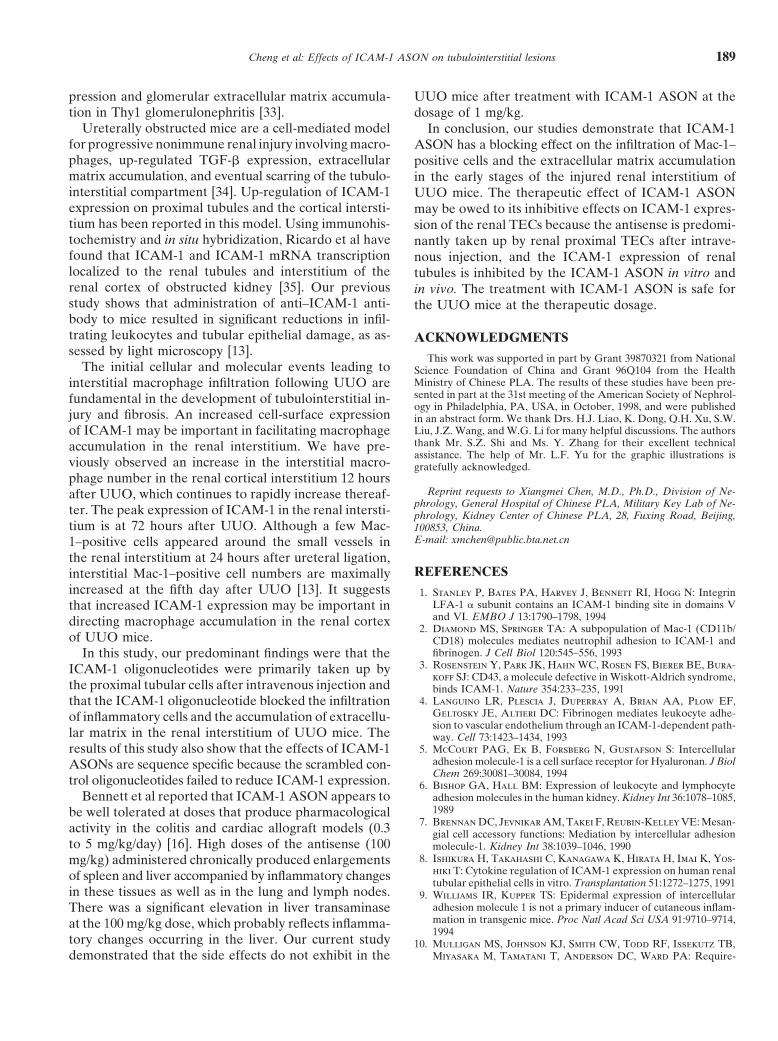
pression and glomerular extracellular matrix accumula- UUO mice after treatment with ICAM-1 ASON at thetion in Thy1 glomerulonephritis [33]. dosage of 1 mg/kg.
Ureterally obstructed mice are a cell-mediated model In conclusion, our studies demonstrate that ICAM-1for progressive nonimmune renal injury involving macro- ASON has a blocking effect on the infiltration of Mac-1–phages, up-regulated TGF-b expression, extracellular positive cells and the extracellular matrix accumulationmatrix accumulation, and eventual scarring of the tubulo- in the early stages of the injured renal interstitium ofinterstitial compartment [34]. Up-regulation of ICAM-1 UUO mice. The therapeutic effect of ICAM-1 ASONexpression on proximal tubules and the cortical intersti- may be owed to its inhibitive effects on ICAM-1 expres-tium has been reported in this model. Using immunohis- sion of the renal TECs because the antisense is predomi-tochemistry and in situ hybridization, Ricardo et al have nantly taken up by renal proximal TECs after intrave-found that ICAM-1 and ICAM-1 mRNA transcription nous injection, and the ICAM-1 expression of renallocalized to the renal tubules and interstitium of the tubules is inhibited by the ICAM-1 ASON in vitro andrenal cortex of obstructed kidney [35]. Our previous in vivo. The treatment with ICAM-1 ASON is safe forstudy shows that administration of anti–ICAM-1 anti- the UUO mice at the therapeutic dosage.body to mice resulted in significant reductions in infil-trating leukocytes and tubular epithelial damage, as as- ACKNOWLEDGMENTSsessed by light microscopy [13].
This work was supported in part by Grant 39870321 from NationalThe initial cellular and molecular events leading to Science Foundation of China and Grant 96Q104 from the Health
Ministry of Chinese PLA. The results of these studies have been pre-interstitial macrophage infiltration following UUO aresented in part at the 31st meeting of the American Society of Nephrol-fundamental in the development of tubulointerstitial in-ogy in Philadelphia, PA, USA, in October, 1998, and were published
jury and fibrosis. An increased cell-surface expression in an abstract form. We thank Drs. H.J. Liao, K. Dong, Q.H. Xu, S.W.Liu, J.Z. Wang, and W.G. Li for many helpful discussions. The authorsof ICAM-1 may be important in facilitating macrophagethank Mr. S.Z. Shi and Ms. Y. Zhang for their excellent technicalaccumulation in the renal interstitium. We have pre-assistance. The help of Mr. L.F. Yu for the graphic illustrations is
viously observed an increase in the interstitial macro- gratefully acknowledged.phage number in the renal cortical interstitium 12 hours
Reprint requests to Xiangmei Chen, M.D., Ph.D., Division of Ne-after UUO, which continues to rapidly increase thereaf-phrology, General Hospital of Chinese PLA, Military Key Lab of Ne-ter. The peak expression of ICAM-1 in the renal intersti- phrology, Kidney Center of Chinese PLA, 28, Fuxing Road, Beijing,
tium is at 72 hours after UUO. Although a few Mac- 100853, China.E-mail: [email protected]–positive cells appeared around the small vessels in
the renal interstitium at 24 hours after ureteral ligation,REFERENCESinterstitial Mac-1–positive cell numbers are maximally
increased at the fifth day after UUO [13]. It suggests 1. Stanley P, Bates PA, Harvey J, Bennett RI, Hogg N: IntegrinLFA-1 a subunit contains an ICAM-1 binding site in domains Vthat increased ICAM-1 expression may be important inand VI. EMBO J 13:1790–1798, 1994directing macrophage accumulation in the renal cortex
2. Diamond MS, Springer TA: A subpopulation of Mac-1 (CD11b/of UUO mice. CD18) molecules mediates neutrophil adhesion to ICAM-1 and
fibrinogen. J Cell Biol 120:545–556, 1993In this study, our predominant findings were that the3. Rosenstein Y, Park JK, Hahn WC, Rosen FS, Bierer BE, Bura-ICAM-1 oligonucleotides were primarily taken up by
koff SJ: CD43, a molecule defective in Wiskott-Aldrich syndrome,the proximal tubular cells after intravenous injection and binds ICAM-1. Nature 354:233–235, 1991
4. Languino LR, Plescia J, Duperray A, Brian AA, Plow EF,that the ICAM-1 oligonucleotide blocked the infiltrationGeltosky JE, Altieri DC: Fibrinogen mediates leukocyte adhe-of inflammatory cells and the accumulation of extracellu-sion to vascular endothelium through an ICAM-1-dependent path-
lar matrix in the renal interstitium of UUO mice. The way. Cell 73:1423–1434, 19935. McCourt PAG, Ek B, Forsberg N, Gustafson S: Intercellularresults of this study also show that the effects of ICAM-1
adhesion molecule-1 is a cell surface receptor for Hyaluronan. J BiolASONs are sequence specific because the scrambled con-Chem 269:30081–30084, 1994trol oligonucleotides failed to reduce ICAM-1 expression. 6. Bishop GA, Hall BM: Expression of leukocyte and lymphocyte
Bennett et al reported that ICAM-1 ASON appears to adhesion molecules in the human kidney. Kidney Int 36:1078–1085,1989be well tolerated at doses that produce pharmacological
7. Brennan DC, Jevnikar AM, Takei F, Reubin-Kelley VE: Mesan-activity in the colitis and cardiac allograft models (0.3 gial cell accessory functions: Mediation by intercellular adhesionto 5 mg/kg/day) [16]. High doses of the antisense (100 molecule-1. Kidney Int 38:1039–1046, 1990
8. Ishikura H, Takahashi C, Kanagawa K, Hirata H, Imai K, Yos-mg/kg) administered chronically produced enlargementshiki T: Cytokine regulation of ICAM-1 expression on human renalof spleen and liver accompanied by inflammatory changestubular epithelial cells in vitro. Transplantation 51:1272–1275, 1991
in these tissues as well as in the lung and lymph nodes. 9. Williams IR, Kupper TS: Epidermal expression of intercellularadhesion molecule 1 is not a primary inducer of cutaneous inflam-There was a significant elevation in liver transaminasemation in transgenic mice. Proc Natl Acad Sci USA 91:9710–9714,at the 100 mg/kg dose, which probably reflects inflamma-1994
tory changes occurring in the liver. Our current study 10. Mulligan MS, Johnson KJ, Smith CW, Todd RF, Issekutz TB,Miyasaka M, Tamatani T, Anderson DC, Ward PA: Require-demonstrated that the side effects do not exhibit in the

Cheng et al: Effects of ICAM-1 ASON on tubulointerstitial lesions190
ments for leukocyte adhesion molecules in nephrotoxic nephritis. oligonucleotide analogs in mice. Anticancer Drug Des 12:1–14,1997J Clin Invest 91:577–587, 1993
11. Nishikawa K, Guo YJ, Miysaka M, Tamatani T, Collins AB, 23. Zamecnik PC, Stephenson ML: Inhibition of Rous sarcoma virusreplication and cell transformation by a specific oligodeoxynucleo-Sy MS, McClaskey RT, Andres G: Antibodies to intercellular
adhesion molecule-1/lymphocyte function-associated antigen 1 tide. Proc Natl Acad Sci USA 75:280–284, 197824. Agrawal S: Antisense oligonucleotides: Towards clinical trials.prevent crescent formation in rat autoimmune glomerulonephritis.
J Exp Med 177:667–677, 1993 Trends Biotechnol 14:376–387, 199625. Oberbauer R, Schreiner GF, Biber J, Murer H, Meyer TW: In12. Cosimi AB, Conti D, Delmonico FL, Preffer FI, Wee SL, Roth-
lein R, Faanes R, Colvin RB: In vivo effects of monoclonal vivo suppression of the renal Na1/Pi cotransporter by antisenseoligonucleotides. Proc Natl Acad Sci USA 93:4903–4906, 1996antibody to ICAM-1 (CD54) in nonhuman primates with renal
allografts. J Immunol 144:4604–4612, 1990 26. Macshima Y, Kashihara N, Sugiyama H, Makino H, Ota Z:Antisense oligonucleotides to proliferating cell nuclear antigen and13. Cheng QL, Li WG, Shi SZ, Chen XM: Renal interstitial lesions
of unilateral ureteral obstruction in mice: The therapeutic efficacy Ki-67 inhibit human mesangial cell proliferation. J Am Soc Nephrol7:2219–2229, 1996of anti-mouse ICAM-1. Chin J Nephrol 12:3–6, 1996
14. Bennett CF, Condon T, Grimm S, Chan H, Chiang MY: Inhibition 27. Rappaport J, Hanss B, Kopp J, Copeland T, Bruggeman L, Coff-man T, Klotman P: Transport of phosphorothioate oligonucleo-of endothelial cell-leukocyte adhesion molecule expression with
antisense oligonucleotides. J Immunol 152:3530–3540, 1994 tides in kidney: Implications for molecular therapy. Kidney Int47:1462–1469, 199515. Stepkowski SM, Tu Y, Condon TP, Bennett CF: Blocking of heart
allograft rejection by intercellular adhesion molecule-1 antisense 28. Oberbauer R, Schreiner GF, Meyer T: Renal uptake of an 18-mer phosphorothioate oligonucleotide. Kidney Int 48:1226–1232,oligonucleotides alone or in combination with other immunosup-
pressive modalities. J Immunol 153:5336–5346, 1994 199529. Goodchild J, Kim B, Zamecnik PC: The clearance and degradation16. Bennett CF, Kornbrust D, Henry S, Stecker K, Howard R,
Cooper S, Dutson S, Hall W, Jacoby HI: An ICAM-1 antisense of oligodeoxynucleotides following intravenous injection into rab-bits. Antisense Res Dev 1:153–160, 1991oligonucleotide prevents and reverses dextran sulfate sodium-
induced colitis in mice. J Pharmacol Exp Ther 280:988–1000, 1997 30. Agrawal S, Temsamani J, Tang JY: Pharmacokinetics, biodistri-bution, and stability of oligodeoxynucleotide phosphorothioates17. Dragun D, Tullins SG, Park JK, Maasch C, Lukitsch I, Lippoldt
A, Gross V, Luft FC, Haller H: ICAM-1 antisense oligodeoxy- in mice. Proc Natl Acad Sci USA 88:7595–7599, 199131. Tomita N, Morishita R, Higaki J, Aoki M, Nakamura Y, Mikaminucleotides prevent reperfusion injury and enhance immediate
graft function in renal transplantation. Kidney Int 54:590–602, 1998 H, Fukamizu A, Murakami K, Kaneda Y, Ogihara T: Transientdecrease in high blood pressure by in vivo transfer of antisense18. Wuthrich RP, Glimcher LH, Yui MA, Jevnikar AM, Dumas
SE, Kelley VE: MHC class II, antigen presentation and tumor oligodeoxynucleotide against rat angiotensinogen. Hypertension26:131–136, 1995necrosis factor in renal tubular epithelial cells. Kidney Int 37:783–
792, 1990 32. Haller H, Dragun D, Miethke A, Park J, Weis A, Lippoldt A,Gross V, Luft F: Antisense oligonucleotides for ICAM-1 attenu-19. Horley FJ, Carpenito C, Baker B, Takei F: Molecular cloning
of murine intercellular adhesion molecule (ICAM-1). EMBO J ate reperfusion injury and renal failure in the rat. Kidney Int50:473–480, 19968:2889–2896, 1989
20. Paolieri F, Battifora M, Riccio AM, Pesce G, Canonica GW, 33. Akagi Y, Isaka Y, Arai M, Kaneko T, Takenaka M, MoriyamaT, Kaneda Y, Ando A, Orita Y, Kamada T, Ueda N, Imai E:Bagnasco M: Intercellular adhesion molecule-1 on cultured human
epithelial cell lines: Influence of proinflammatory cytokines. Al- Inhibition of TGF-b1 expression by antisense oligonucleotides sup-pressed extracellular matrix accumulation in experimental glomer-lergy 52:521–531, 1997
21. Carome MA, Kang YH, Bohen EM, Nicholson DE, Carr FE, ulonephritis. Kidney Int 50:148–155, 199634. Diamond JR: Macrophages and progressive renal disease in experi-Kiandoli LC, Brummel SE, Yuan CM: Distribution of the cellular
uptake of phosphorothioate oligodeoxynucleotides in the rat kid- mental hydronephrosis. Am J Kidney Dis 26:133–140, 199535. Ricardo SD, Levinson ME, DeJoseph MR, Diamond JR: Expres-ney in vivo. Nephron 75:82–87, 1997
22. Henry SP, Zuckerman JE, Rojko J, Hall WC, Robert JH, sion of adhesion molecules in rat renal cortex during experimentalhydronephrosis. Kidney Int 50:2002–2010, 1996Kitchen D, Crooke ST: Toxicological properties of several novel