Embed Size (px)
Citation preview
1
Effects of extremely low frequency electromagnetic fields on morphine analgesia and tolerance in rats
Running title: Electromagnetic fields and morphine tolerance
*Ercan Ozdemir1, MD, Associate Prof,
Ayse Demirkazik2, PhD, Associate Prof,
Sinan Gursoy3, MD, Prof,
Ahmet Sevki Taskıran1, MD, Research assistant,
Olca Kilinc2, MD, Research assistant,
Gokhan Arslan1, MD, Assistant Prof,
1Departments of Physiology, 2Biophysics, and 3Anesthesiology and Reanimation Cumhuriyet
University School of Medicine, Sivas, TURKEY
*Address for correspondence:
Ercan Ozdemir Department of Physiology Cumhuriyet University School of Medicine 58140 Sivas, Turkey E-mail: [email protected] Tel: +90 346 219 1010-2915 Fax: +90 346 219 1602
2
Abstract Morphine is one of the most important drugs used in clinical severe and chronic pain.
However, prolonged use of morphine leads to the development of tolerance to its analgesic
effect. Several studies have demonstrated that the electromagnetic fields produce analgesic
activity. The aim of this study was to investigate the effects of extremely low frequency
(ELF) electromagnetic fields (EMF) on morphine analgesia and tolerance in rats. In the study
was used 78 adult male Wistar albino rats (approximately 240±12 g). The application of 50
Hz magnetic field, each day the same times for 30 minutes for 15 days, and a total of four
times every 15 minute intervals. To constitute morphine tolerance, high dose of morphine (50
mg/kg) were administered for 3 days in rats and tolerance was evaluated on day 4. The
analgesic effect measurement was performed by the tail-flick and hot-plate test equipment.
Prior to analgesia tests, the effective dose (5 mg/kg) of morphine was injected into rats. In the
statistical analyzes of the data, analysis of variance (two-way ANOVA) was used and the
multiple comparison determined by Tukey tests. The maximum analgesic effect of the 5 milli
tesla (mT) magnetic field was determined on 7 days. Administration of morphine (5 mg/kg) in
rats exposed to a magnetic field, the analgesic effect was significantly higher compared to the
morphine group (p<0.05). Morphine tolerant animals exposed to a magnetic field, the
analgesic effect was found significantly higher than morphine tolerant group rats (p<0.05).
Analgesia test data demonstrated that application of ELF-EMFs to rats increases the morphine
analgesia and reduces morphine tolerance.
Key words: Electromagnetic field, analgesia, morphine, morphine tolerance, analgesia tests
3
Introduction Morphine, an opioids drug, is a clinical highly effective treatment for patients with severe and
chronic pain. Nevertheless, the chronic use of morphine is limited by the potential for adverse
effects, addiction and the development of morphine tolerance (Morgan and Cristie 2011;
Dumas and Pollack 2008). The mechanisms of morphine antinociceptive tolerance are
complex and not fully understood. Proposed mechanisms for opioid analgesic tolerance
comprise desensitization of opioid receptors, induction of alpha-2 noradrenergic and
serotonergic systems, inhibition of dopamine and cannabinoid receptors, and altered
intracellular signaling pathways, including nitric oxide and protein kinase C (Nestler and
Aghajanian 1997; Ozdemir et al. 2011; Gursoy et al. 2011; Ozdemir et al. 2013; Altun et al.
2015).
Extremely low frequency (ELF) electromagnetic field (EMF) has been shown to have
a variety of biological effects and induce many different types of cellular changes
(Sienkiewicz et al. 2005). EMF has been used for its analgesic efficacy for several decades.
EMF therapy has verified to be a safe, simple and non-invasive method to treat different types
of pain or inflammation (Fernandez et al. 2007). The pulsed magnetic fields stimulate small
electrical currents in the tissues because of the often changing magnetic influx. It has been
proposed that these currents are the mechanism underlying the alleged analgesia associated
with magnetic field therapy (Basset 1993). Several studies suggest that therapeutic
applications of EMF at a very low frequency level (1–100 Hz) induce the immune system by
repressing inflammatory responses in the cell-membrane (Ross and Harrison 2013). It has also
been demonstrated that they cause induction of nerve transmission, vasodilatation, enhanced
tissue repair, and reduction of oedema (Ramey 1998; Trock 2000).
Clinical studies report, EMF passes through the skin into the conductive tissue,
decreasing nociception and the edema shortly after trauma (Traina et al. 1998, Chalidis et al.
4
2011). The low frequency pulsed EMF therapy (0.1 to 64 Hz) improve mobility, decrease pain
and fatigue in patients with fibromyalgia (Sutbeyaz et al. 2009). The experimental studies
indicate that EMF is effective in the treatment of pain and inflammation in patients with
osteoarthritis (Sadoghi et al. 2013; Pilla et al. 2013), without the unwanted effects of opioids.
Some evidence suggests that administration of cytokine antagonists attenuates the
development of analgesic tolerance and prevents the hyperalgesia in response to systemic
morphine (Song and Zhao 2001). Furthermore, it has been reported that the EMF has an anti-
inflammatory effect by reducing proinflammatory cytokines (Pesce et al. 2013). EMF
exposure (60 Hz, 0.3 mT) amplifies TGF-β signaling and increases the generation of specific
T cell subsets (Lee et al. 2016).
The neural pathway involved in the effects of EMF on pain have not been exactly
understood. The nociceptive perception is the result of complicated interaction between the
primary pain ascending pathways connecting spinal cord and midbrain pain centers, and
descending regulatory pathways (Fields 2004). Animal studies suggest that the analgesic
effects of EMF are centrally integrated in the hot-plate test (Kieffer 1999). In this way, the
EMF-induced decrease of pain sensations probably reflect supra spinal analgesia.
Numerous studies have indicated that EMFs reduce morphine-induced analgesia. For
instance, Kavaliers and Ossenkopp (1988) determined that 15-30 min exposures to magnetic
field (0.1-0.8 mT; 0.5 Hz) inhibited analgesia from opioid agonists in land snails.
Accordingly, exposing the snails to 0.1 mT, 60 Hz magnetic fields produced similar results,
with decreased opioid-induced analgesia. In addition, acute exposure to a variety of magnetic
field conditions has been found to inhibit the analgesic and locomotor effects of morphine in
mice (Ossenkopp and Kavaliers 1988) and magnetic fields abolish the enhanced nocturnal
analgesic response to morphine (Kavaliers et al. 1984). The inhibition of nocturnal morphine
analgesia is a function of magnetic field intensity in mice (Ossenkopp and Kavaliers 1988).
5
Furthermore, a study on the land snail Cepaea nemoralis has shown that the electromagnetic
field reduces opioid induced analgesia. Conversely, Thomas et al. (1997a) suggest that 15-min
exposures to an EMF (100 µT) increase opioid induced-analgesia in land snails. Injection of
naloxone, an opioid antagonist, resulted in a decreased, but not completely removed
antinociceptive effects of EMF. These findings indicate the analgesic action of a specific
EMF via an endogenous opioid mechanism. The ability of naloxone to decrease, but not fully
removed the antinociceptive effect of EMF shows the presence of at least partially δ-opioid
receptor mediation (Thomas et al. 1997b).
Therapeutic uses of magnetic field exposure include a component of pain reduction;
however, there are controversial reports in the literature regarding the effects of magnetic
field exposure on the opioid analgesia. In light of these data, the aim of the current study was
to investigate the effects of ELF-EMF on morphine analgesia and tolerance in rats.
Materials and methods
Animals
This study was conducted at Cumhuriyet University Experimental Research Center with the
permission granted by Local Animal Studies Ethical Board (CUHEK/2014-67). A total of 78
adult male Wistar albino rats (weighing 240±12 g) was used in this study. The rats were
housed four per cage in a room maintained at 21±2 °C with an alternating 12 h dark/12 h light
cycles and free access to water and food. Experimental animals were acclimatized to
laboratory conditions before the analgesia tests. All experiments were carried out blindly
between 10:00 and 17:00 h.
Induction of morphine tolerance
The experimental animals were rendered tolerant to morphine using the method by a previous
study on the induction of morphine analgesic tolerance (Zarrindast et al. 2002). To constitute
6
morphine tolerance, it was used a 3-day cumulative dosing regimen. The treatment schedule
consisted of twice daily s.c. doses of morphine given at 30 mg/kg (a.m.) and 45 mg/kg (p.m.)
on day 1; 60 and 90 mg/kg on day 2; and 120 mg/kg twice on day 3. Animals were assessed
for tolerance on the 4th day. The tail-flick (TF) and hot-plate (HP) tests were done for each rat
to average them as a baseline latency; then the challenge dose of morphine (5 mg/kg; s.c.) was
injected; 30 min after morphine injection other the tail-flick and hot-plate tests were done to
average them to find post-drug latency for each rat for evaluating the development of
morphine tolerance.
Antinociceptive tests
To evaluate thermal nociception, it was used a standardized TF test (May TF 0703 Tail-flick
Unit, Commat) and HP test (May AHP 0603 Analgesic Hotplate Commat, Turkey). In the TF
test, the radiant heat source was focused on the distal portion of the tail at 3 cm after
administration of the vehicle and study drugs. Following vehicle or compound administration,
tail-flick latencies (TFL) were obtained. The infrared intensity was adjusted so that basal TFL
occurred at 2.9±0.5 s. Animals with a baseline TFL below 2.4 or above 3.4 s were excluded
from further testing. The cutoff latency was set at 15 s to avoid tissue damage. Any animal not
responding after 15 s was excluded from the study. The analgesic response is usually
attributed to central mechanisms in this test (Kanaan et al. 1996, Ramabadran et al. 1989).
In the HP test, the rats were individually placed on the HP with the temperature
adjusted to 55±0.5°C. The latency to the first sign of paw licking or jump response to avoid
the heat was taken as an index of the pain threshold; the cut-off time was 30 s in order to
avoid damage to the paws. The antinociceptive response on the HP test is considered to result
from a combination of peripheral and central mechanisms (Kanaan et al. 1996).
Electromagnetic field exposure conditions
7
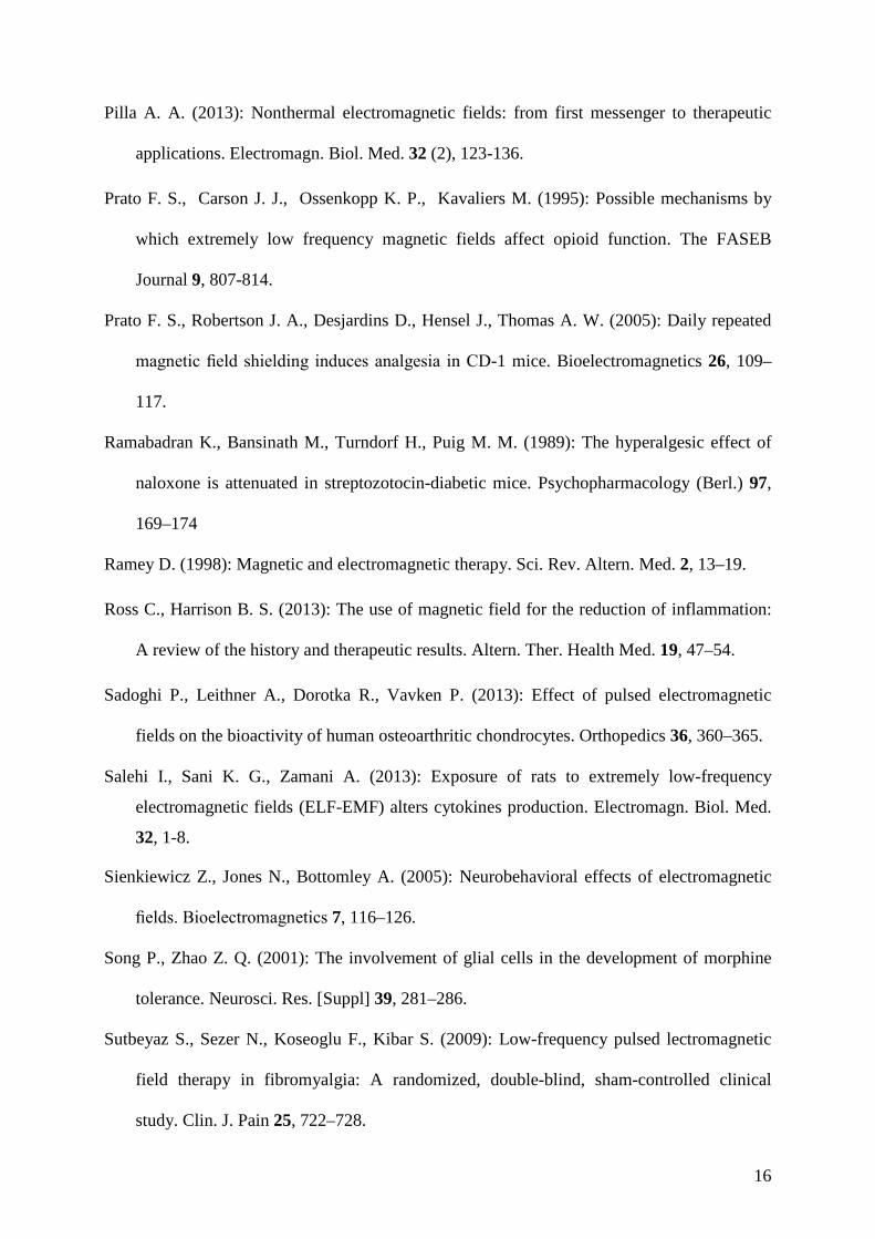
The EMF exposure system consists of 4 sections (Fig. 1): 1. Computer; 2. Power supply; 3.
Digital Teslameter (Phywe, 80010); 4. Solenoid with animal cage. Before the EMF treatment,
all rats were acclimated to their environment for 1 week. Habituation to the treatment
conditions was accomplished by placing the rats in the animal cage at least three times for 30
min. The animals were subjected to repeated exposures of alternating 50 Hz EMF for 15 days.
It was performed in three different magnetic field strengths (1 mT, 5 mT and 10 mT). Thus,
the EMF applied corresponded, according to the extensively used classification, to an
extremely low-frequency range of the electromagnetic phenomena. The EMF was generated
in a specially designed solenoid (500 mm in length and 210 mm in diameter, 1400 turns of
insulated 1.4 mm copper wire). Electrical current (50 Hz, 220 V) was passed through the
device (having a time relay). The alternating EMF was exposed to the rats for four 30-min
episodes halted by 15-min intervals; thus, the entire EMF sessions carried out at the same
time period (9.00-11.00 a.m.) lasted 165 min every day. The EMF intensity in the solenoid
was measured by a digital tesla meter with an axial probe. The solenoid was always kept in a
north-south direction, and its temperature was maintained constant at 22.0 ± 2°C. The
plexiglass rat cage (40×17×13 cm in dimensions) was placed in the solenoid. Three rats were
simultaneously placed in the cage to be exposed to EMF. The control group rats were also
placed in the animal cage, but they were not exposed to EMF. Food and water were provided
ad libitum in this animal cage.
Experimental protocols
Except the control, all group animals were exposed to a magnetic field for 15 days. Analgesic
assays were performed on selected days for 15 days (1, 4, 7, 11, and 15 days). The rats were
subjected to tail-flick and hot-plate tests on the same day. The analgesic test method of rats in
each group was randomly assigned to prevent carry over effects. The antinociceptive effects
of three different EMF strengths (1 mT, 5 mT and 10 mT) were considered at 30-min intervals
8
(0, 30, 60, 90, and 120 min) by tail-flick and hot-plate test in rats (n=6). Initially, the
maximum analgesic effect of EMF was detected in 15 days. Subsequently, the effects of the
magnetic field were determined on morphine analgesia and tolerance by tail-flick and hot-
plate test.
Data analysis
In order to calculate % maximal antinociceptive effects (% MPE), lick/escape latencies (hot-
plate) and tail-withdrawal latencies (tail-flick) were converted to percent antinociceptive
effect using the following equation:
% MPE = [(test latency-baseline) / (cutoff-baseline)] x 100
Statistical analysis
The data (% MPE) were analyzed by two-way analysis of variance (ANOVA) and repeated
measures ANOVA followed by a Tukey post hoc test for multiple comparisons between
groups (SPSS 20.0 for windows). All data are presented as means ±S.E.M. The level of
significance was set at P < 0.05.
Results
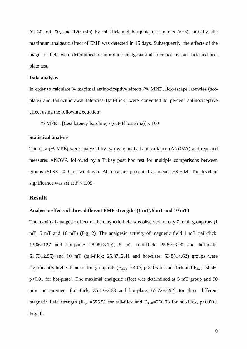
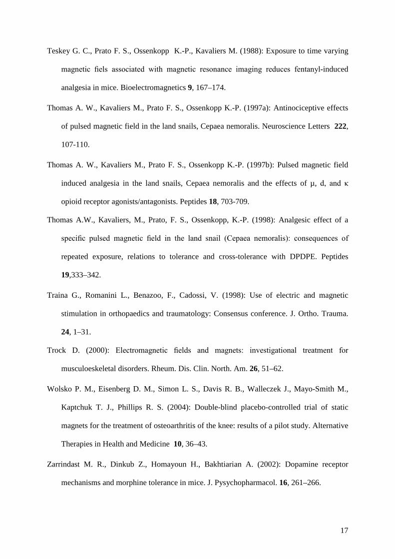
Analgesic effects of three different EMF strengths (1 mT, 5 mT and 10 mT)
The maximal analgesic effect of the magnetic field was observed on day 7 in all group rats (1
mT, 5 mT and 10 mT) (Fig. 2). The analgesic activity of magnetic field 1 mT (tail-flick:
13.66±127 and hot-plate: 28.95±3.10), 5 mT (tail-flick: 25.89±3.00 and hot-plate:
61.73±2.95) and 10 mT (tail-flick: 25.37±2.41 and hot-plate: 53.85±4.62) groups were
significantly higher than control group rats (F3,20=23.13, p<0.05 for tail-flick and F3,20=50.46,
p<0.01 for hot-plate). The maximal analgesic effect was determined at 5 mT group and 90
min measurement (tail-flick: 35.13±2.63 and hot-plate: 65.73±2.92) for three different
magnetic field strength (F3,20=555.51 for tail-flick and F3,20=766.03 for tail-flick, p<0.001;
Fig. 3).
9
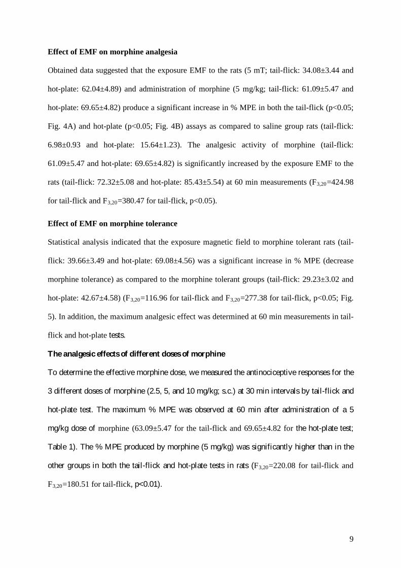
Effect of EMF on morphine analgesia
Obtained data suggested that the exposure EMF to the rats (5 mT; tail-flick: 34.08±3.44 and
hot-plate: 62.04±4.89) and administration of morphine (5 mg/kg; tail-flick: 61.09±5.47 and
hot-plate: 69.65±4.82) produce a significant increase in % MPE in both the tail-flick (p<0.05;
Fig. 4A) and hot-plate (p<0.05; Fig. 4B) assays as compared to saline group rats (tail-flick:
6.98±0.93 and hot-plate: 15.64±1.23). The analgesic activity of morphine (tail-flick:
61.09±5.47 and hot-plate: 69.65±4.82) is significantly increased by the exposure EMF to the
rats (tail-flick: 72.32±5.08 and hot-plate: 85.43±5.54) at 60 min measurements (F3,20=424.98
for tail-flick and F3,20=380.47 for tail-flick, p<0.05).
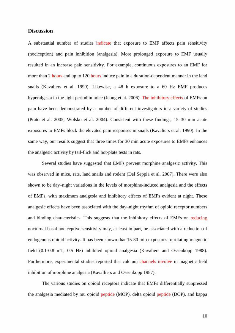
Effect of EMF on morphine tolerance
Statistical analysis indicated that the exposure magnetic field to morphine tolerant rats (tail-
flick: 39.66±3.49 and hot-plate: 69.08±4.56) was a significant increase in % MPE (decrease
morphine tolerance) as compared to the morphine tolerant groups (tail-flick: 29.23±3.02 and
hot-plate: 42.67±4.58) (F3,20=116.96 for tail-flick and F3,20=277.38 for tail-flick, p<0.05; Fig.
5). In addition, the maximum analgesic effect was determined at 60 min measurements in tail-
flick and hot-plate tests.
The analgesic effects of different doses of morphine
To determine the effective morphine dose, we measured the antinociceptive responses for the
3 different doses of morphine (2.5, 5, and 10 mg/kg; s.c.) at 30 min intervals by tail-flick and
hot-plate test. The maximum % MPE was observed at 60 min after administration of a 5
mg/kg dose of morphine (63.09±5.47 for the tail-flick and 69.65±4.82 for the hot-plate test;
Table 1). The % MPE produced by morphine (5 mg/kg) was significantly higher than in the
other groups in both the tail-flick and hot-plate tests in rats (F3,20=220.08 for tail-flick and
F3,20=180.51 for tail-flick, p<0.01).
10
Discussion
A substantial number of studies indicate that exposure to EMF affects pain sensitivity
(nociception) and pain inhibition (analgesia). More prolonged exposure to EMF usually
resulted in an increase pain sensitivity. For example, continuous exposures to an EMF for
more than 2 hours and up to 120 hours induce pain in a duration-dependent manner in the land
snails (Kavaliers et al. 1990). Likewise, a 48 h exposure to a 60 Hz EMF produces
hyperalgesia in the light period in mice (Jeong et al. 2006). The inhibitory effects of EMFs on
pain have been demonstrated by a number of different investigators in a variety of studies
(Prato et al. 2005; Wolsko et al. 2004). Consistent with these findings, 15–30 min acute
exposures to EMFs block the elevated pain responses in snails (Kavaliers et al. 1990). In the
same way, our results suggest that three times for 30 min acute exposures to EMFs enhances
the analgesic activity by tail-flick and hot-plate tests in rats.
Several studies have suggested that EMFs prevent morphine analgesic activity. This
was observed in mice, rats, land snails and rodent (Del Seppia et al. 2007). There were also
shown to be day–night variations in the levels of morphine-induced analgesia and the effects
of EMFs, with maximum analgesia and inhibitory effects of EMFs evident at night. These
analgesic effects have been associated with the day–night rhythm of opioid receptor numbers
and binding characteristics. This suggests that the inhibitory effects of EMFs on reducing
nocturnal basal nociceptive sensitivity may, at least in part, be associated with a reduction of
endogenous opioid activity. It has been shown that 15-30 min exposures to rotating magnetic
field (0.1-0.8 mT; 0.5 Hz) inhibited opioid analgesia (Kavaliers and Ossenkopp 1988).
Furthermore, experimental studies reported that calcium channels involve in magnetic field
inhibition of morphine analgesia (Kavalliers and Ossenkopp 1987).
The various studies on opioid receptors indicate that EMFs differentially suppressed
the analgesia mediated by mu opioid peptide (MOP), delta opioid peptide (DOP), and kappa
11
opioid peptide (KOP) receptors (Kavaliers and Ossenkopp 1986). Analgesia mediated by
MOP and KOP receptors was further shown to be reduced in the snails (Kavaliers and
Ossenkopp 1988). The suppressive effects of EMFs on DOP receptor-mediated
antinociception were also shown in the land snails. In contrast to these studies, further
research has discovered that exposures to magnetic field have positive pain relieving and
analgesic effect. For example, Thomas et al. (1997b) suggest that 15- min exposures to an
EMF (100 µT) in land snails increased opioid analgesia rather than producing inhibitory
effect. Magnetic field used for magnetic resonance imaging (MRI) reduces the nociception
induced in mice by the MOP receptor agonists (Teskey et al. 1988). These evidences suggest
that the possibility of a direct effect of EMFs on opioid receptor numbers or their functional
activity. Consistently, an animal study indicated that 4-day magnetic exposure increased the
levels of beta-endorphin in the hypothalamus (Bao et al. 2006). In addition, our data
demonstrate that the analgesic activity of morphine is significantly increased by the exposure
EMF to the rats. It has been indicated that these findings may explain, in part, the analgesic
effect of repeated exposures to EMFs.
The opioid analgesic tolerance involves associative and non-associative processes
(Bailey and Connor 2005). In the non-associative view, opioid tolerance results from changes
in underlying neurophysiological processes. This includes opioid receptor desensitization,
down-regulation and the induction of endogenous anti-opioid receptor systems. Animal
studies suggested that repeated acute EMF exposures attenuated the development of tolerance
to morphine through in non-associative factors (Kavaliers and Ossenkopp 1985).
Consistently, our findings demonstrated that the exposure magnetic field to morphine tolerant
rats was decreased morphine antinociceptive tolerance. Besides, it has been indicated that
opioid tolerance develops to the analgesic effects of pulsed magnetic field, with cross-
tolerance to the analgesic actions of DOP receptor agonist in the land snail (Thomas et al.
12
1998). In this study, it has been found that the magnitude of the magnetic field-induced
analgesia was reduced following repeated daily (15 or 30 min) exposures: an effect indicative
of the development of opioid tolerance. In addition, snails that received repeated daily
exposures to the specific pulsed EMF (100 µT) displayed decreased sensitivity of the DOP
receptor agonist [D-Pen 2,5] -enkephalin hydrate (DPDPE). This decreased sensitivity
provides evidence for the development of cross-tolerance of the DPDPE to the opioid
component of pulsed EMF.
One of the mechanisms of the magnetic field on the morphine analgesia is to alter the
cytokine levels. Exposure of rats to ELF-EMF decreased proinflammatory cytokine
production (Salehi et al. 2013). Furthermore, chronic morphine administration activates spinal
proinflammatory responses. These responses contribute to the inhibition of acute morphine
analgesia and development of tolerance. In particular, proinflammatory cytokine IL-1 is early
signals in the recruitment of endogenous pain facilitatory mechanisms that modulate opioid
analgesia (Fairbanks and Wilcox 2000). Therefore, the morphine tolerance can be attenuated
by pretreatment with the IL-1 receptor antagonist or EMF.
In conclusions, this study indicated that the extremely low frequency electromagnetic
fields have a significant effect on morphine analgesia and tolerance in analgesia tests.
Application of the extremely low frequency EMF to rats increases the morphine analgesia and
reduces morphine analgesic tolerance. Further research is required for clinical use of the
electromagnetic fields.
Funding: This study was funded by Cumhuriyet University Scientific Research Project (T-
329, CUBAP, Turkey).
Conflict of Interest: The authors declare that they have no conflict of interest.
13
References
Altun A., Ozdemir E., Yildirim K., Gursoy S., Durmus N., Bagcivan I. (2015): The effects of
endocannabinoid receptor agonist anandamide and antagonist rimonabant on opioid
analgesia and tolerance in rats. Gen. Physiol. Biophys. 34, 433–440.
Bailey C. P., Connor M. (2005): Opioids: cellular mechanisms of tolerance and physical
dependence. Current Opinion in Pharmacology 5, 1–9.
Bao X., Shi Y., Huo X., Song T. (2006): A possible involvement of beta-endorphin, substance
P, and serotonin in rat analgesia induced by extremely low frequency magnetic field.
Bioelectromagnetics 27, 467–472.
Bassett C. (1993): Beneficial effects of electromagnetic fields J. Cell. Biochem. 51, 387–393.
Chalidis B., Sachinis N., Assiotis A., Maccauro G. (2011): Stimulation of bone formation and
fracture healing with pulsed electromagnetic fields: Biologic responses and clinical
implications. Int. J. Immunopathol. Pharmacol. 24, 17–20.
Del Seppia C., Ghione S., Luschi P., Ossenkopp K.-P., Choleris E., Kavaliers M. (2007): Pain
perception and electromagnetic fields. Neuroscience and Biobehavioral Reviews 31,
619–642.
Dumas E. O., Pollack G. M. (2008): Opioid tolerance development: a pharmacokinetic
/pharmacodynamic perspective. The AAPS Journal 10(4), 537-551.
Fairbanks C. A., Wilcox G. L. (2000): Spinal plasticity of acute opioid tolerance. J. Biomed.
Sci. 7, 200–212.
Fernandez M. I., Watson P. J., Rowbotham D. J. (2007): Effect of pulsed magnetic field
therapy on pain reported by human volunteers in a laboratory model of acute pain. British
Journal of Anaesthesia 99(2), 266–269.
Fields H. (2004): State-dependent opioid control of pain. Nature Reviews Neuroscience 23,
7255–7261.
14
Gursoy S., Ozdemir E., Bagcıvan I., Altun A., Durmus N. (2011): Effects of alpha 2-
adrenoceptor agonists dexmedetomidine and guanfacine on morphine analgesia and
tolerance in rats. Upsala Journal of Medical Sciences 116, 238–246.
Jeong J. H., Kum C., Choi H. J., Park E. S., Sohn U. D. (2006): Extremely low frequency
magnetic field induces hyperalgesia in mice modulated by nitric oxide synthesis. Life
Science 78, 1407–1412.
Kanaan S. A., Saade N. E., Haddad J. J., Abdelnoor A. M., Atweh S. F., Jabbur S. J.,
Garabedian B. S. (1996): Endotoxin-induced local inflammation and hyperalgesia in rats,
a new model for inflammatory pain. Pharmacology 66, 373–379.
Kavaliers M., Ossenkopp K. P. (1985): Tolerance to morphine-induced analgesia in mice:
magnetic fields function as environmental specific cues and reduce tolerance
development. Life Science 37, 1125–1135.
Kavaliers M., Ossenkopp K. P. (1986): Magnetic fields differentially inhibit mu, delta, kappa
and sigma opiate-induced analgesia in mice. Peptides 7, 449–453.
Kavalliers M., Ossenkopp K.-P. (1987): Calcium channel involvement in magnetic field
inhibition of morphine–induced analgesia. Naunyn-Scheimedeberg’s Archieves of
Pharmacology 336, 308-315.
Kavaliers M., Ossenkopp K. P. (1988): Magnetic field inhibit opioid-mediated ‘‘analgesic’’
behaviors of the terrestrial snail (Cepaea nemoralis). Journal of Comparative Physiology
162A, 551–558.
Kavaliers M., Ossenkopp K. P., Hirst M. (1984): Magnetic fields abolish the enhanced
nocturnal analgesic response to morphine in mice. Physiol. Behav. 32, 261-264.
Kavaliers M., Ossenkopp K. P., Lipa S. M. (1990): Day–night rhythms in the inibitory effects
of 60Hz magnetic fields on opiate-mediated ‘analgesic’ behaviors of the land snail,
Cepaea nemoralis. Brain Research 517, 276–282.
15
Kieffer B. L. (1999): Opioids: first lessons from knockout mice. Trends in Pharmacological
Sciences 20, 19–26.
Lee Y. J., Hyung K. E., Yoo J. S., Jang Y. W., Kim S. J., Lee D. I., et al. (2016): Effects of
exposure to extremely low-frequency electromagnetic fields on the differentiation of
Th17 T cells and regulatory T cells. Gen. Physiol. Biophys 35, 487-495.
Morgan M. M.,. Christie M. J. (2011): Analysis of opioid efficacy, tolerance, addiction and
dependence from cell culture to human. Br. J. Pharmacol. 164, 1322–1334.
Nestler E. J., Aghajanian G. K. (1997): Molecular and cellular basis of addiction. Science
278, 58–63.
Ossenkopp K. P., Kavaliers M. (1987): Morphine-induced analgesia and exposure to low-
intensity 60-Hz magnetic fields: inhibition of nocturnal analgesia in mice is a function of
magnetic field intensity. Brain Research 418, 356–360.
Ossenkopp K. P., Kavaliers M. (1988): Clinical and applied aspects of magnetic field
exposure: possible role for the endogenous opioid systems. Journal of Bioelectricity 7,
189-208.
Ozdemir E., Bağcivan I., Gursoy S. (2013): Role of D1/D2 dopamin receptors antagonist
perphenazine in morphine analgesia and tolerance in rats. Bosn. J. Basic Med. Sci. 13 (2),
1-7.
Ozdemir E., Bagcivan I., Gursoy S., Altun A., Durmus N. (2011): Effects of fluoxetine and
LY 365265 on tolerance to the analgesic effect of morphine in rats. Acta Physiologica
Hungarica 98, 205-213.
Pesce M., Patruno A., Speranza L., Reale M. (2013): Extremely low frequency
electromagnetic field and wound healing: implication of cytokines as biological
mediators. Eur. Cytokine Netw. 24, 1-10.
16
Pilla A. A. (2013): Nonthermal electromagnetic fields: from first messenger to therapeutic
applications. Electromagn. Biol. Med. 32 (2), 123-136.
Prato F. S., Carson J. J., Ossenkopp K. P., Kavaliers M. (1995): Possible mechanisms by
which extremely low frequency magnetic fields affect opioid function. The FASEB
Journal 9, 807-814.
Prato F. S., Robertson J. A., Desjardins D., Hensel J., Thomas A. W. (2005): Daily repeated
magnetic field shielding induces analgesia in CD-1 mice. Bioelectromagnetics 26, 109–
117.
Ramabadran K., Bansinath M., Turndorf H., Puig M. M. (1989): The hyperalgesic effect of
naloxone is attenuated in streptozotocin-diabetic mice. Psychopharmacology (Berl.) 97,
169–174
Ramey D. (1998): Magnetic and electromagnetic therapy. Sci. Rev. Altern. Med. 2, 13–19.
Ross C., Harrison B. S. (2013): The use of magnetic field for the reduction of inflammation:
A review of the history and therapeutic results. Altern. Ther. Health Med. 19, 47–54.
Sadoghi P., Leithner A., Dorotka R., Vavken P. (2013): Effect of pulsed electromagnetic
fields on the bioactivity of human osteoarthritic chondrocytes. Orthopedics 36, 360–365.
Salehi I., Sani K. G., Zamani A. (2013): Exposure of rats to extremely low-frequency
electromagnetic fields (ELF-EMF) alters cytokines production. Electromagn. Biol. Med.
32, 1-8.
Sienkiewicz Z., Jones N., Bottomley A. (2005): Neurobehavioral effects of electromagnetic
fields. Bioelectromagnetics 7, 116–126.
Song P., Zhao Z. Q. (2001): The involvement of glial cells in the development of morphine
tolerance. Neurosci. Res. [Suppl] 39, 281–286.
Sutbeyaz S., Sezer N., Koseoglu F., Kibar S. (2009): Low-frequency pulsed lectromagnetic
field therapy in fibromyalgia: A randomized, double-blind, sham-controlled clinical
study. Clin. J. Pain 25, 722–728.
17
Teskey G. C., Prato F. S., Ossenkopp K.-P., Kavaliers M. (1988): Exposure to time varying
magnetic fiels associated with magnetic resonance imaging reduces fentanyl-induced
analgesia in mice. Bioelectromagnetics 9, 167–174.
Thomas A. W., Kavaliers M., Prato F. S., Ossenkopp K.-P. (1997a): Antinociceptive effects
of pulsed magnetic field in the land snails, Cepaea nemoralis. Neuroscience Letters 222,
107-110.
Thomas A. W., Kavaliers M., Prato F. S., Ossenkopp K.-P. (1997b): Pulsed magnetic field
induced analgesia in the land snails, Cepaea nemoralis and the effects of µ, d, and κ
opioid receptor agonists/antagonists. Peptides 18, 703-709.
Thomas A.W., Kavaliers, M., Prato, F. S., Ossenkopp, K.-P. (1998): Analgesic effect of a
specific pulsed magnetic field in the land snail (Cepaea nemoralis): consequences of
repeated exposure, relations to tolerance and cross-tolerance with DPDPE. Peptides
19,333–342.
Traina G., Romanini L., Benazoo, F., Cadossi, V. (1998): Use of electric and magnetic
stimulation in orthopaedics and traumatology: Consensus conference. J. Ortho. Trauma.
24, 1–31.
Trock D. (2000): Electromagnetic fields and magnets: investigational treatment for
musculoeskeletal disorders. Rheum. Dis. Clin. North. Am. 26, 51–62.
Wolsko P. M., Eisenberg D. M., Simon L. S., Davis R. B., Walleczek J., Mayo-Smith M.,
Kaptchuk T. J., Phillips R. S. (2004): Double-blind placebo-controlled trial of static
magnets for the treatment of osteoarthritis of the knee: results of a pilot study. Alternative
Therapies in Health and Medicine 10, 36–43.
Zarrindast M. R., Dinkub Z., Homayoun H., Bakhtiarian A. (2002): Dopamine receptor
mechanisms and morphine tolerance in mice. J. Pysychopharmacol. 16, 261–266.
18
Figure Legends
Fig. 1. The EMF exposure system. 1) Computer; 2) Power supply; 3) Digital Teslameter
(Phywe, 80010); 4) Solenoid with animal cage.
Fig. 2. Analgesic effects of three different EMF strengths. (A) shows effect of EMF in the
tail-flick test, and (B) shows effect of EMF in the hot-plate test. The maximal analgesic effect
of the magnetic field was observed on day 7 in all group (1 mT, 5 mT and 10 mT) rats. The
analgesic activity of magnetic field 1 mT, 5 mT and 10 mT groups were significantly higher
than control group rats (p<0.01). Each point represents the mean±SEM of % MPE for 6 rats.
*p<0.01 compared to the control and **p<0.05 compared to 1 mT group.
Fig. 3. Time-dependent (Min.) change of EMF analgesic effects. (A) shows effect of EMF in
the tail-flick test, and (B) shows effect of EMF in the hot-plate test. The maximal analgesic
effect was determined at 5 mT group and 90 min measurement for three different magnetic
field strength. Each point represents the mean±SEM of % MPE for 6 rats. *p<0.01 compared
to the control and **p<0.05 compared to 1 mT group.
Fig. 4. Effect of EMF on morphine analgesia. (A) shows effect of EMF in the tail-flick test,
and (B) shows effect of EMF in the hot-plate test. The exposure EMF to the rats and
administration of morphine produce a significant increase in % MPE in both the tail-flick and
hot-plate assays as compared to saline group rats The analgesic activity of morphine is
significantly increased by the exposure EMF to the rats at 60 min measurements. Each point
represents the mean±SEM of % MPE for 6 rats. *p<0.01 compared to the saline and **p<0.05
compared to morphine treated group.
19
Fig. 5. Effect of EMF on morphine tolerance. (A) shows effect of EMF in the tail-flick test,
and (B) shows effect of EMF in the hot-plate test. The exposure EMF to morphine tolerant
rats was a significant increase in % MPE (decrease morphine tolerance) as compared to the
morphine tolerant groups. Each point represents the mean±SEM of % MPE for 6 rats.
*p<0.05 compared to the saline and **p<0.05 compared to morphine tolerant rats.
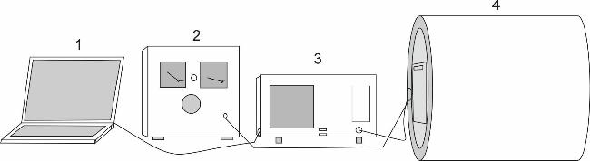
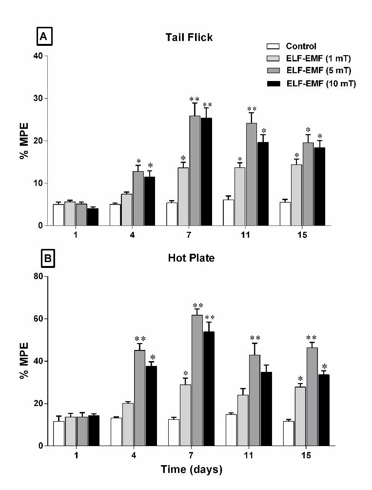
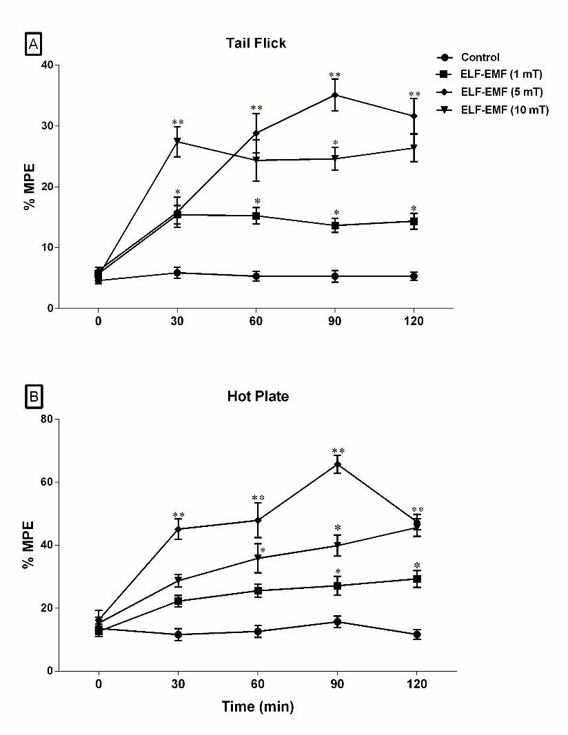
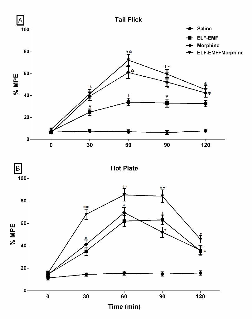
Table 1. The antinociceptive effects of different doses of morphine
Time (min) 0 30 60 90 120
Tail-flick
Saline 6.55±0.92 7.63±0.96 8.47±1.02 6.79±0.98 5.99±0.88
Morphine (2.5 mg/kg) 5.72±0.98 34.60±1.90* 50.32±3.75* 48.78±3.88* 34.38±2.87*
Morphine (5 mg/kg) 6.23±0.84 39.42±3.95* 63.09±5.47** 54.34±4.12** 42.36±3.87*
Morphine (10 mg/kg) 8.09±1.09 45.56±4.09* 61.79±5.02** 57.38±3.95** 45.60±3.83*
Hot-plate
Saline 13.52±1.32 12.42±1.13 15.44±1.07 11.62±1.08 13.18±1.32
Morphine (2.5 mg/kg) 12.89±1.23 35.56±2.84* 54.45±4.15* 37.50±3.22* 32.38±2.56*
Morphine (5 mg/kg) 14.36±1.57 41.29±3.98* 69.65±4.82** 41.98±4.53* 36.31±3.87*
Morphine (10 mg/kg) 15.87±1.85 43.46±3.73* 68.06±4.77** 45.14±4.28* 39.08±3.86*
Data are mean ± SEM. *p < 0.05; **p < 0.01 as compared with its saline group (n = 6 in each group). Analgesia was expressed in % MPE.