Embed Size (px)
Citation preview
Full Terms & Conditions of access and use can be found athttps://www.tandfonline.com/action/journalInformation?journalCode=iebm20
Electromagnetic Biology and Medicine
ISSN: 1536-8378 (Print) 1536-8386 (Online) Journal homepage: https://www.tandfonline.com/loi/iebm20
Exposure to Static and Extremely-Low FrequencyElectromagnetic Fields and Cellular Free Radicals
Henry Lai
To cite this article: Henry Lai (2019): Exposure to Static and Extremely-Low FrequencyElectromagnetic Fields and Cellular Free Radicals, Electromagnetic Biology and Medicine, DOI:10.1080/15368378.2019.1656645
To link to this article: https://doi.org/10.1080/15368378.2019.1656645
View supplementary material
Published online: 26 Aug 2019.
Submit your article to this journal
View related articles
View Crossmark data
Exposure to Static and Extremely-Low Frequency Electromagnetic Fields andCellular Free RadicalsHenry Lai
Department of Bioengineering, University of Washington, Seattle, WA, USA
ABSTRACTThis paper summarizes studies on changes in cellular free radical activities from exposure to static andextremely-low frequency (ELF) electromagnetic fields (EMF), particularly magnetic fields. Changes in freeradical activities, including levels of cellular reactive oxygen (ROS)/nitrogen (RNS) species and endogen-ous antioxidant enzymes and compounds that maintain physiological free radical concentrations incells, is one of the most consistent effects of EMF exposure. These changes have been reported to affectmany physiological functions such as DNA damage; immune response; inflammatory response; cellproliferation and differentiation; wound healing; neural electrical activities; and behavior. An importantconsideration is the effects of EMF-induced changes in free radicals on cell proliferation and differentia-tion. These cellular processes could affect cancer development and proper growth and development inorganisms. On the other hand, they could cause selective killing of cancer cells, for instance, via thegeneration of the highly cytotoxic hydroxyl free radical by the Fenton Reaction. This providesa possibility of using these electromagnetic fields as a non-invasive and low side-effect cancer therapy.Static- and ELF-EMF probably play important roles in the evolution of living organisms. They are cuesused in many critical survival functions, such as foraging, migration, and reproduction. Living organismscan detect and respond immediately to low environmental levels of these fields. Free radical processesare involved in some of these mechanisms. At this time, there is no credible hypothesis or mechanismthat can adequately explain all the observed effects of static- and ELF-EMF on free radical processes. Weare actually at the impasse that there are more questions than answers.
ARTICLE HISTORYReceived 17 April 2019Accepted 4 August 2019
KEYWORDSStatic and extremely-lowfrequency electromagneticfields; free radicals; Fentonreaction; cell proliferation;evolution
Introduction
This is a review of the research on the effects on cellularfree radicals after exposure to static- and extremely-lowfrequency (ELF, 0–300 Hz) non-ionizing electromag-netic field (EMF). In 1997, we first reported that mela-tonin, a potent antioxidant, and the spin-trap compoundN-tert-butyl-alpha-phenylnitrone (that neutralizes freeradicals) blocked a 60-Hz magnetic field-induced DNAstrand break in cells of the rat brain (Lai and Singh1997a, 1997b). Further experiment (Lai and Singh2004) demonstrated similar inhibitory effects of Trolox(a vitamin E-analog anti-oxidant) and 7-nitroindazole (anitric oxide synthase inhibitor). In addition, the effectcould also be blocked by the iron chelator deferipronesuggesting the involvement of the iron-catalyzed FentonReaction that produces the potent cytotoxic hydroxylfree radical. These data indicated that the ELF magneticfield affected free radicals in cells leading to cellularmolecular damages. There are now more than 200papers published showing that static and ELF-EMFaffect cellular free radical processes. A list of the papers
is in the “supplementary material” included in the on-line version of this paper. There are rather strong indi-cations that exposure to static- and ELF-EMF affectsoxidative status in cells and animals. Many of the cellularoxidative and anti-oxidative components have beenshown to be affected by the fields.
Effect on cellular free radical processes is probablythe most consistent biological effect of non-ionizingelectromagnetic fields (EMF). It has been reported inmany different animal and plant species after exposureto EMF from static to radiofrequency (see Yakymenkoet al. (2016) and a 2017-update in the “oxidative effectsof ELF-EMF and radiofrequency radiation (RFR) sec-tion” in the Bioinitiative Report (2012)).
Free radicals
Reactive free radicals (mainly, reactive oxygen species(ROS) and reactive nitrogen species (RNS)) are pro-duced as a result of cellular metabolism, particularly inthe mitochondria. Reactive oxygen species (ROS)include mainly singlet oxygen, superoxide, peroxides,
CONTACT Henry Lai [email protected] Department of Bioengineering, University of Washington, Seattle, WA 98195-5061, USASupplemental data for this article can be accessed here
ELECTROMAGNETIC BIOLOGY AND MEDICINEhttps://doi.org/10.1080/15368378.2019.1656645
© 2019 Taylor & Francis Group, LLC
and hydroxy radical and reactive nitrogen species(RNS) including mainly peroxtnitrite, nitrogen dioxide,which are products of the reaction between nitric oxideand superoxide. Nitric oxide is generated in cell bynitric oxide synthases. Presence of free radicals in cellscan lead to macromolecular damages (in DNA, pro-teins, and lipids), disturbance in cell functions, and celldeath. Damage in DNA is a cause of cancer. Undernormal conditions, free radical levels are kept in checkby various inducible antioxidant enzymes includingsuperoxide dismutase (SOD), catalase (CAT) and glu-tathione peroxidase (GPx). In instances, when there isan excessive increase in free radical production ora deficit in anti-oxidant capacity, oxidative/nitrosativestress results leading to cellular damage and functionaldeficits. However, free radicals also serve importantcellular functions and are involved in cellular signalingcascades that govern normal cell functions and inimmune defense against bacteria. They are alsoinvolved in cellular chemistry that triggers apoptosis.Thus, it is essential to keep free radicals at a criticalphysiological homeostatic level. Any disturbance couldlead to detrimental biological consequences (cf. Pizzinoet al. 2017; Valko et al. 2007).
Cellular free radical processes is a complex physiolo-gical mechanism. It involves feedbacks and compensatoryresponses of different cellular components to maintainhomeostasis. EMF could disturb different components ofthe process leading to a cascade of changes. Since expo-sure to EMF leads to disturbance in free radical produc-tion and excessive presence of free radicals in cells can beconsidered as a stress, living organisms under chronicEMF exposure probably go through the three phases ofthe “general-adaptation-syndrome” of stress, i.e., alarm,resistance, and exhaustion phases (Selye 1951). Thus, thecharacteristic of the free-radical responses depends onhow long exposure has been occurring. In addition,effects observed could depend on the cell type andorgan studied; time when the changes were studied, andexposure conditions (such as intensity, cumulative dura-tion of exposure, and characteristics of the field). Thus, itis not surprising that the changes described in the “sup-plementary material” show a complex pattern, i.e.,changes are not always in the same direction. Researchon the effects of static- and ELF-EMF is mainly on ROS.There are only few studies on RNS (~5%).
Effects of static and ELF EMF-induced changesin cellular free radical processes
Several papers reported changes in biochemistry, phy-siology, and general functions as a consequence ofchanges in cellular oxidative status resulting from
exposure to static- and ELF-EMF. These include:DNA damage (Giorgi et al. 2017; Jajte et al. 2001;Koyama et al. 2008; Lai and Singh 1997a, 1997b; Laiand Singh 2004; Yokus et al. 2005, 2008); immuneresponse (Akan et al., 2010; Kim et al. 2017); inflam-matory response (Kim et al. 2017; Zhang et al. 2017);apoptosis (De Nicola et al. 2006; Ding et al. 2004; Garipand Akan 2010; Ghodbane et al. 2015; Koh et al. 2008;Solek et al. 2017; Wartenberg et al. 2008; Yang and Ye2015); protein misfolding and generation of prions(Lian et al. 2018); cell proliferation and differentiation(Ehnert et al. 2017; Hajipour Verdam et al., 2018; Leeet al. 2010; Patruno et al. 2010; Song et al. 2018; VanHuizen et al. 2019; Wolf et al. 2005); rhythmic slowactivity in hippocampal slices of the brain (Bawin et al.1996); visual evoked potentials (Akpinar et al. 2012);auditory event-related potentials (Akpinar et al., 2016);visual and somatosensory evoked potentials (Akpınaret al. 2016); heart rate (Ciejka and Goraca 2009);wound healing (Glinka et al. 2013; Patruno et al.2010, 2011); bone formation (Zhang et al. 2018); post-stroke recovery (Cichoń et al. 2017a, 2017b, 2018);hyperalgesia (Jeong et al. 2006); opioid-induced anti-nociception (Kavaliers et al. 1998); spatial memory andlearning (Cui et al. 2012; Deng et al. 2013; Karimi et al.2019); cognitive impairment (Duan et al. 2013); mis-match-negativity response (Kantar-Gok et al., 2014);depressive disorder (Ansari et al. 2016); anxiety-likebehavior (Djordjevic et al. 2017); and obsessive com-pulsive disorder-like behavior (Salunke et al. 2014).However, in most of these studies, the cause–effectrelationship was not well established. Do EMF-induced changes in oxidative status cause these effects?Or, are they effects of EMF caused by mechanismsunrelated to oxidative changes? One powerful proof ofa free-radical effect is to establish whether an effect,e.g., DNA damage, could be blocked by antioxidants orpro-oxidants. An effect caused by a change in freeradicals should be able to be blocked by antioxidantsor pro-oxidants. There are several studies thatemployed this strategy (see “Supplementary material”).
In most of the ELF-oxidative effects studies, the inten-sities used were relatively high (i.e., more than 0.1 mT)compared to ambient levels of static- and ELF-EMF (inμT levels) in the human environment. However, effects athigh intensities could possibly occur in occupationalexposure situations where the levels are relatively high.In addition, the exposure durations in most of thesestudies are short-term (from hours to several days),whereas environmental exposure is generally chronic.Can most of the research results applicable to real-lifeexposure situation? Do oxidative changes occur afterexposure to ambient levels of static- and ELF-EMF?
2 H. LAI
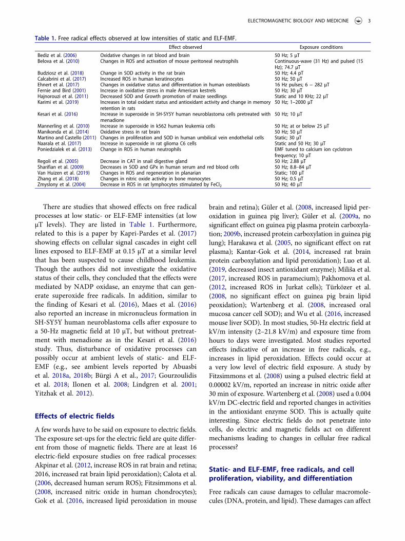
There are studies that showed effects on free radicalprocesses at low static- or ELF-EMF intensities (at lowμT levels). They are listed in Table 1. Furthermore,related to this is a paper by Kapri-Pardes et al. (2017)showing effects on cellular signal cascades in eight celllines exposed to ELF-EMF at 0.15 μT at a similar levelthat has been suspected to cause childhood leukemia.Though the authors did not investigate the oxidativestatus of their cells, they concluded that the effects weremediated by NADP oxidase, an enzyme that can gen-erate superoxide free radicals. In addition, similar tothe finding of Kesari et al. (2016), Maes et al. (2016)also reported an increase in micronucleus formation inSH-SY5Y human neuroblastoma cells after exposure toa 50-Hz magnetic field at 10 μT, but without pretreat-ment with menadione as in the Kesari et al. (2016)study. Thus, disturbance of oxidative processes canpossibly occur at ambient levels of static- and ELF-EMF (e.g., see ambient levels reported by Abuasbiet al. 2018a, 2018b; Bürgi A et al., 2017; Gourzoulidiset al. 2018; Ilonen et al. 2008; Lindgren et al. 2001;Yitzhak et al. 2012).
Effects of electric fields
A few words have to be said on exposure to electric fields.The exposure set-ups for the electric field are quite differ-ent from those of magnetic fields. There are at least 16electric-field exposure studies on free radical processes:Akpinar et al. (2012, increase ROS in rat brain and retina;2016, increased rat brain lipid peroxidation); Calota et al.(2006, decreased human serum ROS); Fitzsimmons et al.(2008, increased nitric oxide in human chondrocytes);Gok et al. (2016, increased lipid peroxidation in mouse
brain and retina); Güler et al. (2008, increased lipid per-oxidation in guinea pig liver); Güler et al. (2009a, nosignificant effect on guinea pig plasma protein carboxyla-tion; 2009b, increased protein carboxylation in guinea piglung); Harakawa et al. (2005, no significant effect on ratplasma); Kantar-Gok et al. (2014, increased rat brainprotein carboxylation and lipid peroxidation); Luo et al.(2019, decreased insect antioxidant enzyme); Miliša et al.(2017, increased ROS in paramecium); Pakhomova et al.(2012, increased ROS in Jurkat cells); Türközer et al.(2008, no significant effect on guinea pig brain lipidpeoxidation); Wartenberg et al. (2008, increased oralmucosa cancer cell SOD); and Wu et al. (2016, increasedmouse liver SOD). In most studies, 50-Hz electric field atkV/m intensity (2–21.8 kV/m) and exposure time fromhours to days were investigated. Most studies reportedeffects indicative of an increase in free radicals, e.g.,increases in lipid peroxidation. Effects could occur ata very low level of electric field exposure. A study byFitzsimmons et al. (2008) using a pulsed electric field at0.00002 kV/m, reported an increase in nitric oxide after30 min of exposure. Wartenberg et al. (2008) used a 0.004kV/m DC-electric field and reported changes in activitiesin the antioxidant enzyme SOD. This is actually quiteinteresting. Since electric fields do not penetrate intocells, do electric and magnetic fields act on differentmechanisms leading to changes in cellular free radicalprocesses?
Static- and ELF-EMF, free radicals, and cellproliferation, viability, and differentiation
Free radicals can cause damages to cellular macromole-cules (DNA, protein, and lipid). These damages can affect
Table 1. Free radical effects observed at low intensities of static and ELF-EMF.Effect observed Exposure conditions
Bediz et al. (2006) Oxidative changes in rat blood and brain 50 Hz; 5 μTBelova et al. (2010) Changes in ROS and activation of mouse peritoneal neutrophils Continuous-wave (31 Hz) and pulsed (15
Hz); 74.7 μTBudziosz et al. (2018) Change in SOD activity in the rat brain 50 Hz; 4.4 pTCalcabrini et al. (2017) Increased ROS in human keratinocytes 50 Hz; 50 μTEhnert et al. (2017) Changes in oxidative status and differentiation in human osteoblasts 16 Hz pulses; 6 − 282 μTFernie and Bird (2001) Increase in oxidative stress in male American kestrels 50 Hz; 30 μTHajnorouzi et al. (2011) Decreased SOD and Growth promotion of maize seedlings Static and 10 KHz; 22 μTKarimi et al. (2019) Increases in total oxidant status and antioxidant activity and change in memory
retention in rats50 Hz; 1–2000 μT
Kesari et al. (2016) Increase in superoxide in SH-SY5Y human neuroblastoma cells pretreated withmenadione
50 Hz; 10 μT
Mannerling et al. (2010) Increase in superoxide in k562 human leukemia cells 50 Hz; at or below 25 μTManikonda et al. (2014) Oxidative stress in rat brain 50 Hz; 50 μTMartino and Castello (2011) Changes in proliferation and SOD in human umbilical vein endothelial cells Static; 30 μTNaarala et al. (2017) Increase in superoxide in rat glioma C6 cells Static and 50 Hz; 30 μTPoniedzialek et al. (2013) Change in ROS in human neutrophils EMF tuned to calcium ion cyclotron
frequency; 10 μTRegoli et al. (2005) Decrease in CAT in snail digestive gland 50 Hz; 2.88 μTSharifian et al. (2009) Decreases in SOD and GPx in human serum and red blood cells 50 Hz; 8.8–84 μTVan Huizen et al. (2019) Changes in ROS and regeneration in planarian Static; 100 μTZhang et al. (2018) Changes in nitric oxide activity in bone monocytes 50 Hz; 0.5 μTZmyslony et al. (2004) Decrease in ROS in rat lymphocytes stimulated by FeCl2 50 Hz; 40 μT
ELECTROMAGNETIC BIOLOGY AND MEDICINE 3
cell functions. Mutation in DNA can lead to cancer devel-opment. However, too much damage to a cell can causecell death. And death to precancerous and cancer cellsdecreases the incidence of cancer. This may be a possiblenon-invasive method for cancer prevention and treat-ment. It is, of course, not known how much EMF expo-sure is needed to push cancerous cell over the edge todeath. It may depend on the type of cancer cell.
On the other hand, the death of cells that cannotreproduce and be replaced leads to dysfunction inorgans. This is particularly true for nerve cells. Theconnection between EMF exposure and neurodegenera-tive diseases are still not yet well established. There areseveral recent studies indicating a possible correlationwith Alzheimer’s disease, amyotrophic lateral sclerosis,dementia, and motor dysfunctions (Gunnarsson andBodin 2018; Huss et al. 2018; Jalilian et al. 2018;Koeman et al. 2017; Pedersen et al. 2017). There are,however, two interesting points that need to be pointedout. First, static- and ELF-EMF have been shown toreverse and improve cognitive performance in animalmodels of neurodegenerative disorders (Akbarnejadet al. 2018; Bobkova et al. 2018; Hu et al. 2016; Li et al.2019; Liu et al. 2015; Sakhaie et al. 2017; Tasset et al.2012). Can these be related to the effects of static- andELF-EMF on protein folding and prion production (Lianet al., 2018) and induction of heat-shock proteins(Laramee et al. 2014; Zeni et al. 2017)? Second, there isan inverse correlation between cancer risk andAlzheimer’s and Parkinson diseases (Poprac et al.2017). An increase in cellular free radicals isa common factor of these diseases. This supports thenotion that static- and ELF-EMF exposure can kill can-cer cells and cause neurodegenerative diseases.
Harnessing cellular oxidative status using static andELF-EMF could also be beneficial in the treatment ofcertain diseases. Several papers have suggested such pos-sibilities including: improvement of immune responses(Akan et al. 2010; Belova et al. 2010; Frahm et al. 2006;Kim et al. 2017); treatment of osteoarthritis (De Matteiet al. 2003); attenuation of ischemic brain injury (Duonget al., 2016; Rauš Balind et al. 2014); increasing antiox-idant properties in cells and tissues (Falone et al. 2016);treatment of myopathies (Vignola et al. 2012); woundhealing and tissue regeneration (Glinka et al. 2013;Patruno et al. 2010, 2011; Van Huizen et al. 2019);cytoprotection (Osera et al. 2015, 2011); inducing differ-entiation of stem cells (Haghighat et al. 2017b, 2017a;Marycz et al. 2018; Park et al. 2013; Van Huizen et al.2019); and protective effect on Huntington’s disease(Tasset et al. 2012; Túnez et al. 2006). One interestingprospect is the use of static and ELF-EMF in the treat-ment of cancer. EMF can selectively kill cancer cells (Lai
and Singh 2010). Many years ago, we (Lai and Singh2004) speculated that cancer cells are more vulnerable toEMF than normal cells and that EMF kills cancer cells byfree radical formation. Since it is much easier to produceELF-EMF than RFR, and ELF-EMF gives a more uni-form distribution and better tissue penetration that RFR,it is more advantageous to use ELF-EMF for cancertreatment. Let us look at the studies on static- and ELF-EMF exposure on free radicals and cell proliferation,differentiation, cell cycle, and cell death in cancer andnormal cells, summarized in Table 2. These are impor-tant cellular processes that determine cancer develop-ment and treatment, growth, development, woundhealing and regeneration in living organisms.
Several studies on cancer cells listed in Table 2 sug-gested a possible beneficial effect on cancer treatmentunder static- or ELF-EMF exposure by increasing apop-tosis and decreasing proliferation and viability (Benassiet al. 2016; Ding et al. 2004; Errico Provenzano et al.2018; Hajipour Verdom et al. 2018; Koh et al. 2008; Laiet al. 2016; Mannerling et al. 2010; Osara et al., 2011;Wartenberg et al. 2008; Yang and Ye 2015). However,others suggested a protective effect by decreasing apop-tosis and increasing proliferation and viability thatwould allow cancer to grow faster (De Nicola et al.2006; Falone et al. 2007, 2017; Garip and Akan 2010;Martinez et al., 2016; Osera et al. 2015; Song et al. 2018;Wolf et al. 2005), whereas no significant effect on cellviability and proliferation was reported by some studies(Consales et al. 2019; Morabito et al. 2010; Naarala et al.2017; Pakhomova et al. 2012; Sadeghipour et al. 2012).Interestingly, A study (Ayşe et al. 2010) showed oppositeeffects depending on the duration of exposure. Thisreflects the discussion above on the dynamic of cellularfree radical processes and their ability to compensate.Cell type probably plays a significant role. Cell-type-specific responses to ELF-EMF have been reported bySullivan et al. (2011), Kesari et al. (2016), Koziorowskaet al. (2018), Makinistian et al. (2019), and Wang et al.(2018). The conditions of exposure probably cause thediversity of responses, but the conditions of exposuredescribed in the table do not reveal a clear pattern onhow different exposure parameters affect cellular freeradical processes and changes in cell proliferation, differ-entiation, and apoptosis. There is a slight tendency of aninverse relationship between free radical activity andcellular proliferation, i.e., an increase in free radicalscauses a decrease in cell proliferation and vice versa.Also, increase in free radical activity tends to enhanceapoptosis. This uncertainty is actually not surprisingbecause, in each study, we are looking at only somecomponents of the free radical processes and not thewhole pattern of changes. Feedback and compensatory
4 H. LAI
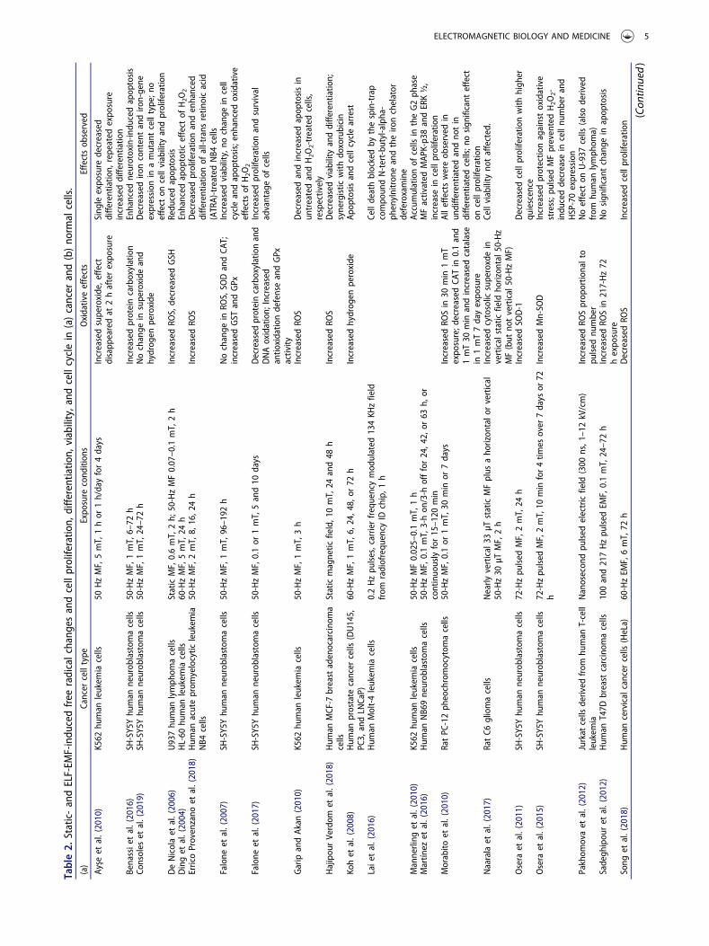
Table2.
Static-andELF-EM
F-indu
cedfree
radicalchang
esandcellproliferatio
n,diffe
rentiatio
n,viability,and
cellcyclein
(a)cancer
and(b)no
rmalcells.
(a)
Cancer
celltype
Expo
sure
cond
ition
sOxidativeeffects
Effectsob
served
Ayşe
etal.(2010)
K562
human
leukem
iacells
50HzMF,5mT,1hor
1h/dayfor4days
Increasedsuperoxide,effe
ctdisapp
earedat
2hafterexpo
sure
Sing
leexpo
sure
decreased
diffe
rentiatio
n,repeated
expo
sure
increaseddiffe
rentiatio
nBenassie
tal.(2016)
SH-SY5Yhu
man
neurob
lastom
acells
50-HzMF,1mT,6–72
hIncreasedproteincarboxylation
Enhanced
neurotoxin-in
ducedapop
tosis
Consoles
etal.(2019)
SH-SY5Yhu
man
neurob
lastom
acells
50-HzMF,1mT,24–72h
Nochange
insuperoxide
and
hydrog
enperoxide
Decreased
ironcontentandiro
n-gene
expression
inamutantcelltype;n
oeffect
oncellviability
andproliferatio
nDeNicolaet
al.(2006)
U937hu
man
lymph
omacells
StaticMF,0.6mT,2h;
50-HzMF0.07–0.1
mT,2h
IncreasedRO
S,decreasedGSH
Redu
cedapop
tosis
Dinget
al.(2004)
HL-60
human
leukem
iacells
60-HzMF,5mT,24
hEnhanced
apop
totic
effect
ofH2O
2ErricoProvenzano
etal.(2018)
Hum
anacuteprom
yelocytic
leukem
iaNB4
cells
50-HzMF,2mT,8,
16,2
4h
IncreasedRO
SDecreased
proliferatio
nandenhanced
diffe
rentiatio
nof
all-trans
retin
oicacid
(ATRA)-treated
NB4
cells
Falone
etal.(2007)
SH-SY5Yhu
man
neurob
lastom
acells
50-HzMF,1mT,96–192
hNochange
inRO
S,SO
DandCA
T;increasedGST
andGPx
Increasedviability,n
ochange
incell
cycleandapop
tosis;enhanced
oxidative
effectsof
H2O
2Falone
etal.(2017)
SH-SY5Yhu
man
neurob
lastom
acells
50-HzMF,0.1or
1mT,5and10
days
Decreased
proteincarboxylationand
DNAoxidation;
Increased
antio
xidatio
ndefenseandGPx
activity
Increasedproliferatio
nandsurvival
advantageof
cells
Garip
andAkan
(2010)
K562
human
leukem
iacells
50-HzMF,1mT,3h
IncreasedRO
SDecreased
andincreasedapop
tosisin
untreatedandH2O
2-treatedcells,
respectively
Hajipou
rVerdom
etal.(2018)
Hum
anMCF-7
breastadenocarcino
ma
cells
Staticmagnetic
field,1
0mT,24
and48
hIncreasedRO
SDecreased
viability
anddiffe
rentiatio
n;synergistic
with
doxorubicin
Kohet
al.(2008)
Hum
anprostate
cancer
cells
(DU145,
PC3,
andLN
CaP)
60-HzMF,1mT,6,
24,4
8,or
72h
Increasedhydrog
enperoxide
Apop
tosisandcellcyclearrest
Laie
tal.(2016)
Hum
anMolt-4leukem
iacells
0.2Hzpu
lses,carrierfrequencymod
ulated
134KH
zfield
from
radiofrequ
ency
IDchip,1
hCelldeathblockedby
thespin-trap
compo
undN-tert-bu
tyl-alpha-
phenylnitron
eandtheiro
nchelator
deferoxamine
Mannerling
etal.(2010)
K562
human
leukem
iacells
50-HzMF0.025–0.1mT,1h
Accumulationof
cells
intheG2ph
ase
Martín
ezet
al.(2016)
Hum
anNB69neurob
lastom
acells
50-HzMF,0.1mT,3-hon
/3-h
offfor24,4
2,or
63h,
orcontinuo
uslyfor15–120
min
MFactivated
MAP
K-p38andERK½,
increase
incellproliferatio
nMorabito
etal.(2010)
RatPC
-12ph
eochromocytom
acells
50-HzMF,0.1or
1mT,30
min
or7days
IncreasedRO
Sin
30min
1mT
expo
sure;d
ecreased
CATin
0.1and
1mT30
min
andincreasedcatalase
in1mT7dayexpo
sure
Alle
ffectswereob
served
inun
diffe
rentiatedandno
tin
diffe
rentiatedcells;n
osign
ificant
effect
oncellproliferatio
nNaaralaet
al.(2017)
RatC6
gliomacells
Nearly
vertical33
μTstaticMFplus
aho
rizon
talo
rvertical
50-Hz30
μTMF,2h
Increasedcytosolic
superoxdein
verticalstaticfield
horizon
tal5
0-Hz
MF(but
notvertical50-HzMF)
Cellviability
notaffected.
Osera
etal.(2011)
SH-SY5Yhu
man
neurob
lastom
acells
72-Hzpu
lsed
MF,2mT,24
hIncreasedSO
D-1
Decreased
cellproliferatio
nwith
high
erqu
iescence
Osera
etal.(2015)
SH-SY5Yhu
man
neurob
lastom
acells
72-Hzpu
lsed
MF,2mT,10
min
for4tim
esover
7days
or72
hIncreasedMn-SO
DIncreasedprotectio
nagainstoxidative
stress;p
ulsedMFpreventedH2O
2-indu
ceddecrease
incellnu
mberand
HSP-70expression
Pakhom
ovaet
al.(2012)
Jurkat
cells
derived
from
human
T-cell
leukem
iaNanosecon
dpu
lsed
electricfield
(300
ns,1
–12kV/cm)
IncreasedRO
Sprop
ortio
nalto
pulsed
number
Noeffect
onU-937
cells
(alsoderived
from
human
lymph
oma)
Sadegh
ipou
ret
al.(2012)
Hum
anT47D
breastcarcinom
acells
100and217Hzpu
lsed
EMF,0.1mT,24–72h
IncreasedRO
Sin
217-Hz72
hexpo
sure
Nosign
ificant
change
inapop
tosis
Song
etal.(2018)
Hum
ancervicalcancer
cells
(HeLa)
60-HzEM
F,6mT,72
hDecreased
ROS
Increasedcellproliferatio
n
(Con
tinued)
ELECTROMAGNETIC BIOLOGY AND MEDICINE 5
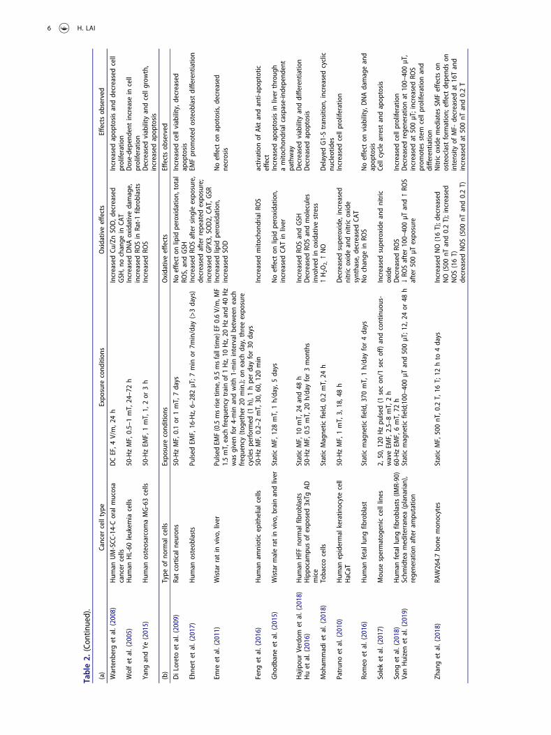
Table2.
(Con
tinued).
(a)
Cancer
celltype
Expo
sure
cond
ition
sOxidativeeffects
Effectsob
served
Wartenb
erget
al.(2008)
Hum
anUM-SCC
-14-Coralmucosa
cancer
cells
DCEF,4
V/m,2
4h
IncreasedCu
/ZnSO
D,d
ecreased
GSH
,nochange
inCA
TIncreasedapop
tosisanddecreasedcell
proliferatio
nWolfet
al.(2005)
Hum
anHL-60
leukem
iacells
50-HzMF,0.5–1mT,24–72h
IncreasedDNAoxidativedamage,
increasedRO
Sin
Rat-1fib
roblasts
Dose-depend
entincrease
incell
proliferatio
nYang
andYe
(2015)
Hum
anosteosarcomaMG-63cells
50-HzEM
F,1mT,1,
2or
3h
IncreasedRO
SDecreased
viability
andcellgrow
th,
increasedapop
tosis
(b)
Type
ofno
rmal
cells
Expo
sure
cond
ition
sOxidativeeffects
Effectsob
served
DiLoretoet
al.(2009)
Ratcorticalneuron
s50-HzMF,0.1or
1mT,7days
Noeffecton
lipid
peroxidatio
n,total
ROS,andGSH
Increasedcellviability,d
ecreased
apop
tosis
Ehnertet
al.(2017)
Hum
anosteob
lasts
Pulsed
EMF,16-Hz,6–282μT;7min
or7m
in/day
(>3days)
IncreasedRO
Saftersing
leexpo
sure,
decreasedafterrepeated
expo
sure;
increasedGPX
3,SO
D2,
CAT,GSR
EMFprom
oted
osteob
lastdiffe
rentiatio
n
Emre
etal.(2011)
Wistarratin
vivo,liver
Pulsed
EMF(0.5msrisetim
e,9.5msfalltim
e)EF
0.6V/m,M
F1.5mT,each
frequencytrainof
1Hz,10
Hz,20
Hzand40
Hz
was
givenfor4-min
andwith
1-min
intervalbetweeneach
frequency(tog
ether20
min.);
oneach
day,threeexpo
sure
cycles
performed
(1h),1
hperdayfor30
days
Increasedlipid
peroxidatio
n,increasedSO
DNoeffect
onapotosis,d
ecreased
necrosis
Feng
etal.(2016)
Hum
anam
nioticepith
elialcells
50-HzMF,0.2–2mT,30,6
0,120min
Increasedmito
chon
drialR
OS
activationof
Aktandanti-apop
totic
effect
Gho
dbaneet
al.(2015)
Wistarmaleratin
vivo,b
rain
andliver
StaticMF,128mT,1h/day,5days
Noeffect
onlipid
peroxidatio
n,increasedCA
Tin
liver
Increasedapop
tosisin
liver
throug
hamito
chon
drialcaspase-in
depend
ent
pathway
Hajipou
rVerdom
etal.(2018)
Hum
anHFF
norm
alfib
roblasts
StaticMF,10
mT,24
and48
hIncreasedRO
SandGSH
Decreased
viability
anddiffe
rentiatio
nHuet
al.(2016)
Hippo
campu
sof
expo
sed3xTg
ADmice
50-HzMF,0.5mT,20
h/dayfor3mon
ths
Decreased
ROSandmolecules
involved
inoxidativestress
Decreased
apop
tosis
Moh
ammadie
tal.(2018)
Tobaccocells
StaticMagnetic
field,0
.2mT,24
h↑H2O
2,↑NO
Delayed
G1-Stransitio
n,increasedcyclic
nucleotid
esPatrun
oet
al.(2010)
Hum
anepidermalkeratin
ocytecell
HaCaT
50-HzMF,1mT,3,
18,4
8h
Decreased
superoxide,increased
nitricoxideandnitricoxide
synthase,d
ecreased
CAT
Increasedcellproliferatio
n
Romeo
etal.(2016)
Hum
anfetallun
gfib
roblast
Staticmagnetic
field,3
70mT,1h/dayfor4days
Nochange
inRO
SNoeffect
onviability,D
NAdamageand
apop
tosis
Soleket
al.(2017)
Mou
sespermatog
eniccelllines
2,50,1
20Hzpu
lsed
(1secon
/1secoff)andcontinuo
us-
waveEM
F,2.5–8mT,2h
Increasedsuperoxide
andnitric
oxide
Cellcyclearrest
andapop
tosis
Song
etal.(2018)
Hum
anfetallun
gfib
roblasts
(IMR-90)
60-HzEM
F,6mT,72
hDecreased
ROS
Increasedcellproliferatio
nVanHuizenet
al.(2019)
Schm
idteamediterranea
(planarian),
regeneratio
nafteram
putatio
nStaticmagnetic
field;100–400
μTand500μT;12,24
or48
h↓RO
Safter100–400μT
and↑RO
Safter500μTexpo
sure
Decreased
regeneratio
nat
100–400μT,
increasedat
500μT;increasedRO
Sprom
otes
stem
cellproliferatio
nand
diffe
rentiatio
nZh
anget
al.(2018)
RAW264.7bo
nemon
ocytes
StaticMF,500nT,0
.2T,16
T;12
hto
4days
IncreasedNO(16T);d
ecreased
NO(500
nTand0.2T);increased
NOS(16T)
decreasedNOS(500
nTand0.2T)
NitricoxidemediatesSM
Feffectson
osteoclast
form
ation;
effect
depend
son
intensity
ofMF-
decreasedat
16Tand
increasedat
500nT
and0.2T
6 H. LAI
mechanisms further complicate the picture. It is like thepredicament of the “blind men and the elephant”: “eachwas partly in the right, and all were in the wrong!” Thus,it is imperative to understand the conditions underwhich static- and ELF-EMF could cause a consistentincrease/decrease in free radical activity in cells.
However, one must also keep in mind that freeradical is by no means the only mechanism by whichstatic- and ELF-EMF affect cell proliferation and viabi-lity. Other mechanisms could be involved, e.g., activa-tion of the ERK1/2 signaling pathway (Qiu et al. 2019);and heat shock proteins (Zeni et al. 2017).
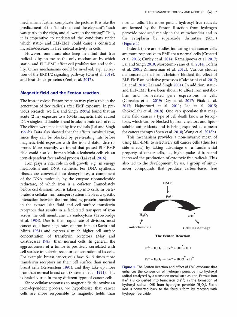
Magnetic field and the Fenton reaction
The iron-involved Fenton reaction may play a role in thegeneration of free radicals after EMF exposure. In pre-vious research, we (Lai and Singh 1997a) found that anacute (2 hr) exposure to a 60-Hz magnetic field causedDNA single and double strand breaks in brain cells of rats.The effects were mediated by free radicals (Lai and Singh1997b). Data also showed that the effects involved iron,since they can be blocked by pre-treating rats beforemagnetic-field exposure with the iron chelator deferri-prone. More recently, we found that pulsed ELF-EMFfield could also kill human Molt-4 leukemia cells via aniron-dependent free radical process (Lai et al. 2016).
Iron plays a vital role in cell growth, e.g., in energymetabolism and DNA synthesis. For DNA synthesis,riboses are converted into deoxyriboses, a componentof the DNA molecule, by the enzyme ribonucleotidereductase, of which iron is a cofactor. Immediatelybefore cell division, iron is taken up into cells. In verte-brates, a cellular iron transport system involves a specificinteraction between the iron-binding protein transferrinin the extracellular fluid and cell surface transferrinreceptors that results in a facilitated transport of ironacross the cell membrane via endocytosis (Trowbridgeet al. 1984). Due to their rapid rate of division, mostcancer cells have high rates of iron intake (Karin andMintz 1981) and express a much higher cell surfaceconcentration of transferrin receptors (May andCuatrecasas 1985) than normal cells. In general, theaggressiveness of a tumor is positively correlated withcell surface transferrin receptor concentration of its cells.For example, breast cancer cells have 5–15 times moretransferrin receptors on their cell surface than normalbreast cells (Reizenstein 1991), and they take up moreiron than normal breast cells (Shterman et al. 1991). Thisis basically true in many different types of cancer cells.
Since cellular responses to magnetic fields involve aniron-dependent process, we hypothesize that cancercells are more responsible to magnetic fields than
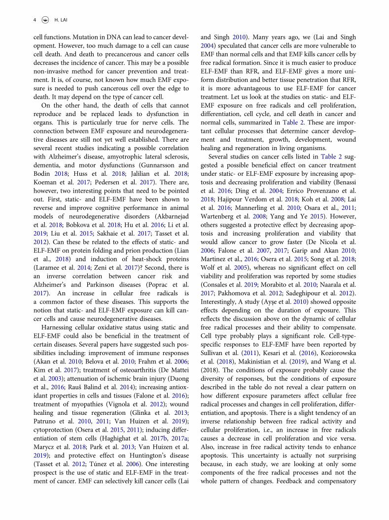
normal cells. The more potent hydroxyl free radicalsare formed by the Fenton Reaction from hydrogenperoxide produced mainly in the mitochondria and inthe cytoplasm by superoxide dismutase (SOD)(Figure 1).
Indeed, there are studies indicating that cancer cellsare more responsive to EMF than normal cells (Crocettiet al. 2013; Curley et al. 2014; Kamalipooya et al. 2017;Lai and Singh 2010; Morotomi-Yano et al. 2014; Tofaniet al. 2001; Zimmerman et al. 2012). Various studiesdemonstrated that iron chelators blocked the effect ofELF-EMF on oxidative processes (Calcabrini et al. 2017;Lai et al. 2016; Lai and Singh 2004). In addition, static-and ELF-EMF have been shown to affect iron metabo-lism and iron-related gene expressions in cells(Consales et al. 2019; Dey et al. 2017; Fitak et al.2017; Hajnorouzi et al. 2011; Lee et al. 2015;Shokrollahi et al. 2018). One can speculate that mag-netic field causes a type of cell death know as ferrop-tosis, which can be blocked by iron chelators and lipid-soluble antioxidants and is being explored as a meanfor cancer therapy (Shen et al. 2018; Wang et al. 2018b).
This mechanism provides a non-invasive mean ofusing ELF-EMF to selectively kill cancer cells (thus lessside effects) by taking advantage of a fundamentalproperty of cancer cells, i.e., high uptake of iron andincreased the production of cytotoxic free radicals. Thisalso led to the development, by us, a group of antic-ancer compounds that produce carbon-based free
Fe2+ + H2O2 Fe3+ + OH.+ OH
Fe3+ + H2O2 Fe2+ + HOO.
+ H+
EMF
Fe2+
mitochondria
H2O2 OH.
Cellular damage
The Fenton Reaction
Figure 1. The Fenton Reaction and effect of EMF exposure thatenhances the conversion of hydrogen peroxide into hydroxylradical catalyzed by a transition metal such as iron. Ferrous iron(Fe2+) is converted into ferric iron (Fe3+) in the formation ofhydroxyl radical (OH) from hydrogen peroxide (H2O2). Ferriciron is converted back to the ferrous form by reacting withhydrogen peroxide.
ELECTROMAGNETIC BIOLOGY AND MEDICINE 7
radicals in the presence of iron (Lai et al. 2013). Thesecompounds have been shown to be very selective andeffective in killing many types of cancer cells. In addi-tion, the concept of the Fenton Reaction also led to theidea of using ELF-MF for treatment of malaria (Feaginet al. 1999; Lai and Singh 2010).
The evolutionary aspects of static- and ELF-EMFin nature and free radicals
For millions of years, living organisms evolved in the pre-sence of environmental static geomagnetic field and naturalextremely low frequency electromagnetic fields, such as thelightning-generated Schumann Resonance. It is not sur-prising that these fields can play important roles in thesurvival of living organisms, e.g., in food foraging, direc-tional cues, and reproduction. Living organisms are verysensitive and responsive to low levels of these EMFs. Thishas been shown in many animal species and affect variousbiological functions, e.g., exploratory behavior in rodents(Malewski et al. 2018); body alignment of dogs (Hart et al.2013a); magnetic alignment in carps (Hart et al. 2012);landing direction of water birds (Hart et al. 2013b); orien-tation of grazing and resting cattle and deer (Begall et al.2008); and cardiovascular and brain activities in humans(Pishchalnikov et al. 2019;Wang et al. 2019). Some animalscan differentiate north and south poles of a magnetic field(known as polarity compass) (Begall et al. 2008; Hart et al.2012, 2013b, 2013a; Malkemper et al. 2015). Another func-tion-related reproduction is the “natal homing behavior”,i.e., an animal returns to its birthplace to reproduce, insome animal species. It is observed in sea turtle (Brothersand Lohmann 2015); eel (Naisbett-Jones et al. 2017); andsalmon (Putman et al. 2014b). Apparently, newborns ofthese animals are imprinted with the memory of the inten-sity and inclination angle of the local geomagnetic field.This information will later be used to locate their place ofbirth. All these traits confer some survival competitivenessto the organisms.
In order for an environmental entity to affect thefunctions of an organism, the following criteria have tobe met. First, the organism should be able to detect theentity. Second, the level of the entity should be similarto those in the normal ambient environment. Third, theorganism must have response mechanisms tuned tocertain parameters of the entity to allow immediatedetection of the presence and changes of the entity.Immediate detection and response to the entity areessential for the survival of the organism.
For the detection of changes in static- and ELF-EMF inthe environment, several mechanisms have been evolved.Some organisms evolved special organs (receptors) todetect environmental EMF. For example, the platypus has
thousands of electric sensors on its bill skin. Using theseelectroreceptors, in interaction with another type of sensorthe mechanoreceptor, the monotreme platypus can detectan electric field of 20 μV/cm (Manger and Pettigrew 1996),which is similar to that produced by the muscles ofa shrimp. The information is processed by the somatosen-sory cortex of the platypus to fix the location of the prey.This type of electroreception is common in all three speciesof monotremes and short bill echidna. Electric fish(Elasmobranch) emits EMF that covers a distance of sev-eral centimeters (Montgomery and Bodznick 1999; von derEmde 1999). Again, this allows the location of a potentialprey by comparing its electrical properties with the vicinity.Their electroreceptors have been shown to detect a field of5 nV/cm. These EMF sensing mechanisms are highly sen-sitive and efficient.
Two other mechanisms have been proposed to accountfor electroreception: magnetite involved in iron-oxidationand radical pair production in certain cellular molecules.In both cases, the generation of reactive oxidative speciesis involved. The radical-pair reaction hypothesis and con-version of the form of radicals (singlet-triplet interconver-sion) in a group of flavoproteins known as cryptochromes(Hore and Mouritsen 2016) in animal species have beenintensively studied. There are reports of the presence ofcryptochromes in plants, which may be responsible forthe effect of EMF on plant growth (Ahmad et al. 2007;Mohammadi et al. 2018). A comprehensible descriptionof this topic is beyond the scope of this paper and theexpertise of this author. Readers are referred to severalpapers on the topic: Barnes and Greenebaum (2015);Binhi and Prato (2017); Galler et al. (2005); Dodsonet al. (2013); Hore (2019); Hore and Mouritsen (2016);Kirschvink et al. (2001); Landler and Keays (2018);Sheppard et al. (2017); and Sherrard et al. (2018).
Thus, the mechanisms described above, electro-recep-tors, magnetites, and radical-pair, enable living organismsto immediately detect the presence and changes in envir-onmental electromagnetic fields of very low intensity. Aneffect that could have dire consequences on species survi-val is that man-made EMFs, with ubiquitous presence inthe recent environment, could disrupt the naturalresponses to nature static- and ELF-EMF. Disruption ofdirectional senses in insects has been reported (Shepherdet al. 2018). Polarity compass also can be disturbed byman-made EMF (Burda etal. 2009; Malkemper et al.2015; Putman et al. 2014a). A study by Engels et al.(2014) showed that magnetic noise (at 2 KHz – 9 MHz,i. e., within the range of AM radio transmission) coulddisrupt magnetic compass orientation in migratoryEuropean robins. The disruption can occur at a very lownoise level of 0.01 V/m (0.0000265 μW/cm2). Similareffects of RFR interference on magnetoreception have
8 H. LAI
also been reported in a night migratory songbird (Aher etal. 2016) and European robin (Wiltschko et al. 2015).
Electro-hypersensitivity in humans (Baliatsas et al.2012) also showed an instantaneous response to EMFand at low intensities. One wonders whether theunderlying mechanisms of electro-hypersensitivityare related to the processes described above. It maybe the remnant of a primordial evolutional response ofhumans to static- and ELF-EMF in the environment.Free radicals may play a role. There is a report ofincreased oxidative stress in electro-hypersensitivityself-reporting patients (Irigaray et al. 2018).
From the discussion above, it is apparent that static-and ELF-EMF that can affect free radical processes atvery low intensities that in turn affect evolution andspecies survival. In the electromagnetic spectrum, theonly other frequency range that biological responsescan occur at very low intensity is the light spectrum.It has been shown that the human visual system issensitive to one photon (Hecht et al. 1942; Pugh2018), i.e., the reaction of one photon with one rho-dopsin molecule in the retina. Apparently, beneficialselection outcomes in evolution have made the livingorganism extremely sensitive to these fields.
Here, let us digress to an unrelated but equally impor-tant topic: i.e., on the biological effects of radiofrequencyradiation (RFR), another segment of the EMF-spectrumthat is being intensively studied. Since the presence of RFRin the ambient environment is new in the evolutionarytime scale, how are living organisms responsive to RFR?No specific cellular detection and response-mechanisms,other than heating, have been discovered. Biologicalresponses to RFR at very low intensities have been reported(e.g., see Table 1 in Levitt and Lai (2010)). But, most ofthose studies were on modulated fields. Is it possible thatthe observed biological responses to RFR are actuallycaused by its ELF-modulations, since almost all environ-mental RFR sources are modulated (see discussion in sec-tion 9 “Effects below 4W/kg: thermal versus nonthermal”in Levitt and Lai (2010))? In the literature, biological effectsof ELF-EMF and RFR are found to be very similar (e.g.,compare the neurological effects of RFR described in Lai(2018) to those of ELF-EMF (see section on “neurologicaleffects of ELF-EMF” Bioinitiative Report (2012))). In thatsense, one can deduce that there is no other significant RFReffect other than thermal effect. Effects of low-level non-modulated continuous-wave (CW) RFR may bea counterargument. However, it is difficult to produce non-modulated RFR in the laboratory, or micro-thermal effectcan occur under CW-RFR exposure. There are only severalstudies showing effects of very low-level CW-RFR (e.g.,with specific absorption rate (SAR) ~10 mW/kg). Positiveresults were reported by de Pomerai et al. (2003)
(aggregation of bovine serum albumin and changes inprotein conformation, 1 GHz CW, 15–20 mW/kg);Marinelli et al. (2004) (cell self-defense responses, 900MHz CW, 3.5 mW/kg); D’Inzeo et al. (1988) (acetylcho-line-related ion channel, 10.75 GHz CW, 8 mW/kg);Persson et al. (1997) (blood-brain barrier permeability,915 MHz CW, 0.4 mW/kg); and Tattersall et al. 2001)(hippocampal functions, 700 MHz CW, 1.6 mW/kg).Somosy et al. (1991) (molecular and structural changes incells of mouse embryos, 2450 MHz) reported that modu-lated radiation (effect observed at 2.4 mW/kg) is morepotent that CW radiation (effect observed at 240 mW/kg). Navakitkia and Tomashevskaya (1994) (behavioraland endocrine changes, 2450 MHz, 2.7 mW/kg with mod-ulation), Schwartz et al. (1990) (calcium movement inheart, 2450 MHz, 0.15 mW/kg with modulation) andWolke et al. (1996) (calcium concentration in heart musclecells, 900 MHz, 1 mW/kg with modulation) reportedeffects with modulated radiation and not with CW radia-tion. The SARs given were averaged SARs. There is a studythat showed DNA damage in human glial cells after expo-sure to a 50 Hz-modulated 900-MHz RFR but not to CWfield. In that study, only an exposure power density of 26μW/cm2 was provided. Other than the Persson et al. (1997)study, all the other studies were carried out in vitro.Interestingly, the Persson et al. (1997) paper reported thatCW- is more potent than modulated-915 MHz radiationon increasing blood-brain barrier permeability. Certainly,the RF-carrier could affect the distribution of energy in theexposed subject. And, the pattern of energy distributioncan affect biological responses to EMF (Lai et al. 1984). Theconcept of interaction of modulation with the RF-carrier isnot new. It was shown in an earlier study (Bawin et al.1978) that 6- and 16-Hz amplitude-modulated 147-MHzRFR (0.8 mW/cm2) increased calcium efflux from chickcerebral tissues, whereas 6- and 16-Hz fields alone causeda decrease in efflux. Lastly, I like to point out that there arenot enough research data to support the popular belief that“modulated is more biologically potent that non-modulated RFR.”More experiments using the same expo-sure set-up with continuous-wave and modulated RFR ofthe same frequency and with intensities that produce thesame averaged SAR are needed to reach such a conclusion.
Concluding remarks
(1) Change in cellular free radical activity is one ofthe most consistent effects of static- and ELF-EMF on living organisms.
(2) The mechanisms by which static- and ELF-EMF affect cellular free radical processes isnot well understood. The “radical pair” hypoth-esis is a likely candidate, particularly the
ELECTROMAGNETIC BIOLOGY AND MEDICINE 9
involvement of cryptochromes. It allowsimmediate detection and response to changesin static- and ELF-EMF in the environment,which likely play important roles in the evolu-tion of living organisms.
(3) Oxidative responses to static- and ELF-EMF areprobably dependent on the characteristic of thefield and exposure (such as frequency, modula-tion, and duration) and the exposed object (suchas cell type, and states of biological activities),
(4) However, chronic exposure that leads to theexcessive and persistent presence of free radi-cals can cause oxidative stress and should beavoided.
(5) Effects of static- and ELF-EMF on free radicalsprobably have beneficial health effects particu-larly relating to cell proliferation, differentia-tion, cell death, and cell cycle. These areeffects that could influence cancer developmentand treatment, growth and development,regeneration, and healing.
(6) In future research, it is imperative to identifythe field parameters that can selectively causebeneficial or detrimental health effects.
Conflict of interest statement
The author declares no conflict of interest.
References
Abuasbi, F., A. Lahham, and I. R. Abdel-Raziq. 2018a.Residential exposure to extremely low frequency electricand magnetic fields in the city of Ramallah-Palestine.Radiat. Prot. Dosimetry. 179:49–57. doi:10.1093/rpd/ncx209.
Abuasbi, F., A. Lahham, and I. R. Abdel-Raziq. 2018b. Levelsof extremely low-frequency electric and magnetic fieldsfrom overhead power lines in the outdoor environmentof Ramallah City- Palestine. Radiat. Prot. Dosimetry.179:229–32. doi:10.1093/rpd/ncx259.
Aher, Y. D., S. Subramaniyan, B. Shanmugasundaram,A. Sase, S. R. Saroja, M. Holy, H. Höger, T. Beryozkina,H. H. Sitte, J. J. Leban, et al. 2016. A novel heterocycliccompound CE-104 enhances spatial working memory inthe radial arm maze in rats and modulates the dopaminer-gic system. Front. Behav. Neurosci. 10:55. doi:10.3389/fnbeh.2016.00020.
Ahmad, M., P. Galland, T. Ritz, and R. Wiltschko. 2007.Magnetic intensity affects cryptochrome-dependentresponses in Arabidopsis thaliana. Planta 225:615–24.doi:10.1007/s00425-006-0416-8.
Akan, Z., B. Aksu, A. Tulunay, S. Bilsel, and A. Inhan-Garip.2010. Extremely low-frequency electromagnetic fieldsaffect the immune response of monocyte-derived
macrophages to pathogens. Bioelectromagnetics31:603–12. doi:10.1002/bem.20607.
Akbarnejad, Z., K. Esmaeilpour, M. Shabani, M. Asadi-Shekaari, M. Saeedi Goraghani, and M. Ahmadi-Zeidabadi.2018. Spatial memory recovery in Alzheimer’s rat model byelectromagnetic field exposure. Int. J. Neurosci. 128:691–96.doi:10.1080/00207454.2017.1411353.
Akpınar, D., D. K. Gok, E. Hidisoglu, M. Aslan, S. Ozen,A. Agar, and P. Yargicoglu. 2016. Effects of pre- and postnatalexposure to extremely low-frequency electric fields on mis-match negativity component of the auditory event-relatedpotentials: Relation to oxidative stress. Electromagn. Biol.Med. 35:245–59. doi:10.3109/15368378.2015.1076727.
Akpinar, D., N. Ozturk, S. Ozen, A. Agar, and P. Yargicoglu.2012. The effect of different strengths of extremelylow-frequency electric fields on antioxidant status, lipidperoxidation, and visual evoked potentials. Electromagn.Biol. Med. 31:436–48. doi:10.3109/15368378.2012.692342.
Ansari, A. M., S. Farzampour, A. Sadr, B. Shekarchi, andK. Majidzadeh-A. 2016. Effects of short term and longterm extremely low frequency magnetic field on depressivedisorder in mice: Involvement of nitric oxide pathway. LifeSci. 146:52–57. doi:10.1016/j.lfs.2015.12.055.
Ayşe, I.-G., A. Zafer, O. Sule, I.-T. Işil, and T. Kalkan. 2010.Differentiation of K562 cells under ELF-EMF applied atdifferent time courses. Electromagn. Biol. Med. 29:122–30.doi:10.3109/15368378.2010.502451.
Baliatsas, C., I. Van Kamp, E. Lebret, and G. J. Rubin. 2012.Idiopathic environmental intolerance attributed to electro-magnetic fields (IEI-EMF): A systematic review of identi-fying criteria. BMC Public Health 12:643. doi:10.1186/1471-2458-12-643.
Barnes, F. S., and B. Greenebaum. 2015. The effects of weakmagnetic fields on radical pairs. Bioelectromagnetics36:45–54. doi:10.1002/bem.21883.
Bawin, S. M., A. Sheppard, and W. R. Adey. 1978. Possiblemechanisms of weak electromagnetic field coupling inbrain tissue. Bioelectrochem. Bioenerg. 5:67–76.doi:10.1016/0302-4598(87)87008-3.
Bawin, S. M., W. M. Satmary, R. A. Jones, W. R. Adey, andG. Zimmerman. 1996. Extremely-low-frequency magneticfields disrupt rhythmic slow activity in rat hippocampalslices. Bioelectromagnetics 17:388–95. doi:10.1002/(SICI)1521-186X(1996)17:5<388::AID-BEM6>3.0.CO;2-#.
Bediz, C. S., A. K. Baltaci, R. Mogulkoc, and E. Oztekin. 2006.Zinc supplementation ameliorates electromagneticfield-induced lipid peroxidation in the rat brain. TohokuJ. Exp. Med. 208:133–40. doi:10.1620/tjem.208.133.
Begall, S., J. Cerveny, J. Neef, O. Vojtech, and H. Burda. 2008.Magnetic alignment in grazing and resting cattle and deer.Proc. Natl. Acad. Sci. U.S.A. 105:13451–5. doi:10.1073/pnas.0803650105.
Belova, N. A., M. M. Potselueva, L. K. Srebnitskaya,A. V. Znobishcheva, and V. V. Lednev. 2010. The influenceof weak magnetic fields on the production of the reactiveoxygen species in peritoneal neutrophils of mice. Biophysics(Biofizika) 55:586–91. doi:10.1134/S0006350910040123.
Benassi, B., G. Filomeni, C. Montagna, C. Merla, V. Lopresto,R. Pinto, C. Marino, and C. Consales. 2016. Extremely lowfrequency magnetic field (ELF-MF) exposure sensitizes SH-SY5Y cells to the Pro-Parkinson’s disease toxin MPP(.).Mol.Neurobiol. 53:4247–60. doi:10.1007/s12035-015-9354-4.
10 H. LAI
Binhi, V. N., and F. S. Prato. 2017. A physical mechanism ofmagnetoreception: Extension and analysis.Bioelectromagnetics38:41–52. doi:10.1002/bem.22011.
Bioinitiative Report 2012. Edited by Sage C and Carpenter D.Accessed April, 2019: https://www.bioinitiative.org/.
Bobkova, N. V., V. V. Novikov, N. I. Medvinskaya,I. Y. Aleksandrova, I. V. Nesterova, and E. E. Fesenko.2018. Effect of weak combined static and extremely low-frequency alternating magnetic fields on spatial memoryand brain amyloid-β in two animal models of Alzheimer’sdisease. Electromagn. Biol. Med. 37:127–37. doi:10.1080/15368378.2018.1471700.
Brothers, J. R., and K. J. Lohmann. 2015. Evidence for geo-magnetic imprinting and magnetic navigation in the natalhoming of sea turtles. Curr. Biol. 25:392–96. doi:10.1016/j.cub.2014.12.035.
Budziosz, J., A. Stanek, A. Sieroń, J. Witkoś, A. Cholewka,and K. Sieroń. 2018. Effects of low-frequency electromag-netic field on oxidative stress in selected structures of thecentral nervous system. Oxid Med Cell Longev 2018:1–8.doi:10.1155/2018/1427412.
Burda, H., S. Begall, J. Cervený, J. Neef, and P. Nemec. 2009.Extremely low-frequency electromagnetic fields disrupt mag-netic alignment of ruminants. Proc. Natl. Acad. Sci. U.S.A.106:5708–13. doi:10.1073/pnas.0811194106.
Bürgi, A., S. Sagar, B. Struchen, S. Joss, and M. Röösli. 2017.Exposure modelling of extremely low-frequency magneticfields from overhead power lines and its validation bymeasurements. Int. J. Environ. Res. Public. Health. 14:pii:E949. doi:10.3390/ijerph14090949.
Calcabrini, C., U. Mancini, R. De Bellis, A. R. Diaz,M. Martinelli, L. Cucchiarini, P. Sestili, V. Stocchi, andL. Potenza. 2017. Effect of extremely low-frequency elec-tromagnetic fields on antioxidant activity in the humankeratinocyte cell line NCTC 2544. Biotechnol. Appl.Biochem. 64:415–22. doi:10.1002/bab.1495.
Calota, V., S. Dragoiu, A. Meghea, and M. Giurginca. 2006.Decrease of luminol chemiluminescence upon exposure ofhuman blood serum to 50 Hz electric fields.Bioelectrochemistry 69:126–27. doi:10.1016/j.bioelechem.2005.12.006.
Cichoń, N., M. Bijak, E. Miller, J. Saluk, C. S. Bediz,A. K. Baltaci, R. Mogulkoc, and E. Oztekin. 2017a.Extremely low frequency electromagnetic field(ELF-EMF) reduces oxidative stress and improves func-tional and psychological status in ischemic stroke patients.Bioelectromagnetics 38:386–96. doi:10.1002/bem.v38.5.
Cichoń, N., P. Czarny, M. Bijak, E. Miller, T. Śliwiński,J. Szemraj, and J. Saluk-Bijak. 2017b. Benign effect ofextremely low-frequency electromagnetic field on brainplasticity assessed by nitric oxide metabolism during post-stroke rehabilitation. Oxid Med Cell Longev 2017:1–9.doi:10.1155/2017/2181942.
Cichoń, N., P. Rzeźnicka, M. Bijak, E. Miller, S. Miller, andJ. Saluk. 2018. Extremely low frequency electromagneticfield reduces oxidative stress during the rehabilitation ofpost-acute stroke patients. Adv. Clin. Exp. Med.27:1285–93. doi:10.17219/acem/73699.
Ciejka, E. B., and A. Goraca. 2009. The influence oflow-frequency magnetic field on plasma antioxidant capa-city and heart rate. Wiad. Lek. 62:81–86.
Consales, C., M. Panatta, A. Butera, G. Filomeni, C. Merla,M. T. Carrì, C. Marino, and B. Benassi. 2019. 50-Hzmagnetic field impairs the expression of iron-relatedgenes in the in vitro SOD1G93A model of amyotrophiclateral sclerosis. Int. J. Radiat. Biol. 95:368–77. doi:10.1080/09553002.2019.1552378.
Crocetti, S., C. Beyer, G. Schade, M. Egli, J. Fröhlich,A. Franco-Obregón, and I. Ulasov. 2013. Low intensityand frequency pulsed electromagnetic fields selectivelyimpair breast cancer cell viability. PLoS ONE 8:e72944.doi:10.1371/journal.pone.0072944.
Cui, Y., Z. Ge, J. D. Rizak, C. Zhai, Z. Zhou, S. Gong, Y. Che, andP. A. Adlard. 2012. Deficits in water maze performance andoxidative stress in the hippocampus and striatum induced byextremely low frequency magnetic field exposure. PLoS ONE7:e32196. doi:10.1371/journal.pone.0032196.
Curley, S. A., F. Palalon, K. E. Sanders, and N. Koshkina.2014. The effects of non-invasive radiofrequency treatmentand hyperthermia on malignant and nonmalignant cells,Int. Int. J. Environ. Res. Public. Health. 11:9142–53.doi:10.3390/ijerph110909142.
D’Inzeo, G., P. Bernardi, F. Eusebi, F. Grassi, C. Tamburello,and B. M. Zani. 1988. Microwave effects onacetylcholine-induced channels in cultured chickmyotubes. Bioelectromagnetics 9:363–72.
De Mattei, M., M. Pasello, A. Pellati, G. Stabellini, L. Massari,D. Gemmati, and A. Caruso. 2003. Effects of electromag-netic fields on proteoglycan metabolism of bovine articularcartilage explants. Connect. Tissue Res. 44:154–59.
De Nicola, M., S. Cordisco, C. Cerella, M. C. Albertini,M. D’Alessio, A. Accorsi, A. Bergamaschi, A. Magrini,and L. Ghibelli. 2006. Magnetic fields protect from apop-tosis via redox alteration. Ann. N. Y. Acad. Sci. 1090:59–68.doi:10.1196/annals.1378.006.
de Pomerai, D. I., B. Smith, A. Dawe, K. North, T. Smith,D. B. Archer, I. R. Duce, D. Jones, and E. P. M. Candido.2003. Microwave radiation can alter protein conformationwithout bulk heating. FEBS Lett. 543:93–97. doi:10.1016/s0014-5793(03)00413-7.
Deng, Y., Y. Zhang, S. Jia, J. Liu, Y. Liu, W. Xu, and L. Liu.2013. Effects of aluminum and extremely low frequencyelectromagnetic radiation on oxidative stress and memoryin brain of mice. Biol. Trace Elem. Res. 156:243–52.doi:10.1007/s12011-013-9847-9.
Dey, S., S. Bose, S. Kumar, R. Rathore, R. Mathur, and S. Jain.2017. Extremely low frequency magnetic field protectsinjured spinal cord from the microglia- and iron-inducedtissue damage. Electromagn. Biol. Med. 36:330–40.doi:10.1080/15368378.2017.1389750.
Di Loreto, S., S. Falone, V. Caracciolo, P. Sebastiani,A. D’Alessandro, A. Mirabilio, V. Zimmitti, and F. Amicarelli.2009. Fifty hertz extremely low-frequency magnetic field expo-sure elicits redox and trophic response in rat-cortical neurons.J. Cell Physiol. 219:334–43. doi:10.1002/jcp.21674.
Ding, G.-R., T. Nakahara, H. Hirose, S. Koyama,Y. Takashima, and J. Miyakoshi. 2004. Extremely lowfrequency magnetic fields and the promotion of H2O2--induced cell death in HL-60 cells. Int. J. Radiat. Biol.80:317–24. doi:10.1080/09553000410001679802.
Djordjevic, N. Z., M. G. Paunović, and A. S. Peulić. 2017.Anxiety-like behavioural effects of extremely low-frequency
ELECTROMAGNETIC BIOLOGY AND MEDICINE 11
electromagnetic field in rats. Environ. Sci. Pollut. Res. Int.24:21693–99. doi:10.1007/s11356-017-9710-1.
Dodson, C. A., P. J. Hore, and M. I. Wallace. 2013. A radicalsense of direction: Signalling and mechanism in crypto-chrome magnetoreception. Trends Biochem. Sci.38:435–46. doi:10.1016/j.tibs.2013.07.002.
Duan, Y., Z. Wang, H. Zhang, Y. He, R. Lu, R. Zhang, G. Sun,and X. Sun. 2013. The preventive effect of lotus seedpodprocyanidins on cognitive impairment and oxidativedamage induced by extremely low frequency electromag-netic field exposure. Food Funct 4:1252–62. doi:10.1039/c3fo60116a.
Duong, C. N., and J. Y. Kim. 2016. Exposure to electromag-netic field attenuates oxygen-glucose deprivation-inducedmicroglial cell death by reducing intracellular Ca2+ andROS. Int. J. Radiat. Biol. 92:195–201. doi:10.3109/09553002.2016.1136851.
Ehnert, S., A.-K. Fentz, A. Schreiner, J. Birk, B. Wilbrand,P. Ziegler, M. K. Reumann, H. Wang, K. Falldorf, andA. K. Nussler. 2017. Extremely low frequency pulsed elec-tromagnetic fields cause antioxidative defense mechanismsin human osteoblasts via induction of •O2
− and H2O2. Sci.Rep. 7:14544. doi:10.1038/s41598-017-14983-9.
Emre, M., S. Cetiner, S. Zencir, I. Unlukurt, I. Kahraman, andZ. Topcu. 2011. Oxidative stress and apoptosis in relationto exposure to magnetic field. Cell. Biochem. Biophys.59:71–77. doi:10.1007/s12013-010-9113-0.
Engels, S., N.-L. Schneider, N. Lefeldt, C. M. Hein, M. Zapka,A. Michalik, D. Elbers, A. Kittel, P. J. Hore, andH. Mouritsen. 2014. Anthropogenic electromagnetic noisedisrupts magnetic compass orientation in a migratory bird.Nature 509:353–56. doi:10.1038/nature13290.
Errico Provenzano, A., S. Amatori, M. G. Nasoni, G. Persico,S. Russo, A. R. Mastrogiacomo, A. Gambarara, andM. Fanelli. 2018. Effects of fifty-hertz electromagneticfields on granulocytic differentiation of ATRA-treatedacute promyelocytic leukemia NB4 cells. Cell. Physiol.Biochem. 46:389–400. doi:10.1159/000488473.
Falone, S., M. R. Grossi, B. Cinque, B. D’Angelo,E. Tettamanti, A. Cimini, C. Di Ilio, and F. Amicarelli.2007. Fifty hertz extremely low-frequency electromagneticfield causes changes in redox and differentiative status inneuroblastoma cells. Int. J. Biochem. Cell. Biol.39:2093–106. doi:10.1016/j.biocel.2007.06.001.
Falone, S., N. Marchesi, C. Osera, L. Fassina, S. Comincini,M. Amadio, and A. Pascale. 2016. Pulsed electromagneticfield (PEMF) prevents pro-oxidant effects of H2O2 inSK-N-BE(2) human neuroblastoma cells. Int. J. Radiat.Biol. 92:281–86. doi:10.3109/09553002.2016.1150619.
Falone, S., S. Santini Jr., V. Cordone, P. Cesare, A. Bonfigli,M. Grannonico, G. Di Emidio, C. Tatone, M. Cacchio, andF. Amicarelli. 2017. Power frequency magnetic field pro-motes a more malignant phenotype in neuroblastoma cellsvia redox-related mechanisms. Sci. Rep. 7:11470.doi:10.1038/s41598-017-11869-8.
Feagin, J. E., M. A. Wurscher, and C. Ramon. 1999. Magneticfields and malaria. In M. F. Holick and E. G. Jung, eds.“Biologic Effects of Light: Proceedings of the BiologicEffects of Light Symposium” (pp.343–49), KluwerAcademic Publishers, Hingham, MA, USA.
Feng, B., C. Ye, L. Qiu, L. Chen, Y. Fu, and W. Sun. 2016.Mitochondrial ROS release and subsequent Akt Activation
potentially mediated the anti-apoptotic effect of a 50-Hzmagnetic field on FL cells. Cell. Physiol. Biochem.38:2489–99. doi:10.1159/000445599.
Fernie, K. J., and D. M. Bird. 2001. Evidence of oxidative stressin American kestrels exposed to electromagnetic fields.Environ. Res. 86:198–207. doi:10.1006/enrs.2001.4263.
Fitak, R. R., B. R. Wheeler, D. A. Ernst, K. J. Lohmann, andS. Johnsen. 2017. Candidate genes mediating magnetore-ception in rainbow trout (Oncorhynchus mykiss). Biol. Lett13:pii: 20170142. doi:10.1098/rsbl.2017.0142.
Fitzsimmons, R. J., S. L. Gordon, J. Kronberg, T. Ganey, andA. A. Pilla. 2008. A pulsing electric field (PEF) increaseshuman chondrocyte proliferation through a transductionpathway involving nitric oxide signaling. J. Orthop. Res.26:854–59. doi:10.1002/jor.20590.
Frahm, J., M. Lantow, M. Lupke, D. G. Weiss, and M. Simkó.2006. Alteration in cellular functions in mouse macro-phages after exposure to 50 Hz magnetic fields. J. Cell.Biochem. 99:168–77. doi:10.1002/jcb.20920.
Galler, S., B. G. Wang, and M. Kawai. 2005. Elementary stepsof the cross-bridge cycle in fast-twitch fiber types fromrabbit skeletal muscles. Biophys. J. 89:3248–60.doi:10.1529/biophysj.104.056614.
Garip, A. I., and Z. Akan. 2010. Effect of ELF-EMF onnumber of apoptotic cells; correlation with reactive oxygenspecies and HSP. Acta Biol. Hung. 61:158–67. doi:10.1556/ABiol.61.2010.2.4.
Ghodbane, S., M. Ammari, A. Lahbib, M. Sakly, andH. Abdelmelek. 2015. Static magnetic fieldexposure-induced oxidative response andcaspase-independent apoptosis in rat liver: Effect of sele-nium and vitamin E supplementations. Environ. Sci. Pollut.Res. Int. 22:16060–66. doi:10.1007/s11356-015-4802-2.
Giorgi, G., C. Pirazzini, M. G. Bacalini, C. Giuliani,P. Garagnani, M. Capri, F. Bersani, and B. Del Re. 2017.Assessing the combined effect of extremely low-frequencymagnetic field exposure and oxidative stress on LINE-1 pro-moter methylation in human neural cells. Radiat. Environ.Biophys. 56:193–200. doi:10.1007/s00411-017-0683-8.
Glinka, M., A. Sieroń, E. Birkner, and G. Cieślar. 2013.Influence of extremely low-frequency magnetic field onthe activity of antioxidant enzymes during skin woundhealing in rats. Electromagn. Biol. Med. 32:463–70.doi:10.3109/15368378.2012.743906.
Gourzoulidis, G. A., P. Tsaprouni, Ν. Skamnakis,C. Tzoumanika, E. Kalampaliki, E. Karastergios,A. Gialofas, A. Achtipis, C. Kappas, and E. Karabetsos.2018. Occupational exposure to electromagnetic fields.The situation in Greece. Phys. Med 49:83–89.doi:10.1016/j.ejmp.2018.05.011.
Guler, G., Z. Turkozer, A. Tomruk, and N. Seyhan. 2008. Theprotective effects of N-acetyl-L-cysteine andepigallocatechin-3-gallate on electric field-induced hepaticoxidative stress. Int. J. Radiat. Biol. 84:669–80. doi:10.1080/09553000802241747.
Güler, G., Z. Türközer, E. Ozgur, A. Tomruk, N. Seyhan, andÇ. Karasu. 2009a. Protein oxidation under extremely lowfrequency electric field in guinea pigs. Effect of N-acetyl-L-cysteine treatment. Gen. Physiol. Biophys. 28:47–55.doi:10.4149/gpb_2009_01_47.
Güler, G., Z. Türközer, E. Ozgur, and N. Seyhan. 2009b.Antioxidants alleviate electric field-induced effects on
12 H. LAI
lung tissue based on assays of heme oxygenase-1, proteincarbonyl content, malondialdehyde, nitric oxide, andhydroxyproline. Sci. Total Environ. 407:1326–32.doi:10.1016/j.scitotenv.2008.10.050.
Gunnarsson, L. G., and L. Bodin. Oct 26 2018. Amyotrophiclateral sclerosis and occupational exposures: A systematicliterature review and meta-analyses. Int. J. Environ. Res.Public Health. 15:pii: E2371. doi:10.3390/ijerph15061188.
Haghighat, N., P. Abdolmaleki, J. Parnian, andM. Behmanesh. 2017a. The expression of pluripotencyand neuronal differentiation markers under the influenceof electromagnetic field and nitric oxide. Mol. Cell.Neurosci. 85:19–28. doi:10.1016/j.mcn.2017.08.005.
Haghighat, N., P. Abdolmaleki, M. Behmanesh, andM. Satari. 2017b. Stable morphological-physiological andneural protein expression changes in rat bone marrowmesenchymal stem cells treated with electromagnetic fieldand nitric oxide. Bioelectromagnetics 38:592–601.doi:10.1002/bem.v38.8.
Hajipour Verdom, B., P. Abdolmaleki, and M. Behmanesh.2018. The static magnetic field remotely boosts the effi-ciency of doxorubicin through modulating ROS behaviors.Sci. Rep. 8:990. doi:10.1038/s41598-018-19247-8.
Hajnorouzi, A., M. Vaezzadeh, F. Ghanati, H. Jamnezhad,and B. Nahidian. 2011. Growth promotion and a decreaseof oxidative stress in maize seedlings by a combination ofgeomagnetic and weak electromagnetic fields. J. PlantPhysiol. 168:1123–28. doi:10.1016/j.jplph.2010.12.003.
Harakawa, S., N. Inoue, T. Hori, K. Tochio, T. Kariya,K. Takahashi, F. Doge, H. Suzuki, and H. Nagasawa.2005. Effects of a 50 Hz electric field on plasma lipidperoxide level and antioxidant activity in rats.Bioelectromagnetics 26:589–94. doi:10.1002/bem.20137.
Hart, V., E. P. Malkemper, T. Kušta, S. Begall, P. Nováková,V. Hanzal, L. Pleskač, M. Ježek, R. Policht, V. Husinec, et al.2013b. Directional compass preference for landing in waterbirds. Front. Zool. 10:38. doi:10.1186/1742-9994-10-38.
Hart, V., P. Nováková, E. P. Malkemper, S. Begall, V. Hanzal,M. Ježek, T. Kušta, V. Němcová, J. Adámková,K. Benediktová, et al. 2013a. Dogs are sensitive to smallvariations of the Earth’s magnetic field. Front. Zool. 10:80.doi:10.1186/1742-9994-10-80.
Hart, V., T. Kušta, P. Němec, V. Bláhová, M. Ježek,P. Nováková, S. Begall, J. Červený, V. Hanzal,E. P. Malkemper, et al. 2012. Magnetic alignment incarps: Evidence from the Czech Christmas fish market.PLoS ONE 7:e51100. doi:10.1371/journal.pone.0051100.
Hecht, S., S. Shlaer, and M. H. Pirenne. 1942. Energy, quanta,and vision. J. Gen. Physiol 25:819–40. doi:10.1085/jgp.25.6.819.
Hore, P. J. Feb 2019. Upper bound on the biological effects of50/60 Hz magnetic fields mediated by radical pairs. Elife25:pii: e44179. doi: 10.7554/eLife.44179.
Hore, P. J., and H. Mouritsen. 2016. The radical-pairmechanism of magnetoreception. Annu. Rev. Biophys.45:299–344. doi:10.1146/annurev-biophys-032116-094545.
Hu, Y., J. Lai, B. Wan, X. Liu, Y. Zhang, J. Zhang, D. Sun,G. Ruan, E. Liu, G.-P. Liu, et al. 2016. Long-term exposureto ELF-MF ameliorates cognitive deficits and attenuates tauhyperphosphorylation in 3xTg AD mice. NeuroToxicology53:290–300. doi:10.1016/j.neuro.2016.02.012.
Huss, A., S. Peters, and R. Vermeulen. 2018. Occupationalexposure to extremely low-frequency magnetic fields andthe risk of ALS: A systematic review and meta-analysis.Bioelectromagnetics 39:156–63. doi:10.1002/bem.22104.
Ilonen, K., A. Markkanen, G. Mezei, and J. Juutilainen. 2008.Indoor transformer stations as predictors of residentialELF magnetic field exposure. Bioelectromagnetics29:213–18. doi:10.1002/bem.20385.
Irigaray, P., D. Caccamo, and D. Belpomme. 2018. Oxidativestress in electrohypersensitivity self-reporting patients:Results of a prospective in vivo investigation with compre-hensive molecular analysis. Int. J. Mol. Med. 42:1885–98.doi:10.3892/ijmm.2018.3774.
Jajte, J., M. Zmyślony, J. Palus, E. Dziubałtowska, andE. Rajkowska. 2001. Protective effect of melatonin againstin vitro iron ions and 7 mT 50 Hz magnetic field-inducedDNA damage in rat lymphocytes. Mutat. Res. 483:57–64.doi:10.1016/s0027-5107(01)00230-5.
Jalilian, H., S. H. Teshnizi, M. Röösli, and M. Neghab. 2018.Occupational exposure to extremely low frequency mag-netic fields and risk of Alzheimer disease: A systematicreview and meta-analysis. NeuroToxicology 69:242–52.doi:10.1016/j.neuro.2017.12.005.
Jeong, J. H., C. Kum, H. J. Choi, E. S. Park, and U. D. Sohn.2006. Extremely low frequency magnetic field induceshyperalgesia in mice modulated by nitric oxide synthesis.Life Sci. 78:1407–12. doi:10.1016/j.lfs.2005.07.006.
Kamalipooya, S., P. Abdolmaleki, Z. Salemi, F. Javani Jouni,J. Zafari, and H. Soleimani. 2017. Simultaneous applicationof cisplatin and static magnetic field enhances oxidativestress in HeLa cell line. In Vitro Cell. Dev. Biol. Anim.53:783–90. doi:10.1007/s11626-017-0148-z.
Kantar Gok, D., D. Akpinar, P. Yargicoglu, S. Ozen,M. Aslan, N. Demir, N. Derin, and A. Agar. 2014. Effectsof extremely low-frequency electric fields at differentintensities and exposure durations on mismatchnegativity. Neuroscience 272:154–66. doi:10.1016/j.neuroscience.2014.04.056.
Kapri-Pardes, E., T. Hanoch, G. Maik-Rachline, M. Murbach,P. L. Bounds, N. Kuster, and R. Seger. 2017. Activation ofsignaling cascades by weak extremely low frequency elec-tromagnetic fields. Cell. Physiol. Biochem. 43:1533–46.doi:10.1159/000481977.
Karimi, S. A., I. Salehi, T. Shykhi, S. Zare, and A. Komaki. 2019.Effects of exposure to extremely low-frequency electromag-netic fields on spatial and passive avoidance learning andmemory, anxiety-like behavior and oxidative stress in malerats. Behav. Brain Res. 359:630–38. doi:10.1016/j.bbr.2018.10.002.
Karin, M., and B. Mintz. 1981. Receptor-mediated endocyto-sis of transferrin in developmentally totipotent mouse ter-atocarcinoma stem cells. J. Biol. Chem. 256:3245–52.
Kavaliers, M., E. Choleris, F. S. Prato, and K. Ossenkopp.1998. Evidence for the involvement of nitric oxide andnitric oxide synthase in the modulation ofopioid-induced antinociception and the inhibitory effectsof exposure to 60-Hz magnetic fields in the land snail.Brain Res. 809:50–57. doi:10.1016/s0006-8993(98)00844-0.
Kesari, K. K., J. Juutilainen, J. Luukkonen, and J. Naarala. Jan2016. Induction of micronuclei and superoxide productionin neuroblastoma and glioma cell lines exposed to weak 50
ELECTROMAGNETIC BIOLOGY AND MEDICINE 13
Hz magnetic fields. J R Soc Interface 13:pii: 20150995. doi:10.1098/rsif.2015.0995.
Kim, S. J., Y. W. Jang, K. E. Hyung, D. K. Lee, K. H. Hyun,S. H. Jeong, K. H. Min, W. Kang, J. H. Jeong, S.-Y. Park,et al. 2017. Extremely low-frequency electromagnetic fieldexposure enhances inflammatory response and inhibitseffect of antioxidant in RAW 264.7 cells.Bioelectromagnetics 38:374–85. doi:10.1002/bem.22049.
Kirschvink, J. L., M. M. Walker, and C. E. Diebel. 2001.Magnetite-based magnetoreception. Curr. Opin. Neurobiol.11:462–67. doi:10.1016/S0959-4388(00)00235-X.
Koeman, T., P. Slottje, L. J. Schouten, S. Peters, A. Huss,J. H. Veldink, H. Kromhout, P. A. van Den Brandt, andR. Vermeulen. 2017. Occupational exposure and amyo-trophic lateral sclerosis in a prospective cohort. Occup.Environ. Med. 74:578–85. doi:10.1136/oemed-2016-103780.
Koh, E. K., B.-K. Ryu, D.-Y. Jeong, I.-S. Bang, M. H. Nam,and K.-S. Chae. 2008. A 60-Hz sinusoidal magnetic fieldinduces apoptosis of prostate cancer cells through reactiveoxygen species. Int. J. Radiat. Biol. 84:945–55. doi:10.1080/09553000802460206.
Koyama, S., T. Sakurai, T. Nakahara, and J. Miyakoshi. 2008.Extremely low frequency (ELF) magnetic fields enhancechemically induced formation of apurinic/apyrimidinic(AP)sites in A172 cells. Int. J. Radiat. Biol. 84:53–59.doi:10.1080/09553000701616064.
Koziorowska, A., M. Romerowicz-Misielak, P. Sołek, andM. Koziorowski. 2018. Extremely low frequency variableelectromagnetic fields affect cancer and noncancerous cellsin vitro differently: Preliminary study. Electromagn. Biol.Med. 37:35–42. doi:10.1080/15368378.2017.1408021.
Lai, H. 2018. A summary of recent literature (2007-2017) onneurobiological effects of radiofrequency radiation. InMobile Communications and Public Health, ed.M. Markov, 187–222. Boca Raton, FL, USA: CRC Press.
Lai, H., A. Horita, C. K. Chou, and A. W. Guy. 1984. Effectsof acute low-level microwaves on pentobarbital-inducedhypothermia depend on exposure orientation.Bioelectromagnetics 5:203–11.
Lai, H., H. W. Chan, and N. P. Singh. 2016. Effects ofradiation from a radiofrequency identification (RFID)microchip on human cancer cells. Int. J. Rad. Biol.92:156–61. doi:10.3109/09553002.2016.1135264.
Lai, H., and N. P. Singh. 1997a. Acute exposure to a 60-Hzmagnetic field increases DNA strand breaks in rat brain cells.Bioelectromagnetics 18:156–65. doi:10.1002/(ISSN)1521-186X.
Lai, H., and N. P. Singh. 1997b. Melatonin and N-tert-butyl-?-phenylnitrone block 60-Hz magnetic field-inducedDNA single and double strand breaks in rat brain cells.J. Pineal Res 22:152–62. doi:10.1111/jpi.1997.22.issue-3.
Lai, H., and N. P. Singh. 2004. Magnetic field-induced DNAstrand breaks in brain cells of the rat. Environ. HealthPerspect. 112:687–94. doi:10.1289/ehp.6355.
Lai, H., and N. P. Singh 2010. Medical applications of elec-tromagnetic fields. Institute of Physics Conference Series:Earth and Environmental Science, London, UK, 10 012006(doi: 10.1088/1755-1315/10/1/012006)
Lai, H., N. P. Singh, and T. Sasaki. 2013. Development ofartemisinin compounds for cancer treatment. Invest NewDrugs 31:230–46. doi:10.1007/s10637-012-9873-z.
Landler, L., and D. A. Keays. 2018. Cryptochrome: The mag-netosensor with a sinister side? PLoS Biol. 16:e3000018.doi:10.1371/journal.pbio.3000018.
Laramee, C. B., P. Frisch, K. McLeod, and G. C. Li. 2014.Elevation of heat shock gene expression from static mag-netic field exposure in vitro. Bioelectromagnetics35:406–13. doi:10.1002/bem.21857.
Lee, H.-M., U.-H. Kwon, H. Kim, H.-J. Kim, B. Kim, J.-O. Park, E.-S. Moon, and S.-H. Moon. 2010. Pulsed elec-tromagnetic field stimulates cellular proliferation inhuman intervertebral disc cells. Yonsei Med. J. 51:954–59.doi:10.3349/ymj.2010.51.6.954.
Lee, H.-N., K.-N. Ko, H.-J. Kim, A. Rosebud Aikins, and C.-W. Kim. 2015. Ferritin is associated with neural differen-tiation of bone marrow-derived mesenchymal stem cellsunder extremely low-frequency electromagnetic field. Cell.Mol. Biol. (noisy-le-grand) 61:55–59.
Levitt, B. B., and H. Lai. 2010. Biological effects from expo-sure to electromagnetic radiation emitted by cell towerbase stations and other antenna arrays. Environ. Rev.18:369–95. doi:10.1139/A10-018.
Li, Y., Y. Zhang, and W. Wang. 2019. Effects of pulsedelectromagnetic fields on learning and memory abilitiesof STZ-induced dementia rats, Electromagn. Biol. Med.doi:10.1080/15368378.2019.1591437.
Lian, H.-Y., K.-W. Lin, C. Yang, and P. Cai. 2018. Generationand propagation of yeast prion [URE3] are elevated underelectromagnetic field. Cell Stress Chaperones 23:581–94.doi:10.1007/s12192-017-0867-9.
Lindgren, M., M. Gustavsson, Y. Hamnerius, and S. Galt.2001. ELF magnetic fields in a city environment.Bioelectromagnetics 22:87–90.
Liu, X., H. Zuo, D. Wang, R. Peng, T. Song, S. Wang,X. Xu, Y. Gao, Y. Li, S. Wang, et al. 2015. Improvementof spatial memory disorder and hippocampal damage byexposure to electromagnetic fields in an Alzheimer’sdisease rat model. PLoS ONE 10:e0126963. doi:10.1371/journal.pone.0126963.
Luo, K., C. Luo, G. Li, X. Yao, R. Gao, Z. Hu, G. Zhang, andH. Zhao. 2019. High-voltage electrostatic field-inducedoxidative stress: Characterization of the physiologicaleffects in Sitobion avenae (Hemiptera: Aphididae) acrossmultiple generations. Bioelectromagnetics 40:52–61.doi:10.1002/bem.22157.
Maes, A., R. Anthonissen, and L. Verschaeve. 2016. On theallerged association between extremely low frequency mag-netic field exposures and increase risk of Alzheimer’s dis-ease. Rad. Applic. 1:151–54.
Makinistian, L., E. Marková, and I. Belyaev. 2019. A highthroughput screening system of coils for ELF magneticfields experiments: Proof of concept on the proliferationof cancer cell lines. BMC Cancer 19:188. doi:10.1186/s12885-019-5699-9.
Malewski, S., E. P. Malkemper, F. Sedláček, R. Šumbera,K. R. Caspar, H. Burda, and S. Begall. 2018. Attracted bya magnet: Exploration behaviour of rodents in the pre-sence of magnetic objects. Behav. Processes. 151:11–15.doi:10.1016/j.beproc.2018.02.023.
Malkemper, E. P., S. H. K. Eder, S. Begall, J. B. Phillips,M. Winklhofer, V. Hart, and H. Burda. 2015.Magnetoreception in the wood mouse (Apodemus
14 H. LAI
sylvaticus): Influence of weak frequency-modulated radiofrequency fields. Sci. Rep. 5:9917. doi:10.1038/srep09917.
Manger, P. R., and J. D. Pettigrew. 1996. Ultrastructure, number,distribution and innervation of electroreceptors and mechan-oreceptors in the bill skin of the platypus, Ornithorhynchusanatinus. Brain Behav. Evol. 48:27–54. doi:10.1159/000113185.
Manikonda, P. K., P. Rajendra, D. Devendranath,B. Gunasekaran, R. S. Aradhya, R. B. Sashidhar, andC. Subramanyam. 2014. Extremely low frequency magneticfields induce oxidative stress in rat brain. Gen. Physiol.Biophys. 33:81–90. doi:10.4149/gpb_2013059.
Mannerling, A.-C., M. Simkó, K. H. Mild, and M.-O. Mattsson. 2010. Effects of 50-Hz magnetic field expo-sure on superoxide radical anion formation and HSP70induction in human K562 cells. Radiat. Environ.Biophys. 49:731–41. doi:10.1007/s00411-010-0306-0.
Marinelli, F., D. La Sala, G. Cicciotti, L. Cattini, C. Trimarchi,S. Putti, A. Zamparelli, L. Giuliani, G. Tomassetti, and C. Cinti.2004. Exposure to 900 MHz electromagnetic field induces anunbalance between pro-apoptotic and pro-survival signals inT-lymphoblastoid leukemia CCRF-CEM cells. J. Cell. Physiol.198:324–32. doi:10.1002/jcp.10425.
Martínez, M. A., A. Úbeda, J. Moreno, and M. Trillo. 2016.Power frequency magnetic fields affect the p38MAPK-mediated regulation of NB69 cell proliferationimplication of free radicals. Int. J. Mol. Sci. 17:510.doi:10.3390/ijms17040510.
Martino, C. F., and P. R. Castello. 2011. Modulation ofhydrogen peroxide production in cellular systems by lowlevel magnetic fields. PLoS ONE 6:e22753. doi:10.1371/journal.pone.0022753.
Marycz, K., K. Kornicka, and M. Röcken. 2018. Static mag-netic field (SMF) as a regulator of stem cell fate - newperspectives in regenerative medicine arising from anunderestimated tool. Stem Cell Rev 14:785–92.doi:10.1007/s12015-018-9847-4.
May, W. S., and W. S. Cuatrecasas. 1985. Transferrin recep-tor: Its biological significance. J. Membrane Biol.88:205–15. doi:10.1007/BF01871086.
Miliša, M., D. Đikić, T. Mandić, D. Grozić, I. Čolić, andA. Ostojić. 2017. Response of aquatic protists to electricfield exposure. Int. J. Radiat. Biol. 93:818–30. doi:10.1080/09553002.2017.1321809.
Mohammadi, F., F. Ghanati, M. Sharifi, and N. A. Chashmi.2018. On the mechanism of the cell cycle control ofsuspension-cultured tobacco cells after exposure to staticmagnetic field. Plant Sci. 277:139–44. doi:10.1016/j.plantsci.2018.08.004.
Montgomery, J. C., and D. Bodznick. 1999. Signals and noisein the elasmobranch electrosensory system. J. Exp. Biol.202:1349–55.
Morabito, C., S. Guarnieri, G. Fanò, and M. A. Mariggiò.2010. Effects of acute and chronic low frequency electro-magnetic field exposure on PC12 cells during neuronaldifferentiation. Cell. Physiol. Biochem. 26:947–58.doi:10.1159/000324003.
Morotomi-Yano, K., H. Akiyama, and K. Yano. 2014.Different involvement of extracellular calcium in twomodes of cell death induced by nanosecond pulsed electricfields. Arch. Biochem. Biophys 555–556:47–54. doi:10.1016/j.abb.2014.05.020.
Naarala, J., K. K. Kesari, I. McClure, C. Chavarriaga,J. Juutilainen, and C. F. Martino. 2017. Direction-dependent effects of combined static and ELF magnetic fieldson cell proliferation and superoxide radical production.Biomed Res Int 2017:1–8. doi:10.1155/2017/5675086.
Naisbett-Jones, L. C., N. F. Putman, J. F. Stephenson,S. Ladak, and K. A. Young. 2017. A magnetic map leadsjuvenile European eels to the gulf stream. Curr. Biol.27:1236–40. doi:10.1016/j.cub.2017.03.015.
Navakatikian, M. A., and L. A. Tomashevskaya. 1994. Phasicbehavioral and endocrine effects of microwaves of non-thermal intensity. In Biological effects of electric and mag-netic fields, ed. D. O. Carpenter, Vol. 1., pp. 333–342. SanDiego, CA, USA: Academic Press.
Osera, C., L. Fassina, M. Amadio, L. Venturini, E. Buoso,G. Magenes, S. Govoni, G. Ricevuti, and A. Pascale. 2011.Cytoprotective response induced by electromagnetic sti-mulation on SH-SY5Y human neuroblastoma cell line.Tissue Eng. Part A. 17:2573–82. doi:10.1089/ten.TEA.2011.0071.
Osera, C., M. Amadio, S. Falone, L. Fassina, G. Magenes,F. Amicarelli, G. Ricevuti, S. Govoni, and A. Pascale.2015. Pre-exposure of neuroblastoma cell line to pulsedelectromagnetic field prevents H2O2 -induced ROS pro-duction by increasing MnSOD activity. Bioelectromagnetics36:219–32. doi:10.1002/bem.21900.
Pakhomova, O. N., V. A. Khorokhorina, A. M. Bowman,R. Rodaitė-Riševičienė, G. Saulis, S. Xiao, andA. G. Pakhomov. 2012. Oxidative effects of nanosecond pulsedelectric field exposure in cells and cell-free media. Arch.Biochem. Biophys. 527:55–64. doi:10.1016/j.abb.2012.08.004.
Park, J.-E., Y.-K. Seo, -H.-H. Yoon, C.-W. Kim, J.-K. Park, andS. Jeon. 2013. Electromagnetic fields induce neural differen-tiation of human bone marrow derived mesenchymal stemcells via ROS mediated EGFR activation. Neurochem. Int.62:418–24. doi:10.1016/j.neuint.2013.02.002.
Patruno, A., P. Amerio, M. Pesce, G. Vianale, S. Di Luzio,A. Tulli, S. Franceschelli, A. Grilli, R. Muraro, and M. Reale.2010. Extremely low frequency electromagnetic fields mod-ulate expression of inducible nitric oxide synthase, endothe-lial nitric oxide synthase and cyclooxygenase-2 in thehuman keratinocyte cell line HaCat: Potential therapeuticeffects in wound healing. Br. J. Dermatol. 162:258–66.doi:10.1111/j.1365-2133.2009.09527.x.
Patruno, A., S. Tabrez, P. Amerio, M. Pesce, G. Vianale,S. Franceschelli, A. Grilli, M. A. Kamal, and M. Reale. 2011.Kinetic study on the effects of extremely low frequencyelectromagnetic field on catalase, cytochrome P450 and indu-cible nitric oxide synthase in human HaCaT and THP-1 celllines. CNS Neurol. Disord. Drug Targets. 10:936–44.
Pedersen, C., A. H. Poulsen, N. H. Rod, P. Frei, J. Hansen,K. Grell, O. Raaschou-Nielsen, J. Schüz, and C. Johansen.2017. Occupational exposure to extremely low-frequencymagnetic fields and risk for central nervous system disease:An update of a Danish cohort study among utility workers.Int. Arch. Occup. Environ. Health. 90:619–28. doi:10.1007/s00420-017-1224-0.
Persson, B. R. R., L. G. Salford, and A. Brun. 1997. Blood-brain barrier permeability in rats exposed to electromag-netic fields used in wireless communication. WirelessNetwork 3:455–61. doi:10.1023/A:1019150510840.
ELECTROMAGNETIC BIOLOGY AND MEDICINE 15
Pishchalnikov, R. Y., Y. I. Gurfinkel, R. M. Sarimov,A. L. Vasin, M. L. Sasonko, T. A. Matveeva, V. N. Binhi,and M. V. Baranov. 2019. Cardiovascular response asa marker of environmental stress caused by variations ingeomagnetic field and local weather. Biomed Signal ProcessControl 51:401–10. doi:10.1016/j.bspc.2019.03.005.
Pizzino, G., N. Irrera, M. Cucinotta, G. Pallio, F. Mannino,V. Arcoraci, F. Squadrito, D. Altavilla, and A. Bitto. 2017.Oxidative stress: Harms and benefits for human health.Oxid. Med. Cell. Longevity 2017:1–13. doi:10.1155/2017/8416763.
Poniedzialek, B., P. Rzymski, H. Nawrocka-Bogusz,F. Jaroszyk, and K. Wiktorowicz. 2013. The effect of elec-tromagnetic field on reactive oxygen species production inhuman neutrophils in vitro. Electromagn. Biol. Med.32:333–41. doi:10.3109/15368378.2012.721845.
Poprac, P., K. Jomova, M. Simunkova, V. Kollar,C. J. Rhodes, and M. Valko. 2017. Targeting free radicalsin oxidative stress-related human diseases. TrendsPharmacol. Sci. 38:592–607. doi:10.1016/j.tips.2017.04.005.
Pugh, E. N. 2018. The discovery of the ability of rod photo-receptors to signal single photons. J. Gen. Physiol.150:383–88. doi:10.1085/jgp.201711970.
Putman, N. F., A. M. Meinke, and D. L. G. Noakes. 2014b.Rearing in a distorted magnetic field disrupts the ‘mapsense’ of juvenile steelhead trout. Biol. Lett 10:pii:20140169. doi:10.1098/rsbl.2014.0169.
Putman, N. F., E. S. Jenkins, C. G. J. Michielsens, andD. L. G. Noakes. 2014a. Geomagnetic imprinting predictsspatio-temporal variation in homing migration of pink andsockeye salmon. J. R. Soc. Interface. 11:20140542.doi:10.1098/rsif.2014.0542.
Qiu, L., L. Chen, X. Yang, A. Ye, W. Jiang, and W. Sun. 2019.S1P mediates human amniotic cells proliferation inducedby a 50-Hz magnetic field exposure via ERK1/2 signalingpathway. J. Cell. Physiol. 234:7734–41. doi:10.1002/jcp.28102.
Rauš Balind, S., V. Selaković, L. Radenović, Z. Prolić,B. Janać, and T. V. Arumugam. 2014. Extremely low fre-quency magnetic field (50 Hz, 0.5 mT) reduces oxidativestress in the brain of gerbils submitted to global cerebralischemia. PLoS ONE 9:e88921. doi:10.1371/journal.pone.0088921.
Regoli, F., S. Gorbi, N. Machella, S. Tedesco, M. Benedetti,R. Bocchetti, A. Notti, D. Fattorini, F. Piva, andG. Principato. 2005. Pro-oxidant effects of extremely lowfrequency electromagnetic fields in the land snail Helixaspersa. Free Radic. Biol. Med. 39:1620–28. doi:10.1016/j.freeradbiomed.2005.08.004.
Reizenstein, P. 1991. Iron, free radicals and cancer. Med.Oncol. Tumor Pharmacother. 8:229–33.
Romeo, S., A. Sannino, M. R. Scarfì, R. Massa, R. d’Angelo, andO. Zeni. 2016. Lack of effects on key cellular parameters ofMRC-5 human lung fibroblasts exposed to 370 mT staticmagnetic field. Sci. Rep 6:19398. doi:10.1038/srep19398.
Sadeghipour, R., S. Ahmadian, B. Bolouri, Y. Pazhang, andM. Shafiezadeh. 2012. Effects of extremely low-frequencypulsed electromagnetic fields on morphological and bio-chemical properties of human breast carcinoma cells(T47D). Electromagn. Biol. Med. 31:425–35. doi:10.3109/15368378.2012.683844.
Sakhaie, M. H., M. Soleimani, B. Pourheydar, Z. Majd,P. Atefimanesh, S. S. Asl, and M. Mehdizadeh. 2017.Effects of extremely low-frequency electromagnetic fieldson neurogenesis and cognitive behavior in an experimentalmodel of hippocampal injury. Behav. Neurol. 2017:1–9.doi:10.1155/2017/9194261.
Salunke, B. P., S. N. Umathe, and J. G. Chavan. 2014.Experimental evidence for involvement of nitric oxide inlow frequency magnetic field induced obsessive compul-sive disorder-like behavior. Pharmacol. Biochem. Behav.122:273–78. doi:10.1016/j.pbb.2014.04.007.
Schwartz, J. L., D. E. House, and G. A. Mealing. 1990.Exposure of frog hearts to CW or amplitude-modulatedVHF fields: Selective efflux of calcium ions at 16 Hz.Bioelectromagnetics 11:349–58.
Selye, H. 1951. The general-adaptation-sydrome. Ann RevMed 2:327–42. doi:10.1146/annurev.me.02.020151.001551.
Sharifian, A., M. Gharavi, P. Pasalar, and O. Aminian. 2009.Effect of extremely low frequency magnetic field on anti-oxidant activity in plasma and red blood cells in spotwelders. Int. Arch. Occup. Environ. Health. 82:259–66.doi:10.1007/s00420-008-0332-2.
Shen, Z., J. Song, B. C. Yung, Z. Zhou, A.Wu, and X. Chen. 2018.Emerging strategies of cancer therapy based on ferroptosis.Adv. Mater. 30:e1704007. doi:10.1002/adma.v30.12.
Shepherd, S., M. A. P. Lima, E. E. Oliveira, S. M. Sharkh,C. W. Jackson, and P. L. Newland. 2018. Extremely lowfrequency electromagnetic fields impair the cognitive andmotor abilities of honey bees. Sci. Rep. 8:7932. doi:10.1038/s41598-018-26185-y.
Sheppard, D. M. W., J. Li, K. B. Henbest, S. R. T. Neil,K. Maeda, J. Storey, E. Schleicher, T. Biskup,R. Rodriguez, S. Weber, et al. 2017. Millitesla magneticfield effects on the photocycle of an animal cryptochrome.Sci. Rep. 7:42228. doi:10.1038/srep42228.
Sherrard, R. M., N. Morellini, N. Jourdan, M. El-Esawi, L.-D. Arthaut, C. Niessner, F. Rouyer, A. Klarsfeld,M. Doulazmi, J. Witczak, et al. 2018. Low-intensity elec-tromagnetic fields induce human cryptochrome to modu-late intracellular reactive oxygen species. PLoS Biol. 16:e2006229. doi:10.1371/journal.pbio.2006229.
Shokrollahi, S., F. Ghanati, and R. H. Sajedi. 2018. Possiblerole of iron containing proteins in physiological responsesof soybean to static magnetic field. J. Plant Physiol.226:163–71. doi:10.1016/j.jplph.2018.04.018.
Shterman, N., B. Kupfer, and C. Moroz. 1991. Comparison oftransferrin receptors, iron content and isoferritin profile innormal and malignant human breast cell lines. Pathobiol59:19–25. doi:10.1159/000163611.
Solek, P., L. Majchrowicz, D. Bloniarz, E. Krotoszynska, andM. Koziorowski. 2017. Pulsed or continuous electromag-netic field induce p53/p21-mediated apoptotic signalingpathway in mouse spermatogenic cells in vitro and thusmay affect male fertility. Toxicology 382:84–92.doi:10.1016/j.tox.2017.03.015.
Somosy, Z., G. Thuroczy, and T. Kubasova, et al. 1991. Effectsof modulated and continuous microwave irradiation onthe morphology and cell surface negative charge of 3T3fibroblasts. Scanning Microsc 5:1145–1155.
Song, K., S. H. Im, Y. J. Yoon, H. M. Kim, H. J. Lee, G. S. Park,and M. R. Scarfi. 2018. A 60 Hz uniform electromagnetic field
16 H. LAI
promotes human cell proliferation by decreasing intracellularreactive oxygen species levels. PLoS ONE 13:e0199753.doi:10.1371/journal.pone.0199753.
Sullivan, K., A. K. Balin, and R. G. Allen. 2011. Effects ofstatic magnetic fields on the growth of various types ofhuman cells. Bioelectromagnetics 32:140–47. doi:10.1002/bem.20624.
Tasset, I., F. J. Medina, I. Jimena, E. Agüera, F. Gascón,M. Feijóo, F. Sánchez-López, E. Luque, J. Peña,R. Drucker-Colín, et al. 2012. Neuroprotective effects ofextremely low-frequency electromagnetic fields ona Huntington’s disease rat model: Effects on neurotrophicfactors and neuronal density. Neuroscience 209:54–63.doi:10.1016/j.neuroscience.2012.02.034.
Tattersall, J. E., I. R. Scott, S. J. Wood, J. J. Nettell,M. K. Bevir, Z. Wang, N. P. Somasiri, and X. Chen.2001. Effects of low intensity radiofrequency electromag-netic fields on electrical activity in rat hippocampal slices.Brain Res. 904:43–53. doi:10.1016/s0006-8993(01)02434-9.
Tofani, S., D. Barone, M. Cintorino, M. M. de Santi,A. Ferrara, R. Orlassino, P. Ossola, F. Peroglio, K. Rolfo,and F. Ronchetto. 2001. Static and ELF magnetic fieldsinduce tumor growth inhibition and apoptosis.Bioelectromagnetics 22:419–28.
Trowbridge, I. S., R. A. Newman, D. L. Domingo, andC. Sauvage. 1984. Transferrin receptors: Structure andfunction. Biochem. Pharmacol. 33:925–32. doi:10.1016/0006-2952(84)90447-7.
Túnez, I., R. Drucker-Colín, I. Jimena, F. J. Medina,M. D. C. Muñoz, J. Peña, and P. Montilla. 2006.Transcranial magnetic stimulation attenuates cell loss andoxidative damage in the striatum induced in the 3-nitro-propionic model of Huntington’s disease. J. Neurochem.97:619–30. doi:10.1111/j.1471-4159.2006.03724.x.
Türközer, Z., G. Güler, and N. Seyhan. 2008. Effects ofexposure to 50 Hz electric field at different strengths onoxidative stress and antioxidant enzyme activities in thebrain tissue of guinea pigs. Int. J. Radiat. Biol. 84:581–90.doi:10.1080/09553000802203606.
Valko, M., D. Leibfritz, J. Moncol, M. T. D. Cronin,M. Mazur, and J. Telser. 2007. Free radicals and antiox-idants in normal physiological functions and humandisease. Int. J. Biochem. Cell. Biol. 39:44–84. doi:10.1016/j.biocel.2006.07.001.
Van Huizen, A. V., J. M. Morton, L. J. Kinsey, D. G. VonKannon, M. A. Saad, and T. R. Birkholz. 2019. Weakmagnetic fields alter stem cell-mediated growth. Sci. Adv5:eaau7201. doi:10.1126/sciadv.aax7333.
Vignola, M. B., S. Dávila, D. Cremonezzi, J. C. Simes,J. A. Palma, and V. R. Campana. 2012. Evaluation ofinflammatory biomarkers associated with oxidative stressand histological assessment of magnetic therapy on experi-mental myopathy in rats. Electromagn. Biol. Med.31:320–32. doi:10.3109/15368378.2011.641706.
von der Emde, G. 1999. Active electrolocation of objects inweakly electric fish. Exp. Biol. 202:1205–15.
Wang, C. X., I. A. Hilburn, and D.-A. Wu. 2019.Transduction of the geomagnetic field as evidenced fromalpha-band activity in the human brain. eNeuro, 6(2): e0483-18 2019 1–23.
Wang, D., L. Zhang, G. Shao, S. Yang, S. Tao, K. Fang, andX. Zhang. 2018a. 6-mT 0-120-Hz magnetic fields
differentially affect cellular ATP levels. Environ. Sci. Pollut.Res. Int. 25:28237–47. doi:10.1007/s11356-018-2868-3.
Wang, S., J. Luo, Z. Zhang, D. Dong, Y. Shen, Y. Fang, L. Hu,M. Liu, C. Dai, S. Peng, et al. 2018b. Iron and magnetic:New research direction of the ferroptosis-based cancertherapy. Am. J. Cancer Res. 8:1933–46.
Wartenberg, M., N. Wirtz, and A. Grob. 2008. Direct currentelectrical fields induce apoptosis in oral mucosa cancercells by NADPH oxidase-derived reactive oxygen species.Bioelectromagnetics 29:47–54. doi:10.1002/bem.20361.
Wiltschko, R., P. Thalau, D. Gehring, C. Nießner, T. Ritz, andW. Wiltschko. 2015. Magnetoreception in birds: The effectof radio-frequency fields. J. R. Soc. Interface 12:pii:20141103. doi:10.1098/rsif.2014.1103.
Wolf, F. I., A. Torsello, B. Tedesco, S. Fasanella, A. Boninsegna,M. D’Ascenzo, C. Grassi, G. B. Azzena, and A. Cittadini.2005. 50-Hz extremely low frequency electromagnetic fieldsenhance cell proliferation and DNA damage: Possible invol-vement of a redox mechanism. Biochim. Biophys. Acta.1743:120–29. doi:10.1016/j.bbamcr.2004.09.005.
Wolke, S., U. Neibig, R. Elsner, F. Gollnick, and R. Meyer.1996. Calcium homeostasis of isolated heart muscle cellsexposed to pulsed high-frequency electromagnetic fields.Bioelectromagnetics 17:144–53. doi:10.1002/(SICI)1521-186X(1996)17:2<144::AID-BEM9>3.0.CO;2-3.
Wu, S. X., Y. Q. Xu, and G. Q. Di. 2016. Influence ofenvironmental static electric field on antioxidant enzymesactivities in hepatocytes of mice. Genet. Mol. Res. 15.doi:10.4238/gmr.15038800.
Yakymenko, I., O. Tsybulin, E. Sidorik, D. Henshel,O. Kyrylenko, and S. Kyrylenko. 2016. Oxidative mechan-isms of biological activity of low-intensity radiofrequencyradiation. Electromagn. Biol. Med. 35:186–202.doi:10.3109/15368378.2015.1043557.
Yang, M. L., and Z. M. Ye. 2015. Extremely low frequencyelectromagnetic field induces apoptosis of osteosarcomacells via oxidative stress. Zhejiang Da Xue Xue Bao YiXue Ban 44:323–28. [Article in Chinese].
Yitzhak, N.-M., R. Hareuveny, S. Kandel, and R. Ruppin.2012. Time dependence of 50 Hz magnetic fields in apart-ment buildings with indoor transformer stations. Radiat.Prot. Dosimetry. 149:191–95. doi:10.1093/rpd/ncr226.
Yokus, B., D. U. Cakir, M. Z. Akdag, C. Sert, and N. Mete.2005. Oxidative DNA damage in rats exposed to extremelylow frequency electromagnetic fields. Free Radic. Res.39:317–23. doi:10.1080/10715760500043603.
Yokus, B., M. Z. Akdag, S. Dasdag, D. U. Cakir, and M. Kizil.2008. Extremely low frequency magnetic fields cause oxi-dative DNA damage in rats. Int. J. Radiat. Biol. 84:789–95.doi:10.1080/09553000802348203.
Zeni, O., M. Simkó, and M. R. Scarfi. 2017. Cellular responseto ELF-MF and heat: Evidence for a common involvementof heat shock proteins? Front Public Health 18:280.
Zhang, D., Y. Zhang, B. Zhu, H. Zhang, Y. Sun, and C. Sun.2017. Resveratrol may reverse the effects of long-termoccupational exposure to electromagnetic fields on work-ers of a power plant. Oncotarget 8:47497–506.doi:10.18632/oncotarget.17668.
Zhang, J., C. Ding, X. Meng, and P. Shang. 2018. Nitric oxidemodulates the responses of osteoclast formation to staticmagnetic fields. Electromagn. Biol. Med. 37:23–34.doi:10.1080/15368378.2017.1414057.
ELECTROMAGNETIC BIOLOGY AND MEDICINE 17
Zimmerman, J. W., M. J. Pennison, I. Brezovich, N. Yi,C. T. Yang, R. Ramaker, D. Absher, R. M. Myers,N. Kuster, F. P. Costa, et al. 2012. Cancer cell proliferationis inhibited by specific modulation frequencies. Br.J. Cancer 106:307–13. doi:10.1038/bjc.2011.523.
Zmyslony, M., E. Rajkowska, P. Mamrot, P. Politanski, andJ. Jajte. 2004. The effect of weak 50 Hz magnetic fields onthe number of free oxygen radicals in rat lymphocytes invitro. Bioelectromagnetics 25:607–12. doi:10.1002/bem.20045.
18 H. LAI