Embed Size (px)
Citation preview

Journal of the American Society of Nephrology 357
Effect of Acute pH Change on Serum Anion Gap1’2’3William D. Paulson4
W.D. Paulson, Section of Nephrology and Hyperfen-sion, Department of Medicine, Louisiana State Univer-sity Medical Center, Shreveport. LA
(J. Am. Soc. Nephrol. 199#{243};7:357-363)
ABSTRACTThe serum anion gap is decreased in hyperchloremic(HCI) acidosis and increased in diuretic-induced al-kalosis. These anion gap changes have been largely
attributed to titration-induced variations in the net
negative charge of the serum proteins, which are thepredominant non-HCO3 buffers of serum. It has re-
cently been shown, however, that albumin has all of
the net protein charge, and titration-induced changesin charge are smaller than have been widely believed.
Because the non-HCO3 buffers are also titrated in acutehypocapnia and hypercapnia, these disorders wereinduced in 16 anesthetized dogs for 10 mm in order toassess the effect of acute changes in pH on the aniongap. Although the mean arterial pH varied from 7.04 to7.65, the calculated mean albumin charge only variedfrom 6.8 to 9.0 mEq/L When the anion gap was corn-puted with HCO3 (AGHCO3 Na + K - Cl - HCO3), thechange in AGHCO3 per 0.1 change in pH (�AGHco3/z�pH) was only 0.15 mEqIL per 0.1 pH. When the anion
gap was computed with total CO2 content (AGTcQ2 =
Na + K - Cl - TCO2), �AG��02ThpH was larger (0.51mEq/L per 0.1 pH) because of the effect of variable
�#{176}2 levels on TCO2. In a review of 22 previous studies inhumans and dogs, similar estimates of �AGThpH wereobtained (after adjusting for the lower albumin level in
dogs). These results show that simple titration processesthat occur within 10 mm of a change in pH causeminimal changes in the anion gap. Titralion of theknown non-HCO3 buffers of serum does not explain the
much larger anion gap changes of HCI acidosis anddiuretic alkalosis.
Key Words: Acid-base imbalance, serum albumin charge,
buffers, acidosis, alkalosis
1 Received March 14, 1995. Accepted October 10, 1995.
2 This study was presented in part at the American Federation for Clinical
Research Southern Regional Meeting, February 1991, and published in abstractform (Gin Res 1990;38:986A).
3 some of this work originated at the Departments of Medicine and ClinicalInvestigation, Tripler Army Medical center, Honolulu, HI. The opinions or asser-tions contained herein are the private views of the author and are not to beconstrued as official or as reflecting the views of the Department of the Army orthe Department of Defense.
4 Correspondence to Dr. W.D. Paulson, Louisiana State University Medical Center,
P.O. Box 33932, Shreveport, LA 71130.
1046.6673/0702.0357$03.00/0Journal of the American Society of NephroiogyCopyright © 1996 by the American Society of Nephrology
T he serum anion gap is decreased in hyperchbor-
emic (HC1) metabolic acidosis and increased in
diuretic-induced metabolic alkalosis ( 1 ). These anion
gap variations have been largely attributed to titra-
tion-induced changes in the net negative charge of
serum proteins. which are the predominant non-
HCO3 buffers of serum ( 1 .2). As the pH rises, theserum proteins raise their negative charge by rebeas-
ing hydrogen ions. Thus, the anion gap should in-
crease in alkalemia and decrease in acidemia. In
diuretic alkalosis, fluid-volume depletion may furtherraise the anion gap by increasing the serum protein
level ( 1 ,2). Alkalemia may also raise the anion gap bystimulating organic acid production (3).
The notion that the serum proteins have a large
effect on the anion gap followed from the work of Van
Slyke et al. , who reported in 1928 that the net serum
protein charge in humans is - 17 mEq/L (4). Figge et
a!. have recently shown by newer methods that the net
charge is only - 12 mEq/L and that titration-induced
changes in net charge are smaller than have beenwidely accepted for so long (5-7). They also found that
albumin has all of the net protein charge. whereas theglobulins have no net charge. The serum proteins (i.e.,
albumin) should therefore have a smaller effect on the
anion gap than has been generally believed.
Because the non-HCO3 buffers of serum are also
titrated in acute hypocapnia and hypercapnia. these
disorders provide an opportunity to assess the effect of
acute changes in pH on the anion gap. Although it is
widely accepted that the anion gap varies in acutehypocapnia and hypercapnia. to our knowledge, theanion gap has not yet been systematically analyzed in
these disorders. We have found, however, 22 studies
of these disorders in humans and dogs that mnciden-
tally included anion gap data or ion bevels from which
the anion gap can be computed (8-29). These studies
show little agreement on how much the anion gap
varies with pH. Also, to our knowledge. no studies
have reconsidered the effect of albumin charge on the
anion gap in bight of the new data of Figge et a!.
This study assessed the effect of acute changes in
pH on the anion gap by inducing acute hypocapnia
and hypercapnia in dogs. The anion gap was com-
puted with the HCO3 level (AGHCO3) and, as in clinical
practice, with the total CO2 content (AGTCO2) in order
to determine the effect of variable PCO2 levels onAGTCO2. Data from this and previous studies of un-
complicated acute hypocapnia and hypercapnia (8-
29) were then compared with the anion gap changes
observed in HC1 acidosis and diuretic alkalosis (1).
METHODS
Animals and Experimental Protocol
This study was conducted in accord with the NIH Guide for
the Care and Use of Laboratory Animals. Sixteen dogs.
Anion Gap and pH
358 Volume 7 . Number 2 . 1996
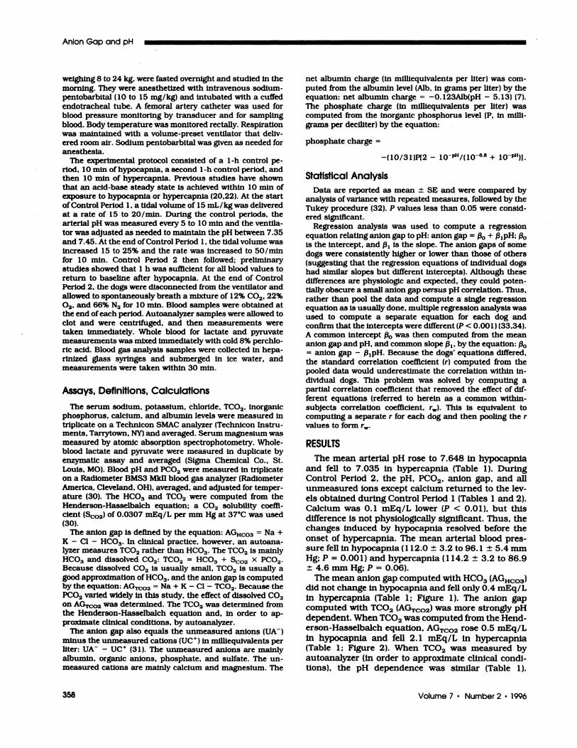
weighing 8 to 24 kg. were fasted overnight and studied in themorning. They were anesthetized with intravenous sodium-pentobarbital (10 to 15 mg/kg) and intubated with a cuffedendotracheal tube. A femorab artery catheter was used forblood pressure monitoring by transducer and for samplingblood. Body temperature was monitored rectally. Respirationwas maintained with a volume-preset ventilator that deliv-ered room air. Sodium pentobarbital was given as needed foranesthesia.
The experimental protocol consisted of a 1 -h control pe-nod, 10 mm ofhypocapnia. a second 1-h control period. and
then 10 mm of hypercapnia. Previous studies have shownthat an acid-base steady state is achieved within 10 mm ofexposure to hypocapnia or hypercapnia (20.22). At the startofControl Period 1 , a tidal volume of 15 mL/kg was deliveredat a rate of 15 to 20/mm. During the control periods. thearterial pH was measured every 5 to 1 0 mm and the ventila-tor was adjusted as needed to maintain the pH between 7.35and 7.45. At the end ofControl Period 1 , the tidal volume wasincreased 15 to 25% and the rate was increased to SO/mmfor 10 min. Control Period 2 then followed; preliminarystudies showed that 1 h was sufficient for all blood values toreturn to baseline after hypocapnia. At the end of ControlPeriod 2. the dogs were disconnected from the ventilator andallowed to spontaneously breath a mixture of 1 2% CO2. 22%
02. and 66% N2 for 10 mm. Blood samples were obtained at
the end ofeach period. Autoanalyzer samples were allowed toclot and were centrifuged. and then measurements weretaken immediately. Whole blood for lactate and pyruvatemeasurements was mixed immediately with cold 8% perchlo-nc acid. Blood gas analysis samples were collected in hepa-rinized glass syringes and submerged in ice water, andmeasurements were taken within 30 min.
Assays, Definitions, Calculations
The serum sodium, potassium. chloride, TCO2. inorganicphosphorus. calcium, and albumin levels were measured intriplicate on a Technicon SMAC analyzer (Technicon Instru-ments. Tarrytown, NY) and averaged. Serum magnesium wasmeasured by atomic absorption spectrophotometry. Whole-blood lactate and pyruvate were measured in duplicate byenzymatic assay and averaged (Sigma Chemical Co., St.Louis, MO). Blood pH and PCO2 were measured in triplicateon a Radiometer BMS3 MkII blood gas analyzer (RadiometerAmerica. Cleveland, OH), averaged. and adjusted for temper-
ature (30). The HCO3 and TCO2 were computed from theHenderson-Hasselbalch equation; a CO2 solubility coeffi-cient (Sco2) of 0.0307 mEq/L per mm Hg at 37#{176}Cwas used(30).
The anion gap is defined by the equation: AGHC03 = Na +
K - Cl - HCO3. In clinical practice. however. an autoana-lyzer measures TCO2 rather than HCO3. The TCO2 is mainlyHCO3 and dissolved CO2: TCO2 HCO3 + S�02 >< PCO2.Because dissolved CO2 is usually small, TCO2 is usually agood approximation of HCO3, and the anion gap is computedby the equation: AGTCO2 = Na + K - Cl - TCO2. Because thePCO2 varied widely in this study, the effect of dissolved CO2on AGTCO2 was determined. The TCO2 was determined fromthe Henderson-Hasselbalch equation and, in order to ap-proximate clinical conditions. by autoanalyzer.
The anion gap also equals the unmeasured anions (UA)minus the unmeasured cations (UC�) in mibliequivalents perliter: UA - UC� (3 1). The unmeasured anions are mainlyalbumin, organic anions, phosphate, and sulfate. The Un-measured cations are mainly calcium and magnesium. The
net albumin charge (in mffiiequivalents per biter) was com-puted from the albumin level (Abb. in grams per biter) by theequation: net albumin charge = -0. 123Alb(pH - 5. 13) (7).The phosphate charge (in mffiiequivalents per liter) wascomputed from the inorganic phosphorus level (P. in mdli-grams per deciliter) by the equation:
phosphate charge =
Statistical Analysis
-(1O/31)P[2 - 10PH/(106.8 + 10P”)].
Data are reported as mean ± SE and were compared byanalysis ofvariance with repeated measures, followed by theTukey procedure (32). P values less than 0.05 were consid-ered significant.
Regression analysis was used to compute a regressionequation rebating anion gap to pH: anion gap = /3� + /31pH; (3�is the intercept, and (3� is the slope. The anion gaps of some
dogs were consistently higher or bower than those of others(suggesting that the regression equations of individual dogshad similar slopes but different intercepts). Although thesedifferences are physiologic and expected, they could poten-tially obscure a small anion gap versus pH correlation. Thus,rather than pool the data and compute a single regressionequation as is usually done. multiple regression analysis wasused to compute a separate equation for each dog andconfirm that the intercepts were different (P < 0.00 1)(33.34).A common intercept /3� was then computed from the meananion gap and pH. and common slope (3k. by the equation: (3�= anion gap - (31pH. Because the dogs’ equations differed,the standard correlation coefficient (r) computed from thepooled data would underestimate the correlation within in-dividual dogs. This problem was solved by computing apartial correlation coefficient that removed the effect of dii-ferent equations (referred to herein as a common within-subjects correlation coefficient. r�). This is equivalent tocomputing a separate r for each dog and then pooling the rvalues to form r�.
RESULTS
The mean arterial pH rose to 7.648 in hypocapniaand fell to 7.035 in hypercapnia (Table 1). DuringControl Period 2, the pH, PCO2, anion gap. and allunmeasured ions except calcium returned to the 1ev-els obtained during Control Period 1 (Tables 1 and 2).Calcium was 0. 1 mEq/L lower (P < 0.01), but thisdifference is not physiologically significant. Thus, thechanges induced by hypocapnia resolved before theonset of hypercapnla. The mean arterial blood pres-sure fell in hypocapnla ( 1 1 2.0 ± 3.2 to 96. 1 ± 5.4 mm
Hg; P = 0.001) and hypercapnia (1 14.2 ± 3.2 to 86.9± 4.6 mm Hg; P = 0.06).
The mean anion gap computed with HCO3 (AGHC03)did not change in hypocapnla and fell only 0.4 mEq/Lin hypercapnia (Table 1 ; Figure 1). The anion gapcomputed with TCO2 (AGTCO2) was more strongly pHdependent. When TCO2 was computed from the Hend-erson-Hasselbalch equation. AGTCO2 rose 0.5 mEq/Lin hypocapnla and fell 2. 1 mEq/L in hypercapnia(Table 1 ; Figure 2). When TCO2 was measured by
autoanalyzer (in order to approximate clinical condi-tions), the pH dependence was similar (Table 1).

ANION GAP = Na + K - ci - HCO3
p = NS
28
�24
wE 20
� 16
�12
z� 8
p = NS
28
‘-I 24
0�wE 20
0�< 160z 120z48
ANION GAP � Na + K - cI - TCO2
p = 0.55 p < 0.001
-..� ....�...
Paulson
Journal of the American Society of Nephrology 359
TABLE 1 . Changes in arterial pH, Pco2, and serum anion gap in acute hypocapnia and hypercapnia(mean ± SE)
Control . ControlParameter Period 1 Hypocapnia Period 2 Hypercapnia
pH 7.404 ± 0.005 7.648 ± 0.012#{176} 7.398 ± 0.007 7.035 ± 0,006b
Pco2 (mm Hg) 32.7 ± 0.7 15.5 ± 0.5#{176} 32.8 ± 0.8 89.5 ± 1#{149}4b
AGHCO3C (mEq/L) 15.7 ± 0.5 15.7 ± 0.6 15.1 ± 0.6 14.7 ± 0.6
AG1co2d (mEq/L) 14.7 ± 0.5 15.2 ± 0.6 14.2 ± 0.6 12.1 ± 0,6b
AG1�02e (mEq/L) 16.6 ± 0.5 18.3 ± 0.4#{176} 16.1 ± 0.5 15.2 ± 0.5
0 p < 0.01 versus Control Period 1.b p < 0.01 versus Control Period 2.C AGHCO3, anion gap computed as: Na + K - Cl - HCO3. HCO3 was computed from the Henderson-Hasselbaich equation.d AG1�02. anion gap computed as: Na + K - Cl - 1002. TOO2 was computed from the Henderson-Hasselbalch equation.e AG1�02, anion gap computed as: Na + K - Cl - TCO2. TCO2 was measured by autoanatyzer.
TABLE 2. Changes in serum unmeasured anion and unmeasured cation levels in acute hypocapnia andhypercapnia (mean ± SE)
Parameter Control . Control(mEq/L) Period 1 Hypocapnia Period 2 Hypercapnia
Unmeasured Anions
Albumin0 8.12 ± 0.17 9.01 ± 020d 8.02 ± 0.18 6.84 ± 0.15�Phosphateb 2.53 ± 0.13 2.30 ± 014d 2.56 ± 0.16 2.88 ± 0.15�
Lactatec 1.69±0.18 2�#{247}028d 1.63±0.19 1.20± 0.16Pyruvatec 0.09 ± 0.01 0.13 ± 0.02d 0.08 ± 0.01 0.06 ± 0.01
Unmeasured CationsCalcium 5.22 ± 0.05 5.17 ± 0.06 5.10 ± 005d 5.08 ± 0.05
Magnesium 1 .54 ± 0.03 1 .40 ± 005d � .46 ± 0.04 1 .50 ± 0.04
a Net albumin charge (in mEq/L)was computed from albumin level(Alb. in grams perliter) by the equation (7): net charge = -O.123Alb (pH - 5.13).b Phosphate charge was computed from phosphorus level (in milligrams per deciliter) by using pK0’ of H2P04 � = 6.8.C Whole blood.d p < 0.01 versus Control Period 1.e p < o.oi versus Control Period 2.
.-.� ....�
CONTROL 1 HVPOCAPNIA CONTROL 2 HYPERCAPNIA
Figure 1 . Serum anion gap (AGHCO3) in Control Period 1,hypocapnia, Control Period 2, and hypercapnia. HCO3 wascomputed from the Henderson-Hasselbalch equation. Thedashed lines denote means. Changes were not significant
(NS).
AGTCO2 was now somewhat higher, however, probably
because anaerobic metabolism during the clotting of
blood samples lowered the TCO2.
Regression analysis was used to determine the re-
bationship between anion gap and pH. Because the
-p . � - .
CONTROL 1 HYPOCAPNIA CONTROL 2 HVPERCAPNIA
Figure 2. Serum anion gap (AGTCO2) in Control Period 1,hypocapnia, Control Period 2, and hypercapnia. TCO2 wascomputed from the Henderson-Hasselbalch equation. Thedashed lines denote means.
correlation was weak, it was strongly Influenced by
normal physiologic anion gap differences between
dogs. The effect of these differences was removed by
computing a common within-subjects correlation co-
efficient (r�) (Figure 3). The regression equation shows
that the change in AG11�03 per 0. 1 change in pH

28
-I 24
�20
Q- 164C., 12z08
z44
ANION GAP = Na + K - cI - HCO3
0
ANION GAP = 4.05 + 1.53pH
r� = 0.344, p = 0.02
Na #{247}K = 186.2 - 4.6lpH
r� = 0.837, p < 0.001
-I
C-wE
7.0 7.2 7.5 7.8
7.0 7.2 7.5 7.8
pH
28
224
�20
0_ 1640 12z08z44
0
ANION GAP = -23.83 + 5.l4pH
r.., = 0.760. o < 0.001
7.0 7.2 7.5 7.8
Anion Gap and pH
360 Volume 7 ‘ Number 2 . 1996
pH
Figure 3. Correlation of serum anion gap (AGHCO3) with pH.The slope of the regression line is z�AGHCO3/�pH (in mil-liequlvalents per liter per 1 .0 pH).
(�AGHCO3/z�pH) was only 0. 15 mEq/L per 0. 1 pH. The
correlation of AGTCO2 with pH was stronger whetherTCO2 was computed from the Henderson-Hasselbalchequation (Figure 4) or measured by autoanalyzer (r� =
0.761 ; P < 0.001). The two �.AGTCO2/z�pH ratios were
nearby identical: 0.5 1 and 0.50 mEq/L per 0. 1 pH.
respectively.
To help explain the weak AGHCO3 versus pH corre-
lation, regression equations relating the measured
cation ([Na] + [K]) and anion ([Cl] + [HCO3]) levels topH were computed (Figure 5). Unlike the anion gap.the measured ions were negatively correlated with pH.
Because the distance between the two regression lines
widened slightly as the pH increased, the anion gap
(computed from their difference) was weakly pH de-pendent (Figure 3).
Small changes in unmeasured ion levels also helpedexplain the weak pH dependence 0fAGHCO3 (Table 2).
ANION GAP = Na + K - cI - TCO2
pH
Figure 4. Correlation of serum anion gap (AG1�02) with pH.TCO2 was computed from the Henderson-Hasselbalch equa-tion. The slope of the regression line is z�AGTCO2/�pH (in
milliequivalents per liter per 1 .0 pH). �AG1�02/z�pH wasnearly identical when TCO2 was measured by autoanalyzer(5.0 mEq/L per 1 .0 pH).
Figure 5. Correlations of measured cation ((Na) + (K)) andanion ((Cl) + (HCO3)) levels with pH. The vertical distance
between the two regression lines is the anion gap (Figure 3).
The calculated net albumin charge rose 0.9 mEq/L in
hypocapnia and fell 1 .2 mEq/L in hypercapnia. Lac-
tate and pyruvate also rose and fell, but these changeswere partially offset by opposite changes in phos-phate. The phosphate charge (in milliequivalents per
liter) fell with increasing pH (despite an increase in
phosphate ionization) because the phosphorus level
fell in hypocapnia ( 1 .40 ± 0.07 to 1 .22 ± 0.07
mmol/L) and rose in hypercapnia ( 1 .43 ± 0.09 to 1.76± 0.09 mmol/L) (both P < 0.01). Both calcium and
magnesium varied minimally with pH.
DISCUSSION
This study shows that simple titration processes
that occur within 1 0 min of a change in pH cause
minimal changes in the serum anion gap computed
with HCO3 (AGHCO3) (Figure 3). This result agrees with
the recent discovery that titration-induced changes in
the charge of the predominant non-HCO3 buffers of
serum, the serum proteins, are smaller than have
been widely believed (5-7). Albumin has all of the net
protein charge. and this charge (in milliequivalents
per liter) is computed from the albumin level (Alb. ingrams per liter) by the equation (7): net albumin
charge = -0.123Alb(pH - 5.13).At the albumin level of 29 g/L in the dogs in this
study. the net charge was only -8 mEq/L at pH =
7.4. The change in net charge per 0. 1 change in pH
(�(albumin charge)/z�pH) was only 0.36 mEq/L per
0. 1 pH. Small and partially offsetting changes in otherunmeasured ions contributed to the weak relation-
ship of AGHCO3 with pH.
Analysis of measured ion levels also helped explain
the minimal pH dependence ofAGHco3. The measured
cations ([Na] + [K]) and anions ([Cl] + [HCO3]) were
negatively correlated with pH because they participate
in the buffering of acid-base changes by body fluids
(Figure 5) (35). The mechanisms ofbuffering are com-
plex and involve the movement of measured ionsacross cell membranes. Because the changes in mea-
sured cations and anions were similar, the anion gap

ANION GAP = Na + K - CI - HCO3
5
I
I-
0
-a
w
I0�“1
‘l
3
2
1
0
-1
-2.
Paulson
Journal of the American Society of Nephrology 361
(computed from their difference) was weakly pH de-
pendent.
In clinical practice. the anion gap Is usually com-
puted with TCO2 (AGTCO2). AGTCO2 was more strongly
pH dependent than AGHCO3 when TCO2 was com-puted from the Henderson-Hasselbalch equation ormeasured by autoanalyzer. and both methods gavethe same MGTco2/�pH ratio (Figure 4). This strongerpH dependence was the result of the effect of dissolved
CO2 (5co2 X PCO2) on AGTCO2, as described by the
relatjon of AGTCO2 to AGHCO3: AGTCO2 = AGHC03 -
5C02 X PCO2. If the PCO2 rises, for example, subtrac-tion by 5C02 X PCO2 augments the anion gap decreasepredicted by the fall in albumin charge.
In a literature review, we found 22 studies of acute
hypocapnia and hypercapnla in humans and dogs Inwhich the ratio of the AGHCO3 change to the pHchange can be computed (Figure 6). Ratios above thedashed line indicate an AGHC03 increase in hypocap-
nia and a decrease in hypercapnia, as predicted by
titration-Induced changes in net albumin charge. Be-
cause the serum albumin level averages 28 g/L in
dogs and 43 g/L in humans (36,37), the albumin
charge equation predicts that �.AGHco3/�pH shouldbe approximately 0. 18 mEq/L per 0. 1 pH higher inhumans than in dogs (7). To adjust for this difference,
0. 18 was added to z1AGHCO3/�pH from each dogstudy. This adjustment increased this study’s ratio to
0.33 mEq/L per 0. 1 pH (also shown In Figure 6).The scatter in Figure 6 is wide, largely because a
7
6
4
1--- ;HYPOCAPNIA HYPERCAPNIA
Figure 6. Change in serum anion gap per 0.1 change in pH
(z�AGHCO3/z�pH) in uncomplicated acute hypocapnia andhypercapnia from this study and References 8 to 29. Studies
were included more than once if data were obtained at
different levels or durations (up to 6 h) of hypocapnia orhypercapnia. There were a total of 143 measurements Inhumans and 251 in dogs. The solid lines denote means, witheach study weighted according to its number of subjects (N= 3to39).
limited number of subjects In most studies made themeasurement of a small anion gap change unreliable
(N was as low as 3). For example. if z�AG/�pH = 0.5mEq/L per 0. 1 pH. then a pH change of 0.4 will alter
the anion gap only 2 mEq/L. At a typical anion gapstandard deviation of approximately 2.5 mEq/L, afairly barge N is required to measure such a small
anion gap change with precision. Differing experimen-
tal protocols also probably contributed to the scatter.
In order to summarize the data in Figure 6. mean
z�AGHCO3/�pH ratios were computed with each study
weighted according to its N. The ratio was 0.7 mEq/Lper 0. 1 pH in hypocapnia and 0.4 mEq/L per 0. 1 pH in
hypercapnia. These mean ratios are similar to the
anion gap change attributed to albumin at 43 g/L:
�(albumin charge)/z�pH = 0.53 mEq/L per 0. 1 pH (7).The anion gap is far more pH dependent in HC1
acidosis and diuretic alkalosis, where z�AGHCO3/z�pH
in dogs is 2.54 and 5.04 mEq/L per 0. 1 pH. respec-
tively ( 1). Albumin’s role in these anion gap changes
can be estimated from its charge equation: at a normal
dog albumin level of 28 g/L (36). �(albumin charge)/
z�pH is 0.34 mEq/L per 0. 1 pH (7). Thus, In HC1acidosis, albumin explains only 13% of AAGHco3/z�pH
(0.34/2.54). In diuretic alkalosis, the albumin levelrises 1 4.3% per 0. 1 increase in pH ( 1 ). The increase inalbumin level and pH wIll raise the net albumin charge
only 1 .5 mEq/L per 0. 1 pH. Even if a lactate Increase
of 0.75 mEq/L per 0. 1 pH is included ( 1 ). only 45% of
z�.AGHCO3/z�pH is explained ([ 1 .5 + 0.75]/5.04).These calculations show that the anion gap changes
in HC1 acidosis and diuretic alkalosis are largely un-
explaIned. An unexplained anion gap is a recurringtheme. Others have been unable to explain high aniongaps in many patients despite determining the levels
0 LXX� of as many unmeasured ions as is technically feasible
HUMANS (38,39). Unfortunately, the presence of at least 29
different organic anions in normal serum makes suchan approach difficult (40).
The recently reduced estimate of protein charge alsoyields an unexplained anion gap in this study. Be-
cause of the large number of organic anions in serum,
the total organic anion level (OA) is uncertain. Thus.the widely recognized OA of approximately 5 mEq/L
has been obtained Indirectly by solving for OA In the
equation: anion gap = OA + protein + HPO4 + SO4 -
Mg - Ca (3 1). Assuming that OA is 5 mEq/L andsulfate is 1 mEq/L (31), and using the other unmea-
sured ion levels in Table 2 (the average of the control
periods). then AGHCO3 should equal: unmeasured an-ions - unmeasured cations = 10.0 mEq/L. Becausethe average control period AGHCO3 was 15.4 mEq/L,approximately 5 mEq/L of anion gap is unexplained.
The cause of the unexplained anion gap is un-
known, but there are several possibilities. One possi-
ble cause is errors or analytical bias In measured ion
levels. This study determined measured ions with the
Technicon SMAC analyzer. which is the source of thetraditional anion gap reference value of 12 mEq/L (for
anion gap = Na - Cl - TCO2) (4 1). We recently

Anion Gap and pH
362 Volume 7 - Number 2 ‘ 1996
compared the reference values of analyzers that are in
widest current use and found that they are lower,
ranging from 5 to 1 1 mEq/L (42). Thus, this study’s
unexplained anion gap might be the result of an
analytical bias causing anion gap measurements to behigher than their true values. The large differences
between analyzers also suggest that analytical bias is
a widespread problem and might account for the
unexplained anion gap in other studies.
Another possible cause of the unexplained anion
gap is errors or incorrect assumptions concerning
unmeasured ion bevels. In HC1 acidosis and diuretic
alkalosis, these might include unrecognized changes
in serum protein composition or charge or the pres-
ence of unidentified ions. In this study, the unex-
plained anion gap would be reduced if the total or-
ganic anion level was higher than 5 mEq/L. It is also
possible that the unexplained anion gap is the result
of a combination of the above factors.
ACKNOWLEDGMENTS
The author thanks Katharine A. Kirk, PhD (Professor of Biostatistics.
School of Public Health, University of Alabama at Birmingham.
Birmingham, AL). for reviewing the statistical methods in this study.
Samuel A. Cucinell. MD, for his encouragement and support during
the study. and Merit F. Gadallah. MD, for his helpful comments
concerning the manuscript.
REFERENCES
1 . Adrogue HJ, Brensilver J, Madias NE: Changes in theplasma anion gap during chronic metabolic acid-basedisturbances. Am J Physiol 1978;235:F29 1 -F297.
2. Madias NE, Ayus JC, Adrogu#{233} HJ: Increased anion gapin metabolic alkalosis: The role of plasma protein equiv-
alency. N Engl J Med 1979:300:1421-1423.3. Relman AS: Metabolic consequences of acid-base disor-
ders. Kidney Int 1972:1:347-359.4. Van Slyke DD, Hastings AB, Hiller A, Sendroy J: Studies
of gas and electrolyte equilibria in blood: XIV; Theamounts ofalkali bound by serum albumin and globulin.J Biol Chem 1928:79:769-780.
5. Figge J, Rossing TH, Fend V: The role of serum proteinsin acid-base equilibria. J Lab Clin Med 199 1 ; 117:4S3-467.
6. Reeves RB: Error proved and corrected: Net anioniccharge on serum albumin [Editorial]. J Lab Clin Med1991;1 17:437.
7. Figge J, Mydosh T, Fend V: Serum proteins and acid-base equilibria: A follow-up. J Lab Clin Med 1992:120:713-719.
8. Rapoport S. Stevens CD. Engel GL, Ferris EB, Logan M:The effect of voluntary overbreathing on the electrolyteequilibrium of arterial blood in man. J Biol Chem 1946;163:411-427.
9. Stanbury SW, Thomson AE: The renal response to respi-ratory alkalosis. Clin Sd 1952; 1 1:3�7-374.
10. Franglen GT, McGarry E, Spencer AG: Renal functionand the excretion of potassium in acute alkalosis. JPhysiol 1953:121:35-45.
1 1 . Scribner BH, Fremont-Smith K, Burnell JM: The effectof acute respiratory acidosis on the internal equilibriumof potassium. J Clin Invest 19��:34:1276-128�.
1 2. Elkinton JR. Singer RB, Barker ES, Clark JK: Effects inman of acute experimental respiratory alkalosis andacidosis on ionic transfers in the total body fluids. J ClinInvest 1955:34: 167 1-1690.
13. Giebisch GL, Berger L, Pitts RF: The extrarenal re-sponse to acute acid-base disturbances of respiratoryorigin. J Clin Invest 1955:34:231-245.
14. Platts MM, Greaves MS: The composition ofthe blood in
respiratory acidosis. Cbin Sci 1957; 16:695-708.15. Papadopoulos CN, Keats AS: Metabolic acidosis of hy-
perventilation induced by controlled ventillation. Anes-thesiology 1959:20:156-161.
16. Brown EB: Plasma electrolyte composition in dogsbreathing high CO2 mixtures: Source of bicarbonatedeficit in severe respiratory acidosis. J Lab Clin Med1960:55:767-775.
1 7. Epstein RM, Nahas GG, Mark LC: Circulatory changesfollowing rapid correction of severe hypercapnic acidosisby 2-ammno-2-hydroxymethyl- 1 .3-propanediol. Ann NY
Acad Sci 196 1:92:500-508.18. Sigaard-Andersen 0: Acute experimental acid-base dis-
turbances in dogs: An investigation of the acid-base andelectrolyte content of blood and urine. Scand J Clin LabInvest 1962; l4ESuppl 661:1-20.
19. Cohen JJ, Brackett NC, Schwartz WE: The nature of thecarbon dioxide titration curve in the normal dog. J CbinInvest 1964:43:777-786.
20. Brackett NC, Cohen JJ, Schwartz WB: Carbon dioxidetitration curve of normal man: Effect of increasing de-grees of acute hypercapnia on acid-base equilibrium. NEngb J Med 1965;272:6-l2.
2 1 . Boning D: Shifts of the CO2-dissociation curve in acuterespiratory acidosis and its causes. Pfl[umlaut]ugersArch 1968:302:133-148.
22. Arbus GS, Hebert LA, Levesque PR, Etsten BE,Schwartz WE: Characterization and clinical applicationof the “significance band” for acute respiratory alkalosis.N Engl J Med 1969;280: 117-123.
23. Goldstein MB, Gennari FJ, Schwartz WE: The influenceof graded degrees of chronic hypercapnia on the acutecarbon-dioxide titration curve. J Cbin Invest l97l;50:208-2 16.
24. Held DR. Steiner CA: Effect of acute respiratory acidosison arterial plasma osmolality. Respir Physiol 1971 ; 12:25-35.
25. Ferlinz R, Heymer G, Stadeler HJ: Electrolyte change inacute respiratory alkalosis. Pneumonologie 1 97 1 ; 144:
120-138.26. Gledhill N, Beirne GJ, Dempsey JA: Renal response to
short-term hypocapnia in man. Kidney Int 1975:8:376-386.
27. Javaheri 5, Kazemi H: Electrolyte composition of cere-brospmnal fluid in acute acid-base disorders. RespirPhysiol 1981;45:141-151.
28. Javaheri S. Weyne J. Demeester G, Leusen I: Effects ofacetazolamide on ionic composition of cisternal fluidduring acute respiratory acidosis. J Appl Physiob 1984;57:85-91.
29. Freidel JF, Javaheri 5: Bumetanide and cerebrospinalfluid acid-base variables during acute CO2 elevation.Respir Physiol 1990:79:91-100.
30. Burnett RW, Noonan DC: Calculations and correctionfactors used in determination of blood pH and bloodgases. Clin Chem 1974:20:1499-1S06.
3 1 . Oh MS. Carroll HJ: Current concepts: the anion gap. NEngl J Med 1977:297:814-8 17.
32. Neter J, Wasserman W, Kutner MH. Applied LinearStatistical Models. 2nd Ed. Boston: Irwin: 1990:103S-1047.
33. Draper NR, Smith H. Applied Regression Analysis. 2ndEd. New York: John Wiley & Sons; 1981:241-246.
34. Kleinbaum DG, Kupper LL. Applied Regression Analysisand Other Multivariable Methods. 2nd Ed. Boston: Dux-bury Press: 1988: 144-157, 260-281.
35. Pitts RF. Physiology of the Kidney and Body Fluids. 2ndEd. Chicago: Year Book Medical Publishers: 1968:174-177.
36. Bentinck-Smith J, French TW. A roster ofnormal valuesfor dogs and cats. In: Kirk RW, Ed. Current VeterinaryTherapy; X: Small Animal Practice. Philadelphia: WBSaunders; 1989:133S-134�.
37. Elm RJ. Reference intervals and laboratory values ofclinical importance. In: Wyngaarden JB, Smith LH, Ben-nett JC, Eds. Cecil Textbook of Medicine. 19th Ed.Philadelphia: WB Saunders; 1992:2370-2380.

Paulson
Journal of the American Society of Nephrology 363
38. Gabow PA, Kaehny WD. Fennesey PV, Goodman SI,Gross PA, Schrier RW: Diagnostic importance of anincreased serum anion gap. N Engl J Med 1980:303:854-858.
39. Mecher C, Rackow EC, Astiz ME, Well MH: Unaccountedfor anion in metabolic acidosis during severe sepsis inhumans. Crit Care Med 199 1 ; 1 9:705-7 1 1.
40. Issachar D, Holland JF, Sweeley CC: Metabolic profiles
of organic acids from human plasma. Anal Chem 1982;54:29 -32.
4 1 . Winter SD, Pearson JR. Gabow PA, Schultz AL, LepoffRB: The fall of the serum anion gap. Arch Intern Med1990 1�031-3 13.
42. Paulson WD, Lurie AA: Comparison of the serum aniongap measurements of analyzers [Abstract]. J Am SocNephrol 1995:6:442.