Embed Size (px)
Citation preview
Effect of the MTHFR 677C/T Polymorphism on Homocysteinemia in Response to
Creatine Supplementation: a case study
MIROSLAV PETR1, MICHAL ŠTEFFL1, EVA KOHLÍKOVÁ 1
1Department of Physiology and Biochemistry, Faculty of Physical Education and Sport,
Charles University in Prague, José Martího, 162 52 Prague 6, Czech Republic
Corresponding author
Miroslav Petr
Department of Physiology and Biochemistry, Faculty of Physical Education and Sport,
Charles University in Prague, José Martího, 162 52 Prague 6, Czech Republic
Short title
Homocysteinemia and response to creatine supplementation
Summary
Creatine (Cr) is recommended as a dietary supplement especially for athletes but its
therapeutic potential is also discussed. It is assumed that human body uses Cr for the
formation of phosphocreatine, which is necessary for muscular work as a source of energy.
Production of Cr in a body is closely connected to methionine cycle where guanidinoacetate
(GAA) is in a final step methylated from S-adenosylmethionine (SAM). Increased availability
of SAM for phosphatidylcholine (PC) and sarcosine synthesis can potentially stimulate
endogenous production of betain a thus methylation of homocysteine (HCy) to form
methionine. Our subject who was methylentetrahydrofolate reductase (MTHFR) 677TT
homozygote lowered plasma HCy from 33.3 µmol/L to 17.1 µmol/L following 1-month Cr
supplementation (5 g/day) opposite to 677CC and CT genotypes whose HCy levels tended to
increase (but still in normal ranges). We suppose that Cr supplementation stimulates pathways
leading to production of sarcosine which can serve to regenerate tetrahydrofolate (THF) to
form 5, 10-methylene-THF. This could potentially increase MTHFR enzyme activity which
may later result in increased HCy methylation. Cr supplementation significantly effects
metabolism of one carbon unit and potentially lower body´s demands for methyl groups. This
could be beneficial as in the case of reduced enzyme activity such as MTHFR 677C/T
polymorphism.
Key words
Creatine (Cr), Homocysteine (HCy), Supplementation, MTHFR gene, 677C/T
Introduction
Creatine (Cr) supplementation has been widely used in athletes to support recovery in
short term bouts of high-intensity exercise enabling more effective training and performance.
Beneficial effect of Cr supplementation in young, healthy males is the enhanced muscle fiber
size, strength and increased lean body mass (Greenhaff et al. 1994, Kreider et al. 1998). The
greatest improvements in performance have been found in activities, which possibly stress the
phosphocreatine (PCr) system such as series of high-power output exercises (Terjung et al.
2000). Studies have shown improved anaerobic exercise performance (Law et al. 2009), and
specific performance in many sports such as fin swimming (Juhasz et al. 2009), swimming,
all-out cycling, sprinting, repeated jumping, and resistance training (Juhn and Tarnopolsky
1998).
The potential therapeutic value of Cr supplementation has recently been investigated
with respect to various neurodegenerative disorders which include; Alzheimer´s, Parkinson´s,
amyotrophic lateral sclerosis, and Huntington´s disease patients (Adhihetty and Beal 2008). In
patients with diseases that result in atrophy or muscle fatigue secondary to impaired energy
production there have shown beneficial effects from Cr supplementation. Cr supplementation
has also been investigated in various neuromuscular diseases including mitochondrial
cytopathies, neuropathic disorders, dystrophies, congenital myopathies, and inflammatory
myopathies (Tarnopolsky and Martin 1999).
The daily turnover of Cr (1 – 2 g/ day) is replaced either through dietary Cr intake
(from animal-derived tissues such as meat) or through endogenous synthesis in the liver from
amino acid precursors (arginine, glycine and methionine). At the same time and
approximately the rate (2 g/day), Cr is broken down to creatinine and excreted to the urine
(Stead et al. 2006).The first step of Cr synthesis is the formation of guanidinoacetate (GAA)
and ornithine in a reaction catalyzed by glycine amidinotransferase (AGAT), GAA can then
be methylated on the original glycine nitrogen using S-adenosylmethionine (SAM) as the
methyl donor. This reaction yields Cr and S-adenosylhomocysteine (SAH) and is catalyzed by
the enzyme guanidinoacetate N-methyltransferase (GAMT) (Wyss and Kaddurah-Daouk
2000). The rate limiting step in Cr synthesis is the formation of guanidinoacetate by AGAT
and it has been shown that Cr supplementation down-regulates AGAT expression (Guthmiller
et al. 1994). An increase in serum levels of Cr resulted in a decrease in AGAT enzyme
activity, enzyme level, and mRNA expression in rat kidney (McGuire et al. 1984). On the
other hand, the growth hormone up-regulates AGAT expression in rats (Guthmiller et al.
1994). AGAT is highly active in the kidneys, whereas GAMT is highly active in the liver.
Therefore it is suggested that GAA is synthesized primarily in the kidney and then transported
to the liver where it is methylated to form Cr (Wyss and Kaddurah-Daouk 2000). In rats, Van
Pilsum et al. (1972) documented high activity of AGAT apart from kidney in pancreas, brain,
spleen, and testes. In a study Edison et al. (2007) authors suggested that renal GAA
production in a human may account only for about 20% of total GAA synthesis.
Cr metabolism disorders have so far been described at the level of two synthetic steps,
GAMT and AGAT, and at the level of the Cr transporter 1 (CrT1). The most common GAMT
and AGAT deficiency symptoms and signs are delayed language development, learning
disorders, autistic behavior, epileptic seizures, and movement disorders (Gordon 2010).
GAMT and AGAT deficiency are treatable by oral Cr supplementation, but patients with Cr
transporter deficiency do not respond to this type of treatment (Evangeliou et al. 2009).
Neither nine months of L-arginine supplementation did not showed effectiveness in the four
patients affected with Cr transporter deficiency (Fons et al. 2008).
In a human body, more than 90% of SAM is used for methylation reactions by at least
50 different methyltransferases (Stead et al. 2006). There are three major methyltransferases
playing an important role in the generation of homocysteine (HCy) and regulation of methyl
group metabolism; except for GAMT they include phosphatidylethanolamine N-
methyltransferase (PEMT), and glycine N-methyltransferase (GNMT) (Mudd et al. 2007).
Production of Cr via GAMT and phosphatidylcholine (PC) via PEMT is considered to be the
largest consumer of methyl groups derived from SAM. Stead et al. (2006) suggests opposite
of original investment where GAMT has been proposed to consume up to 70% of methyl
groups (Mudd et al. 1980), and considers PEMT to be the primary consumer of methyl
groups, having the greatest impact on HCy levels. It was found that PEMT -/- mice have
lower choline pools in liver despite being fed sufficient or supplemental amounts of dietary
choline (Zhu et al. 2003). When PEMT is deleted in mice, plasma HCy concentrations fall
50% and, when it is over expressed, plasma HCy concentrations increase 40%, demonstrating
that PEMT activity is a very major consumer of SAM (and thereby a producer of HCy)
(Jacobs et al. 2005). Furthermore, methylation of macromolecules such as DNA, RNA,
histones, and other proteins play critical roles in cellular metabolism. Therefore, the level of
SAM must be carefully regulated to maintain cellular homeostasis (Luka et al. 2009).
The serum concentration of HCy is positively associated with the risk of ischemic
heart disease, deep vein thrombosis and pulmonary embolism, and stroke (Boushey et al.
1995). A meta-analysis by Boushey et al. (1995) of 27 studies showed that HCy was an
independent, graded risk factor for atherosclerotic disease. Total HCy (tHCy) measured in
blood is usually the sum of free (reduced) HCy and protein-bound HCy (Brosnan et al. 2004).
There are two methylation pathways to form methionine from HCy, both of which result in
lowering HCy concentrations (Olthof et al. 2003). In the first, vitamins B12 and folic acid are
involved in a reaction catalyzed by methionine synthase (MTR) (Weisberg et al. 2001).
Deficiencies of these vitamins can result in elevated plasma HCy concentrations (Bailey et al.
2002, He et al. 2010). Also single nucleotide polymorphisms in genes coding enzymes
involved in this pathway can result in elevated HCy in blood. Examples are substitutions as
MTR 2756A/G (Barbosa et al. 2008), methionine synthase reductase (MTRR 66A/G)
(Naushad et al. 2008), methylenetetrahyrofolate reductase (MTHFR 677C/T and 1298A/C)
(Weisberg et al. 2001), or methylenetetrahydrofolate dehydrogenase (MTHFD1 1958G/A)
(Brody et al. 2002). The most profound effect on HCy levels in blood was found in MTHFR
677C/T and/or in MTR 2756A/G (Barbosa et al. 2008). The 677C/T variant which leads to the
substitution of Ala-222 by valine produces MTHFR enzyme with reduced activity, resulting
in an elevation of serum HCy concentrations of about 20% (Brattstrom et al. 1998). The
677C/T polymorphism is surprisingly common, with about 10% of people in the population
being homozygous affected (TT), 47% homozygous unaffected (CC), and 43% heterozygotes
(CT) (Brattstrom et al. 1998). In the Czech population similar frequencies were elicited (CC
– 41%, CT – 49%, TT – 10%) (Vesela et al. 2005).
An association with hyperhomocysteinemia was also found in MTHFR 1298A/C
polymorphism (Barbosa et al. 2008), although its contribution seems to be lesser compared to
677C/T (Bailey et al. 2002, Barbosa et al. 2008). In additional studies, the activity of enzyme
produced by the heterozygotes for both mutations was lower compared to individuals who
carried only 677T/T (Chango et al. 2000). The genetic influence of the MTHFR
polymorphism on HCy levels is attenuated in females in premenopausal age and is not
significant in subjects who exhibit serum levels of folate and/or vitamin B12 above the 50th
percentile of distribution in the general population (Cortese and Motti 2001). A decrease in
serum HCy of 3 mmol/l (achievable by daily intake of about 0,8 mg folic acid) should reduce
the risk of ischemic heart disease by 16%, deep vein thrombosis by 25% and stroke by 24%
(Wald et al. 2002).
In the second pathway, the methylation of HCy to form methionine is catalyzed by
betaine homocysteine methyltransferase (BHMT) which is in addition to choline
dehydrogenase (CHDH), and PEMT the most important enzyme in choline metabolism (da
Costa et al. 2006). Betaine, once formed from choline via CHDH, donates its methyl group to
HCy via BHMT to form methionine. Administration of betaine can lower plasma HCy
concentrations (Steenge et al. 2003). Activity of BHMT is increased during methionine

excess and plasma betaine was shown to be a strong determinant after methionine increase in
tHCy in subjects not supplemented with B-vitamins (Holm et al. 2004). On the other hand,
neither a common variant 742G/A, nor other variants in BHMT gene seem to play a
significant role in plasma HCy (Heil et al. 2000, Morin et al. 2003).
The aim of the present post-hoc analysis is to evaluate homocysteinemia following Cr
supplementation in relation to MTHFR 677C/T genotype.
Methods
Subjects
This is a secondary analysis of data from 11 athletes participating in our previous
study (Navratil et al. 2010) who provided DNA from buccal cells. Of those participants, 10
submitted DNA testing. All subjects were young, aged 24 – 28 y old, healthy, physically
active persons, dealing with sportive activities (ice hockey, football, horsemanship, and
athletics) on a professional level. Subjects' height, weight, and body composition via
Bioelectrical Impedance Analyse (Multi-frequency analyzer In Body 3.0, Korea) was
measured. All 10 men were Caucasian. Subject characteristics are shown in Table 1. Written
informed consent was obtained from all subjects under protocols approved by the Institutional
Ethics Committee of the Charles University of Faculty of Physical Education and Sport.
Intervention
As previously published, all subjects ingested 5 g of CR-monohydrate (Plutino, Czech
Republic) a day, diluted in tepid water. These doses were administered every morning (at
about 8 a.m.) for 30 days. Participants were not allowed to consume any other supplement,
especially those containing vitamin B and folic acid, and they were advised to maintain their
usual dietary habits and physical activity during the study.
Biochemical Assays
Fasting blood and urine samples were collected at baseline and the next morning after
completion 30-day Cr supplementation and further analyzed for several metabolites including
HCy as described previously (Navratil et al. 2010, Petr et al. 2011).
Genotyping
Genomic DNA was isolated from buccal cells collected with cheek brushes
(Whatman, USA). Samples were lysed and DNA was stabilized with DNA Extract All
Reagents Kit (Applied Biosystems, USA) according to the manufacturer´s protocol. Samples
(5 µL) were genotyped according to the manufacturer's protocol on an Illumina BeadStation
500G Golden Gate genotyping platform using a custom panel (GS0011351-OPA) of 384
candidate single-nucleotide polymorphisms. From the whole set the genotypes for the
MTHFR C677T polymorphism (rs1801133) were extracted.
Results
Of 10 subjects, 9 individuals were carrying 677CC+CT, and 1 individual the 677TT
genotype. Pre-test levels of plasma HCy were normal among those carrying 677CC+CT
genotype (6.3±1.3 umol/l), but strongly elevated in 677TT carrier (33.2 umol/l). After 30-day
Cr supplementation individuals with 677CC+CT genotype mildly elevated HCy levels, but
completely different response was registered in 677TT carrier who lowered HCy almost to
normal levels (Tab. 2).
Discussion
We found an individual response of plasma HCy to Cr supplementation which was
associated with genotypes in MTHFR 677C/T polymorphism. Our results cannot be
considered as statistically significant due to the low number of subjects and the presence of
just one TT carrier. In spite of that, we assume our outcomes to be interesting, as they should
contribute to better understanding how supplemented Cr can affect methyl donor balance and
consequent plasma HCy levels.
A meta-analysis of published cohort studies states that hyperhomocysteinemia
moderately increases the risk of a first cardiovascular event, regardless of age and follow-up
duration (Bautista et al. 2002). In homocystinuria, which is a rare inherited disorder (most
often due to cystathionine ß-synthase deficiency, which occurs in 1 from 200.000 people),
plasma HCy levels are markedly elevated (>50 µmol/L; normal range, 5 to 15 µmol/L), and
patients have severe, widespread vascular disease (Bellamy et al. 1998). In the general
population, mild to moderate elevations in plasma HCy (15 to 35 µmol/L) are common and
may occur due to inherited enzyme variants and/or a relative deficiency of folate, vitamin B12,
or vitamin B6, which are required for the normal metabolism of HCy (Ubbink et al. 1993).
Our subjects with CC and CT genotypes had pre-test HCy concentration in normal
ranges 6.1±1.3 µmol/L with milder individual differences. The only carrier of TT genotype
had elevated HCy (33.3 µmol/L). After 30-day Cr supplementation all CC and CT carriers
increased plasma HCy to 10.9±3.2 µmol/L opposite to TT carrier who significantly lowered
HCy levels to 17.1 µmol/L. An interpretation of these changes is not simple to explain.
Reliable description of addressed biochemical pathways should be supported by analysis of
associated intermediates e.g. sarkosine in urine.
We assume that supplemented Cr inhibited the synthesis of endogenous Cr and thus
increased availability of SAM for PC and sarcosine synthesis via PEMT and GNMT
respectively. Concomitantly, the methylation of HCy relied more on BHMT where methyl
donor is betain, stimulated by increased production of PC and choline via increased PEMT
activity. Although PEMT is considered to be a major consumer of SAM and a producer of
HCy by some authors (Jacobs et al. 2005), provided that PC and choline levels are sufficient
from diet, HCy methylation from betaine could be stimulated. Moreover, resulting
dimethylglycine demands tetrahydrofolate (THF) for sarcosine formation via
dimethylglycine-dehydrogenase. THF is also needed for glycine formation from sarcosine via
sarcosine-dehydrogenase (SARDH). Sarcosine production can also be accelerated due to
better availability of SAM for GNMT. Based on these facts TFH is dynamically converted to
5,10-methylene-THF (methylene tetrahydrofolate) stimulating MTHFR enzyme to 5-methyl-
THF production which is later used for HCy conversion to methionine via MTR. Higher
availability of 5, 10-methylene-THF could possibly effectively stimulate MTHFR even in
677TT carriers. This can have quite a strong effect on HCy levels as we registered in our one
677TT carrier.
In fact, studies evaluated effects of Cr on HCy levels are not consistent. Cr
administration has been shown to decrease plasma tHCy by 25% in rats (Stead et al. 2001)
and by 10% in humans, according to one report (Korzun 2004); in another report, this
reduction in humans was not significant (Steenge et al. 2001). Opposite to these results, Cr
supplementation (alone or in combination with L-arginine) was associated with an 11 – 20%
increase in HCy concentration, which was not attributable to worsened renal function,
providing evidence against an effect of Cr on decreasing methylation demand (Jahangir et al.
2009). In rats, plasma HCy was increased up to 2 hours after intense anaerobic exercise, but
Cr supplementation decreased plasma HCy independent on exercise intensity (Deminice et al.
2011). Recent study shows interesting results about the prevalence of hyperhomocysteinaemia
(>15 µmol /l) in elite athletes which was 47% compared to 17% in controls without any
correlation between HCy and any of the other investigated variables, including plasma folate,
vitamin B12, blood pressure, lactate dehydrogenase (LDH), creatine kinase (CrK), total and
high-density lipoprotein (HDL) cholesterol and interleukin-6 (IL-6) (Borrione et al. 2008).
Our findings demonstrate an increase of HCy following Cr supplementation in
MTHFR 677C allele carriers but an average and individual augmentation were all in normal
ranges (<15 µmol /l). We suppose, when levels of dietary choline (PC) are sufficient to cover
physical needs, a great part of choline from endogenous production of PC should be used to
form betain for further HCy methylation. Provided that choline from diet does not meet body
demands, endogenous choline production (via PEMT) is very important. Under these
conditions most probably less betain can be produced to methylate HCy. Pathways following
methylation with betain seem to have a secondary methylation potential in regeneration of
THF to form 5,10-methylene-THF. Cr supplementation significantly effects metabolism of
one carbon unit and potentially lower body demands for methyl groups. This could be
beneficial as in the case of reduced enzyme activity like MTHFR 677C/T polymorphism.
Acknowledgements
This study was supported by research grant No. MSM 002160864, SVV 2012-265603
and PRVOUK n. 38 founded by Faculty of Physical Education and Sport, Charles University
in Prague.
References
ADHIHETTY PJ, BEAL MF: Creatine and its potential therapeutic value for targeting cellular energy impairment in neurodegenerative diseases. Neuromolecular Med 10: 275-290, 2008.
BAILEY LB, DUHANEY RL, MANEVAL DR, KAUWELL GP, QUINLIVAN EP, DAVIS SR, CUADRAS A, HUTSON AD, GREGORY JF, 3RD: Vitamin B-12 status is inversely associated with plasma homocysteine in young women with C677T and/or A1298C methylenetetrahydrofolate reductase polymorphisms. J Nutr 132: 1872-1878, 2002.
BARBOSA PR, STABLER SP, MACHADO AL, BRAGA RC, HIRATA RD, HIRATA MH, SAMPAIO-NETO LF, ALLEN RH, GUERRA-SHINOHARA EM: Association between decreased vitamin levels and MTHFR, MTR and MTRR gene polymorphisms as determinants for elevated total homocysteine concentrations in pregnant women. Eur J Clin Nutr 62: 1010-1021, 2008.
BAUTISTA LE, ARENAS IA, PENUELA A, MARTINEZ LX: Total plasma homocysteine level and risk of cardiovascular disease: a meta-analysis of prospective cohort studies. J Clin Epidemiol 55: 882-887, 2002.
BELLAMY MF, MCDOWELL IF, RAMSEY MW, BROWNLEE M, BONES C, NEWCOMBE RG, LEWIS MJ: Hyperhomocysteinemia after an oral methionine load acutely impairs endothelial function in healthy adults. Circulation 98: 1848-1852, 1998.
BORRIONE P, RIZZO M, SPACCAMIGLIO A, SALVO RA, DOVIO A, TERMINE A, PARISI A, FAGNANI F, ANGELI A, PIGOZZI F: Sport-related hyperhomocysteinaemia: a putative marker of muscular demand to be noted for cardiovascular risk. Br J Sports Med 42: 894-900, 2008.
BOUSHEY CJ, BERESFORD SA, OMENN GS, MOTULSKY AG: A quantitative assessment of plasma homocysteine as a risk factor for vascular disease. Probable benefits of increasing folic acid intakes. JAMA 274: 1049-1057, 1995.
BOUSHEY CJ, BERESFORD SA, OMENN GS, MOTULSKY AG: A quantitative assessment of plasma homocysteine as a risk factor for vascular disease. Probable benefits of increasing folic acid intakes. JAMA : the journal of the American Medical Association 274: 1049-1057, 1995.
BRATTSTROM L, WILCKEN DE, OHRVIK J, BRUDIN L: Common methylenetetrahydrofolate reductase gene mutation leads to hyperhomocysteinemia but not to vascular disease: the result of a meta-analysis. Circulation 98: 2520-2526, 1998.
BRODY LC, CONLEY M, COX C, KIRKE PN, MCKEEVER MP, MILLS JL, MOLLOY AM, O'LEARY VB, PARLE-MCDERMOTT A, SCOTT JM, SWANSON DA: A polymorphism, R653Q, in the trifunctional enzyme methylenetetrahydrofolate dehydrogenase/methenyltetrahydrofolate cyclohydrolase/formyltetrahydrofolate synthetase is a maternal genetic risk factor for neural tube defects: report of the Birth Defects Research Group. Am J Hum Genet 71: 1207-1215, 2002.
BROSNAN JT, JACOBS RL, STEAD LM, BROSNAN ME: Methylation demand: a key determinant of homocysteine metabolism. Acta Biochim Pol 51: 405-413, 2004.
CORTESE C, MOTTI C: MTHFR gene polymorphism, homocysteine and cardiovascular disease. Public Health Nutr 4: 493-497, 2001.
DA COSTA KA, KOZYREVA OG, SONG J, GALANKO JA, FISCHER LM, ZEISEL SH: Common genetic polymorphisms affect the human requirement for the nutrient choline. FASEB J 20: 1336-1344, 2006.
DEMINICE R, VANNUCCHI H, SIMOES-AMBROSIO LM, JORDAO AA: Creatine supplementation reduces increased homocysteine concentration induced by acute exercise in rats. Eur J Appl Physiol 2011.
EDISON EE, BROSNAN ME, MEYER C, BROSNAN JT: Creatine synthesis: production of guanidinoacetate by the rat and human kidney in vivo. Am J Physiol Renal Physiol 293: F1799-1804, 2007.
EVANGELIOU A, VASILAKI K, KARAGIANNI P, NIKOLAIDIS N: Clinical applications of creatine supplementation on paediatrics. Curr Pharm Biotechnol 10: 683-690, 2009.
FONS C, SEMPERE A, ARIAS A, LOPEZ-SALA A, POO P, PINEDA M, MAS A, VILASECA MA, SALOMONS GS, RIBES A, ARTUCH R, CAMPISTOL J: Arginine supplementation in four patients with X-linked creatine transporter defect. J Inherit Metab Dis 31: 724-728, 2008.
GORDON N: Guanidinoacetate methyltransferase deficiency (GAMT). Brain Dev 32: 79-81, 2010.
GREENHAFF PL, BODIN K, SODERLUND K, HULTMAN E: Effect of oral creatine supplementation on skeletal muscle phosphocreatine resynthesis. Am J Physiol 266: E725-730, 1994.
GUTHMILLER P, VAN PILSUM JF, BOEN JR, MCGUIRE DM: Cloning and sequencing of rat kidney L-arginine:glycine amidinotransferase. Studies on the mechanism of regulation by growth hormone and creatine. J Biol Chem 269: 17556-17560, 1994.
HE JA, HU XH, FAN YY, YANG J, ZHANG ZS, LIU CW, YANG DH, ZHANG J, XIN SJ, ZHANG Q, DUAN ZQ: Hyperhomocysteinaemia, low folate concentrations and methylene tetrahydrofolate reductase C677T mutation in acute mesenteric venous thrombosis. Eur J Vasc Endovasc Surg 39: 508-513, 2010.
HEIL SG, LIEVERS KJ, BOERS GH, VERHOEF P, DEN HEIJER M, TRIJBELS FJ, BLOM HJ: Betaine-homocysteine methyltransferase (BHMT): genomic sequencing and relevance to hyperhomocysteinemia and vascular disease in humans. Mol Genet Metab 71: 511-519, 2000.
HOLM PI, BLEIE O, UELAND PM, LIEN EA, REFSUM H, NORDREHAUG JE, NYGARD O: Betaine as a determinant of postmethionine load total plasma homocysteine before and after B-vitamin supplementation. �Arterioscler Thromb Va c Biol 24: 301-307, 2004.
CHANGO A, BOISSON F, BARBE F, QUILLIOT D, DROESCH S, PFISTER M, FILLON-EMERY N, LAMBERT D, FREMONT S, ROSENBLATT DS, NICOLAS JP: The effect of 677C-->T and 1298A-->C mutations on plasma homocysteine and 5,10-methylenetetrahydrofolate reductase activity in healthy subjects. Br J Nutr 83: 593-596, 2000.
JACOBS RL, STEAD LM, DEVLIN C, TABAS I, BROSNAN ME, BROSNAN JT, VANCE DE: Physiological regulation of phospholipid methylation alters plasma homocysteine in mice. J Biol Chem 280: 28299-28305, 2005.
JAHANGIR E, VITA JA, HANDY D, HOLBROOK M, PALMISANO J, BEAL R, LOSCALZO J, EBERHARDT RT: The effect of L-arginine and creatine on vascular function and homocysteine metabolism. Vasc Med 14: 239-248, 2009.
JUHASZ I, GYORE I, CSENDE Z, RACZ L, TIHANYI J: Creatine supplementation improves the anaerobic performance of elite junior fin swimmers. Acta Physiol Hung 96: 325-336, 2009.
JUHN MS, TARNOPOLSKY M: Oral creatine supplementation and athletic performance: a critical review. Clin J Sport Med 8: 286-297, 1998.
KORZUN WJ: Oral creatine supplements lower plasma homocysteine concentrations in humans. Clin Lab Sci 17: 102-106, 2004.
KREIDER RB, FERREIRA M, WILSON M, GRINDSTAFF P, PLISK S, REINARDY J, CANTLER E, ALMADA AL : Effects of creatine supplementation on body composition, strength, and sprint performance. Med Sci Sports Exerc 30: 73-82, 1998.
LAW YL, ONG WS, GILLIANYAP TL, LIM SC, VON CHIA E: Effects of two and five days of creatine loading on muscular strength and anaerobic power in trained athletes. J Strength Cond Res 23: 906-914, 2009.
LUKA Z, MUDD SH, WAGNER C: Glycine N-methyltransferase and regulation of S-adenosylmethionine levels. J Biol Chem 284: 22507-22511, 2009.
MCGUIRE DM, GROSS MD, VAN PILSUM JF, TOWLE HC: Repression of rat kidney L-arginine:glycine amidinotransferase synthesis by creatine at a pretranslational level. J Biol Chem 259: 12034-12038, 1984.
MORIN I, PLATT R, WEISBERG I, SABBAGHIAN N, WU Q, GARROW TA, ROZEN R: Common variant in betaine-homocysteine methyltransferase (BHMT) and risk for spina bifida. Am J Med Genet A 119A: 172-176, 2003.
MUDD SH, EBERT MH, SCRIVER CR: Labile methyl group balances in the human: the role of sarcosine. Metabolism 29: 707-720, 1980.
MUDD SH, BROSNAN JT, BROSNAN ME, JACOBS RL, STABLER SP, ALLEN RH, VANCE DE, WAGNER C: Methyl balance and transmethylation fluxes in humans. Am J Clin Nutr 85: 19-25, 2007.
NAUSHAD SM, JAIN JAMAL MN, PRASAD CK, RAMA DEVI AR: Relationship between methionine synthase, methionine synthase reductase genetic polymorphisms and deep vein thrombosis among South Indians. Clin Chem Lab Med 46: 73-79, 2008.
NAVRATIL T, KOHLIKOVA E, PETR M, PELCLOVA D, HEYROVSKY M, PRISTOUPILOVA K: Supplemented creatine induces changes in human metabolism of thiocompounds and one- and two-carbon units. Physiol Res 59: 431-442, 2010.
OLTHOF MR, VAN VLIET T, BOELSMA E, VERHOEF P: Low dose betaine supplementation leads to immediate and long term lowering of plasma homocysteine in healthy men and women. J Nutr 133: 4135-4138, 2003.
PETR M, NAVRATIL T, HEYROVSKY M, KOHLIKOVA E: The role of supplemented creatine in human metabolism. Current Organic Chemistry 15: 3029-3042, 2011.
STEAD LM, AU KP, JACOBS RL, BROSNAN ME, BROSNAN JT: Methylation demand and homocysteine metabolism: effects of dietary provision of creatine and guanidinoacetate. Am J Physiol Endocrinol Metab 281: E1095-1100, 2001.
STEAD LM, BROSNAN JT, BROSNAN ME, VANCE DE, JACOBS RL: Is it time to reevaluate methyl balance in humans? Am J Clin Nutr 83: 5-10, 2006.
STEENGE GR, VERHOEF P, GREENHAFF PL: The effect of creatine and resistance training on plasma homocysteine concentration in healthy volunteers. Arch Intern Med 161: 1455-1456, 2001.
STEENGE GR, VERHOEF P, KATAN MB: Betaine supplementation lowers plasma homocysteine in healthy men and women. J Nutr 133: 1291-1295, 2003.
TARNOPOLSKY M, MARTIN J: Creatine monohydrate increases strength in patients with neuromuscular disease. Neurology 52: 854-857, 1999.
TERJUNG RL, CLARKSON P, EICHNER ER, GREENHAFF PL, HESPEL PJ, ISRAEL RG, KRAEMER WJ, MEYER RA, SPRIET LL, TARNOPOLSKY MA, WAGENMAKERS AJ, WILLIAMS MH: American College of Sports Medicine roundtable. The physiological and health effects of oral creatine supplementation. Med Sci Sports Exerc 32: 706-717, 2000.
UBBINK JB, VERMAAK WJ, VAN DER MERWE A, BECKER PJ: Vitamin B-12, vitamin B-6, and folate nutritional status in men with hyperhomocysteinemia. Am J Clin Nutr 57: 47-53, 1993.
VAN PILSUM JF, STEPHENS GC, TAYLOR D: Distribution of creatine, guanidinoacetate and the enzymes for their biosynthesis in the animal kingdom. Implications for phylogeny. Biochem J 126: 325-345, 1972.
VESELA K, PAVLIKOVA M, JANOSIKOVA B, ANDEL M, ZVAROVA J, HYANEK J, KOZICH V: Genetic determinants of folate status in Central Bohemia. Physiological research / Academia Scientiarum Bohemoslovaca 54: 295-303, 2005.
WALD DS, LAW M, MORRIS JK: Homocysteine and cardiovascular disease: evidence on causality from a meta-analysis. BMJ 325: 1202, 2002.
WEISBERG IS, JACQUES PF, SELHUB J, BOSTOM AG, CHEN Z, CURTIS ELLISON R, ECKFELDT JH, ROZEN R: The 1298A-->C polymorphism in methylenetetrahydrofolate reductase (MTHFR): in vitro expression and association with homocysteine. Atherosclerosis 156: 409-415, 2001.
WYSS M, KADDURAH-DAOUK R: Creatine and creatinine metabolism. Physiol Rev 80: 1107-1213, 2000.
ZHU X, SONG J, MAR MH, EDWARDS LJ, ZEISEL SH: Phosphatidylethanolamine N-methyltransferase (PEMT) knockout mice have hepatic steatosis and abnormal hepatic choline metabolite concentrations despite ingesting a recommended dietary intake of choline. Biochem J 370: 987-993, 2003.
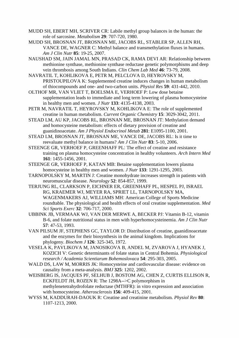
Table 1. Descriptive characteristics of 10 exercise-trained men
Variable Value
Age (y) 24.6±2.1
Height (cm) 181.7±4.8
Weight (kg) 82.8±11.0
Body fat (%) 12.0±2.9
BMI (kg.m-2) 25.0±2.4
Fat Free Mass (kg) 75.6±8.0
Values are means ± SD.
Table 2. Pre-test and post-test HCy levels in different MTHFR 677C/T genotypes
677CC
(n = 4)
677CT
(n = 5)
677TT
(n = 1)
677CC+CT
HCy (umol/l)
pre-test 5.9±1.3 6.6±1.3 33.2 6.3±1.3
post-test 9.9±2.9 11.6±3.3 17.1 10.9±3.2
Values are means ± SD.
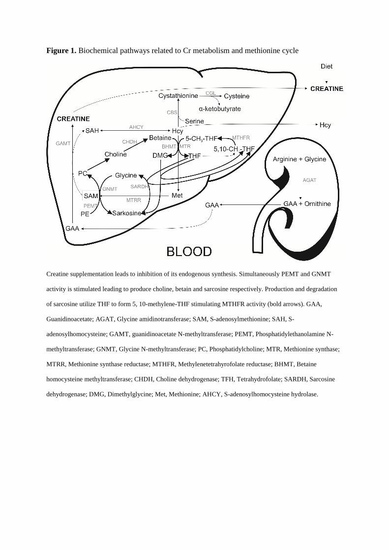
Figure 1. Biochemical pathways related to Cr metabolism and methionine cycle
Creatine supplementation leads to inhibition of its endogenous synthesis. Simultaneously PEMT and GNMT
activity is stimulated leading to produce choline, betain and sarcosine respectively. Production and degradation
of sarcosine utilize THF to form 5, 10-methylene-THF stimulating MTHFR activity (bold arrows). GAA,
Guanidinoacetate; AGAT, Glycine amidinotransferase; SAM, S-adenosylmethionine; SAH, S-
adenosylhomocysteine; GAMT, guanidinoacetate N-methyltransferase; PEMT, Phosphatidylethanolamine N-
methyltransferase; GNMT, Glycine N-methyltransferase; PC, Phosphatidylcholine; MTR, Methionine synthase;
MTRR, Methionine synthase reductase; MTHFR, Methylenetetrahyrofolate reductase; BHMT, Betaine
homocysteine methyltransferase; CHDH, Choline dehydrogenase; TFH, Tetrahydrofolate; SARDH, Sarcosine
dehydrogenase; DMG, Dimethylglycine; Met, Methionine; AHCY, S-adenosylhomocysteine hydrolase.


![Schizophrenia Functional Medicine Approachchoicesfoundation.us.com/wp-content/uploads/2014/... · MTHFR GENE [METHYLENE TETRAHYDROFOLATE REDUCTASE] MTHFR C677T (Nisha et al., 2014)](https://img.pdfslide.us/doc/110x75/5f0ad3ed7e708231d42d89e6/schizophrenia-functional-medicine-appro-mthfr-gene-methylene-tetrahydrofolate-reductase.jpg)