Embed Size (px)
Citation preview
J. Biosci., Vol. 6, Supplement 2, July 1984, pp. 75–82. © Printed in India. Effect of super-ovulatory doses of pregnant mare serum gonadotropin and human chorionic gonadotropin in blastocyst implantation in golden hamsters
J. PETER and J. MENEZES
Unit of Medical Genetics, Institute for Research in Reproduction, Jehangir Merwanji Street, Parel, Bombay 400012, India Abstract. The effect of super-ovulatory dose of pregnant mare serum gonadotropin and human chorionic gonadotropin on ovulation, advancement of ovulation, subsequent embryo development and implantation were studied in the hamster. Groups of hamsters received pregnant mare serum gonadotropin injection on day 1 of the estrous cycle followed by human chorionic gonadotropin injection either at 56 or 76 h later, pregnant mare serum gonadotropin alone on day 1 or human chorionic gonadotropin alone on day 3.
The combination therapy (pregnant mare serum gonadotropin and human chorionicgonadotropin) resulted in super-ovulation (an average of 40 mature ova/animal) while human chorionic gonadotropin alone yielded an average of 10 mature ova/animal. Ovulation was advanced by 24 h by giving human chorionic gonadotropin at 56 h instead of 76 h after pregnant mare serum gonadotropin. Subsequent embryo development and implantation occurring under different hormonal regimens were studied. The ova obtained by giving human chorionic gonadotropin injection at 56 h were poorly fertilizable in vivo and hence the pregnancy rate was low (6 %). These ova however, were fertilizable in vitro, suggesting that the low fertilization rate and developmental failure may be due to inhibition of sperm capacitation/transport because of premature human chorionic gonadotropin administration. In the group receiving human chorionic gonadotropin alone on day 3 there was fertilization and cleavage, but no implantation occurred due to failure of functional corpora lutea. However, administration of progesterone and estrone from day 2 of gestation resulted in 80% implantation and sustenance of pregnancy. On the other hand, the pregnant mare serum gonadotropin and human chorionic gonadotropin combination therapy resulted in super- pregnancy. The number of fetuses present at term was higher in the group receiving pregnant mare serum gonadotropin alone than in the group receiving the combination therapy. Embryo resorption however was higher (37%) in the latter group compared with the former (9·5%). However, preimplantation embryos were found to be viable as evidenced by fluorescein diacetate staining. Keywords. Gonadotropins; embryo viability; implantation.
Introduction In contrast to the normal process of ovulation, induction of ovulation by exogenous administration of hormones results in a large number of eggs being released. However, it is questionable whether all these eggs are fertilizable in vivo and whether subsequent embryo development can continue till term. Abbreviations used: PMSG, Pregnant mare serum gonadotropin; hCG, human chorionic gonadotropin, HSA, human serum albumin; PBS, phosphate buffered saline, 0·01 M, pH 7·4,0·9 NaCl, normal saline; FDA, fluorescein diacetate.
75
76 Peter and Menezes
In the present paper we have evaluated a number of biological parameters in hamsters exogenously treated with the hormones, pregnant mare serum gonadotropin (PMSG) and human chorionic gonadotropin (hCG). The parameters studied were ovum maturity, in vivo fertilization, normality and viability of embryos, ability of the blastocysts to implant and development of the fetuses to term. Materials and methods Golden hamsters were maintained under a light:dark schedule of 14:10h. Females 2 to 4 months old showing at least two consecutive estrous cycles were moated with 4–6months old male breeders. The day of the post estrous vaginal discharge was designated as day 1 of cycle.
Four different hormone regimens were used. Females in group 1, 2 and 3 received 30 I.U. of PMSG (NIH) on day 1 of the estrous cycle at 10·00 h. Those in group 1received 30 I.U. of hCG (Sigma) on day 4 (76 h after the PMSG injection). While those in group 3 received 30 I.U. hCG on day 3 (56 h after the PMSG injection). Females in group 2 were not administered hCG. Females in group 4 did not receive PMSG but received 30 I.U. hCG on day 3 at 15·00 h. Ovulation was advanced by 24 h in groups 3and 4.
In the first experiment the maturity of the ova was assessed. Females in group 1,3 and 4 were sacrificed 16–18 h after the hCG injection; those in group 2 were sacrificedbetween 08·00 and 10·00 h on the day of ovulation. The oviducts were dissected out andthe cumulus masses were flushed with BWW medium (Biggers et al., 1971), containing 0·3 % human serum albumin (HSA-Sigma) using a number 30 gauge blunt needleinserted through the fimbrial end. The cumulus cells were dispersed using 0·1% hyaluronidase solution (Sigma). After removal of the cumulus, the ova were counted and checked for their maturity.
In the second experiment, the normality and viability of the superovulated embryos was studied. Females in groups 1 and 2 were mated overnight from the evening of day 4 of cycle. Females in group 3 and 4 were mated overnight from the evening of day 3 of cycle. Vaginal smears were taken the following morning to check for mating. This day was considered as day 1 of pregnancy.
On day 3 of pregnancy, between 17·00 and 19·00 h the animals were sacrificed and the oviducts were flushed with 0·01 M phosphate buffered saline (PBS) pH 7·4 containing 0·4% bovine serum albumin (Sigma). Embryos were assessed for normality by their morphology and expected developmental stage. Viability of embryos obtained in group 1 and 2 was assessed by fluorescein diacetate (FDA) (Sigma) staining. Embryos were incubated in FDA solution for approximately one minute at room temperature (27–33°C). The concentration of FDA used was 2·5 μg per ml PBS. Viable embryosfluoresce brightly while nonviable ones show absence of fluorescence or markedly diminished fluorescence.
In the third experiment, mated females from each group were sacrificed on day 8 or day 14 of pregnancy. The number of implantation sites or fetuses as well as the numberof resorption sites or unilateral pregnancies were recorded.
In the fourth experiment exogenous progesterone was administered to animals in
Effect of superovulation on implantation in golden hamsters 77 group 4 at different dose levels according to the protocol of Sehgal and Diamond (1977) soon after mating. Additionally 2 other regimens were used. The first comprised of daily administration of 2 mg of progesterone from day 2 of pregnancy. The latter was a combination therapy of 2 mg progesterone and 1 µg estrone (Sigma). Results In groups 1,2 and 3 an average of 40 mature ova (as evidenced by release of the 1st polar body) were obtained per animal. An average of 10 mature ova per animal was obtained in group 4.
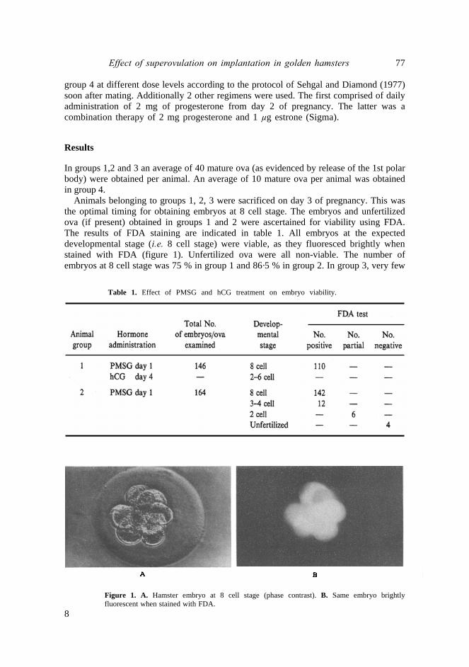
Animals belonging to groups 1, 2, 3 were sacrificed on day 3 of pregnancy. This was the optimal timing for obtaining embryos at 8 cell stage. The embryos and unfertilized ova (if present) obtained in groups 1 and 2 were ascertained for viability using FDA. The results of FDA staining are indicated in table 1. All embryos at the expected developmental stage (i.e. 8 cell stage) were viable, as they fluoresced brightly whenstained with FDA (figure 1). Unfertilized ova were all non-viable. The number of embryos at 8 cell stage was 75 % in group 1 and 86·5 % in group 2. In group 3, very few
Table 1. Effect of PMSG and hCG treatment on embryo viability.
Figure 1. A. Hamster embryo at 8 cell stage (phase contrast). B. Same embryo brightly fluorescent when stained with FDA.
8
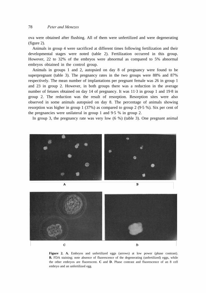
78 Peter and Menezes ova were obtained after flushing. All of them were unfertilized and were degenerating (figure 2).
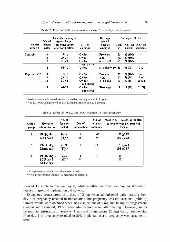
Animals in group 4 were sacrificed at different times following fertilization and their developmental stages were noted (table 2). Fertilization occurred in this group. However, 22 to 32% of the embryos were abnormal as compared to 5% abnormal embryos obtained in the control group.
Animals in groups 1 and 2, autopsied on day 8 of pregnancy were found to be superpregnant (table 3). The pregnancy rates in the two groups were 88% and 87% respectively. The mean number of implantations per pregnant female was 26 in group 1 and 23 in group 2. However, in both groups there was a reduction in the average number of fetuses obtained on day 14 of pregnancy. It was 11·3 in group 1 and 19·8 in group 2. The reduction was the result of resorption. Resorption sites were also observed in some animals autopsied on day 8. The percentage of animals showing resorption was higher in group 1 (37%) as compared to group 2 (9·5 %). Six per cent of the pregnancies were unilateral in group 1 and 9·5 % in group 2.
In group 3, the pregnancy rate was very low (6 %) (table 3). One pregnant animal
Figure 2. A. Embryos and unfertilized eggs (arrows) at low power (phase contrast). B. FDA staining; note absence of fluorescence of the degenerating (unfertilized) eggs, while the other embryos are fluorescent. C and D. Phase contrast and fluorescence of an 8 cell embryo and an unfertilized egg.
Effect of superovulation on implantation in golden hamsters 79
Table 2. Effect of hCG administration on day 3 on embryo development.
* No hormone administered. Animals mated on evening of day 4 of cycle. ** 30 I.U. hCG administered on day 3, Animals mated on day 3 evening.
Table 3. Effect of PMSG and hCG treatment in super-pregnancy,
* Complete resorption in the other three animals, ** No. in parenthesis indicate % pregnancies obtained.
showed 11 implantations on day 8, while another sacrificed on day 14 showed 16 fetuses. In group 4 implantation did not occur.
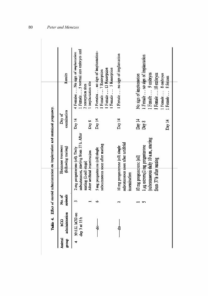
Exogenous progesterone at a dose of 2 mg when administered daily, starting from day 2 of pregnancy resulted in implantation, but pregnancy was not sustained (table 4). Similar results were obtained when single injections of 5 mg and 10 mg of progesterone (Sehgal and Diamond, 1977) were administered soon after mating. However, simul- taneous administration of estrone (1 μg) and progesterone (2 mg) daily, commencing from day 2 of pregnancy resulted in 80% implantation and pregnancy was sustained to term.
80 Peter and Menezes
Effect of superovulation on implantation in golden hamsters 81 Discussion The results of this study establish the maturity of the ova obtained with all 4 hormone regimens used. Super-ovulated preimplantation embryos obtained using hormone regimens 1 and 2 were viable as evidenced by FDA staining. FDA is a non-polar compound and hence can readily cross the cell membrane. Once within the cell, it is hydrolysed by esterases to yield fluorescein. The latter being polar cannot pass out of the cell. Thus, the presence of fluorescence within the blastomeres indicates both esterase activity which is present only in living cells (Rotman and Papermaster, 1966), as well as membrane integrity. This technique is useful because the stained embryos can be transferred back to the uterus and they are not affected by the staining.
There was a decline from the average number of embryos present on day 8 of pregnancy to that obtained on day 14 of pregnancy in groups 1 and 2. The resorption of embryos is probably due to the inability of the uterus to accomodate a large number of embryos to term. At present it is not clear if the higher resorption rate in group 1, is due to the presence of a hostile environment in their uteri. Perhaps transfer of viable 8 cell embryos from groups 1 and 2 to synchronized albino recepients, may provide an answer.
The unilateral pregnancies which occurred in groups 1 and 2 may be due to asynchrony between embryo development and uterine receptivity to implantation.
The average number of fetuses obtained on day 14 was 11·3 in group 2. This figure is lower than that reported by Fleming and Yanagimachi (1980). In group 2, the average number of implantations was 23 which is lower than the average number of 29 implantations reported by Greenwald (1979). However, the number of fetuses present at term was 19·8 which is comparable to the value of 20·8 fetuses reported by Greenwald (1979).
Hormone regimen 3 resulted in low rates of fertilization and sustained pregnancy. Super-ovulated ova obtained by this hormone regimen are fertilizable in vitro (Yanagimachi and Chang, 1964). Therefore the causes for developmental failure probably include failure of sperm transport or capacitation due to hCG administration on day 3 (Chang 1970). Endocrine disturbances in the maternal or fetal environment may have also resulted due to elevated steroid concentrations. Artificial insemination is yet to be carried out to ascertain if the fertilization rate increases.
With hormone regimen 4, 68–78% of the preimplantation embryos were normal but they all failed to implant, perhaps due to failure of corpus luteum function. Only administration of progesterone and estrone from day 2 to day 14 of pregnancy sustained pregnancy to term. This finding is surprising since implantation in the hamster has been reported to occur with progesterone alone (Orsini and Psychoyos 1965).
Of the four hormone regimens used, the results of administration of hCG alone on day 3 are of interest. Advancement of ovulation does not seem to affect fertilization, but results in a higher percentage of abnormal cleavage compared to the control. Further, implantation is prevented due to failure of corpus luteum function. The results of embryo transfer experiment in all these groups perhaps, would provide the reason behind the observation. This would help in understanding the effect of the donor’s endocrine environment to which the embryos have been subjected, on further development. 9
82 Peter and Menezes Acknowledgements Part of the study reported here was carried out in the laboratory of Prof. R. Yanagimachi at the University of Hawaii, Honolulu, USA, by Dr. J. Peter who was on a Ford Foundation Fellowship. The authors are also grateful to the N.I.H. for the supply of PMSG. References Biggers, J. D., Whitten, W. K., Whittingham, D. G. (1971) in Methods in Mammalian Embryology (ed. J. C.
Daniel) (San Francisco: Freeman) p. 86. Chang, M. C. (1970) in Advances in the Bioscience (ed. G. Raspe) (Germany: Brown Schweig, F. Vieweg and
Sohn) VoI 4, p. 13. Fleming, A. D. and Yanagimachi, R. (1980) Dev. Growth Differ., 22, 103. Greenwald, G. S. (1979) Ann, Biol. Anim. Biochem. Biophys., 19, 1483. Mohr, L. R. and Trounson, A. O. (1980) J. Reprod. Fertil., 58, 189. Orsini, M. W. and Psychoyos, A. (1965) J. Reprod. Fertil., 10, 300. Rotman, B. and Papermaster, B. W. (1966) Proc. Natl. Acad. Sci. USA., 55, 134. Sehgal, S. and Diamond, M. (1977) Biol. Reprod., 16, 370. Yanagimachi, R. and Chang, M. C. (1964). J. Exp. Zool., 156, 361.