Embed Size (px)
Citation preview
Peri-ovulatory endocrine regulation of the prostanoidpathways in the bovine uterus at early dioestrus
Milena Lopes OliveiraA, Fabio Luiz D’AlexandriA, Guilherme PugliesiA,Veerle Van HoeckA, Fernando Silveira MesquitaB, Claudia M. B. MembriveC,Joao Alberto NegraoD, Craig E. WheelockE and Mario BinelliA,F
ADepartment of Animal Reproduction, School of Veterinary Medicine and Animal Science,
University of Sao Paulo – Avenida Duque de Caxias Norte, 225, 13630-000, Pirassununga,
SP, Brazil.BUniversidade Federal do Pampa, School of Veterinary Medicine – BR472, Km592, 97508-000,
Uruguaiana, RS, Brazil.CUniversidade Estadual Paulista ‘Julio de Mesquita Filho’ – Rodovia Comandante Joao Ribeiro de
Barros, Km65, 17900-000, Dracena, SP, Brazil.DCollege of Animal Science and Food Engineering, University of Sao Paulo – Avenida Duque de
Caxias Norte, 225, 13630-000, Pirassununga, SP, Brazil.EDepartment of Medical Biochemistry and Biophysics, Division of Physiological Chemistry II,
Karolinska Institutet – SE-171 77 Stockholm, Sweden.FCorresponding author. Email: [email protected]
Abstract. We hypothesised that different endocrine profiles associated with pre-ovulatory follicle (POF) size wouldimpact on uterine prostanoid pathways and thereby modulate the histotroph composition. Beef cows (n¼ 15 per group)were hormonally manipulated to have small (SF-SCL group) or large (LF-LCL group) pre-ovulatory follicles (POF) and
corpora lutea (CL). Seven days after induction of ovulation, animals were slaughtered and uterine tissues and flushingswere collected for quantification of prostanoids. The POF and CL size and the circulating progesterone concentrationsat Day 7 were greater (P, 0.05) in the LF-LCL cows than in the SF-SCL group, as expected. The abundance of 5 out of
19 genes involved in prostanoid regulation was different between groups. Transcript abundance of prostaglandin F2a, E2and I2 synthases was upregulated (P, 0.05) and phospholipase A2 was downregulated (P, 0.05) in endometrium of theLF-LCL group. No difference (P. 0.1) in prostanoid concentrations in the endometrium or in uterine flushings wasdetected between groups. However, prostaglandin F2a and E2 concentrations in the uterine flushings were positively
correlated with the abundance of transcripts for prostaglandin endoperoxide synthase 2 (0.779 and 0.865, respectively;P, 0.002). We conclude that endometrial gene expression related to prostanoid synthesis is modulated by the peri-ovulatory endocrine profile associated with POF size, but at early dioestrus differences in transcript abundance were not
reflected in changes in prostanoid concentrations in the uterine tissue and fluid.
Additional keywords: endometrium, oestrogen, physiology, prostaglandins.
Received 2 December 2014, accepted 16 August 2015, published online 14 October 2015
Introduction
A significant proportion of bovine females fail to becomepregnant after insemination (Diskin et al. 2012; Pohler et al.
2012) and this has a negative economic impact on beef cattleoperations. This high proportion of non-pregnant animals ismainly caused by early embryo loss (Diskin and Sreenan 1980;
Diskin andMorris 2008). Therefore, necessary improvements inthe reproductive efficiency depend on a greater understandingof the endocrine, cellular and molecular mechanisms involved
in reproductive events during early dioestrus. The oviductaland uterine environments play a relevant role during the
establishment and maintenance of pregnancy (Bauersachs et al.2003; El-Sayed et al. 2006; Ulbrich et al. 2013). Indeed, pre-vious studies determined that specific transcriptomic profiles at
early dioestrus are necessary for adequate uterine receptivity(Forde et al. 2009; Mansouri-Attia et al. 2009; Walker et al.2012; Beltman et al. 2014; Binelli et al. 2015; Mesquita et al.
2015). It is known that the timing and magnitude of oestradiol(E2) exposure during pro-oestrus and oestrus, followed byprogesterone (P4) at dioestrus, modulate gene expression in the
endometrium and histotroph composition and function (Fordeet al. 2009; Bridges et al. 2012; Ramos et al. 2015). In this
CSIRO PUBLISHING
Reproduction, Fertility and Development
http://dx.doi.org/10.1071/RD15269
Journal compilation � CSIRO 2015 www.publish.csiro.au/journals/rfd
context, Mesquita et al. (2014) also showed that the size ofthe pre-ovulatory follicle (POF) alters the peri-ovulatory
endocrine milieu (i.e. the concentrations of E2 in pro-oestrusand P4 in dioestrus) and acts on the uterus to alter endometrialgene expression.
It is proposed that the uterine environment and receptivitymight be modulated in response to peri-ovulatory endocrinechanges. Several molecules and associated pathways have been
proposed as key factors to determine uterine receptivity andthe endocrine regulation of these pathways has been studiedin detail by our group (Ramos et al. 2014, 2015; Araujo et al.
2015; Franca et al. 2015) and others (Bauersachs et al. 2006).
However, the search continues for regulatory signals that mightbe involved in the critical processes of maternal receptivity inorder to understand and subsequently tackle the possible causes
for high rates of early embryonic death in beef cattle. In thiscontext, reports have focussed on unravelling the complexrole of the endometrial eicosanoid lipid mediators in the control
of a range of reproductive processes (Weems et al. 2006).Prostanoids are well-described eicosanoids, which exert pivotalroles in regulation of reproductive processes such as ovulation,implantation, luteolysis and parturition in mammals (Lim et al.
1997; Wiltbank and Ottobre 2003). However, prostaglandin(PG) synthesis pathways are also important and active beforematernal recognition of pregnancy in ruminants.
Previous studies have shown that bovine embryos at morulaand blastocyst stages were susceptible to elevated prostaglandinF2a (PGF2a) concentrations in the uterine lumen, which
could negatively influence embryo viability and pregnancy rates(Schrick et al. 1993; Buford et al. 1996; Seals et al. 1998;Hockett et al. 2004). Regarding prostaglandin E2 (PGE2), the
expression of its main synthase (prostaglandin E synthase(PTGES)) was downregulated in the endometrium of heiferswith a retarded embryo at Day 7 after oestrus (Beltman et al.
2010), indicating that lack of PGE2 embryotrophic stimulus
(Arosh et al. 2004; Ulbrich et al. 2009) could have contributed tothe decreased fertility in these beef heifers. In addition, PGE2is known to stimulate embryo implantation, luteal function and
to modulate the uterine immune response and embryo develop-ment mainly by exerting anti-inflammatory effects (Arosh et al.2004; Cong et al. 2006; Mosher et al. 2012; Vilella et al. 2013).
Prostaglandin I2 (PGI2) improves the developmental compe-tence of embryos, as the supplementation of in vitro culturemedium with a PGI2 analogue improved embryonic quality byincreasing the proportion of bovine embryos that developed to
the expanded blastocyst stage (Song et al. 2009). Expression ofgenes involved in prostaglandin synthesis was reported byDorniak et al. (2011). These authors concluded that PGF2aand PGE2 are important regulators of conceptus elongation andmediators of endometrial responses to P4 in sheep. Therefore,because of critical effects of prostaglandins on embryo devel-
opment during early dioestrus, deregulation of their biosynthesismay be one of the mechanisms associated with early embryonicloss in cattle.
Herein, we propose that prostanoids are a possible class ofendocrine-modulated molecules that are important for embryoreceptivity and thus female fertility at early dioestrus. In thepresent study, we are the first to evaluate the endocrine
influences on prostanoid pathways in Day-7 endometrial tissueand uterine flushings; a timing that coincides with the moment
of embryo reception by the maternal uterus. Therefore, we useda bovine fertility model as previously described by Mesquitaet al. (2014, 2015), Ramos et al. (2014, 2015), Araujo et al.
(2015) and Franca et al. (2015) and associated with fertility(Pugliesi et al. 2015) in order to evaluate whether peri-ovulatoryvariations in circulating steroids, positively associated with the
ovulatory follicle size, regulate: (1) the expression of endome-trial genes involved in the synthesis, transport, signalling andcatabolism of eicosanoids and (2) the concentration of eicosa-noids in endometrial tissue and uterine secretions.
Materials and methods
Animal procedures
This study was conducted at the Universidade de Sao Paulo,Pirassununga, Sao Paulo, Brazil. Animal procedures were
approved by the Ethics and Animal Handling Committee of theSchool of Veterinary Medicine and Animal Science of theUniversity of Sao Paulo with protocol number 2287/2011.Thirty multiparous Nelore cows (Bos taurus indicus) without
reproductive abnormalities and with body-condition scoresbetween 3 and 4 (0 being emaciate, 5 being obese) were kept ongrazing conditions supplemented with sugarcane or corn silage
(or both), concentrate and mineral salt. Animals received waterad libitum.
In order to form two groups of cows with different POF sizes
and subsequent corpus luteum (CL) volume and P4 concentra-tion, a hormonal protocol was used in all cows to manageendocrine patterns of the peri-ovulatory period as described by
Mesquita et al. (2014). The model was based on the pharmaco-logical control of follicle growth to result in a group exhibitinglarger (LF-LCL group) or smaller (SF-SCL group) POF and CLand consequently resulting in different circulating P4 concen-
trations during early dioestrus. To reach this goal, cows (n¼ 15per group) received two intramuscular (i.m.) doses of prosta-glandin F2a (PGF; 0.5mg; sodium cloprostenol; Sincrocio;
Ourofino, Cravinhos, Brazil) 14 days apart. Following thispre-synchronisation procedure, ovaries were visualised usingtransrectal ultrasound scanning in order to confirm the presence
of a PGF-responsible CL 10 days after the second PGF admin-istration, (Day �10 of the experiment; D–10). On D–10, allcows were treated with 2mg of oestradiol benzoate (Sincrodiol;Ourofino) and received a P4 intravaginal releasing device
(1 g; Sincrogest; Ourofino) to stimulate recruitment of a newfollicular wave. Females assigned to the larger POF and subse-quent larger CL group (LF-LCL) additionally received a PGF
injection on D–10 (0.5mg; sodium cloprostenol; Sincrocio;Ourofino) to induce CL regression during follicle development,whereas cows assigned to the smaller POF followed by smaller
CL group (SF-SCL) did not. Sixty hours before the inductionof ovulation, P4 devices were removed and a PGF dose (0.5mg;Cloprostenol) was administered i.m. to cows in the LF-LCL
group, whereas cows in the SF-SCL group received the sametreatment 12 h later (D–2.5 and D–2, respectively). Ovulationwas induced onDay 0 by an i.m. administration of gonadotrophin-releasing hormone (GnRH; 10mg buserelin acetate, Sincroforte;
B Reproduction, Fertility and Development M. L. Oliveira et al.
Ourofino). Seven days after induction of ovulation, cows thatovulated in response to GnRH within 48 h (n¼ 11 cows in the
SF-SCL group and 12 cows in LF-LCL group) were slaughteredand their reproductive tracts were collected for further analysis.More details about the animal model are available in previous
publications by our group (Mesquita et al. 2014; Franca et al.
2015).
Ultrasonography
The ovaries were evaluated by transrectal ultrasonography onD–10, D–6 and every 24 h, starting on D–2 until D7, using bothan Aloka SSD-500 attached to a 5-MHz linear probe (HitachiAloka, Tokyo, Japan) and an Esaote MyLab 30 attached to
a multi-frequency probe (Esaote, Genoa, Italy) set between 6and 7.5MHz. Ultrasonography was performed to evaluate thepresence and size of dominant follicles and CL. The maximum
diameter and perpendicular diameter of the largest ovarianfollicle were measured using a B-mode still image and anaveraged diameter was calculated. Ovulation was defined as the
disappearance of the largest ovarian follicle followed by thepresence of a new CL at the same location. For evaluation ofsize of the CL, the maximum CL area was determined using a
B-mode still image and the tracing function. For CL with ananechoic fluid-filled cavity, the area of the cavity was subtractedfrom the total area (Kastelic et al. 1990; Pugliesi et al. 2014).
Sample collection and processing
At D7 the animals were slaughtered for collection of thereproductive tract. The reproductive tissues were transported onice to the laboratory within 15min. The uterine horn ipsilateral
to the ovulation was flushed with 20mL of phosphate-bufferedsaline (PBS). The uterine flushing was centrifuged at 3000gfor 30min at 48C and the supernatant was stored at �808C for
quantification of eicosanoids. After the flushing, the ipsilateraluterine horn was dissected and fragments of the intercarunculararea were taken. This region was chosen because most endo-metrial glands responsible for secretion of histotroph are found
in this region on the uterine tissue (Dhaliwal et al. 2002). Theuterine samples were stored at�808C for quantification of RNAand prostanoid metabolites.
Quantification of progesterone concentrations
Blood samples were taken from the jugular vein on the day of
slaughter (D7). Plasma was removed after blood centrifugationat 1500g for 30min at 48C. P4 plasma concentrations weremeasured in the samples using a commercial kit (Coat-A-Count;
SiemensMedical Solutions Diagnostics, Los Angeles, CA, USA),previously validated for bovine plasma samples (Garbarinoet al. 2004). The intra- and inter-assay CV and sensitivity for
P4, were 0.3%, 7.0% and 0.076 ngmL�1, respectively.
Transcript quantification by real-time reverse transcriptionpolymerase chain reaction (PCR)
Approximately 30mg of endometrial tissue was macerated inliquid nitrogen (SF-SCL, n¼ 11; LF-LCL, n¼ 12) and sub-mitted to total RNA extraction using the RNeasy Mini columnskit (Qiagen Laboratories, Valencia, CA, USA) according to the
manufacturer’s instructions. The RNA concentration was mea-sured spectrophotometrically (NanoDrop; Thermo Scientific,
Wilmington, MA, USA). Before the reverse transcription (RT),the RNA samples were treatedwithDNase I (deoxyribonucleaseI, Pure Link Genomic DNA Purification; Invitrogen, Carlsbad,
CA, USA) as per the manufacturer’s instructions. Briefly, thetreatment with DNase was done at room temperature using1.0mg of RNA in a 10-mL reaction volume. After 15min of
incubation, 1.0mL of ethylenediamine tetraacetic acid (EDTA,25mM; Invitrogen) was added and warmed to 658C for 10min.Synthesis of cDNA was performed using a High-CapacitycDNA Reverse Transcription Kit (Life Technologies Corpora-
tion, Frederick, MD, USA). A master mix (9.0 mL) was added tothe 11.0 mL of the treated samples. The samples were incubatedat 258C for 10min and then at 378C for 2 h, followed by an
enzymatic inactivation period at 858C for 5min.The primers were designed using the Oligo Analyzer 3.1
software (Integrated DNA Technologies, Inc., Coralville, IA,
USA, http://www.idtdna.com/calc/analyzer, accessed 15 June2012) and Software Primer Express 3.0.1 (Life Technologies,Frederick, MA, USA) or were obtained from previous reports.The qPCR reactions were performed using SYBR Green Chem-
istry for the amplification analysis in a thermocycler (Step OnePlus Real Time System; Life Technologies, Frederick, MA,USA). The thermocycler was programmed to start in a holding
stage (958C for 10min), followed by 40 cycles. Each cycle hada denaturation step (958C for 15 s) and an annealing phase(608C for 1min). A dissociation (‘melting’) curve was obtained
immediately after the amplification and thenmaintained at 958Cfor 15min, at 608C for 1min and then at 958C for 15min. Thecriteria for validation of primers were: amplification efficiency
between 85 and 110%, absence of amplification of the negativecontrol, a single peak in the melting curve and the smallest cyclethreshold. Standard curves for each primer were validated usingfive progressive dilutions and using duplicates. This was
obtained using a pool of endometrial cDNA samples in dilutionsof 1 : 20, 1 : 40, 1 : 80, 1 : 160 and 1 : 320 (cDNA :H2O). Deter-mination of PCR efficiency and Cq (quantification cycle) values
per sample was performed with LinReg PCR software (http://linregpcr.nl/, verified 28 August 2015). Quantification wasobtained after normalisation of the target gene expression values
(Cq values) by the endogenous control expression in triplicatevalues of peptidylprolyl isomerase A (cyclophilin A, PPIA),using the equation described by Pfaffl (2001) and expressed as aratio of target gene-to-endogenous control. PCR products of
reactions using the primers designed were submitted to electro-phoresis and sequencing. Details of primers are provided inTable 1 and the validation data is given in Table S1, available as
Supplementary Material to this paper.
Quantification of prostanoid abundance
Liquid chromatography –mass spectrometry (LC–MS/MS)
The prostanoid concentrations were measured in the endo-
metrial tissue and in the uterine flushings from a subgroup ofcows selected randomly from each experimental group (n¼ 4–6samples per group). The oxylipin analysis was basically per-formed as described by Lundstrom et al. (2013) using the
Uterine prostanoid pathways at early dioestrus Reproduction, Fertility and Development C
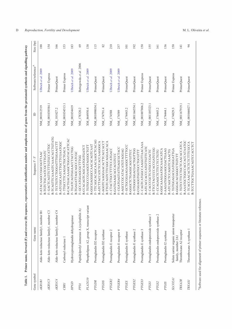
Table1.
Primer
name,forw
ard
(F)andreverse
(R)sequence,representativeidentificationnumber
andampliconsize
ofgenes
from
theprostanoid
synthesisandsignallingpathway
Genesymbol
Genenam
eSequence
50 –30
IDSoftware/reference
ASize(bp)
AKR1B1
Aldo–ketoreductasefamily1,mem
ber
B1
F-A
TACAAGCCGGCGGTTAAC
NM_001012519
Ulbrich
etal.2009
188
R-TGTCTGCAATCGCTTTGATC
AKR1C3
Aldo–ketoreductasefamily1,mem
ber
C3
F-GACTCAGTTCTTTGTGCCATTGC
NM_001038584.1
Primer
Express
154
R-TCAGTTCAAAGTCAAACACCTGTATG
AKR1C4
Aldo–ketoreductasefamily1,mem
ber
C4
F-TCCTGTCCTGGGATTTGGAACCTT
NM_181027.2
PrimerQuest
166
R-A
TCGGCAATCTTGCTTCGAATGGC
CBR1
Carbonylreductase1
F-TTGCCTTCAAGACTGCTGACA
NM_001034513.1
Primer
Express
153
R-CACTGACAAAGCTGGATACATTCAC
HPGD
Hydroxyprostaglandin
dehydrogenase
F-TGATCAGTGGAACCTACCTGG
NM_001034419
Ulbrich
etal.2009
183
R-TGAGATTAGCAGCCATCGC
PPIA
PeptidylprolylisomeraseA(cyclophilin
A)
F-GCCATGGAGCGCTTTGG
NM_178320.2
Bettegowdaetal.2006
69
R-CCACAGTCAGCAATGGTGATCT
PLA2G10
PhospholipaseA2,groupX,transcriptvariant
F-TGTGCCCGAAGGTAGGGCTGTT
XM_864950.4
Ulbrich
etal.2009
138
R-G
GCGAGGGCCAACACAGTCAAT
PTGDR
Prostaglandin
D2receptor
F-TTCAGCACAGCAACAAGCTCACAG
NM_001098034.1
PrimerQuest
115
R-A
TCTTACCATCTCCACCAAGGGCA
PTGDS
Prostaglandin
D2synthase
F-A
GGTCAAGGAACACTTCACCACCT
NM_174791.4
PrimerQuest
82
R-TTGTCAGTCTTCGGCAGGAACACA
PTGER2
Prostaglandin
Ereceptor2
F-CTACTTGCCTTTTCCATGACC
NM_174588
Ulbrich
etal.2009
210
R-G
ATGAAGCACCACGTCCC
PTGER4
Prostaglandin
Ereceptor4
F-CGATGAGTATTGAGCGCTACC
NM_174589
Ulbrich
etal.2009
237
R-A
GCCCGCATACATGTAGGAG
PTGES
Prostaglandin
Esynthase
F-GCTGCGGAAGAAGGCTTTTGCC
NM_174443.2
PrimerQuest
101
R-G
GGCTCTGAGGCAGCGTTCC
PTGES2
Prostaglandin
Esynthase2
F-GTGGGCGGACGACTGGTTGG
NM_001166554.1
PrimerQuest
192
R-CGGAGGTGGTGCCTGCGTTT
PTGES3
Prostaglandin
Esynthase3
F-CAGTCATGGCCAAGGTTAACAAA
NM_001007806.2
Primer
Express
150
R-A
TCACCACCCATGTTGTTCATC
PTGS1
Prostaglandin
endoperoxidesynthase1
F-CACCCGCTCATGCCCGACTC
NM_001105323.1
PrimerQuest
155
R-TTCCTACCCCCACCGATCCGG
PTGS2
Prostaglandin
endoperoxidesynthase2
F-CCAGAGCTCTTCCTCCTGTG
NM_174445.2
PrimerQuest
161
R-G
GCAAAGAATGCAAACATCA
PTGIS
Prostaglandin
I2synthase
F-A
AGATGGGAAGCGACTGAAG
NM_174444.1
PrimerQuest
136
R-A
TCAGCTCCAGGTCAAACTG
SLCO2A1
Solutecarrierorganicaniontransporter
family,mem
ber
2A1
F-TGTGGAGACGATGGGATTGA
NM_174829.3
Primer
Express
150
R-G
GGACACGGGCCTGTCTT
TBXA2R
ThromboxaneA2receptor
F-TGTCCTTCCTGCTCAACACCATCA
NM_001167919.1
PrimerQuest
141
R-A
AATGCTGGCCACCACCATAATGC
TBXAS1
ThromboxaneAsynthase1
F-TCACCAACACTCTCTCTTTCGCCA
NM_001046027.1
PrimerQuest
94
R-TCCTTGCTGAAACAGTCCACCTCT
ASoftwareusedforalignmentofprimer
sequencesorliterature
reference.
D Reproduction, Fertility and Development M. L. Oliveira et al.
LC–MS/MS approach and is only briefly described here. Theanalytical standards and deuterated surrogates were obtained
from Cayman Chemical (Ann Arbor, MI, USA), LarodanFine Chemicals AB (Malmo, Sweden) or Biomol International(Plymouth Meeting, PA, USA). The oxylipins were extracted
from 2mL of uterine flushing using Waters Oasis-HBL car-tridges (Waters, Milford, MA, USA) preconditioned with washsolution (H2O :MeOH; 95 : 5, in 0.1% acetic acid). The uterine
flushing aliquots, 200mL of wash solution, 10 mL of surrogatestandards (400 nM per standard in MeOH), 10 mL anti-oxidantand enzyme inhibitor solution (0.2mgmL�1 of butylated hydro-xytoluene (BHT), EDTA, thiamine pyrophosphate and indo-
methacin) were applied to the cartridge, rinsed with washsolution, eluted with 500mL of methanol and then with 1.5mLof ethyl acetate and collected into polypropylene tubes contain-
ing 6mL of 30% glycerol in methanol. The solvent was strippedand the sample was suspended in 50mL of methanol containingthe technical standard 1-cyclohexyl-dodecanoic acid urea
(CUDA; 800 nM). The samples were then centrifuged at10 000g for 30min at 48C and the supernatants were stored at�208C until analysis. Oxylipin profiling was performed using10-mL sample injections on a Waters ACQUITY UPLC system
via a 2.1� 150mm, 1.7-mm Waters Acquity BEH columnmaintained at 608C coupled to an XEVO TQ triple quadrupolemass spectrometer (Waters). The samples were maintained at
48C before injection. Solvents A (0.1% acetic acid in water) andB (acetonitrile : methanol : acetic acid, 88 : 12 : 0.1) were used inthe following gradient: 15% B for 0.74min, 30% B at 1.5min,
47% B at 3.5min, 54% B at 6min, 60% B at 10.5min, 70% B at15min, 80% B at 16min, 100% B from 17 to 19min, 30% Bfrom 19.3 to 21min. The oxylipins detected above the limit of
quantitation (LOQ) were quantified, recalculated based on theoriginal uterine flushing concentrations and normalised to theuterine flushing recovery (V[recovered volume]/V[instilledvolume]). The normalisation to uterine flushing recovery did
not affect the overall trends in the samples. For the endometrialtissue, the samples were previously extracted with organicsolvents before solid-phase extraction. For this step, 100mg of
cryo-pulverised endometrial tissue was added to amber vials(2mL, polytetrafluoroethylene (PTFE) caps; National ScientificCo., Rockwood, TN, USA) prepared with 5mL of BHT–EDTA
(0.2mgmL�1 in 1 : 1 MeOH :H2O) and 20mL of the surrogatestandards (1000 nM per standard in MeOH). Then, 500mL ofMeOH was added and the vials were capped and then brieflyvortexed. The samples were then centrifuged at 3.000g and 08Cfor 5min. The supernatant was collected and saved. Then,350mL of isopropyl alcohol (IPA) was added to the remainingtissue. The samples were treated in an identical manner using
methanol and the IPA extract was added to the MeOH fraction.The remaining tissue was mixed with 350mL of cyclohexane.The cyclohexane extract was treated as described above and the
supernatant was pooled with IPA and MeOH. The combinedfraction was dried at reduced pressure (Genevac Inc., StoneRidge, NY, USA) for ,1 h. The dried samples were reconsti-
tuted in 200mL of MeOH : toluene (1 : 1) and 100mL of a sub-aliquot of the extract was loaded into the solid phase extraction(SPE) cartridges and extracted as described above for the uterineflushing.
Enzyme-linked immunosorbent assay (ELISA)
Because a reduced number of samples was used for massspectrometry and considering that PGE2 and PGF2a are the
most important prostanoids in the uterus, enzyme-linked immu-nosorbent assays (ELISAs) to measure the concentrations ofPGE2 and PGF2a in uterine flushing samples were validated
using commercial kits for PGE2 and PGF2a (both fromCaymanChemical Co.). Initially, a uterine flushing pool was treatedwith activated charcoal to remove prostaglandins (Turzillo and
Fortune 1990). Briefly, 500mg of activated charcoal was addedfor each mL of uterine flushing and this mixture was incubatedfor 45min and then centrifuged at 12 000g and 48C for 1 h. Thesupernatant was filtered and stored at�808C. This prostaglandin-freematrixwas used only to prepare PGE2 and PGF2a standardsfrom 15.6 to 1000 pgmL�1 and from 7.8 to 500 pgmL�1,respectively. The matrix volume added to each standard was
equal at each standard-curve point (five points were used in eachcurve). The standard curves were compared with the curvesproduced using only the manufacturer’s enzyme immunoassay
(EIA) buffer and validated by parallelism. All of the assay-specific reagents were prepared as described and suggested bythe manufacturers. After the plate setup and incubations, the
absorptions were read spectrophotometrically at a wavelengthof 414 nm (Labsystems Multiskan – MS; Thermo Fisher Scien-tific, Waltham, MA, USA). After validation, the concentrationsof PGE2 and PGF2a in the uterine flushings collected from a
subset of cows in the LF-CL group (n¼ 10) and the SF-SCLgroup (n¼ 10) were assayed in duplicate. Concentrations werecalculated in reference to a regression equation generated from a
standard curve preparedwith increasing concentrations of PGE2or PGF2a, diluted in prostaglandin-free uterine flushing.
Statistical analyses
Outlying observations greater than two standard deviationranges from the mean were not used in the statistical analyses.The data were tested for normality of the residues using theShapiro–Wilk test and for homogeneity of variance using the
F-max test and natural log-transformed if needed. The ovarianand endocrine variables were analysed by one-way ANOVAto test the effect of the treatment using the PROC GLM proce-
dure of the SAS software (Version 9.2; SAS Institute, Cary,NC, USA). The eicosanoid concentrations and relative geneexpression levels were analysed through non-paired Students’
t-test. Pearson’s correlation coefficients were calculated bet-ween P4 concentrations, POF and CL size or P4 concentrationsand abundance of transcripts and concentrations of prostanoids
in the uterus, and between abundance of transcripts and con-centrations of prostanoids in the endometrial tissues or uterineflushings. A probability of P# 0.05 indicated that an effect wassignificant and a probability of P. 0.05 to P# 0.1 indicated
that significance was approached.
Results
Ovarian responses and circulating P4 concentrations:animal model
The hormonal treatments successfully resulted in two groupsof cows with distinct ovarian characteristics, as previously
Uterine prostanoid pathways at early dioestrus Reproduction, Fertility and Development E
described by Mesquita et al. (2014). More specifically, cowsassigned to the LF-LCL group had larger POF diameters when
compared with animals from the SF-SCL group (12.7� 0.3mmand 11.2� 0.4mm, respectively; P, 0.05). Furthermore, the
larger POFs resulted in larger (2.5� 0.31 vs 1.6� 0.1 cm3;P, 0.05) and heavier (2.9� 0.4 vs 2.0� 0.1 g; P, 0.05) cor-
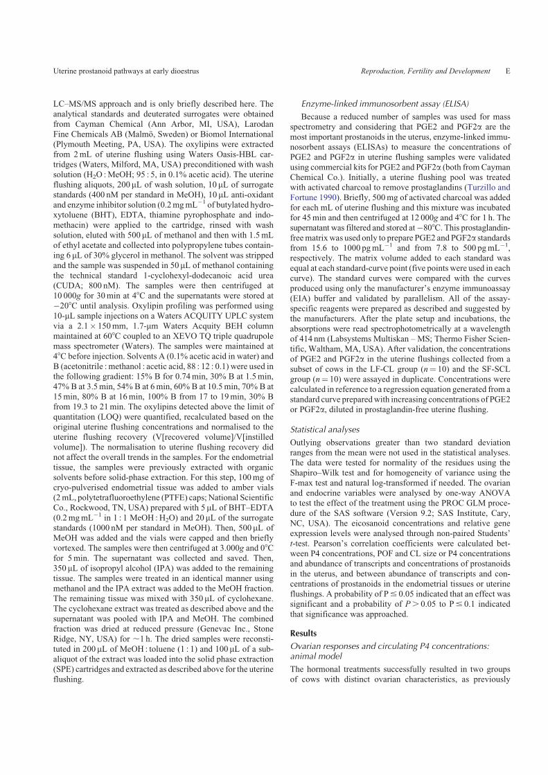
pora lutea on Day 7 after induction of ovulation. Mean P4concentrations were higher in cows from the LF-LCL groupwhen compared with cows from the SF-SCL group (4.4� 0.4 vs
3.5� 0.3 ngmL�1; P, 0.05; Fig. 1).
Transcript abundance in endometrial tissue
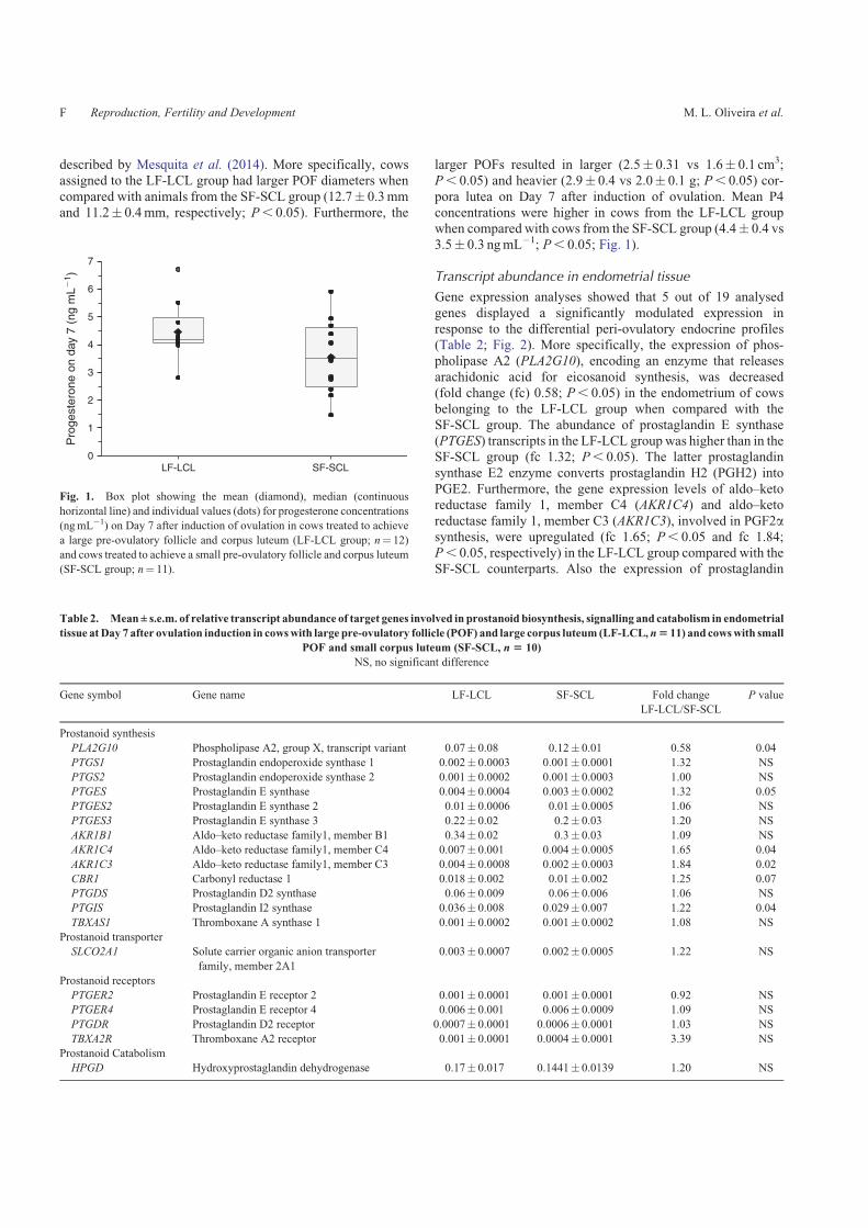
Gene expression analyses showed that 5 out of 19 analysed
genes displayed a significantly modulated expression inresponse to the differential peri-ovulatory endocrine profiles(Table 2; Fig. 2). More specifically, the expression of phos-pholipase A2 (PLA2G10), encoding an enzyme that releases
arachidonic acid for eicosanoid synthesis, was decreased(fold change (fc) 0.58; P, 0.05) in the endometrium of cowsbelonging to the LF-LCL group when compared with the
SF-SCL group. The abundance of prostaglandin E synthase(PTGES) transcripts in the LF-LCL group was higher than in theSF-SCL group (fc 1.32; P, 0.05). The latter prostaglandin
synthase E2 enzyme converts prostaglandin H2 (PGH2) intoPGE2. Furthermore, the gene expression levels of aldo–ketoreductase family 1, member C4 (AKR1C4) and aldo–keto
reductase family 1, member C3 (AKR1C3), involved in PGF2asynthesis, were upregulated (fc 1.65; P, 0.05 and fc 1.84;P, 0.05, respectively) in the LF-LCL group compared with theSF-SCL counterparts. Also the expression of prostaglandin
Table 2. Mean± s.e.m. of relative transcript abundance of target genes involved in prostanoid biosynthesis, signalling and catabolism in endometrial
tissue atDay 7 after ovulation induction in cowswith large pre-ovulatory follicle (POF) and large corpus luteum (LF-LCL,n5 11) and cowswith small
POF and small corpus luteum (SF-SCL, n 5 10)
NS, no significant difference
Gene symbol Gene name LF-LCL SF-SCL Fold change
LF-LCL/SF-SCL
P value
Prostanoid synthesis
PLA2G10 Phospholipase A2, group X, transcript variant 0.07� 0.08 0.12� 0.01 0.58 0.04
PTGS1 Prostaglandin endoperoxide synthase 1 0.002� 0.0003 0.001� 0.0001 1.32 NS
PTGS2 Prostaglandin endoperoxide synthase 2 0.001� 0.0002 0.001� 0.0003 1.00 NS
PTGES Prostaglandin E synthase 0.004� 0.0004 0.003� 0.0002 1.32 0.05
PTGES2 Prostaglandin E synthase 2 0.01� 0.0006 0.01� 0.0005 1.06 NS
PTGES3 Prostaglandin E synthase 3 0.22� 0.02 0.2� 0.03 1.20 NS
AKR1B1 Aldo–keto reductase family1, member B1 0.34� 0.02 0.3� 0.03 1.09 NS
AKR1C4 Aldo–keto reductase family1, member C4 0.007� 0.001 0.004� 0.0005 1.65 0.04
AKR1C3 Aldo–keto reductase family1, member C3 0.004� 0.0008 0.002� 0.0003 1.84 0.02
CBR1 Carbonyl reductase 1 0.018� 0.002 0.01� 0.002 1.25 0.07
PTGDS Prostaglandin D2 synthase 0.06� 0.009 0.06� 0.006 1.06 NS
PTGIS Prostaglandin I2 synthase 0.036� 0.008 0.029� 0.007 1.22 0.04
TBXAS1 Thromboxane A synthase 1 0.001� 0.0002 0.001� 0.0002 1.08 NS
Prostanoid transporter
SLCO2A1 Solute carrier organic anion transporter
family, member 2A1
0.003� 0.0007 0.002� 0.0005 1.22 NS
Prostanoid receptors
PTGER2 Prostaglandin E receptor 2 0.001� 0.0001 0.001� 0.0001 0.92 NS
PTGER4 Prostaglandin E receptor 4 0.006� 0.001 0.006� 0.0009 1.09 NS
PTGDR Prostaglandin D2 receptor 0.0007� 0.0001 0.0006� 0.0001 1.03 NS
TBXA2R Thromboxane A2 receptor 0.001� 0.0001 0.0004� 0.0001 3.39 NS
Prostanoid Catabolism
HPGD Hydroxyprostaglandin dehydrogenase 0.17� 0.017 0.1441� 0.0139 1.20 NS
7
5
6
4
3
2
1
0LF-LCL SF-SCL
Pro
gest
eron
e on
day
7 (
ng m
L�1 )
Fig. 1. Box plot showing the mean (diamond), median (continuous
horizontal line) and individual values (dots) for progesterone concentrations
(ngmL�1) on Day 7 after induction of ovulation in cows treated to achieve
a large pre-ovulatory follicle and corpus luteum (LF-LCL group; n¼ 12)
and cows treated to achieve a small pre-ovulatory follicle and corpus luteum
(SF-SCL group; n¼ 11).
F Reproduction, Fertility and Development M. L. Oliveira et al.
I synthase (PTGIS), an enzyme that converts PGH2 into PGI2,was greater (fc 1.22; P, 0.05) in the endometrium from the
LF-LCL group than in the SF-SCL group. The transcriptabundance of carbonyl reductase 1 (CBR1), which uses PGE2 asa substrate for the synthesis of PGF2a, tended to be upregulatedin the LF-LCL compared with the SF-SCL tissue (fc 1.25;P¼ 0.07). No difference (P. 0.1) between groups was detectedfor the transcripts of the main gene related to PGF2a synthesis
(aldo–keto reductase family 1, member B1 (AKR1B1); Fig. 2).
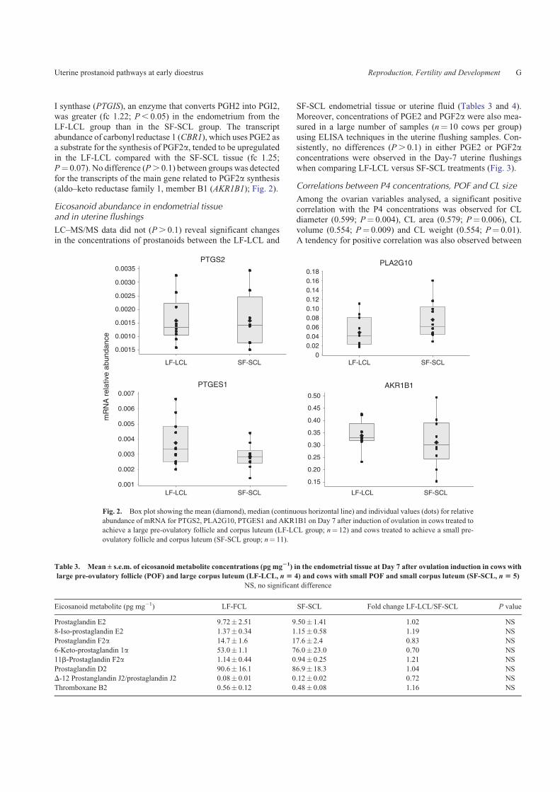
Eicosanoid abundance in endometrial tissueand in uterine flushings
LC–MS/MS data did not (P. 0.1) reveal significant changesin the concentrations of prostanoids between the LF-LCL and
SF-SCL endometrial tissue or uterine fluid (Tables 3 and 4).Moreover, concentrations of PGE2 and PGF2a were also mea-
sured in a large number of samples (n¼ 10 cows per group)using ELISA techniques in the uterine flushing samples. Con-sistently, no differences (P. 0.1) in either PGE2 or PGF2aconcentrations were observed in the Day-7 uterine flushingswhen comparing LF-LCL versus SF-SCL treatments (Fig. 3).
Correlations between P4 concentrations, POF and CL size
Among the ovarian variables analysed, a significant positivecorrelation with the P4 concentrations was observed for CLdiameter (0.599; P¼ 0.004), CL area (0.579; P¼ 0.006), CL
volume (0.554; P¼ 0.009) and CL weight (0.554; P¼ 0.01).A tendency for positive correlation was also observed between
Table 3. Mean ± s.e.m. of eicosanoid metabolite concentrations (pg mg21) in the endometrial tissue at Day 7 after ovulation induction in cows with
large pre-ovulatory follicle (POF) and large corpus luteum (LF-LCL, n 5 4) and cows with small POF and small corpus luteum (SF-SCL, n 5 5)
NS, no significant difference
Eicosanoid metabolite (pg mg�1) LF-FCL SF-SCL Fold change LF-LCL/SF-SCL P value
Prostaglandin E2 9.72� 2.51 9.50� 1.41 1.02 NS
8-Iso-prostaglandin E2 1.37� 0.34 1.15� 0.58 1.19 NS
Prostaglandin F2a 14.7� 1.6 17.6� 2.4 0.83 NS
6-Keto-prostaglandin 1a 53.0� 1.1 76.0� 23.0 0.70 NS
11b-Prostaglandin F2a 1.14� 0.44 0.94� 0.25 1.21 NS
Prostaglandin D2 90.6� 16.1 86.9� 18.3 1.04 NS
D-12 Prostanglandin J2/prostaglandin J2 0.08� 0.01 0.12� 0.02 0.72 NS
Thromboxane B2 0.56� 0.12 0.48� 0.08 1.16 NS
0.0035
0.0030
0.0025
0.0020
0.0015
0.0010
0.0015
0.007
0.006
0.005
0.004
0.003
0.002
0.001
0.50
0.45
0.40
0.35
0.30
0.25
0.20
0.15
0.180.160.140.120.100.080.060.040.02
0LF-LCL SF-SCL LF-LCL SF-SCL
LF-LCL SF-SCL
PTGES1
PTGS2
AKR1B1
PLA2G10
LF-LCL SF-SCL
mR
NA
rel
ativ
e ab
unda
nce
Fig. 2. Box plot showing the mean (diamond), median (continuous horizontal line) and individual values (dots) for relative
abundance of mRNA for PTGS2, PLA2G10, PTGES1 and AKR1B1 on Day 7 after induction of ovulation in cows treated to
achieve a large pre-ovulatory follicle and corpus luteum (LF-LCL group; n¼ 12) and cows treated to achieve a small pre-
ovulatory follicle and corpus luteum (SF-SCL group; n¼ 11).
Uterine prostanoid pathways at early dioestrus Reproduction, Fertility and Development G
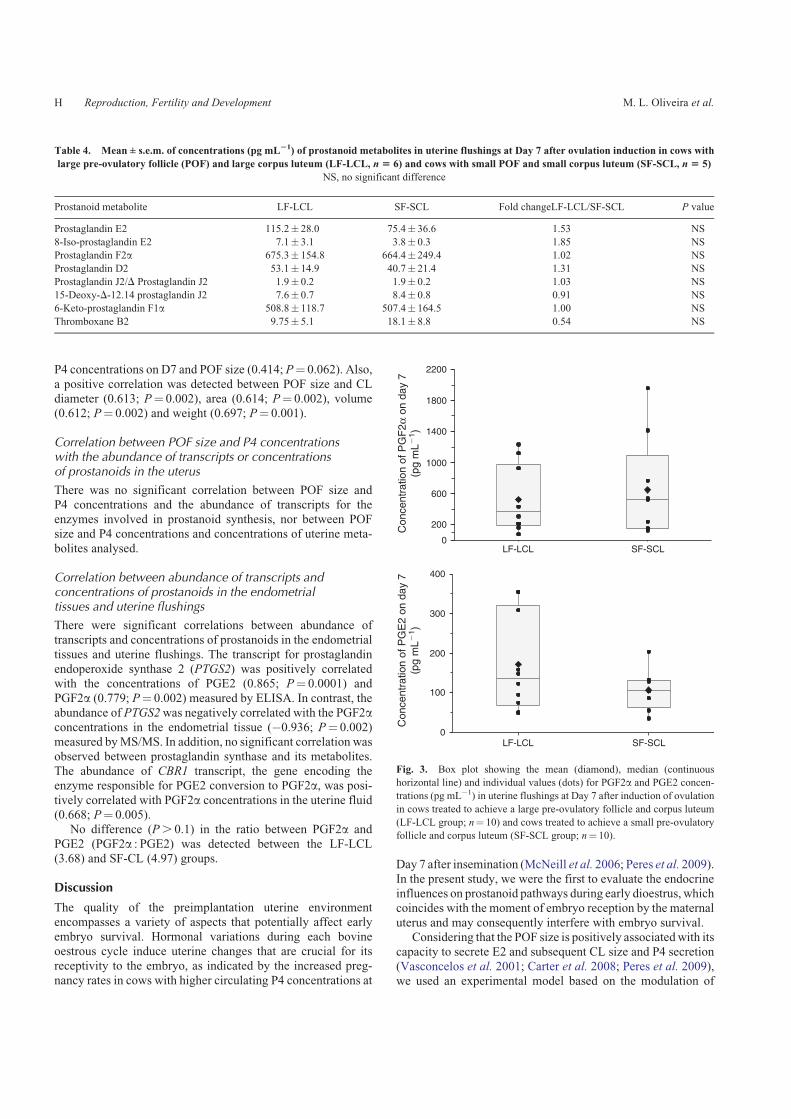
P4 concentrations on D7 and POF size (0.414; P¼ 0.062). Also,
a positive correlation was detected between POF size and CLdiameter (0.613; P¼ 0.002), area (0.614; P¼ 0.002), volume(0.612; P¼ 0.002) and weight (0.697; P¼ 0.001).
Correlation between POF size and P4 concentrationswith the abundance of transcripts or concentrationsof prostanoids in the uterus
There was no significant correlation between POF size andP4 concentrations and the abundance of transcripts for theenzymes involved in prostanoid synthesis, nor between POFsize and P4 concentrations and concentrations of uterine meta-
bolites analysed.
Correlation between abundance of transcripts andconcentrations of prostanoids in the endometrialtissues and uterine flushings
There were significant correlations between abundance oftranscripts and concentrations of prostanoids in the endometrial
tissues and uterine flushings. The transcript for prostaglandinendoperoxide synthase 2 (PTGS2) was positively correlatedwith the concentrations of PGE2 (0.865; P¼ 0.0001) and
PGF2a (0.779; P¼ 0.002) measured by ELISA. In contrast, theabundance of PTGS2was negatively correlated with the PGF2aconcentrations in the endometrial tissue (�0.936; P¼ 0.002)measured byMS/MS. In addition, no significant correlation was
observed between prostaglandin synthase and its metabolites.The abundance of CBR1 transcript, the gene encoding theenzyme responsible for PGE2 conversion to PGF2a, was posi-tively correlated with PGF2a concentrations in the uterine fluid(0.668; P¼ 0.005).
No difference (P. 0.1) in the ratio between PGF2a and
PGE2 (PGF2a : PGE2) was detected between the LF-LCL(3.68) and SF-CL (4.97) groups.
Discussion
The quality of the preimplantation uterine environmentencompasses a variety of aspects that potentially affect early
embryo survival. Hormonal variations during each bovineoestrous cycle induce uterine changes that are crucial for itsreceptivity to the embryo, as indicated by the increased preg-
nancy rates in cows with higher circulating P4 concentrations at
Day 7 after insemination (McNeill et al. 2006; Peres et al. 2009).In the present study, we were the first to evaluate the endocrineinfluences on prostanoid pathways during early dioestrus, whichcoincides with the moment of embryo reception by the maternal
uterus and may consequently interfere with embryo survival.Considering that the POF size is positively associatedwith its
capacity to secrete E2 and subsequent CL size and P4 secretion
(Vasconcelos et al. 2001; Carter et al. 2008; Peres et al. 2009),we used an experimental model based on the modulation of
2200
1800
1400
1000
600
400
300
200
100
0
200
0LF-LCL SF-SCL
LF-LCL SF-SCL
Con
cent
ratio
n of
PG
E2
on d
ay 7
(pg
mL�
1 )C
once
ntra
tion
of P
GF
2α o
n da
y 7
(pg
mL�
1 )
Fig. 3. Box plot showing the mean (diamond), median (continuous
horizontal line) and individual values (dots) for PGF2a and PGE2 concen-
trations (pg mL�1) in uterine flushings at Day 7 after induction of ovulation
in cows treated to achieve a large pre-ovulatory follicle and corpus luteum
(LF-LCL group; n¼ 10) and cows treated to achieve a small pre-ovulatory
follicle and corpus luteum (SF-SCL group; n¼ 10).
Table 4. Mean ± s.e.m. of concentrations (pg mL21) of prostanoid metabolites in uterine flushings at Day 7 after ovulation induction in cows with
large pre-ovulatory follicle (POF) and large corpus luteum (LF-LCL, n 5 6) and cows with small POF and small corpus luteum (SF-SCL, n 5 5)
NS, no significant difference
Prostanoid metabolite LF-LCL SF-SCL Fold changeLF-LCL/SF-SCL P value
Prostaglandin E2 115.2� 28.0 75.4� 36.6 1.53 NS
8-Iso-prostaglandin E2 7.1� 3.1 3.8� 0.3 1.85 NS
Prostaglandin F2a 675.3� 154.8 664.4� 249.4 1.02 NS
Prostaglandin D2 53.1� 14.9 40.7� 21.4 1.31 NS
Prostaglandin J2/D Prostaglandin J2 1.9� 0.2 1.9� 0.2 1.03 NS
15-Deoxy-D-12.14 prostaglandin J2 7.6� 0.7 8.4� 0.8 0.91 NS
6-Keto-prostaglandin F1a 508.8� 118.7 507.4� 164.5 1.00 NS
Thromboxane B2 9.75� 5.1 18.1� 8.8 0.54 NS
H Reproduction, Fertility and Development M. L. Oliveira et al.
follicle growth and CL size, as has been previously describedby our group (Mesquita et al. 2014, 2015; Franca et al. 2015;Ramos et al. 2014). In the present study, positive correlations
between P4 concentrations on D7 and POF and CL size wereobserved. This confirmed that our experimental model not onlymodulated POF growth but efficiently altered CL growth andfunction, based on the P4 concentrations during early dioestrus.
However, there was no significant correlation between POF sizeand P4 concentrations with the transcripts involved in thesynthesis of PGE2 and PGF2a.
Based on the present results, at Day 7 after induction ofovulation the expression of several enzymes responsible forprostaglandin synthesis was upregulated in the endometrial
tissue of cows that ovulated larger follicles compared with thetissue from cows ovulating small follicles and consequentlysmall CLs. Using LC–MS/MS and ELISA techniques, the
relevance of the differently expressed genes have been studiedin detail, by eicosanoid identification and quantification in theDay-7 endometrial tissue and associated uterine flushings fromcows ovulating large or small POF. Interestingly, no differences
in concentrations of prostanoids could be observed either inendometrial tissue or in associated uterine flushings whencomparing the experimental groups. In Fig. 4, an overview of
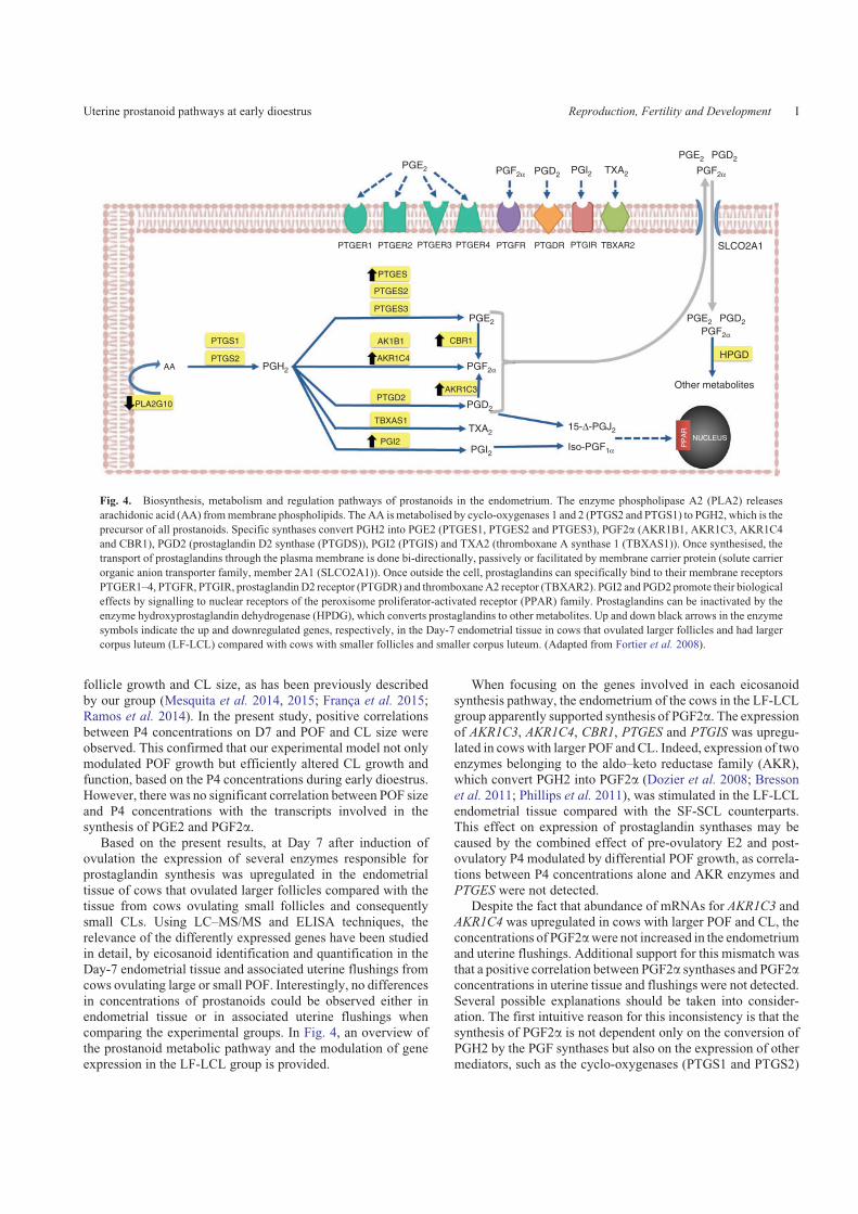
the prostanoid metabolic pathway and the modulation of geneexpression in the LF-LCL group is provided.
When focusing on the genes involved in each eicosanoidsynthesis pathway, the endometrium of the cows in the LF-LCLgroup apparently supported synthesis of PGF2a. The expressionof AKR1C3, AKR1C4, CBR1, PTGES and PTGIS was upregu-lated in cowswith larger POF and CL. Indeed, expression of twoenzymes belonging to the aldo–keto reductase family (AKR),which convert PGH2 into PGF2a (Dozier et al. 2008; Bresson
et al. 2011; Phillips et al. 2011), was stimulated in the LF-LCLendometrial tissue compared with the SF-SCL counterparts.This effect on expression of prostaglandin synthases may be
caused by the combined effect of pre-ovulatory E2 and post-ovulatory P4 modulated by differential POF growth, as correla-tions between P4 concentrations alone and AKR enzymes and
PTGES were not detected.Despite the fact that abundance of mRNAs for AKR1C3 and
AKR1C4 was upregulated in cows with larger POF and CL, the
concentrations of PGF2awere not increased in the endometriumand uterine flushings. Additional support for this mismatch wasthat a positive correlation between PGF2a synthases and PGF2aconcentrations in uterine tissue and flushings were not detected.
Several possible explanations should be taken into consider-ation. The first intuitive reason for this inconsistency is that thesynthesis of PGF2a is not dependent only on the conversion of
PGH2 by the PGF synthases but also on the expression of othermediators, such as the cyclo-oxygenases (PTGS1 and PTGS2)
PGE2
PGE2
PGH2
PTGER1 PTGER2
PTGES
PTGES2
PTGES3
PTGD2
PTGS1
AA
PLA2G10
PTGS2
TBXAS1
AK1B1
PGI2
CBR1
AKR1C4
AKR1C3
PTGER3 PTGER4 PTGFR PTGDR PTGIR TBXAR2
PGF2α
PGF2α
PGF2α
PGD2
PGD2
15-Δ-PGJ2
Iso-PGF1α
TXA2
PGI2
PGE2 PGD2
HPGD
PGl2
PGE2 PGD2
SLCO2A1
Other metabolites
NUCLEUS
PP
AR
PGF2αTXA2
Fig. 4. Biosynthesis, metabolism and regulation pathways of prostanoids in the endometrium. The enzyme phospholipase A2 (PLA2) releases
arachidonic acid (AA) frommembrane phospholipids. The AA ismetabolised by cyclo-oxygenases 1 and 2 (PTGS2 and PTGS1) to PGH2, which is the
precursor of all prostanoids. Specific synthases convert PGH2 into PGE2 (PTGES1, PTGES2 and PTGES3), PGF2a (AKR1B1, AKR1C3, AKR1C4
and CBR1), PGD2 (prostaglandin D2 synthase (PTGDS)), PGI2 (PTGIS) and TXA2 (thromboxane A synthase 1 (TBXAS1)). Once synthesised, the
transport of prostaglandins through the plasma membrane is done bi-directionally, passively or facilitated by membrane carrier protein (solute carrier
organic anion transporter family, member 2A1 (SLCO2A1)). Once outside the cell, prostaglandins can specifically bind to their membrane receptors
PTGER1–4, PTGFR, PTGIR, prostaglandin D2 receptor (PTGDR) and thromboxaneA2 receptor (TBXAR2). PGI2 and PGD2 promote their biological
effects by signalling to nuclear receptors of the peroxisome proliferator-activated receptor (PPAR) family. Prostaglandins can be inactivated by the
enzyme hydroxyprostaglandin dehydrogenase (HPDG), which converts prostaglandins to other metabolites. Up and down black arrows in the enzyme
symbols indicate the up and downregulated genes, respectively, in the Day-7 endometrial tissue in cows that ovulated larger follicles and had larger
corpus luteum (LF-LCL) compared with cows with smaller follicles and smaller corpus luteum. (Adapted from Fortier et al. 2008).
Uterine prostanoid pathways at early dioestrus Reproduction, Fertility and Development I
to convert the arachidonic acid (AA) into PGH2. In this regard,the production of PGH2 by the cyclo-oxygenase PTGS1 (con-
stitutive) and PTGS2 (regulatory) is considered the rate-limitingstep of prostaglandin biosynthesis in the endometrium (Smithet al. 2000; Parent et al. 2003). This was supported in the present
study by the strong positive correlations betweenPTGS2 and theconcentrations of PGF2a and PGE2 in the uterine flushings.Secretion of PGF2a by endometrial explants is also correlated
with their PTGS2 content, suggesting that the increase in theability of the uterus to produce prostaglandin during the lutealphase of the oestrous cycle is due to the increase in PTGS2 levels(Charpigny et al. 1997). In addition, the expression of PTGS2
increases 70–100 times before PGF2a elevation at parturition,whereas PGF2a synthase (AKR1B1) increases only 2.6 times(Schuler et al. 2006). Steroid hormones may modulate the
expression of PTGS2 in endometrial cells (Madore et al. 2003),but the significant increase in P4 concentrations in cows withlarge POF and CL did not result in altered expression of this gene
in the present study.A second consideration is that the abundance ofAKR1B1was
also similar between cows with large and small POF and CL.AKR1B1 is considered to be the main synthase enzyme in the
ARK family responsible for PGF2a biosynthesis in the humanand bovine endometrium (Madore et al. 2003; Bresson et al.
2011) and its expression is positively associated with PTGS2
abundance (Charpigny et al. 1997; Xiao et al. 1998; Schuleret al. 2006). Consequently, the similar abundance of transcriptsforPTGS2 andAKR1B1 in cowswith large or small POF andCL
and the absence of a significant correlation between P4 con-centrations and the abundance of these transcripts may be themain explanations for the lack of difference in PGF2a concen-
trations in the endometrium and uterine flushings betweengroups. Therefore, the upregulation of AKR1C3 and AKR1C4
in cows with large POF and CL was possibly a response to thegreater P4 concentrations on D7, as enzymes in the AKR family
have a double function of prostaglandin synthesis and P4catabolism (Pelletier et al. 1999; Madore et al. 2003; Ito et al.
2006). This result is also a novel finding, as a previous study
reported that AKR1C familymembers were not expressed in thebovine endometrium during dioestrus (Madore et al. 2003).
Similarly, the concentrations of PGE2 were not increased
in consequence of the greater abundance of PTGES1 transcriptin cows with larger POF and CL. This mismatch between asynthase and its prostanoid may also be caused by the absenceof the modulation of PTGS2 by the different peri-ovulatory
endocrine profiles. In line with this, Arosh et al. (2002) sug-gested that the increased PGE2 production in endometrial cellsis mainly caused by the associative upregulation of PTGES1
with PTGS2. In addition, lower levels of PGE synthase andPTGS2 in the bovine endometriumwere detected between Days1 and 12 of the oestrous cycle (Arosh et al. 2002), indicating a
limited capacity of the uterus to secrete PGE2 during early andmid dioestrus. This also suggested that the cyclo-oxygenasesmight be the key component monitoring final prostaglandin
concentrations in the bovine endometrium at early dioestrus.Furthermore, there was an increased abundance of CBR1 tran-scripts in the LF-LCL group. This enzyme uses PGE2 as asubstrate for the synthesis of PGF2a (Kankofer and Wiercinski
1999; Asselin and Fortier 2000; Kankofer et al. 2002). There-fore, part of the PGE2 converted by PTGES1 could be instantly
transformed into PGF2a by CBR1 activity. The concentrationsof PGF2a were greater than the concentrations of PGE2 in theuterine flushings and in the endometrial tissue on D7 of the
oestrous cycle. Thus, at least part of this abundance of PGF2amay be caused by the conversion of PGE2 into PGF2a throughCBR1, as indicated by the positive correlation between PGF2aand CBR1 in the uterine flushings.
A third consideration is related to the gene expression resultsof the PLA2G10 enzyme. This phospholipase comes intoview as a potential regulator of eicosanoid homeostasis, as
its downregulated expression in the LF-LCL compared withSF-SCL tissue might result in a limited substrate provisiontowards effective production of prostaglandins. In this regard,
the PLA2 acts on the release of AA, the primary precursor ofprostanoids (Godkin et al. 2008).
Another consideration regarding the mismatch between
gene expression and prostaglandin concentrations is that ourresults are primarily based on the transcript abundance data.It is not clear whether all transcripts will be translated or evenpost-translationally modified (Robert 2010). A previous report
(Ulbrich et al. 2009) documented a similar mismatch whencomparing eicosanoid transcripts and metabolite concentrationsin the uterus, although in a reverse way and during a later time
window during dioestrus. Finally, post-transcriptional effectsregulating activities should be considered as well. The impor-tance of the latter assumption has been recently emphasised
by Walker et al. (2013); DNA methylation is involved in earlypregnancy events, which might point towards potential post-transcriptional alterations.
The complete role of prostaglandins in the fertility of cowsstill needs to be elucidated, but during early embryo develop-ment the evidence is that specific prostanoids are needed foradequate embryonic viability during early dioestrus. Previous
research has revealed that development of bovine embryos isimpaired by increased PGF2a levels (Scenna et al. 2004, 2005)and is stimulated by PGE2 (Arosh et al. 2004; Ulbrich et al.
2009). Prostaglandins are also essential for elongation ofthe conceptus, as intrauterine infusions of a selective PTGS2inhibitor prevented conceptus elongation in early-pregnant
sheep (Simmons et al. 2010; Dorniak et al. 2011). In the presentstudy, a bovine model was used in order to screen for endocrinepreparation of maternal receptivity without the presence of theembryo. Considering the previous studies and our working
model where cows with large POF and CL had an 80% increasein pregnancy rates (Pugliesi et al. 2015), the expectation wasthat cows in the LF-LCL group could stimulate PGE2 synthesis
and inhibit PGF2a in the endometrium. However, as the concen-trations of PGE2 and PGF2awere correlated onlywith abundanceof transcripts for PTGS2, the study of other important metabolic
pathways in uterine tissue at early dioestrus are indicated tounderstand the positive effects of greater steroid concentrationsduring the peri-ovulatory period on bovine fertility.
In conclusion, the peri-ovulatory endocrine changes associ-ated with the size of the POF regulate transcript abundanceof genes belonging to prostanoid synthesis pathways in thebovine endometrium at early dioestrus (at Day 7 after induction
J Reproduction, Fertility and Development M. L. Oliveira et al.
of ovulation). Specifically, cows that ovulated larger follicleshave increased abundance of AKR1C4, AKR1C3, PTGIS,
PTGES and CBR1 transcripts in the endometrium, whereas theexpression of PLA2G10 was reduced. These changes in tran-scription do not result in modifications in the prostanoid con-
centrations in the endometrium nor in the uterine flushings,which probably result from the lack of modulation of PTGS2,the regulatory rate-limiting enzyme in prostaglandin biosynthe-
sis. Indeed, the abundance of transcripts forPTGS2 is highly andpositively correlated with PGF2a and PGE2 concentrations inthe uterine flushings. Although the concentrations of prosta-noids are not affected by the peri-ovulatory endocrine profiles at
this time point, these novel results characterising the prostanoidconcentrations at early dioestrus point towards maintenance ofhomeostasis at the time of early embryo development.
Acknowledgements
This work was supported by LFEM (Projects #: 204 and 206), CNPq
(481199/2012–8) and FAPESP (2011/03226–4). The authors thank S. C.
Scolari, R. Ramos, M. Sponchiado, M. Franca, Everton Lopes and Estela R.
Araujo for technical assistance, the administration of the Pirassununga
campus of the University of Sao Paulo and CAPES (Coordination for the
Improvement of Higher Education Personnel), Brazil for a scholarship to the
first author.
References
Araujo, E. R., Sponchiado,M., Pugliesi, G., Van Hoeck, V., Mesquita, F. S.,
Membrive, C. M., and Binelli, M. (2015). Spatio-specific regulation of
endocrine-responsive gene transcription by peri-ovulatory endocrine
profiles in the bovine reproductive tract. Reprod. Fertil. Dev.
doi:10.1071/RD14178
Arosh, J. A., Parent, J., Chapdelaine, P., Sirois, J., and Fortier, M. A. (2002).
Expression of cyclo-oxygenases 1 and 2 and prostaglandin E synthase
in bovine endometrial tissue during the oestrous cycle. Biol. Reprod.
67, 161–169. doi:10.1095/BIOLREPROD67.1.161
Arosh, J. A., Banu, S. K., Chapdelaine, P., Madore, E., Sirois, J., and Fortier,
M. A. (2004). Prostaglandin biosynthesis, transport and signalling in
corpus luteum: a basis for autoregulation of luteal function. Endocrinol-
ogy 145, 2551–2560. doi:10.1210/EN.2003-1607
Asselin, E., and Fortier, M. A. (2000). Detection and regulation of the
messenger for a putative bovine endometrial 9-keto-prostaglandin
E(2) reductase: effect of oxytocin and interferon-tau. Biol. Reprod.
62, 125–131. doi:10.1095/BIOLREPROD62.1.125
Bauersachs, S., Blum, H., Mallok, S., Wenigerkind, H., Rief, S., Prelle, K.,
and Wolf, E. (2003). Regulation of ipsilateral and contralateral bovine
oviduct epithelial cell function in the postovulation period: a transcrip-
tomics approach. Biol. Reprod. 68, 1170–1177. doi:10.1095/BIOLRE
PROD.102.010660
Bauersachs, S., Ulbrich, S. E., Gross, K., Schmidt, S. E., Meyer, H. H.,
Wenigerkind, H., Vermehren, M., Sinowatz, F., Blum, H., and Wolf, E.
(2006). Embryo-induced transcriptome changes in bovine endometrium
reveal species-specific and common molecular markers of uterine
receptivity. Reproduction 132, 319–331. doi:10.1530/REP.1.00996
Beltman, M. E., Forde, N., Furney, P., Carter, F., Roche, J. F., Lonergan, P.,
and Crowe, M. A. (2010). Characterization of endometrial gene expres-
sion and metabolic parameters in beef heifers yielding viable or
non-viable embryos on D 7 after insemination. Reprod. Fertil. Dev.
22, 987–999. doi:10.1071/RD09302
Beltman, M. E., Mullen, M. P., Elia, G., Hilliard, M., Diskin, M. G., Evans,
A. C., and Crowe, M. A. (2014). Global proteomic characterisation of
uterine histotroph recovered from beef heifers yielding good-quality and
degenerate Day-7 embryos. Domest. Anim. Endocrinol. 46, 49–57.
doi:10.1016/J.DOMANIEND.2013.10.003
Binelli, M., Scolari, S. C., Pugliesi, G., Van Hoeck, V., Gonella-Diaza,
A. M., Andrade, S. C., Gasparin, G. R., and Coutinho, L. L. (2015). The
transcriptome signature of the receptive bovine uterus determined at
early gestation. PLoS One 10, e0122874. doi:10.1371/JOURNAL.
PONE.0122874
Bresson, E., Boucher-Kovalik, S., Chapdelaine, P., Madore, E., Harvey, N.,
Laberge, P. Y., Leboeuf, M., and Fortier, M. A. (2011). The human
aldose reductase AKR1B1 qualifies as the primary prostaglandin F
synthase in the endometrium. J. Clin. Endocrinol. Metab. 96, 210–219.
doi:10.1210/JC.2010-1589
Bridges, G. A.,Mussard,M. L., Pate, J. L., Ott, T. L., Hansen, T. R., andDay,
M. L. (2012). Impact of pre-ovulatory oestradiol concentrations on
conceptus development and uterine gene expression. Anim. Reprod.
Sci. 133(1–2), 16–26. doi:10.1016/J.ANIREPROSCI.2012.06.013
Buford, W. I., Ahmad, N., Schrick, F. N., Butcher, R. L., Lewis, P. E., and
Inskeep, E. K. (1996). Embryotoxicity of a regressing corpus luteum in
beef cows supplemented with progestogen. Biol. Reprod. 54, 531–537.
doi:10.1095/BIOLREPROD54.3.531
Carter, F., Forde, N., Duffy, P., Wade, M., Fair, T., Crowe, M. A., Evans, A.
C., Kenny, D. A., Roche, J. F., and Lonergan, P. (2008). Effect of
increasing progesterone concentration from Day 3 of pregnancy on
subsequent embryo survival and development in beef heifers. Reprod.
Fertil. Dev. 20, 368–375. doi:10.1071/RD07204
Charpigny, G., Reinaud, P., Tamby, J. P., Creminon, C., Martal, J., Maclouf,
J., and Guillomot,M. (1997). Expression of cyclo-oxygenase-1 and -2 in
ovine endometrium during the oestrous cycle and early pregnancy.
Endocrinology 138, 2163–2171.
Cong, J., Diao, H. L., Zhao, Y. C., Ni, H., Yan, Y. Q., and Yang, Z. M.
(2006). Differential expression and regulation of cyclo-oxygenases,
prostaglandin E synthases and prostacyclin synthase in rat uterus
during the peri-implantation period. Reproduction 131(1), 139–151.
doi:10.1530/REP.1.00861
Dhaliwal, G. S., Murray, R. D., Rees, E. M., Howard, C. V., and Beech, D. J.
(2002). Quantitative unbiased estimates of endometrial gland surface
area and volume in cycling cows and heifers. Res. Vet. Sci. 73, 259–265.
doi:10.1016/S0034-5288(02)00098-X
Diskin, M. G., and Morris, D. G. (2008). Embryonic and early fetal losses
in cattle and other ruminants. Reprod. Domest. Anim. 43, 260–267.
doi:10.1111/J.1439-0531.2008.01171.X
Diskin, M. G., and Sreenan, J. M. (1980). Fertilisation and embryonic
mortality rates in beef heifers after artificial insemination. J. Reprod.
Fertil. 59, 463–468. doi:10.1530/JRF.0.0590463
Diskin,M. G., Parr,M. H., andMorris, D. G. (2012). Embryo death in cattle:
an update. Reprod. Fertil. Dev. 24, 244–251. doi:10.1071/RD11914
Dorniak, P., Bazer, F.W., and Spencer, T. E. (2011). Prostaglandins regulate
conceptus elongation and mediate effects of interferon tau on the ovine
uterine endometrium. Biol. Reprod. 84, 1119–1127. doi:10.1095/BIOL
REPROD.110.089979
Dozier, B. L., Watanabe, K., and Duffy, D. M. (2008). Two pathways for
prostaglandin F2 alpha synthesis by the primate peri-ovulatory follicle.
Reproduction 136, 53–63. doi:10.1530/REP-07-0514
El-Sayed, A., Hoelker, M., Rings, F., Salilew, D., Jennen, D., Tholen, E.,
Sirard, M. A., Schellander, K., and Tesfaye, D. (2006). Large-scale
transcriptional analysis of bovine embryo biopsies in relation to
pregnancy success after transfer to recipients. Physiol. Genomics
28(1), 84–96. doi:10.1152/PHYSIOLGENOMICS.00111.2006
Forde, N., Carter, F., Fair, T., Crowe, M. A., Evans, A. C., Spencer, T. E.,
Bazer, F.W.,McBride, R., Boland,M. P., O’Gaora, P., Lonergan, P., and
Roche, J. F. (2009). Progesterone-regulated changes in endometrial gene
expression contribute to advanced conceptus development in cattle.Biol.
Reprod. 81, 784–794. doi:10.1095/BIOLREPROD.108.074336
Uterine prostanoid pathways at early dioestrus Reproduction, Fertility and Development K
Fortier, M. A., Krishnaswamy, K., Danyod, G., Boucher-Kovalik, S., and
Chapdalaine, P. (2008). A postgenomic integrated view of prostaglan-
dins in reproduction: implications for other body systems. J. Physiol.
Pharmacol. 59(Suppl 1), 65–89.
Franca, M. R., Mesquita, F. S., Lopes, E., Pugliesi, G., Van Hoeck, V.,
Chiaratti, M. R., Membrive, C. B., Papa, P. C., and Binelli, M. (2015).
Modulation of peri-ovulatory endocrine profiles in beef cows: conse-
quences for endometrial glucose transporters and uterine fluid glucose
levels. Domest. Anim. Endocrinol. 50, 83–90. doi:10.1016/J.DOMA
NIEND.2014.09.005
Garbarino, E. J., Hernandez, J. A., Shearer, J. K., Risco, C. A., and Thatcher,
W. W. (2004). Effect of lameness on ovarian activity in postpartum
hostein cows. J. Dairy Sci. 87, 4123–4131. doi:10.3168/jds.S0022-0302
(04)73555-9
Godkin, J. D., Roberts, M. P., Elgayyar, M., Guan, W., and Tithof, P. K.
(2008). PhospholipaseA2 regulation of bovine endometrial (BEND) cell
prostaglandin production. Reprod. Biol. Endocrinol. 6, 44. doi:10.1186/
1477-7827-6-44
Hockett, M. E., Rohrbach, N. R., and Schrick, F. N. (2004). Alterations in
embryo development in progestogen-supplemented cows administered
prostaglandin F2alpha. Prostaglandins Other Lipid Mediat. 73, 227–236.
doi:10.1016/J.PROSTAGLANDINS.2004.02.002
Ito, K., Utsunomiya, H., Suzuki, T., Saitou, S., Akahira, J., Okamura, K.,
Yaegashi, N., and Sasano, H. (2006). 17Beta-hydroxysteroid dehydro-
genases in human endometrium and its disorders.Mol. Cell. Endocrinol.
248, 136–140. doi:10.1016/J.MCE.2005.11.038
Kankofer, M., and Wiercinski, J. (1999). Prostaglandin E2 9-keto reductase
from bovine term placenta. Prostaglandins Leukot. Essent. Fatty Acids
61(1), 29–32. doi:10.1054/PLEF.1999.0069
Kankofer, M., Wiercinski, J., and Zerbe, H. (2002). Prostaglandin E(2)
9-keto reductase activity in bovine retained and not-retained placenta.
Prostaglandins Leukot. Essent. Fatty Acids 66, 413–417. doi:10.1054/
PLEF.2002.0367
Kastelic, J. P., Pierson, R. A., and Ginther, O. J. (1990). Ultrasonic
morphology of corpora lutea and central luteal cavities during the estrous
cycle and early pregnancy in heifers. Theriogenology 34, 487–498.
doi:10.1016/0093-691X(90)90006-F
Lim, H., Paria, B. C., Das, S. K., Dinchuk, J. E., Langenbach, R., Trzaskos,
J. M., and Dey, S. K. (1997). Multiple female reproductive failures in
cyclo-oxygenase 2-deficient mice. Cell 91, 197–208. doi:10.1016/
S0092-8674(00)80402-X
Lundstrom, S. L., Saluja, R., Adner, M., Haeggstrom, J. Z., Nilsson, G., and
Wheelock, C. E. (2013). Lipid mediator metabolic profiling demon-
strates differences in eicosanoid patterns in two phenotypically distinct
mast cell populations. J. Lipid Res. 54, 116–126. doi:10.1194/JLR.
M030171
Madore, E., Harvey, N., Parent, J., Chapdelaine, P., Arosh, J. A., and Fortier,
M. A. (2003). An aldose reductase with 20 alpha-hydroxysteroid
dehydrogenase activity is most likely the enzyme responsible for the
production of prostaglandin F2 alpha in the bovine endometrium. J. Biol.
Chem. 278, 11205–11212. doi:10.1074/JBC.M208318200
Mansouri-Attia, N., Aubert, J., Reinaud, P., Giraud-Delville, C., Taghouti, G.,
Galio, L., Everts, R. E., Degrelle, S., Richard, C., Hue, I., Yang, X.,
Tian, X. C., Lewin, H. A., Renard, J. P., and Sandra, O. (2009). Gene
expression profiles of bovine caruncular and intercaruncular endometrium
at implantation Physiol. Genomics 39, 14–27. doi:10.1152/PHYSIOLGE
NOMICS.90404.2008
McNeill, R. E., Diskin, M. G., Sreenan, J. M., and Morris, D. G. (2006).
Associations betweenmilk progesterone concentration on different days
and with embryo survival during the early luteal phase in dairy cows.
Theriogenology 65, 1435–1441. doi:10.1016/J.THERIOGENOLOGY.
2005.08.015
Mesquita, F. S., Pugliesi, G., Scolari, S. C., Franca, M. R., Ramos, R. S.,
Oliveira, M., Papa, P. C., Bressan, F. F., Meirelles, F. V., Silva, L. A.,
Nogueira, G. P., Membrive, C. M., and Binelli, M. (2014). Manipulation
of the peri-ovulatory sex steroidal milieu affects endometrial but not
luteal gene expression in early dioestrus Nelore cows. Theriogenology
81, 861–869. doi:10.1016/J.THERIOGENOLOGY.2013.12.022
Mesquita, F. S., Ramos, R. S., Pugliesi, G., Andrade, S. C., Van Hoeck, V.,
Langbeen, A., Oliveira, M. L., Gonella-Diaza, A. M., Gasparin, G.,
Fukumasu, H., Pulz, L. H., Membrive, C. M., Coutinho, L. L., and
Binelli, M. (2015). The receptive endometrial transcriptomic signature
indicates an earlier shift from proliferation tometabolism at early dioestrus
in the cow. Biol. Reprod. doi:10.1095/BIOLREPROD.115.129031
Mosher, A. A., Rainey, K. J., Giembycz, M. A., Wood, S., and Slater, D. M.
(2012). Prostaglandin E2 represses interleukin 1 beta-induced inflam-
matory mediator output from pregnant human myometrial cells through
the EP2 and EP4 receptors. Biol. Reprod. 87(1), 7. doi:10.1095/BIOL
REPROD.112.100099
Parent, J., Villeneuve, C., and Fortier, M. A. (2003). Evaluation of the
contribution of cyclo-oxygenase 1 and cyclo-oxygenase 2 to the produc-
tion of PGE2andPGF2alpha in epithelial cells frombovine endometrium.
Reproduction 126, 539–547. doi:10.1530/REP.0.1260539
Pelletier, G., Luu-The, V., Tetu, B., and Labrie, F. (1999). Immunocyto-
chemical localisation of Type 5 17beta-hydroxysteroid dehydrogenase
in human reproductive tissues. J. Histochem. Cytochem. 47, 731–737.
doi:10.1177/002215549904700602
Peres, R. F., Claro, I., Sa Filho, O.G., Nogueira, G. P., andVasconcelos, J. L.
(2009). Strategies to improve fertility inBos indicus post-pubertal heifers
and non-lactating cows submitted to fixed-time artificial insemination.
Theriogenology 72, 681–689. doi:10.1016/J.THERIOGENOLOGY.
2009.04.026
Pfaffl, M. W. (2001). A new mathematical model for relative quantification
in real-time RT-PCR. Nucleic Acids Res. 29, e45. doi:10.1093/NAR/29.
9.E45
Phillips, R. J., Al-Zamil, H., Hunt, L. P., Fortier,M. A., and Lopez Bernal, A.
(2011). Genes for prostaglandin synthesis, transport and inactivation are
differentially expressed in human uterine tissues and the prostaglandin
F synthase AKR1B1 is induced in myometrial cells by inflammatory
cytokines.Mol. Hum.Reprod. 17, 1–13. doi:10.1093/MOLEHR/GAQ057
Pohler, K. G., Geary, T. W., Atkins, J. A., Perry, G. A., Jinks, E. M., and
Smith,M. F. (2012). Follicular determinants of pregnancy establishment
and maintenance. Cell Tissue Res. 349, 649–664. doi:10.1007/S00441-
012-1386-8
Pugliesi, G., Miagawa, B. T., Paiva, Y. N., Franca, M. R., Silva, L. A., and
Binelli, M. (2014). Conceptus-induced changes in the gene expression
of blood immune cells and the ultrasound-accessed luteal function in
beef cattle: how early can we detect pregnancy? Biol. Reprod. 91, 95.
doi:10.1095/BIOLREPROD.114.121525
Pugliesi, G., Santos, F. B., Lopes, E., Nogueira, E., Maio, J. R. G., and
Binelli,M. (2015). Fertility response in suckled beef cows supplemented
with long-acting progesterone after timed artificial insemination.
Reprod. Fertil. Dev. 27, 98. doi:10.1071/RDV27N1AB11
Ramos, R. S., Mesquita, F. S., D’Alexandri, F. L., Gonella-Diaza, A. M.,
Papa Pe, C., and Binelli, M. (2014). Regulation of the polyamine
metabolic pathway in the endometrium of cows during early dioestrus.
Mol. Reprod. Dev. 81, 584–594. doi:10.1002/MRD.22323
Ramos, R. S., Oliveira, M. L., Izaguirry, A. P., Vargas, L. M., Soares, M. B.,
Mesquita, F. S., Santos, F.W., andBinelli,M. (2015). The peri-ovulatory
endocrine milieu affects the uterine redox environment in beef cows.
Reprod. Biol. Endocrinol. 13, 39. doi:10.1186/S12958-015-0036-X
Robert, C. (2010). Microarray analysis of gene expression during early
development: a cautionary overview. Reproduction 140, 787–801.
doi:10.1530/REP-10-0191
L Reproduction, Fertility and Development M. L. Oliveira et al.
Scenna, F. N., Edwards, J. L., Rohrbach, N. R., Hockett, M. E., Saxton,
A. M., and Schrick, F. N. (2004). Detrimental effects of prostaglandin
F2alpha on preimplantation bovine embryos. Prostaglandins Other Lipid
Mediat. 73, 215–226. doi:10.1016/J.PROSTAGLANDINS.2004.02.001
Scenna, F. N., Hockett,M. E., Towns, T.M., Saxton, A.M., Rohrbach,N. R.,
Wehrman,M. E., and Schrick, F. N. (2005). Influence of a prostaglandin
synthesis inhibitor administered at embryo transfer on pregnancy rates
of recipient cows. Prostaglandins Other Lipid Mediat. 78, 38–45.
doi:10.1016/J.PROSTAGLANDINS.2005.02.003
Schrick, F. N., Inskeep, E. K., and Butcher, R. L. (1993). Pregnancy rates for
embryos transferred from early postpartum beef cows into recipients
with normal oestrous cycles. Biol. Reprod. 49, 617–621. doi:10.1095/
BIOLREPROD49.3.617
Schuler, G., Teichmann, U., Kowalewski, M. P., Hoffmann, B., Madore, E.,
Fortier, M. A., and Klisch, K. (2006). Expression of cyclo-oxygenase-II
(COX-II) and 20alpha-hydroxysteroid dehydrogenase (20alpha-HSD)/
prostaglandin F-synthase (PGFS) in bovine placentomes: implications
for the initiation of parturition in cattle. Placenta 27, 1022–1029.
doi:10.1016/J.PLACENTA.2005.11.001
Seals, R. C., Lemaster, J. W., Hopkins, F. M., and Schrick, F. N. (1998).
Effects of elevated concentrations of prostaglandin F2 alpha on preg-
nancy rates in progestogen-supplemented cattle. Prostaglandins Other
Lipid Mediat. 56, 377–389. doi:10.1016/S0090-6980(98)00063-X
Simmons, R.M., Satterfield,M. C.,Welsh, T. H., Bazer, F.W., and Spencer,
T. E. (2010). HSD11B1, HSD11B2, PTGS2 and NR3C1 expression in
the peri-implantation ovine uterus: effects of pregnancy, progesterone
and interferon tau. Biol. Reprod. 82, 35–43. doi:10.1095/BIOLRE
PROD.109.079608
Smith,W. L., DeWitt, D. L., and Garavito, R.M. (2000). Cyclo-oxygenases:
structural, cellular and molecular biology. Annu. Rev. Biochem. 69,
145–182. doi:10.1146/ANNUREV.BIOCHEM.69.1.145
Song, B. S., Kim, J. S., Kim, C. H., Han, Y. M., Lee, D. S., Lee, K. K., and
Koo, D. B. (2009). Prostacyclin stimulates embryonic development
via regulation of the cAMP response element-binding protein-cyclo-
oxygenase-2 signalling pathway in cattle. Reprod. Fertil. Dev. 21,
400–407. doi:10.1071/RD08180
Turzillo, A. M., and Fortune, J. E. (1990). Suppression of the secondary
FSH surge with bovine follicular fluid is associated with delayed
ovarian follicular development in heifers. J. Reprod. Fertil. 89, 643–653.
doi:10.1530/JRF.0.0890643
Ulbrich, S. E., Schulke, K., Groebner, A. E., Reichenbach, H. D., Angioni, C.,
Geisslinger, G., and Meyer, H. H. (2009). Quantitative characterisation
of prostaglandins in the uterus of early pregnant cattle. Reproduction
138, 371–382. doi:10.1530/REP-09-0081
Ulbrich, S. E., Wolf, E., and Bauersachs, S. (2013). Hosting the preimplan-
tation embryo: potentials and limitations of different approaches for
analysing embryo–endometrium interactions in cattle. Reprod. Fertil.
Dev. 25, 62–70. doi:10.1071/RD12279
Vasconcelos, J. L., Sartori, R., Oliveira, H. N., Guenther, J. G., and
Wiltbank, M. C. (2001). Reduction in size of the ovulatory follicle
reduces subsequent luteal size and pregnancy rate. Theriogenology
56, 307–314. doi:10.1016/S0093-691X(01)00565-9
Vilella, F., Ramirez, L., Berlanga, O., Martınez, S., Alama, P., Meseguer, M.,
Pellicer, A., and Simon, C. (2013). PGE2 and PGF2a concentrations in
human endometrial fluid as biomarkers for embryonic implantation.
J. Clin. Endocrinol. Metab. 98, 4123–4132. doi:10.1210/JC.2013-2205
Walker, C. G., Littlejohn,M. D., Mitchell, M. D., Roche, J. R., andMeier, S.
(2012). Endometrial gene expression during early pregnancy differs
between fertile and subfertile dairy cow strains. Physiol. Genomics 44,
47–58. doi:10.1152/PHYSIOLGENOMICS.00254.2010
Walker, C. G., Littlejohn,M. D., Meier, S., Roche, J. R., andMitchell, M. D.
(2013). DNA methylation is correlated with gene expression during
early pregnancy in Bos taurus. Physiol. Genomics 45, 276–286.
doi:10.1152/PHYSIOLGENOMICS.00145.2012
Weems, C. W., Weems, Y. S., and Randel, R. D. (2006). Prostaglandins and
reproduction in female farm animals. Vet. J. 171, 206–228. doi:10.1016/
J.TVJL.2004.11.014
Wiltbank, M. C., and Ottobre, J. S. (2003). Regulation of intraluteal produc-
tion of prostaglandins. Reprod. Biol. Endocrinol. 1, 91. doi:10.1186/
1477-7827-1-91
Xiao, C. W., Liu, J. M., Sirois, J., and Goff, A. K. (1998). Regulation of
cyclo-oxygenase-2 and prostaglandin F synthase gene expression
by steroid hormones and interferon-tau in bovine endometrial cells.
Endocrinology 139, 2293–2299.
www.publish.csiro.au/journals/rfd
Uterine prostanoid pathways at early dioestrus Reproduction, Fertility and Development M