Embed Size (px)
Citation preview
Effect of Platelet-Rich Plasma on CellAdhesion, Cell Migration, andMyofibroblastic Differentiation in HumanGingival FibroblastsMonica Caceres,* Rodrigo Hidalgo,† Antonio Sanz,† Jorge Martınez,* Paula Riera,†
and Patricio C. Smith†
Background: Platelet-rich plasma is a blood-derived frac-tion that contains a high concentration of platelets and growthfactors. It was proposed that the use of this platelet concen-trate stimulates tissue repair. However, little is known aboutthe biologic response of gingival fibroblasts to platelet’s de-rived growth factors. In the present study, we evaluatedwhether platelet-rich plasma modulated cell adhesion, cell mi-gration, and myofibroblastic differentiation in primary culturesof human gingival fibroblasts.
Methods: We studied the response of primary cultures ofgingival fibroblasts to thrombin-activated platelet-rich plasmafractions. Cell adhesion was evaluated through a colorimetricassay. Cell spreading, actin cytoskeleton remodeling, and fo-cal adhesion distribution were assessed through light and im-munofluorescence microscopy. Cell migration was analyzedusing a bicameral cell culture system. Smooth muscle actinproduction was studied through Western blotting.
Results: Exposure of gingival fibroblasts to platelet-richplasma stimulated adhesion and spreading of cells on fibro-nectin matrices, the development of actin-enriched cellularextensions, and formation of focal adhesions. Platelet-richplasma also promoted cell migration and invasion througha reconstituted basement membrane matrix. Differentiationinto the myofibroblastic phenotype, assessed through the pro-duction of smooth muscle actin, was also stimulated by plate-let-rich plasma preparations.
Conclusion: Platelet-rich plasma may modulate several cellresponses potentially involved in wound healing such as celladhesion, cell migration, and myofibroblastic differentiation.J Periodontol 2008;79:714-720.
KEY WORDS
Adhesion; migration; myofibroblast; platelet-rich plasma.
Wound healing requires the co-ordination of a variety of phys-iological processes that follow
a specific time sequence. After tissueinjury, several cell responses, such ascell adhesion and migration, extracellu-lar matrix synthesis and remodeling, cellproliferation, and differentiation are ob-served in a highly coordinated fashion.1
It is generally accepted that growth fac-tors play a significant role in wound heal-ing.2 In fact, almost all stages of tissuerepair are controlled by a wide varietyof cytokines and growth factors actinglocally as regulators of basic cell func-tions.1,2
Cell adhesion and migration are strik-ing events observed during wound heal-ing, and these responses are modulatedsignificantly by several polypeptidicgrowth factors released into the woundedtissue after injury.1,2 A key event ob-served in this phenomenon is the dif-ferentiation of specialized cells termedmyofibroblasts.1 It has been postulatedthat this cell phenotype is derived fromresident tissue fibroblasts, which are ac-tivated by growth factors released byplatelets and activated macrophagespresent in the wound.2 Transforminggrowth factor b1 (TGF-b1) has been iden-tified as an inducer of the myofibroblastmarker smooth muscle actin (a-sma),an actin isoform that takes part in
* Institute of Nutrition and Food Technology, University of Chile, Santiago, Chile.† Faculty of Odontology, University of Chile.
doi: 10.1902/jop.2008.070395
Volume 79 • Number 4
714
cell-mediated granulation tissue contraction duringtissue repair.3-5
Platelets contribute to hemostasis by preventingblood loss at sites of vascular injury, and they containa large number of growth factors and cytokines thatplay a key role in inflammation and tissue repair.1,6
This has led to the idea of using platelets as therapeu-tic tools to improve wound healing, particularly in pa-tients or conditions in which tissue repair is impairedor delayed significantly.6 Platelet-rich plasma (PRP) isa fraction of concentrated human platelets in a smallvolume of plasma.6 It contains concentrated amountsof at least eight growth factors, including three iso-forms of platelet-derived growth factor (PDGF-AA,-BB, and -AB), two forms of transforming growth fac-tor (TGF-b1 and -b2), insulin-like growth factor-I,vascular endothelial growth factor, and epidermalgrowth factor (EGF).7,8 Although initial results werepromising regarding the use of PRP in oral woundrepair, a subsequent study9 demonstrated contra-dictory results. In fact, although some investigatorsreported significant improvements in wound healing,others failed to detect those effects.10-14 Moreover, invitro studies15-21 analyzing the response of cells toPRP demonstrated variable results. These discrep-ancies may be explained by the still poorly character-ized cellular responses to PRP fractions.
Several studies7,19-23 have assessed the biologicresponse of oral cells to PRP. Most of them evaluatedthe capacity of these preparations to induce cell pro-liferation and bone differentiation. However, studiesdesigned to analyze other cellular responses, suchas modulation of cell–matrix interactions and migra-tion, are limited. The main aim of the present studywas to assess, in primary cultures of human gingivalfibroblasts (GFs), the potential of PRP to modulaterelevant mechanisms potentially involved in woundrepair including cell adhesion and spreading, cellmigration, and myofibroblastic differentiation.
MATERIALS AND METHODS
Cell CulturePrimary cultures of human GFs were established bythe explant method.24 Tissue explants were obtainedfrom the retromolar tissue of three individuals (two fe-males and one male; age range: 20 to 28 years) under-going extraction of third molars at a private dentalpractice in Santiago, Chile between June and Decem-ber 2006. Tissue samples were harvested with the in-formed consent of the patients, and the protocol fortissue obtainment was approved by the Ethical Com-mittee of the Faculty of Dentistry, University of Chile.No history of inflammation of the retromolar tissuewas reported. Patients reported no relevant preexist-ing medical or drug histories during the last 6 months.Cells were cultured in Dulbecco’s modified Eagle’s
medium‡ (DMEM) containing 10% fetal bovine serum,§
100 mg/ml penicillin,i 100 mg/ml streptomycin,¶ and50 mg/ml gentamicin# at 37�C in a 5% CO2 atmosphere.All experiments were performed using cells from thesethree donors and with cells expanded between pas-sages four and 10.
Collection and Preparation of PRPPRP was obtained from two healthy male volunteersusing a commercially available system** followingthe method recommended by the manufacturer as de-scribed previously.25 PRP was prepared from bloodderived from these volunteers on two separate occa-sions, and all experiments were performed with thesePRP samples. The average platelet count of donorswas 230,000 (– 35,000) platelets/mm3. The averageplatelet count in the PRP preparation was 1,560,000platelets/mm3 corresponding to 678.3% of the normalvenous blood count. To induce platelet activation,PRP was added to serum-free DMEM at a 1:10 ratio,and bovine thrombin†† (10 units/ml) was added tothe diluted PRP solution and incubated at 37�C for30 minutes. Incubation with thrombin resulted in theformation of a gel that was centrifuged at 1,800 revo-lutions per minute for 7 minutes at 4�C. The superna-tant was used to stimulate cells in all experiments. Afraction of platelet-poor plasma (PPP) was obtained ac-cording to the manufacturer’s instructions and mixedwith serum-free medium and thrombin as describedfor the PRP fractions. Activated PRP was diluted withDMEM to reach concentrations corresponding to 10%,25%, and 50% of the PRP/DMEM mixture.
Cell Migration AssayThe ability of GFs to migrate was assayed using trans-well chambers‡‡ with 8.0 mm–pore polycarbonate fil-ters coated with 10 mg/ml reconstituted basementmembrane§§ on the upper side of the filter.ii Cells wereresuspended in serum-free medium and seeded onthe upper compartment of the chamber. DifferentPRP concentrations were added to the lower chamber.Migration was allowed to occur for 16 hours. Follow-ing the removal of the non-invading cells from the up-per surface of the reconstituted basement membranewith a cotton swab, the invading cells were fixed andstained with 0.2% crystal violet. Cell migration wasevaluated by counting five (·20) fields per chamberas described previously.26 All assays were performedin quadruplicate in three separate sets of experiments.
‡ Gibco BRL, Grand Island, NY.§ Gibco BRL.i Sigma, St. Louis, MO.¶ Sigma.# Sigma.** Harvest Technologies, Munich, Germany.†† Calbiochem, San Diego, CA.‡‡ Matrigel, Costar, Cambridge, MA.§§ BDBioscience USA, Bedford, MA.ii Collaborative Research, Bedford, MA.
J Periodontol • April 2008 Caceres, Hidalgo, Sanz, Martınez, Riera, Smith
715
Cell AdhesionFor cell adhesion, tissue culture 96-well plates werecoated overnight at 4�C with 10 mg/ml fibronectin¶¶
in phosphate buffered saline (PBS), and non-specificbinding sites were blocked by denatured bovine se-rum albumin## (BSA). GFs were stimulated previ-ously with different concentrations of PRP for 16hours. Cells were detached from the cell culture platesby a brief exposure to trypsin/EDTA and counted;1 · 103 cells were seeded onto fibronectin-coatedplates for 7 minutes. Cell adhesion was stopped bypouring off the medium, fixing cells with methanolfor 2 minutes, and incubating with 0.2% crystal violetfor 5 minutes. After removing excess dye, cells weresolubilized in 0.1 M NaH2PO4 in 50% methanol for10 minutes at room temperature. The absorbanceat 570 nm was analyzed on a microplate reader***as described previously.26 All assays were performedin quadruplicate in three separate sets of experiments.
Evaluation of Cell SpreadingAfter stimulation with PPP or PRP, cells were detachedfrom the substratum by a brief exposure to trypsin/EDTA and plated onto fibronectin-coated plates (10mg/ml) for 20 minutes. Cell spreading was stoppedby pouring off the medium, washing adhered cellsonce with PBS, fixing them with methanol for 2 min-utes, and incubating with 0.2% crystal violet for 5 min-utes. After several washes, spread cells were scoredfrom at least four fields of different regions of the dish.Images were captured with a camera††† through an in-verted microscope.‡‡‡ These assays were performedin three separate sets of experiments.
Actin Remodeling and FocalAdhesion DistributionAs described for the cell adhesion assay, cells werestimulated previously with PPP and PRP. After this,cells were plated on fibronectin-coated coverslips,washed with PBS and fixed with 4% paraformaldehydefor 10 minutes, permeabilized with 0.25% TritonX-100 for 4 minutes, and incubated with PBS contain-ing 5% BSA for 30 minutes at 37�C. Primary antibodiesdiluted in PBS containing 1% BSA§§§ were used ina dilution of 1:100 for anti-paxillin.iii The antigen-antibody complex was washed and incubated withfluorescein 5-isothiocynate conjugated anti-rabbitimmunoglobulinG.¶¶¶ F-actinwasstainedwithphalloi-din-rhodamine.### Fluorescence images were exam-ined with a microscope**** and photographed usinga 63· immersion objective and a camera.†††† Theseexperiments were done on three separate occasions.
Western Blotting to Detect a-sma and b-ActinCells were lysed with a buffer containing 50 mM HepespH 7.4, 150 mM NaCl, 2 mM MgCl2, 2 mM ethyleneglycol tetra acetic acid, 1% Triton X-100, 10% glyc-
erol, 2 mM phenylmethylsulphonyl fluoride, 2 mg/mlpepstatin, 2 mg/ml leupeptin, and 1 mM orthovana-date at 4�C. Proteins were resolved by 10% sodiumdodecyl sulfate-polyacrylamide gel electrophoresisand transferred to polyvinylidene difluoride transfermembrane.‡‡‡‡ Membranes were exposed to primaryantibodies (a-sma§§§§ or b-actiniiii) and secondaryantibodies coupled to horseradish peroxidase and de-veloped.¶¶¶¶ Smooth muscle actin production, stimu-lated by PRP, was assessed in three independentexperiments.
Statistical AnalysisThe statistical significance for each data set wastested using the Student t and Tukey tests with the sig-nificance level set at P <0.05.
RESULTS
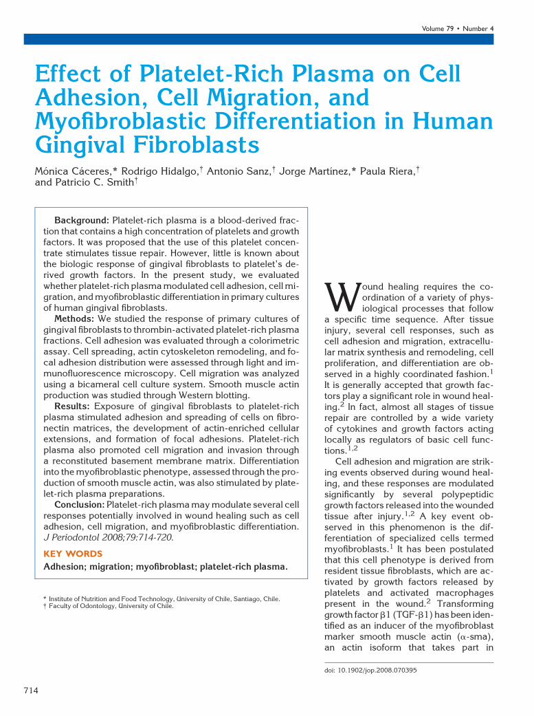
Effect of PRP on Cell AdhesionThe ability of cells to adhere to a fibronectin matrixwas evaluated in GFs stimulated with PPP and differ-ent concentrations of PRP (10%, 25%, 50%, and100%). As shown in Figure 1, after 7 minutes of cellattachment, cell adhesion was stimulated in cells pre-treated with PPP and all of the PRP preparations in adose-dependent manner. Statistically significant in-creases in cell adhesion were only observed in cellsstimulated with 25%, 50%, and 100% PRP comparedto control (non-stimulated) cells (P <0.01).
PRP Stimulates Cell Spreading and Formationof Focal AdhesionsTo further characterize the morphological and molec-ular basis of the enhancement in cell adhesion stimu-lated by PPP and PRP fractions, we evaluated theability of fibroblasts to spread over a fibronectin ma-trix, to develop focal adhesions, and to polymerizeF-actin fibers.
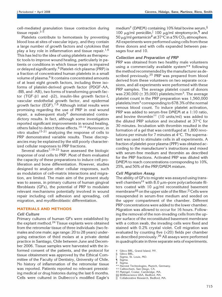
After 20 minutes of cell adhesion, cells stimulatedwith 25% PRP spread well over a fibronectin matrixcompared to control or PPP-stimulated cells (Fig. 2A).Quantification of the number of spread cells demon-strated that PRP significantly stimulated this responsecompared to PPP or no stimulation (P <0.01) (Fig.2B).
¶¶ Calbiochem.## Rockland Immunochemicals, Gilbertsville, PA.*** ELX 800, Bio-Tek Instruments, Winooski, VT.††† Nikon Coolpix 4500, Nikon, Tokyo, Japan.‡‡‡ Zeiss Axiovert 25, Zeiss, Bernried, Germany.§§§ Rockland Immunochemicals.iii Santa Cruz Biotechnology, Santa Cruz, CA.¶¶¶ Invitrogen Molecular Probes, Carlsbad, CA.### Alexa fluor 594, Invitrogen Molecular Probes.**** Zeiss Axioplan, Zeiss.†††† Zeiss Axiocam, Zeiss.‡‡‡‡ PerkinElmer Life Sciences, Boston, MA.§§§§ Sigma.iiii Sigma.¶¶¶¶ ECL kit, Amersham Biosciences, Piscataway, NJ.
Fibroblast Response to Platelet-Rich Plasma Volume 79 • Number 4
716
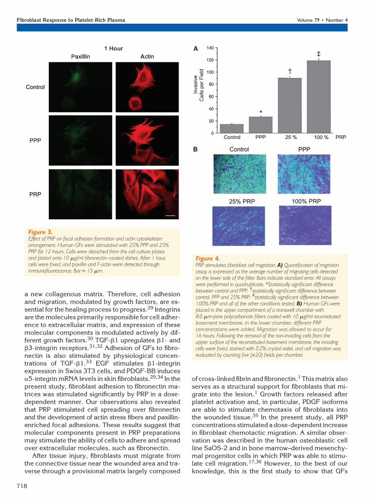
We also evaluated the distribution of paxillin-enriched focal adhesions and the formation of F-actinfibers through immunofluorescence. After1 hourof celladhesion, unstimulated cells showed a rounded mor-phology with no signs of spreading (Fig. 3). However,at equivalent time-points, PPP- and PRP-stimulatedcells showed an elongated morphology, more actinstress fibers, and a higher number of paxillin-enrichedfocal adhesions (Fig. 3). Under the stimulus of PRPfractions, these results suggest that, GFs developeda more active cytoskeleton arrangement that predis-posed cells to adhere.
PRP Promotes Cell MigrationTo analyze whether PRP also constitutes a chemotac-tic stimulus to GFs, we evaluated the ability of fibro-blasts to migrate toward a PRP gradient by a cellmigration assay using a bicameral cell culture sys-tem. As shown in Figure 4, in every condition tested(PPP and 25% and 100% PRP), cell migration wasstimulated at statistically significant levels comparedto untreated cells (P <0.01).
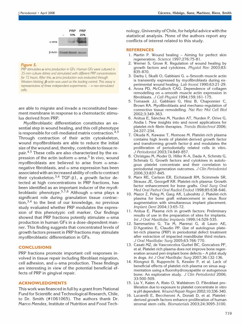
PRP Stimulates a-sma ProductionFinally, we assessed whether PRP has a role in myofi-broblastic differentiation. To this end, we measuredthe production of the actin isoform a-sma, a long-termmarker of this phenomenon, by exposing GFs to 10%
and 25% PRP for 72 hours. Figure 5 shows that a-smaproduction,assessedthroughWesternblotting,wasstim-ulated strongly by PRP in a dose-dependent manner.
DISCUSSION
Although PRP and platelet concentrates have beenproposed as a therapeutic tool to improve tissue re-pair, the underlying cellular-level mechanisms re-main poorly characterized. In the present study, weused primary cultures of human GFs to evaluatewhether PRP fractions may modulate several cell re-sponses that have been involved in wound healing.Our results showed that PRP may stimulate adhesionto fibronectin, cell spreading, focal adhesion forma-tion, cell migration, and differentiation into the myofi-broblastic phenotype.
During wound healing, cells must attach to a newlyformed matrix structure to migrate.27 Soon afterwounding, a provisional matrix of fibronectin, fibrino-gen, fibrin, and vitronectin is formed in the woundarea.28 Activated fibroblasts must move from sur-rounding collagenous connective tissues into a fibrin/fibronectin-filled wound and subsequently synthesize
Figure 2.Effect of PRP on cell spreading. A) Human GFs were stimulated with25% PPP and 25% PRP for 12 hours. Cells were detached, counted(1 · 103), and plated onto 10 mg/ml fibronectin–coated dishes. After20 minutes, attached cells were stained with crystal violet, and imageswere recorded for each experimental condition. Bar = 15 mm.B) Spreading of cells was calculated by counting at least four fieldsfrom different regions of the culture dish. Data are the mean value ofthe number of spread cells detected in three separate experiments.Bars indicate standard error. *Statistically significant differencesbetween control and PRP-stimulated cells (P <0.01).
Figure 1.Effect of PRP fractions on fibroblast adhesion. Human GFs werestimulated with 100% PPP and different PRP concentrations (10%,25%, 50%, and 100%) for 16 hours. Cells were detached from the cellculture plates, counted (1 · 103), and plated onto 10 mg/ml-fibronectin–coated dishes. After 7 minutes, unattached cells wereremoved, and adhered cells were fixed with methanol and stainedwith 0.2% crystal violet. Cells were solubilized, and the released crystalviolet was read in a microplate reader. The y axis indicates meanabsorbance (optical density [OD]) at 590 nm. Bars indicate standarderror. All assays were performed in quadruplicate. *Statisticallysignificant difference between control and 25% PRP; †statisticallysignificant difference between control, PPP, and 50% PRP;‡statistically significant difference between 100% PRP and all of theother conditions tested.
J Periodontol • April 2008 Caceres, Hidalgo, Sanz, Martınez, Riera, Smith
717
a new collagenous matrix. Therefore, cell adhesionand migration, modulated by growth factors, are es-sential for the healing process to progress.29 Integrinsare the molecules primarily responsible for cell adher-ence to extracellular matrix, and expression of thesemolecular components is modulated actively by dif-ferent growth factors.30 TGF-b1 upregulates b1- andb3-integrin receptors.31,32 Adhesion of GFs to fibro-nectin is also stimulated by physiological concen-trations of TGF-b1.33 EGF stimulates b1-integrinexpression in Swiss 3T3 cells, and PDGF-BB inducesa5-integrin mRNA levels in skin fibroblasts.29,34 In thepresent study, fibroblast adhesion to fibronectin ma-trices was stimulated significantly by PRP in a dose-dependent manner. Our observations also revealedthat PRP stimulated cell spreading over fibronectinand the development of actin stress fibers and paxillin-enriched focal adhesions. These results suggest thatmolecular components present in PRP preparationsmay stimulate the ability of cells to adhere and spreadover extracellular molecules, such as fibronectin.
After tissue injury, fibroblasts must migrate fromthe connective tissue near the wounded area and tra-verse through a provisional matrix largely composed
of cross-linked fibrin and fibronectin.1 This matrix alsoserves as a structural support for fibroblasts that mi-grate into the lesion.1 Growth factors released afterplatelet activation and, in particular, PDGF isoformsare able to stimulate chemotaxis of fibroblasts intothe wounded tissue.35 In the present study, all PRPconcentrations stimulated a dose-dependent increasein fibroblast chemotactic migration. A similar obser-vation was described in the human osteoblastic cellline SaOS-2 and in bone marrow–derived mesenchy-mal progenitor cells in which PRP was able to stimu-late cell migration.17,36 However, to the best of ourknowledge, this is the first study to show that GFs
Figure 3.Effect of PRP on focal adhesion formation and actin cytoskeletonarrangement. Human GFs were stimulated with 25% PPP and 25%PRP for 12 hours. Cells were detached from the cell culture platesand plated onto 10 mg/ml fibronectin–coated dishes. After 1 hour,cells were fixed, and paxillin and F-actin were detected throughimmunofluorescence. Bar = 15 mm.
Figure 4.PRP stimulates fibroblast cell migration. A) Quantification of migrationassay is expressed as the average number of migrating cells detectedon the lower side of the filter. Bars indicate standard error. All assayswere performed in quadruplicate. *Statistically significant differencebetween control and PPP; †statistically significant difference betweencontrol, PPP and 25% PRP; ‡statistically significant difference between100% PRP and all of the other conditions tested. B) Human GFs wereplaced in the upper compartment of a transwell chamber with8.0 mm-pore polycarbonate filters coated with 10 mg/ml reconstitutedbasement membrane. In the lower chamber, different PRPconcentrations were added. Migration was allowed to occur for16 hours. Following the removal of the non-invading cells from theupper surface of the reconstituted basement membrane, the invadingcells were fixed, stained with 0.2% crystal violet, and cell migration wasevaluated by counting five (·20) fields per chamber.
Fibroblast Response to Platelet-Rich Plasma Volume 79 • Number 4
718
are able to migrate and invade a reconstituted base-ment membrane in response to a chemotactic stimu-lus derived from PRP.
Myofibroblastic differentiation constitutes an es-sential step in wound healing, and this cell phenotypeis responsible for cell-mediated matrix contraction.4,5
Through contraction of their actin cytoskeleton,wound myofibroblasts are able to reduce the initialsize of the wound and, thereby, contribute to tissue re-pair.4,5 These cells are also characterized by the ex-pression of the actin isoform a-sma.5 In vivo, woundmyofibroblasts are believed to arise from a-sma–negative fibroblasts, and a-sma expression has beenassociated with an increased ability of cells to contracttheir cytoskeleton.3,4 TGF-b1, a growth factor de-tected at high concentrations in PRP fractions, hasbeen identified as an important inducer of the myofi-broblastic phenotype.5,7,8 Although a-sma plays asignificant role during granulation tissue contrac-tion,3,4 to the best of our knowledge, no previousstudy evaluated whether PRP modulates the expres-sion of this phenotypic cell marker. Our findingsshowed that PRP fractions potently stimulate a-smaproduction in human GFs in a dose-dependent man-ner. This finding suggests that concentrated levels ofgrowth factors present in PRP fractions may stimulatemyofibroblastic differentiation in GFs.
CONCLUSIONS
PRP fractions promote important cell responses in-volved in tissue repair including fibroblast migration,cell adhesion, and a-sma production. These findingsare interesting in view of the potential beneficial ef-fects of PRP in gingival repair.
ACKNOWLEDGMENTS
This work was financed in full by a grant from NationalFund for Scientific and Technological Research, Chile,to Dr. Smith (#1061065). The authors thank Dr.Marco Mendez, Institute of Nutrition and Food Tech-
nology, University of Chile, for helpful advice with thestatistical analysis. None of the authors report anyconflicts of interest related to this study.
REFERENCES1. Martin P. Wound healing – Aiming for perfect skin
regeneration. Science 1997;276:75-81.2. Werner S, Grose R. Regulation of wound healing by
growth factors and cytokines. Physiol Rev 2003;83:835-870.
3. Darby I, Skalli O, Gabbiani G. a-Smooth muscle actinis transiently expressed by myofibroblasts during ex-perimental wound healing. Lab Invest 1990;63:21-29.
4. Arora PD, McCulloch CAG. Dependence of collagenremodelling on a-smooth muscle actin expression infibroblasts. J Cell Physiol 1994;159:161-175.
5. Tomasek JJ, Gabbiani G, Hinz B, Chaponnier C,Brown RA. Myofibroblasts and mechano-regulation ofconnective tissue remodelling. Nat Rev Mol Cell Biol2002;3:349-363.
6. Anitua E, Sanchez M, Nurden AT, Nurden P, Orive G,Andıa I. New insights into and novel applications forplatelet-rich fibrin therapies. Trends Biotechnol 2006;24:227-234.
7. Okuda K, Kawase T, Momose M. Platelet-rich plasmacontains high levels of platelet-derived growth factorand transforming growth factor-b and modulates theproliferation of periodontally related cells in vitro.J Periodontol 2003;74:849-857.
8. Christgau M, Moder D, Hiller K-A, Dada A, Schmitz G,Schmalz G. Growth factors and cytokines in autolo-gous platelet concentrate and their correlation toperiodontal regeneration outcomes. J Clin Periodontol2006;33:837-845.
9. Marx RE, Carlson ER, Eichstaedt RM, Scimmele SR,Strauss JE, Georgeff KR. Platelet-rich plasma: Growthfactor enhancement for bone grafts. Oral Surg OralMed Oral Pathol Oral Radiol Endod 1998;85:638-646.
10. Mazor Z, Peleg M, Garg AK, Luboshitz J. Platelet-richplasma for bone graft enhancement in sinus flooraugmentation with simultaneous implant placement.Implant Dent 2004;13:65-72.
11. Anitua E. Plasma rich in growth factors: Preliminaryresults of use in the preparation of sites for implants.Int J Oral Maxillofac Implants 1999;14:529-535.
12. Sammartino G, Tia M, Marenzi G, di Lauro AE,D’Agostino E, Claudio PP. Use of autologous plate-let-rich plasma (PRP) in periodontal defect treatmentafter extraction of impacted mandibular third molars.J Oral Maxillofac Surg 2005;63:766-770.
13. Casati MZ, de Vasconcelos Gurbel BC, Goncalves PF,et al. Platelet-rich plasma does not improve bone regen-eration around peri-implant bone defects – A pilot studyin dogs. Int J Oral Maxillofac Surg 2007;36:132-136.
14. Klongnoi B, Rupprecht S, Kessler P, et al. Lack ofbeneficial effects of platelet-rich plasma on sinus aug-mentation using a fluorohydroxyapatite or autogenousbone: An explorative study. J Clin Periodontol 2006;33:500-509.
15. Liu Y, Kalen A, Risto O, Wahlstrom O. Fibroblast pro-liferation due to exposure to platelet concentrate in vitrois pH dependent. Wound Repair Regen 2002;10:336-340.
16. Lucarelli E, Beccheroni A, Donati D, et al. Platelet-derived growth factors enhance proliferation of humanstromal stem cells. Biomaterials 2003;24:3095-3100.
Figure 5.PRP stimulates a-sma production in GFs. Human GFs were cultured in35-mm culture dishes and stimulated with different PRP concentrationsfor 72 hours. After this, a-sma production was evaluated throughWestern blotting. b-actin was used as the loading control. This assay isrepresentative of three independent experiments. – = non-stimulatedcells.
J Periodontol • April 2008 Caceres, Hidalgo, Sanz, Martınez, Riera, Smith
719
17. Gruber R, Karreth F, Kandler B, et al. Platelet-releasedsupernatants increase migration and proliferation, anddecrease osteogenic differentiation of bone marrow-derived mesenchymal progenitor cells under in vitroconditions. Platelets 2004;15:29-35.
18. Kilian O, Flesch I, Wenisch S, et al. Effects of plateletgrowth factors on human mesenchymal stem cells andhuman endothelial cells in vitro. Eur J Med Res 2004;9:337-344.
19. Soffer E, Ouhayoun JP, Dosquet C, Meunier A,Anagnostou F. Effects of platelet lysates on selectbone cell functions. Clin Oral Implants Res 2004;15:581-588.
20. Kanno T, Takahashi T, Tsujisawa T, Ariyoshi W,Nishihara T. Platelet-rich plasma enhances humanosteoblast-like cell proliferation and differentiation.J Oral Maxillofac Surg 2005;63:362-369.
21. Graziani F, Ivanovski S, Cei S, Ducci F, Tonetti M,Gabriele M. The in vitro effect of different PRP con-centrations on osteoblasts and fibroblasts. Clin OralImplants Res 2006;17:212-219.
22. Goto H, Matsuyama T, Miyamoto M, Yonamine I, IzumiY. Platelet-rich plasma/osteoblasts complex inducesbone formation via osteoblastic differentiation follow-ing subcutaneous transplantation. J Periodontal Res2006;41:455-462.
23. Kawase T, Okuda K, Wolff LF, Yoshie H. Platelet-richplasma-derived fibrin clot formation stimulates colla-gen synthesis in periodontal ligament and osteoblasticcells in vitro. J Periodontol 2003;74:858-864.
24. Larjava H, Heino J, Kahari V-M, Krusius T, Vuorio E.Characterization of one phenotype of human granula-tion-tissue fibroblasts. J Dent Res 1989;68:20-25.
25. Weibrich G, Kleis WKG, Buch R, Hitzler WE, Hafner G.The Harvest Smart PreP system versus the Friadent-Schutze platelet-rich plasma kit. Clin Oral ImplantsRes 2003;14:233-239.
26. Caceres M, Guerrero J, Martinez J. Overexpression ofRhoA-GTP induces activation of the epidermal growthfactor receptor, dephosphorylation of focal adhesionkinase and increased motility in breast cancer cells.Exp Cell Res 2005;309:229-238.
27. Schaffer CJ, Nanney LB. Cell biology of wound heal-ing. Int Rev Cytol 1996;169:151-181.
28. Gailit J, Clark RAF. Wound repair in the context of ex-tracellular matrix. Curr Opin Cell Biol 1994;6:717-725.
29. Xu J, Clark RAF. Extracellular matrix alters PDGFregulation of fibroblast integrins. J Cell Biol 1996;132:239-249.
30. Hynes RO. Integrins: Versatility, modulation and sig-naling in cell adhesion. Cell 1992;69:11-25.
31. Heino J, Massague J. Transforming growth factor-b
switches the pattern of integrins expressed in MG-63human osteosarcoma cells and causes a selective lossof cell adhesion to laminin. J Biol Chem 1989;264:21806-21811.
32. Heino J, Ignotz RA, Hemler ME, Crouse C, MassagueJ. Regulation of cell adhesion receptors by trans-forming growth factor-b. Concomitant regulation ofintegrins that share a common b1 subunit. J BiolChem 1989;264:380-388.
33. Smith PC, Caceres M, Martinez J. Induction of themyofibroblastic phenotype in human gingival fibro-blasts by transforming growth factor-b1: Role ofRhoA-ROCK and c-Jun N-terminal kinase signalingpathway. J Periodontal Res 2006;41:418-425.
34. Bellas RE, Bendori R, Farmer SR. Epidermal growthfactor activation of vinculin and b1-integrin geneexpression in quiescent Swiss 3T3 cells: Regulationthrough a protein kinase C-independent pathway.J Biol Chem 1991;266:12008-12014.
35. Seppa H, Grotendorst G, Seppa S, Schiffmann E,Martin GR. Platelet-derived growth factor is chemo-tactic for fibroblasts. J Cell Biol 1982;92:584-588.
36. Celotti F, Colciago A, Negri-Cesi P, Pravettoni A,Zaninetti R, Sacchi MC. Effect of platelet-rich plasmaon migration and proliferation of SaOS-2 osteoblasts:Role of platelet-derived growth factor and transform-ing growth factor-beta. Wound Repair Regen 2006;14:195-202.
Correspondence: Dr. Patricio C. Smith, Faculty of Odon-tology, University of Chile, Casilla 1903, Santiago, Chile.Fax: 56-2-221-4030; e-mail: [email protected].
Submitted July 17, 2007; accepted for publication August14, 2007.
Fibroblast Response to Platelet-Rich Plasma Volume 79 • Number 4
720