Embed Size (px)
Citation preview

ORIGINAL ARTICLE
Effect of HHH-Therapy on Regional CBF afterSevere Subarachnoid Hemorrhage Studied by BedsideXenon-Enhanced CT
Henrik Engquist1,2• Elham Rostami1 • Elisabeth Ronne-Engstrom1
•
Pelle Nilsson1• Anders Lewen1
• Per Enblad1
Published online: 5 October 2017
� The Author(s) 2017. This article is an open access publication
Abstract
Background Management of delayed cerebral ischemia
(DCI) following subarachnoid hemorrhage (SAH) is diffi-
cult and still carries controversies. In this study, the effect
of therapeutic hypervolemia, hemodilution, and hyperten-
sion (HHH-therapy) on cerebral blood flow (CBF) was
assessed by xenon-enhanced computerized tomography
(XeCT) hypothesizing an increase in CBF in poorly per-
fused regions.
Methods Bedside XeCT measurements of regional CBF in
mechanically ventilated SAH patients were routinely
scheduled for day 0–3, 4–7, and 8–12. At clinical suspicion
of DCI, patients received 5-day HHH-therapy. For inclu-
sion, XeCT was required at 0–48 h before start of HHH
(baseline) and during therapy. Data from corresponding
time-windows were also collected for non-DCI patients.
Results Twenty patients who later developed DCI were
included, and twenty-eight patients without DCI were
identified for comparison. During HHH, there was a slight
nonsignificant increase in systolic blood pressure (SBP)
and a significant reduction in hematocrit. Median global
cortical CBF for the DCI group increased from 29.5 (IQR
24.6–33.9) to 38.4 (IQR 27.0–41.2) ml/100 g/min
(P = 0.001). There was a concomitant increase in regional
CBF of the worst vascular territories, and the proportion of
area with blood flow below 20 ml/100 g/min was signifi-
cantly reduced. Non-DCI patients showed higher CBF at
baseline, and no significant change over time.
Conclusions HHH-therapy appeared to increase global and
regional CBF in DCI patients. The increase in SBP was
small, while the decrease in hematocrit was more pro-
nounced, which may suggest that intravascular volume
status and rheological effects are of importance. XeCT may
be potentially helpful in managing poor-grade SAH
patients.
Keywords Subarachnoid hemorrhage (SAH) �Delayed cerebral ischemia (DCI) �Cerebral blood flow (CBF) � HHH-therapy (Triple-H) �Xenon CT (XeCT)
Introduction
The complex mechanisms leading to secondary brain
injury and the sometimes devastating course of delayed
cerebral ischemia (DCI) [1] after aneurysmal subarachnoid
hemorrhage (SAH) are still not well understood [2].
Improved techniques for early surgical or endovascular
interventions and general improvement of the neurointen-
sive care (NIC) have reduced mortality and morbidity in
these patients during the last decades [3]. Still there is little
evidence supporting prophylactic strategies to minimize
the risk of DCI and also concerning therapeutic interven-
tions when DCI is suspected [4]. In modern NIC, clinical
surveillance combined with multimodal systemic and
cerebral monitoring is used to aid the early detection of
avoidable factors and also DCI [5]. There is, however, no
consensus regarding the best tools to guide the therapy at
suspicion of DCI [6].
& Henrik Engquist
1 Dept of Neuroscience/Neurosurgery, Uppsala University,
Uppsala, Sweden
2 Dept of Surgical Sciences/Anesthesia and Intensive Care,
Uppsala University Hospital, 751 85 Uppsala, Sweden
123
Neurocrit Care (2018) 28:143–151
https://doi.org/10.1007/s12028-017-0439-y

After early repair of the aneurysm, most guidelines
propose fluid therapy to ensure normovolemia or mild
hypervolemia, monitoring of blood pressure and vasoactive
support if necessary to avoid hypotension [7, 8]. At sus-
picion of DCI, therapeutic hypervolemia, hemodilution,
and hypertension (HHH-therapy) [9] has been widely used
aiming to increase cerebral blood flow (CBF) in ischemic
regions. Controversy exists concerning the actual effects of
the different components of this intervention [10]. In the
standard NIC protocol for SAH patients at Uppsala
University Hospital, therapeutic interventions on suspicion
of DCI include cautious institution of HHH-therapy with
moderate blood pressure targets and frequent reevaluation
to avoid side effects and complications.
The aim of this study was to evaluate the effects of
HHH-therapy on CBF, testing the hypothesis that HHH-
therapy increases global CBF and also regional CBF
(rCBF) in areas at risk of ischemia. Bedside xenon-en-
hanced computerized tomography (XeCT) was used to
assess rCBF prior to clinical suspicion of DCI and during
the treatment to resolve DCI.
Materials and Methods
Patients
Patients diagnosed with severe spontaneous SAH admitted
to Uppsala University Hospital during the time period
2013–2016 were prospectively enrolled in the study. The
diagnosis of SAH was verified by CT. As ventilator treat-
ment is a prerequisite for bedside XeCT in our setting, only
patients requiring intubation due to their neurological state
at admission or neurological deterioration day 0–1 were
included, i.e., patients with more severe SAH. Exclusion
criteria were severe intracranial hypertension, deep seda-
tion with thiopental, respiratory problems requiring FiO2
>0.6, the absence of informed consent, and futility or ‘‘do
not resuscitate’’ order.
CBF Measurements: protocol
According to our standard NIC protocol, CBF measure-
ments were scheduled for three time intervals: day 0–3,
4–7, and 8–12. Patients with clinical diagnose of DCI
received HHH-therapy as described below. For inclusion in
this study, a XeCT measurement within 0–48 h before the
start of HHH-therapy was required (used as baseline), and
the intention was to perform the next XeCT measurement
during ongoing therapy when logistically possible. Data
were also collected for non-DCI patients with corre-
sponding XeCT measurements in the same time-windows
as a reference group for comparison.
Xenon CT CBF Measurements: Method
The XeCT procedures in our NIC unit are conducted as
described by Gur et al. and Yonas et al. [11–14], using
inhaled xenon as an inert contrast agent during the CT
scans. Since xenon is radiopaque, lipid soluble, and crosses
the blood brain barrier, its concentration in the cerebral
tissue can be measured by CT. Blood flow calculation is
based on Kety’s [15, 16] application of the Fick principle;
blood flow is proportional to inert gas uptake in tissue.
Mobile equipment allows bedside measurements in the
NIC unit, which facilitates the clinical use of XeCT [17].
Stable (non-radioactive) xenon at a concentration of 28% is
administered to the patients breathing circuit for 4.5 min
using the Enhancer 3000 and computer software (Diversi-
fied Diagnostic Products Inc, Huston, USA). CT scans
during the xenon inhalation are obtained by the CereTom
(Neurologica, Boston, USA). The xenon delivery and CT
scans are synchronized by the computer software, and the
resulting radiologic tissue enhancement of the xenon wash-
in enables CBF to be calculated (ml/100 g/min) and plotted
as color-coded maps in scans at four different levels of the
brain (eight scans per level, two at baseline and six
enhanced during xenon wash-in). Levels with extensive
artifacts from bone structures or coils/clips were excluded
from calculation. Typically, three scan levels could be used
for further calculations in each patient. Mean blood flow in
each of twenty evenly distributed cortical regions of
interest (ROIs) was calculated (Fig. 1) for every level [13],
resulting in a total of sixty ROIs. Typical area of each ROI
was 350–450 mm2. ROIs in areas of hematoma or with
artifacts from ventricular drains were excluded.
Neurointensive Care
Management of all patients followed our standard NIC
procedures for SAH as earlier described by Ryttlefors et al.
[5], where the cornerstones are continuous multimodal
monitoring of physiological and biochemical parameters
and cautious clinical observation for avoidable factors with
prompt interventions to minimize secondary brain injury.
Sedation with propofol (Propolipid�, Fresenius Kabi AB,
Uppsala, Sweden) is titrated in the interval 0–4 mg/kg/h,
and morphine (Morfin, Meda AB, Solna, Sweden) is
administered as needed. Patients with suspicion of hydro-
cephalus or altered level of consciousness receive a
ventriculostomy catheter for monitoring of intracranial
pressure (ICP) and drainage of cerebrospinal fluid. Ele-
vated ICP (>20 mmHg) is treated with open ventricular
drainage against a pressure level of 15 mmHg. The general
policy is to treat aneurysms early with endovascular coil
embolization as first choice when possible or surgical
clipping. After the aneurysm repair, patients are kept
144 Neurocrit Care (2018) 28:143–151
123

mildly hypervolemic if not contraindicated by intracranial
mass effect or elevated ICP. The volume status is main-
tained by fluid administration in the higher normal range
with addition of albumin infusion (Flexbumin� 200 mg/ml,
Baxter AG, Vienna, Austria) if needed and monitored by
central venous pressure, clinical evaluation and rigorous
fluid balance calculation. Nimodipine (Nimotop�, Bayer
Pharma AG, Berlin, Germany) is given from day one and
onwards.
Detection of DCI and HHH-Therapy
To detect DCI, patients were monitored for general and
focal neurological changes by repeated wake-up tests [18].
In the case of neurological deterioration, CT and laboratory
tests were performed to rule out intracranial mass, manifest
infarction, hydrocephalus, and meningitis. If DCI was
concluded in the absence of other causes of deterioration,
HHH-therapy to augment CBF was initiated for 5 days if
not contraindicated by the intracranial situation, congestive
heart failure, fluid overload or severe respiratory problems.
Patients received daily infusions of 500 ml dextran solution
(Rheomacrodex�, Meda AB, Solna, Sweden) and 200 ml
albumin (Flexbumin� 200 mg/ml, Baxter AG, Vienna,
Austria) and were kept in supine position. In our standard
NIC protocol, emphasis is on avoiding severe side effects
and the blood pressure targets during HHH-therapy are
moderate. The systolic blood pressure (SBP) was closely
monitored and kept above 140 mmHg using vasoactive
agents if needed. As first line, dobutamine (Hospira, Lake
Forest, IL, USA) was used for inotropy or, if insufficient,
norepinephrine (Noradrenalin, Hospira Nordic AB, Stock-
holm, Sweden) was added as vasopressor. The clinical
effect of the HHH-therapy was repeatedly evaluated.
XeCT Parameters
Global cortical CBF (ml/100 g/min) was calculated as
mean of all ROIs at all scan levels used (Fig. 1). Regional
CBF for each vascular territory was calculated as mean of
the corresponding ROIs at all scan levels used (Fig. 1):
right anterior cerebral artery—ROI 1–2, right middle
cerebral artery—ROI 3–8, right posterior cerebral artery—
ROI 9–10 and equally for ROI 11–20 on the left side. The
vascular territory with lowest CBF was identified as the
worst vascular territory in each patient.
To detect and quantify the extent of areas with low
blood flow and near-ischemic flow, thresholds for local
CBF were set to 20 and 10 ml/100 g/min [19, 20]. The
percentage of cortical ROI area with local CBF below these
thresholds was then calculated as sum of ROI area (mm2)
with local CBF below the threshold divided by the total
analyzed ROI area in each patient.
Short-Term Outcome Parameters
Clinical course outcome was defined as neurological state
at discharge from the NIC unit and determined as good
(responding to commands, GCS motor 6), poor (uncon-
scious, GCS motor B5), or dead.
Fig. 1 Example of XeCT measurements before and during HHH-
therapy (one of three scan levels). Global and regional cortical CBF
(ml/100 g/min) was calculated as mean of CBF in the corresponding
regions of interest (ROIs) at three scan levels of the brain for each
patient. Global CBF—ROI 1–20. Right anterior cerebral artery
territory—ROI 1–2, right middle cerebral artery territory—ROI 3–8,
right posterior cerebral artery territory—ROI 9–10 and equally for
ROI 11–20 on the left side. CBF cerebral blood flow, HHH-
therapy therapeutic hypervolemia, hemodilution and hypertension,
XeCT xenon-enhanced computerized tomography
Neurocrit Care (2018) 28:143–151 145
123

Cerebral infarcts visible on follow-up CT at 12 days or
later after admission to the NIC unit were categorized as
<20 mm, 20–39 mm, >40 mm, or multiple.
Statistical Methods
SPSS statistics 23.0 software (IBM Corp, Armonk, NY,
USA) was used for statistical analyses of the collected data.
Differences in systemic physiological parameters between
measurements were tested by paired samples t test. CBF
data for groups of patients are presented as median values
and interquartile range because of non-normal distribution.
Differences in CBF parameters between related samples
were tested by Wilcoxon signed-ranks test, and between
independent samples by Mann–Whitney U test. Statistical
significance level was set at P < 0.05.
Results
A total of 119 intubated SAH patients underwent one or more
XeCT measurements during the time period 2013–2016.
During this period, 20 patients with clinical suspicion of DCI
were identified where XeCT measurements of CBF had been
taken within 0–48 h before the initiation of HHH-therapy
(baseline), and during the 5-day HHH-therapy (Fig. 2).
Twenty-eight SAH patients with no suspicion of DCI, who
had corresponding XeCT measurements in the same time-
windows, were identified as a group for comparison of the
natural course (Fig. 2). The characteristics of these groups are
presented in Table 1. The median time-point for initiation of
HHH-therapy was 4.38 days (IQR 2.75–5.97) after the
admission to the NIC unit.
Hemodynamics and Ventilation
Hemodynamic parameters and ventilation were clinically
stable during the XeCT procedures. The alterations from
start to end in all procedures (N = 96) were small; mean
arterial pressure (MAP) mean 92.5 mmHg (CI 90.0–95.0)
versus 89.9 (CI 87.6–92.2), ICP mean 14.3 mmHg (CI
13.2–15.4) versus 14.4 (CI 13.5–15.4), PCO2 mean
Fig. 2 Time for XeCT measurements in relation to the start of HHH-
therapy for DCI patients (above). Time for XeCT at baseline and
during HHH-therapy in relation to admission to neurosurgical
intensive care for DCI patients (gray) and for non-DCI patients
(white) at corresponding time-windows (below). DCI delayed cere-
bral ischemia, HHH-therapy therapeutic hypervolemia, hemodilution
and hypertension, XeCT xenon-enhanced computerized tomography
Table 1 Characteristics of
patients in the DCI group who
received HHH-therapy and the
non-DCI group
DCI No DCI
Characteristic n (%) n (%)
Patients n 20 28
Gender F 12 (60) 19 (68)
Age, years Mean [range] 58 [40–75] 60 [28–84]
Hunt and Hess grade at admission/first XeCT1 I 0/0 2/0 (7/0)
II 5/0 (25/0) 1/0 (4/0)
III 5/1 (25/5) 7/8 (25/29)
IV 7/18 (35/90) 14/18 (50/64)
V 3/1 (15/5) 4/2 (14/7)
CT Fisher group 2 0 1 (4)
3 6 (30) 5 (18)
4 14 (70) 22 (79)
Treatment modality Endovascular 18 (90) 18 (64)
Surgical clip 1 (5) 9 (32)
None 1 (5) 1 (4)
1 Neurological state according to Hunt and Hess assessed both at admission and at the time of the first
XeCT. DCI delayed cerebral ischemia, HHH-therapy therapeutic hypervolemia, hemodilution and hyper-
tension, XeCT xenon-enhanced computerized tomography
146 Neurocrit Care (2018) 28:143–151
123

5.41 kPa (CI 5.29–5.53) versus 5.51 (CI 5.40–5.63). No
adverse events were observed in relation to the procedures.
SBP, MAP, cerebral perfusion pressure (CPP), pCO2,
and hematocrit at start of the XeCT measurements at the
different time-windows for the DCI and non-DCI groups
are presented in Table 2. The mean SBP was slightly ele-
vated during HHH-therapy in the DCI group, from
151.2 mmHg (CI 142.1–160.3) to 157.3 mmHg (CI
150.7–163.8), but the difference did not reach statistical
significance. Among the DCI patients, hematocrit was
significantly reduced during HHH-therapy compared to
baseline (Table 2).
The use of inotropes and vasopressors was low in all
groups. The number of patients and dose range for each
group are also presented in Table 2.
Global Cortical CBF
Results of the XeCT measurements are presented in
Table 2 and Fig. 3. Global cortical CBF at baseline was
significantly lower in the DCI group compared to the non-
DCI group; median 29.5 ml/100 g/min (IQR 24.6–33.9)
versus 34.9 (IQR 29.0–41.7) (P = 0.005).
During HHH-therapy, the DCI group showed an
increase in median global cortical CBF from 29.5 (IQR
24.6–33.9) to 38.4 (IQR 27.0–41.2) ml/100 g/min
(P = 0.001), while no significant change over time was
seen in the non-DCI group. No statistical analyses were
performed for differences between DCI patients during
HHH-therapy and non-DCI patients at the corresponding
time-window.
Table 2 Systemic hemodynamic parameters, ventilation, sedation, and vasoactive medication at the time of XeCT measurements (upper
part). Calculated XeCT CBF parameters (lower part)
DCI (n = 20) Baseline DCI During HHH No DCI (n = 28) Baseline No DCI Day 5–8
Mean (CI) Mean (CI) Mean (CI) Mean (CI)
SBP mmHg 151.2 (142.1–160.3) 157.3 (150.7–163.8) 150.0 (143.5–156.4) 152.9 (145.9–159.8)
P = 0.105
MAP mmHg 95.4 (87.1–103.7) 94.0 (88.7–99.4) 90.3 (87.0–93.6) 92.5 (88.2–96.7)
CPP mmHg 80.9 (72.4–89.3) 79.7 (74.3–85.1) 78.6 (74.3–82.9) 79.0 (74.8–83.3)
Hematocrit % 36.4 (34.7–38.0) 31.7 (30.2–33.2) 33.9 (32.6–35.3) 32.5 (31.4–33.6)
P < 0.001*
pCO2 kPa 5.20 (4.96–5.44) 5.34 (5.10–5.57) 5.35 (5.14–5.57) 5.71 (5.45–5.97)
Sedation dose, propofol mg/kg/h 2.58 (2.15–3.02) 2.57 (2.16–2.98) 2.70 (2.13–3.26) 2.81 (2.22–3.40)
n [range] n [range] n [range] n [range]
Dobutamine, n [range ug/kg/min] 0 [–] 5 [1.1–12.0] 4 [1.6–4.0] 3 [1.0–6.2]
Norepinephrine, n [range ug/kg/
min]
1 [0.06] 2 [0.05–0.15] 3 [0.01–0.12] 2 [0.05–0.08]
Median (IQR) Median (IQR) Median (IQR) Median (IQR)
glob CBF ml/100 g/min 29.5 (24.6–33.9) 38.4 (27.0–41.2) 34.9 (29.0–41.7) 36.5 (28.0–42.3)
P = 0.001*
% ROI area [rCBF < 20 ml/100 g/min] 26.2 (13.4–44.5) 8.55 (2.4–34.8) 11.9 (3.2–23.0) 9.1 (2.1–29.2)
P = 0.019*
% ROI area [rCBF < 10 ml/100 g/min] 6.7 (0.0–11.0) 0.0 (0.0–5.0) 0.7 (0.0–4.6) 0.7 (0.0–4.9)
P = 0.056
rCBF worst territory ml/100 g/min 19.6 (15.0–24.2) 27.3 (17.8–34.1) 27.2 (20.8–35.4) 25.8 (17.4–31.4)
P = 0.006*
Index [rCBFworst/best hemisph] 0.59 (0.46–0.75) 0.70 (0.55–0.83) 0.70 (0.60–0.79) 0.63 (0.52–0.73)
P = 0.040* P = 0.029*
* Indicates P < 0.05. CBF cerebral blood flow, CI confidence interval, CPP cerebral perfusion pressure, DCI delayed cerebral ischemia, HHH-
therapy therapeutic hypervolemia, hemodilution and hypertension, IQR interquartile range, MAP mean arterial pressure, rCBF regional cerebral
blood flow, ROI region of interest, SBP systolic blood pressure
Neurocrit Care (2018) 28:143–151 147
123
Regional CBF and CBF Heterogeneity
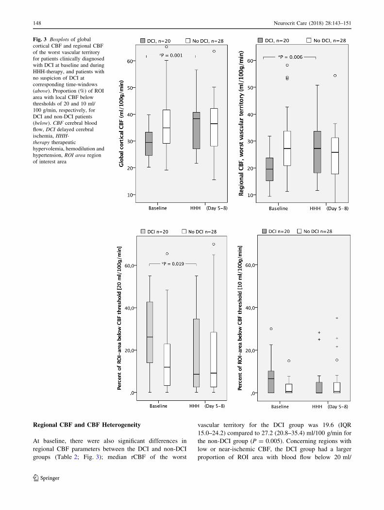
At baseline, there were also significant differences in
regional CBF parameters between the DCI and non-DCI
groups (Table 2; Fig. 3); median rCBF of the worst
vascular territory for the DCI group was 19.6 (IQR
15.0–24.2) compared to 27.2 (20.8–35.4) ml/100 g/min for
the non-DCI group (P = 0.005). Concerning regions with
low or near-ischemic CBF, the DCI group had a larger
proportion of ROI area with blood flow below 20 ml/
Fig. 3 Boxplots of global
cortical CBF and regional CBF
of the worst vascular territory
for patients clinically diagnosed
with DCI at baseline and during
HHH-therapy, and patients with
no suspicion of DCI at
corresponding time-windows
(above). Proportion (%) of ROI
area with local CBF below
thresholds of 20 and 10 ml/
100 g/min, respectively, for
DCI and non-DCI patients
(below). CBF cerebral blood
flow, DCI delayed cerebral
ischemia, HHH-
therapy therapeutic
hypervolemia, hemodilution and
hypertension, ROI area region
of interest area
148 Neurocrit Care (2018) 28:143–151
123

100 g/min (median 26.2 vs 11.9%, P = 0.005) and flow
below 10 ml/100 g/min (median 6.7 vs 0.7%, P = 0.023)
compared to the non-DCI group.
During HHH-therapy, median rCBF of the worst vascular
territory for the DCI group increased from 19.6 (IQR
15.0–24.2) to 27.3 (IQR 17.8–34.1) ml/100 g/min
(P = 0.006) (Table 2; Fig. 3), and the proportion of ROI
area with local blood flow below the threshold of 20 ml/
100 g/min was reduced from median 26.2% at baseline to
8.55% (P = 0.019). The area with flow below 10 ml/100 g/
min was small already at baseline but tended to decrease
(from 6.7 to 0.0%, P = 0.056). The relative blood flow in
the worst vascular territory compared to the best hemi-
spheric blood flow (CBF index of worst vascular territory)
showed a small but statistically significant increase during
HHH-therapy. In the non-DCI group, there were no signif-
icant changes from baseline to the second time-window
regarding rCBF in the worst vascular territory or the pro-
portion of ROI area with local blood flow below the
specified thresholds. In this group, there was a small, but
statistically significant reduction in the CBF index of worst
vascular territory between the two measurements.
Short-Term Outcome; Clinical Course and Cerebral
Infarcts
Short-term outcome parameters are presented in Fig. 4.
The proportion of patients with favorable clinical course
outcome was 65% in the HHH-treated DCI group and 57%
in the non-DCI group. The proportion of patients where no
infarcts >20 mm were detected at follow-up CT was 65%
in the DCI group and 46% in the non-DCI group. Since the
non-DCI group is not considered a valid control group for
HHH-therapy, no statistical analyses were performed on
the differences in outcome parameters.
Discussion
Poor-grade, unconscious SAHpatients still suffer high risk of
deterioration and development of DCI during the acute
course after the initial bleed, despite early aneurysm repair
and modern specialized NIC. The main therapeutic option
remains the use of one or several components of HHH-ther-
apy. The effectiveness of the HHH-therapy has been
questioned, and according to recent literature, the limited
scientific support favors only augmentation of systemic blood
pressure [10]. Moreover, the use of excessive fluid overload
and high doses of vasopressors in these patients carries the
risk of cardiopulmonary and cerebral complications [21–23].
The use of HHH-therapy to resolve DCI, according to
our standardized NIC protocol, is instituted cautiously to
avoid negative side effects, and the treatment is frequently
reevaluated in each patient. Systemic blood pressure targets
are moderate to avoid excessive use of vasopressors. The
establishment of bedside CBF XeCT in routine NIC
Fig. 4 Clinical course outcome
for patients in the DCI and non-
DCI groups; neurological state
at discharge from the NIC unit
(good—responding to
commands and GCS motor 6,
poor—unconscious and GCS
motor B5, dead). Infarcts
visible on follow-up CT > day
12. CT computerized
tomography, DCI delayed
cerebral ischemia, GCS
motor motor component of
Glasgow coma scale, NIC neuro
intensive care
Neurocrit Care (2018) 28:143–151 149
123

provided an excellent possibility to evaluate the effects of
HHH-therapy on CBF in our setting. A general problem in
the scientific evaluation of HHH-therapy is the difficulty to
obtain a relevant control group, since it is unethical to leave
one group of patients with signs of DCI without treatment.
To overcome this shortcoming to some extent, we identi-
fied non-DCI patients with XeCT measurements in the
same time-windows to serve as a reference group, although
CBF dynamics in these patients may differ from patients
with suspicion of DCI.
At baseline, the subsequently HHH-treated DCI patients
had lower CBF than non-DCI patients. During HHH treat-
ment there was a significant elevation of global CBF,
whereas CBF in non-DCI patients remained unchanged over
time. There is a possibility that the observed increase during
HHH-therapy may represent a natural spontaneous recovery
of the initially low CBF, but there are several indications in
this study that there was a real effect of the HHH-therapy on
CBF, which is discussed in the following sections.
The most important therapeutic goal is to achieve better
perfusion in near-ischemic regions, and therefore, analysis
of regional blood flow is of special importance. There was
an increase in regional CBF in the previously worst per-
fused vascular regions during HHH-therapy, and the
proportion of ROI area with blood flow below the threshold
of 20 ml/100 g/min decreased significantly. These findings
indicate that the increase in CBF is not only luxury per-
fusion of already well-perfused regions, but also a true
restoration of CBF in poorly perfused regions associated
with HHH-therapy.
The restoration of CBF found in the group of patients
receiving HHH-therapy does not necessarily correlate with
better outcome. The absence of a strict DCI control group
and the small number of patients make conclusions
regarding outcome uncertain, and no long-term outcome
data were analyzed. However, concerning short-term out-
come, the frequencies of infarctions, and good clinical
course outcome at discharge from the NIC unit were at
similar levels for HHH-treated DCI patients and non-DCI
patients. This observation also indicates that the HHH-
therapy may have been effective.
One of the strengths of using XeCT with a mobile CT-
scanner is the possibility to conduct CBF measurements
bedside, which minimizes alterations in the physiological
situation of the patients. The hemodynamic and respiratory
parameters proved to be stable in this study, and the
amount of sedation given was low (Table 2), which indi-
cates that the results obtained were not confounded by
those factors.
From this study, it is difficult to assess which component
of the HHH-therapy was most important for the observed
increase in CBF. However, the results indicate that the
controlled hypervolemia and resulting reduction in
hematocrit may be of importance, since the elevation of
SBP was small and CPP not significantly increased. This
indication is in contrast with previous studies suggesting
that the hypertensive treatment is most important [10],
which calls for further studies.
In evaluating the effect of HHH-therapy, baseline
measurement of CBF should ideally be performed imme-
diately prior to the initiation of the therapy. However, this
was not possible for logistic reasons and the risk of
delaying treatment. Baseline CBF in our study thus rep-
resents measurements performed at 0–48 h prior to the start
of HHH-therapy, which is not optimal but reasonably close
to the start of therapy. The second CBF measurement,
during the five-day therapy, was performed at median
1.82 days (range 0.45–5.04 days) after the start of HHH-
therapy, suggesting a sustained increase in CBF after ini-
tiation of the therapy. Early and repeated measurements
would be preferred to better evaluate a direct relationship
between HHH-therapy and CBF changes, but an increased
number of measurements might be questionable due to
radiation exposure and also logistically difficult.
We find bedside XeCT feasible for clinical use in NIC to
estimate rCBF in unconscious, mechanically ventilated
patients, but the procedure takes the resources of a trained
team of intensive care and radiology personnel. Regarding
safety issues, XeCT using 28% inhaled xenon is previously
reported safe [24], and all patients in our study were
physiologically stable during the procedures. Since inert
and rapidly washed-out xenon is used as contrast agent, the
method allows repeated measurements, although taking
radiation exposure into consideration.
Conclusions
The initial low global CBF found in patients diagnosed
with DCI was significantly elevated during HHH-therapy.
A concomitant increase in rCBF was also found in the
worst perfused regions and the proportion of ROI area with
blood flow below the threshold of 20 ml/100 g/min
decreased significantly, which indicate a true effect of
HHH-therapy. The increase in SBP was small, while the
decrease in hematocrit was more pronounced, which may
suggest that intravascular volume status and rheological
effects are of importance. We found bedside XeCT to be a
feasible and potentially clinically valuable tool in NIC.
Compliance with Ethical Standards
Conflict of interest The authors declare that they have no conflict of
interest.
Ethical approval and informed consent The study protocol was
approved by the Uppsala University Regional Ethical Review Board,
150 Neurocrit Care (2018) 28:143–151
123

and informed consent was obtained from the patients included or their
next of kin. All procedures performed in the study were in compliance
with the 1964 Helsinki Declaration and its later amendments or
comparable ethical standards. The study was also approved by the
local Radiation Safety Authority.
Open Access This article is distributed under the terms of the
Creative Commons Attribution 4.0 International License (http://
creativecommons.org/licenses/by/4.0/), which permits unrestricted
use, distribution, and reproduction in any medium, provided you give
appropriate credit to the original author(s) and the source, provide a
link to the Creative Commons license, and indicate if changes were
made.
References
1. Frontera JA, Fernandez A, Schmidt JM, Claassen J, Wartenberg
KE, Badjatia N, Connolly ES, Mayer SA. Defining vasospasm
after subarachnoid hemorrhage: what is the most clinically rele-
vant definition? Stroke. 2009;40(6):1963–8. doi:10.1161/
STROKEAHA.108.544700.
2. Carr KR, Zuckerman SL, Mocco J. Inflammation, cerebral
vasospasm, and evolving theories of delayed cerebral ischemia.
Neurol Res Int. 2013;2013:506584. doi:10.1155/2013/506584.
3. Ronne-Engstrom E, Enblad P, Gal G, Norback O, Ryttlefors M,
Cesarini KG, Bolander H, Tovi M, Persson L. Patients with
spontaneous subarachnoid haemorrhage–presentation of a
10-year hospital series. Br J Neurosurg. 2009;23(5):499–506.
doi:10.1080/02688690902874901.
4. Brathwaite S, Macdonald RL. Current management of delayed
cerebral ischemia: update from results of recent clinical trials.
Transl Stroke Res. 2014;5(2):207–26. doi:10.1007/s12975-013-
0316-8.
5. Ryttlefors M, Howells T, Nilsson P, Ronne-Engstrom E, Enblad
P. Secondary insults in subarachnoid hemorrhage: occurrence and
impact on outcome and clinical deterioration. Neurosurgery.
2007;61(4):704–14. doi:10.1227/01.NEU.0000298898.38979.E3
discussion 714–705.6. Sarrafzadeh AS, Vajkoczy P, Bijlenga P, Schaller K. Monitoring
in neurointensive Care—the challenge to detect delayed cerebral
ischemia in high-grade aneurysmal SAH. Front Neurol.
2014;5:134. doi:10.3389/fneur.2014.00134.
7. Connolly ES, Rabinstein AA, Carhuapoma JR, Derdeyn CP, Dion
J, Higashida RT, Hoh BL, Kirkness CJ, Naidech AM, Ogilvy CS,
Patel AB, Thompson BG, Vespa P, AHASCouncil, CoCRaInter-
vention, CoCNursing, CoCSaAnesthesia, CoCCardiology.
Guidelines for the management of aneurysmal subarachnoid hem-
orrhage: a guideline for healthcare professionals from the American
Heart Association/american Stroke Association. Stroke.
2012;43(6):1711–37. doi:10.1161/STR.0b013e3182587839.
8. Steiner T, Juvela S, Unterberg A, Jung C, Forsting M, Rinkel G,
Organization ES. European stroke organization guidelines for the
management of intracranial aneurysms and subarachnoid haem-
orrhage. Cerebrovasc Dis. 2013;35(2):93–112. doi:10.1159/
000346087.
9. Origitano TC, Wascher TM, Reichman OH, Anderson DE. Sus-
tained increased cerebral blood flow with prophylactic
hypertensive hypervolemic hemodilution (‘‘triple-H’’ therapy)
after subarachnoid hemorrhage. Neurosurgery. 1990;27(5):729–
39 discussion 739–740.
10. Dankbaar JW, Slooter AJ, Rinkel GJ, Schaaf IC. Effect of dif-
ferent components of triple-H therapy on cerebral perfusion in
patients with aneurysmal subarachnoid haemorrhage: a system-
atic review. Crit Care. 2010;14(1):R23. doi:10.1186/cc8886.
11. Gur D, Good WF, Wolfson SK Jr, Yonas H, Shabason L. In vivo
mapping of local cerebral blood flow by xenon-enhanced com-
puted tomography. Science (New York, NY).
1982;215(4537):1267–8.
12. Yonas H, Gur D, Wolfson SK, Good WF, Good BC, Latchaw RE.
Xenon-enhanced computerised tomographic cerebral blood flow
mapping. Lancet. 1984;1(8390):1357.
13. Yonas H, Darby JM, Marks EC, Durham SR, Maxwell C. CBF
measured by Xe-CT: approach to analysis and normal values.
J Cereb Blood Flow Metab. 1991;11(5):716–25. doi:10.1038/
jcbfm.1991.128.
14. Yonas H, Pindzola RP, Johnson DW. Xenon/computed tomog-
raphy cerebral blood flow and its use in clinical management.
Neurosurg Clin N Am. 1996;7(4):605–16.
15. Kety S, Schmidt C. The determination of cerebral blood flow in
man by the use of nitrous oxide in low concentrations. Am J
Physiol. 1945;143:53–66.
16. Kety SS. The measurement of cerebral blood flow by means of
inert diffusible tracers. Keio J Med. 1994;43(1):9–14.
17. Sturnegk P, Mellergard P, Yonas H, Theodorsson A, Hillman J.
Potential use of quantitative bedside CBF monitoring (Xe-CT) for
decision making in neurosurgical intensive care. Br J Neurosurg.
2007;21(4):332–9. doi:10.1080/02688690701411574.
18. Skoglund K, Hillered L, Purins K, Tsitsopoulos PP, Flygt J,
Engquist H, Lewen A, Enblad P, Marklund N. The neurological
wake-up test does not alter cerebral energy metabolism and
oxygenation in patients with severe traumatic brain injury. Neu-
rocrit Care. 2014;20(3):413–26. doi:10.1007/s12028-013-9876-4.
19. Fainardi E, Tagliaferri MF, Compagnone C, Tanfani A, Cocciolo
F, Battaglia R, Frattarelli M, Pascarella R, Targa L, Chieregato A.
Regional cerebral blood flow levels as measured by xenon-CT in
vascular territorial low-density areas after subarachnoid hemor-
rhage are not always ischemic. Neuroradiology.
2006;48(9):685–90. doi:10.1007/s00234-006-0111-2.
20. Chieregato A, Tanfani A, Noto A, Fronza S, Cocciolo F, Fainardi
E. Cerebral blood flow thresholds predicting new hypoattenuation
areas due to macrovascular ischemia during the acute phase of
severe and complicated aneurysmal subarachnoid hemorrhage: a
preliminary study. Acta Neurochir Suppl. 2008;102:311–6.
21. Walid MS, Sahiner G, Robinson DR, Robinson JS. The rela-
tionship between pulmonary dysfunction and age in vasospasm
patients receiving triple H therapy. J Vasc Interv Neurol.
2011;4(2):29–33.
22. Muhammad S, Guresir A, Greschus S, Scorzin J, Vatter H,
Guresir E. Posterior reversible encephalopathy syndrome as an
overlooked complication of induced hypertension for cerebral
vasospasm: systematic review and illustrative case. Stroke.
2016;47(2):519–22. doi:10.1161/STROKEAHA.115.011697.
23. Lee KH, Lukovits T, Friedman JA. ‘‘Triple-H’’ therapy for
cerebral vasospasm following subarachnoid hemorrhage. Neuro-
crit Care. 2006;4(1):68–76. doi:10.1385/NCC:4:1:068.
24. Carlson AP, Brown AM, Zager E, Uchino K, Marks MP,
Robertson C, Sinson GP, Marmarou A, Yonas H. Xenon-en-
hanced cerebral blood flow at 28% xenon provides uniquely safe
access to quantitative, clinically useful cerebral blood flow
information: a multicenter study. AJNR Am J Neuroradiol.
2011;32(7):1315–20. doi:10.3174/ajnr.A2522.
Neurocrit Care (2018) 28:143–151 151
123