Embed Size (px)
Citation preview
Submitted 24 November 2014Accepted 25 October 2016Published 22 November 2016
Corresponding authorSiouxsie Wiles,[email protected]
Academic editorPaul Tulkens
Additional Information andDeclarations can be found onpage 8
DOI 10.7717/peerj.2717
Copyright2016 Dalton et al.
Distributed underCreative Commons CC-BY 4.0
OPEN ACCESS
Effect of common and experimental anti-tuberculosis treatments on Mycobacteriumtuberculosis growing as biofilmsJames P. Dalton1,2,3, Benedict Uy1,2, Narisa Phummarin4, Brent R. Copp3,4,William A. Denny3,5, Simon Swift1 and Siouxsie Wiles1,2,3
1Molecular Medicine and Pathology, University of Auckland, Auckland, New Zealand2Bioluminescent Superbugs Lab, University of Auckland, Auckland, New Zealand3Maurice Wilkins Centre for Molecular Biodiscovery, Auckland, New Zealand4 School of Chemical Sciences, University of Auckland, Auckland, New Zealand5Auckland Cancer Society Research Centre, University of Auckland, Auckland, New Zealand
ABSTRACTMuch is known regarding the antibiotic susceptibility of planktonic cultures ofMycobacterium tuberculosis, the bacterium responsible for the lung disease tuberculosis(TB). As planktonically-grownM. tuberculosis are unlikely to be entirely representativeof the bacterium during infection, we set out to determine how effective a range ofanti-mycobacterial treatments were against M. tuberculosis growing as a biofilm, abacterial phenotype known to be more resistant to antibiotic treatment. Light levelsfrom bioluminescently-labelled M. tuberculosis H37Rv (strain BSG001) were used asa surrogate for bacterial viability, and were monitored before and after one week oftreatment. After treatment, biofilms were disrupted, washed and inoculated into freshbroth and plated onto solid media to rescue any surviving bacteria. We found thatin this phenotypic stateM. tuberculosis was resistant to the majority of the compoundstested.Minimum inhibitory concentrations (MICs) increased by 20-fold to greater than1,000-fold, underlying the potential of this phenotype to cause significant problemsduring treatment.
Subjects Microbiology, Infectious DiseasesKeywords Mycobacterium tuberculosis, MTB, TB, Biofilm, Pellicle, Ascorbic acid,Nitroimidazole, Fluoroanthranilic acid, Tryptophan biosynthesis
INTRODUCTIONThe bacterium Mycobacterium tuberculosis is responsible for the lung disease tuberculosis(TB). It is estimated that one-third of the world’s population is infected with this deadlypathogen (World Health Organization, 2015). While TB represents a huge burden on healthcare systems in its own right, it also complicates other serious illnesses such as HIV/AIDS(World Health Organization, 2015). New compounds are desperately required to shortenthe current TB treatment regimes, which routinely last longer than 6 months, and tocombat the rise of resistant M. tuberculosis strains. Drug-resistant TB leads to extendedhospital stays and treatment times and, in the worst case scenario, untreatable disease(Klopper et al., 2013;World Health Organization, 2011).
How to cite this article Dalton et al. (2016), Effect of common and experimental anti-tuberculosis treatments onMycobacterium tubercu-losis growing as biofilms. PeerJ 4:e2717; DOI 10.7717/peerj.2717

Bacterial cells present in an infected host can display a range of phenotypes andoccupy several divergent physiological niches (Sendi et al., 2009; Tuchscherr et al., 2011).For example, during infection, cells of M. tuberculosis can be both replicative and non-replicative (Wayne & Sohaskey, 2001) and occupy a number of different niches, includingmacrophages (Rengarajan, Bloom & Rubin, 2005) and necrotic and non-necrotic lesions(Fenhalls et al., 2002). M. tuberculosis growing in such diverse environments is unlikelyto be accurately reflected by planktonically-grown laboratory cultures. Many bacteriacan form microcolonies called biofilms, which can contain a mixture of replicating andnon-replicating cells, and cells in different metabolic states (Rice, Hamilton & Camper,2003; Stewart & Franklin, 2008). Bacteria can form biofilms at the interface between asurface and the surrounding air or liquid. Alternatively, floating biofilms can form at aliquid/air interface. These floating biofilms are also known as pellicles. Within a biofilm orpellicle, bacterial cells are more resistant to disinfection and drug treatment and thereforerepresent a much harder target to sterilise (Kulka, Hatfull & Ojha, 2012; Ceri et al., 1999).As such, the biofilm/pellicle represents a useful model for investigating the efficacy ofantibacterial treatments.
M. tuberculosis can form pellicles in vitro (Sambandan et al., 2013) and the presence ofmicrocolonies of extracellular M. tuberculosis in animal models has led to speculationthat these are biofilms formed in vivo (Lenaerts et al., 2007). Some sources point tothe possible presence of pellicles in the lung-air interface present in secondary TB inhumans; (Hunter et al., 2006; Hunter et al., 2013; Hunter et al., 2014) and indicate that thesusceptibility of this phenotype to antibacterial compounds is of particular relevance from atreatment standpoint. Here we describe the use of bioluminescently-taggedM. tuberculosisto investigate the susceptibility of pellicle cells to a range of anti-mycobacterial compounds,including those in current clinical use as well as a selection of experimental compounds.
MATERIALS AND METHODSStrains and growth conditionsIn this study we used M. tuberculosis BSG001 (Wang et al., 2016), a stable bioluminescentderivative of H37Rv transformed with the integrating plasmid pMV306hspLuxABG13CDE(Andreu et al., 2010). Cultures of BSG001 were grown at 37 ◦C with gentle shaking(100 rpm) in Middlebrook 7H9 broth (Fort Richard, Auckland) supplemented with 10%Middlebrook ADC enrichment media (Fort Richard) and 0.5% glycerol (Sigma-Aldrich),or on 7H11 agar (Fort Richard) supplemented with 10% Middlebrook OADC enrichmentmedia (Fort Richard) and 0.5% glycerol. We grew pellicles in sterile, black 96 well plates(Nunc) using a previously described method (Kulka, Hatfull & Ojha, 2012). Briefly, wegrew M. tuberculosis BSG001 in liquid culture for 2 weeks at 37 ◦C and then adjusted thecultures to give an optical density at 600 nm (OD600) of 1.0, before diluting them 1:100in modified Sauton’s media (0.5 g/L KH2PO4, 0.5 g/L MgSO4, 4 g/L L-Asparagine, 2 g/LCitric acid, 0.05 g/L Ferric Ammonium Citrate, 60 mL/L glycerol, 0.1% ZnSO4, pH 7.0(all chemicals from Sigma-Aldrich)) and adding 100 µL aliquots to each inner well of a 96
Dalton et al. (2016), PeerJ, DOI 10.7717/peerj.2717 2/12
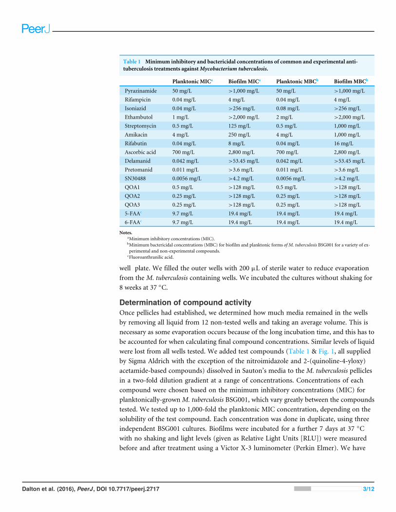
Table 1 Minimum inhibitory and bactericidal concentrations of common and experimental anti-tuberculosis treatments againstMycobacterium tuberculosis.
Planktonic MICa BiofilmMICa Planktonic MBCb BiofilmMBCb
Pyrazinamide 50 mg/L >1,000 mg/L 50 mg/L >1,000 mg/LRifampicin 0.04 mg/L 4 mg/L 0.04 mg/L 4 mg/LIsoniazid 0.04 mg/L >256 mg/L 0.08 mg/L >256 mg/LEthambutol 1 mg/L >2,000 mg/L 2 mg/L >2,000 mg/LStreptomycin 0.5 mg/L 125 mg/L 0.5 mg/L 1,000 mg/LAmikacin 4 mg/L 250 mg/L 4 mg/L 1,000 mg/LRifabutin 0.04 mg/L 8 mg/L 0.04 mg/L 16 mg/LAscorbic acid 700 mg/L 2,800 mg/L 700 mg/L 2,800 mg/LDelamanid 0.042 mg/L >53.45 mg/L 0.042 mg/L >53.45 mg/LPretomanid 0.011 mg/L >3.6 mg/L 0.011 mg/L >3.6 mg/LSN30488 0.0056 mg/L >4.2 mg/L 0.0056 mg/L >4.2 mg/LQOA1 0.5 mg/L >128 mg/L 0.5 mg/L >128 mg/LQOA2 0.25 mg/L >128 mg/L 0.25 mg/L >128 mg/LQOA3 0.25 mg/L >128 mg/L 0.25 mg/L >128 mg/L5-FAAc 9.7 mg/L 19.4 mg/L 19.4 mg/L 19.4 mg/L6-FAAc 9.7 mg/L 19.4 mg/L 19.4 mg/L 19.4 mg/L
Notes.aMinimum inhibitory concentrations (MIC).bMinimum bactericidal concentrations (MBC) for biofilm and planktonic forms of M. tuberculosis BSG001 for a variety of ex-perimental and non-experimental compounds.
cFluoroanthranilic acid.
well plate. We filled the outer wells with 200 µL of sterile water to reduce evaporationfrom the M. tuberculosis containing wells. We incubated the cultures without shaking for8 weeks at 37 ◦C.
Determination of compound activityOnce pellicles had established, we determined how much media remained in the wellsby removing all liquid from 12 non-tested wells and taking an average volume. This isnecessary as some evaporation occurs because of the long incubation time, and this has tobe accounted for when calculating final compound concentrations. Similar levels of liquidwere lost from all wells tested. We added test compounds (Table 1 & Fig. 1, all suppliedby Sigma Aldrich with the exception of the nitroimidazole and 2-(quinoline-4-yloxy)acetamide-based compounds) dissolved in Sauton’s media to the M. tuberculosis pelliclesin a two-fold dilution gradient at a range of concentrations. Concentrations of eachcompound were chosen based on the minimum inhibitory concentrations (MIC) forplanktonically-grownM. tuberculosis BSG001, which vary greatly between the compoundstested. We tested up to 1,000-fold the planktonic MIC concentration, depending on thesolubility of the test compound. Each concentration was done in duplicate, using threeindependent BSG001 cultures. Biofilms were incubated for a further 7 days at 37 ◦Cwith no shaking and light levels (given as Relative Light Units [RLU]) were measuredbefore and after treatment using a Victor X-3 luminometer (Perkin Elmer). We have
Dalton et al. (2016), PeerJ, DOI 10.7717/peerj.2717 3/12

Figure 1 Chemical structures of the experimental compounds used in this study.
defined the MIC as causing a 1 log reduction in light production, as previously described(Andreu et al., 2012).
To determine the minimum bactericidal concentration (MBC), pellicles were removedfrom the wells, disrupted by pipetting and washed 3 times in Sauton’s media supplementedwith 0.05% tween 80. The cells were then resuspended in fresh 7H9 broth (5 ml,supplemented as described above) and plated onto 7H11 agar (supplemented as describedabove). We have defined the MBC as the concentration which resulted in no growth.We incubated broth cultures for 2 weeks and plate cultures for eight weeks to recoverany surviving bacteria. All experiments were performed using three biological replicatesof M. tuberculosis BSG001 and two technical replicates. Biological replicates were grownseparately and tested on different days.
Dalton et al. (2016), PeerJ, DOI 10.7717/peerj.2717 4/12

RESULTSDecreased susceptibility of pellicle-grown M. tuberculosis tofront-line and experimental compoundsOf the four main first line drugs only rifampicin was seen to inhibit pellicles ofM. tuberculosis at the concentrations tested (Table 1, Figs. 2C and 3B). Isoniazid alsoled to some inhibition but below the threshold defined (Figs 2C and 3C). In the case ofrifampicin the MIC and MBC for pellicle-grown BSG001 were determined to be 4 mg/L,100 times the concentration required to produce a similar result with planktonic cells(Table 1). High levels of pyrazinamide, isoniazid and ethambutol (20, 6,000 and 1,000times the MIC’s for planktonic cells, respectively) failed to sufficiently reduce light to beclassed as inhibitory (Table 1, Figs. 2A–2D and 3A–3D). More success was observed withnon-first line antibiotics, with MICs obtained for pellicle-grown BSG001 for streptomycin(125mg/L), amikacin (250mg/L) and rifabutin (8mg/L) (Table 1, Figs. 2E–2G and 3E–3G).These pellicle-MICs represent an increase of 250, 62.5 and 200 times the MIC’s obtainedfor planktonic cultures, respectively (Table 1).
When novel and experimental compounds were examined, none of the currentderivations of the nitroimidazole based compounds (Olaru et al., 2015; Palmer et al., 2010)(delamanid, pretomanid and SN30488) were able to reduce light from the pellicle-grownM. tuberculosis at the concentrations tested (Table 1, Figs. 2H–2J and 3H–3J). The sameresistance to drug-killing was seen with experimental compounds based on 2-(quinoline-4-yloxy) acetamides (Phummarin et al., 2016) (QOA 1, QOA 2 and QOA 3) (Table 1,Figs. 2K–2M and 3K–3M). In contrast, the fluoroanthranilic-acid based compounds,5-fluoroanthranilic acid (5-FAA) and 6-fluoroanthranilic acid (6-FAA), which targetthe tryptophan biosynthetic pathway (Toyn et al, 2000) were seen to be quite effective atinhibiting light from M. tuberculosis BCG001 growing as a pellicles (Table 1, Figs. 2N/Oand 3N/O). Ascorbic acid was also seen to cause inhibition at 2.8 g/L, 4 times the MIC forplanktonically-grownM. tuberculosis (Table 1, Figs. 2P and 3P).
DISCUSSIONMany infectious bacteria form biofilms within their host. Bacteria living within a biofilm arenotoriously difficult to treat and can persist for extended periods of time, as they have theability to resist the immune system (Donlan & Costerton, 2002), display increased virulence(Safadi et al., 2012; Wand et al., 2012) and can become phenotypically more resistant toantibiotics. It is common for antibiotic concentrations required to control bacteria withinbiofilms to be 100–1,000 fold greater than those needed to treat planktonic forms (Ceriet al., 1999). This was seen to be true of the majority of compounds tested in this study.In many cases this is not too surprising. Biofilms can affect drug activity by acting as aphysical barrier to entry into the cell. The phenotypic state of the cells within the biofilmcould also make the cells less susceptible. Isoniazid’s mode of action relates to mycolic acidsynthesis. When growing as a pellicle, it is possible that mycolic acid synthesis is minimalor nonessential. Similarly ethambutol, delamanid and pretomanid are also thought toaffect various steps in cell wall biosynthesis and formation; none of these were seen to
Dalton et al. (2016), PeerJ, DOI 10.7717/peerj.2717 5/12

Figure 2 The effect of clinically-used and experimental compounds onM. tuberculosis BSG001 pellicles. The inhibitory effect of first line (A–D) and second line (E–G) anti-tuberculosis drugs used in the clinic and experimental compounds (H–P), including those based on nitroimidazole(H–J), 2-(quinoline-4-yloxy) acetamides (K–M) and fluoroanthranilic-acid (N, O), is presented as a reduction in bioluminescence plotted as relativelight units (RLU) per well on day 7 of treatment. The dashed line indicates the limit of detection. The solid and open arrows indicate the MBC’s (theconcentration which resulted in the recovery of no bacterial colonies) obtained for planktonically-grown cells and pellicles, respectively. All com-pounds were tested in three biological replicates on separate days with multiple technical replicates. Results are given as box whisker plots with thebox representing values from the lower to upper quartile and the whiskers representing the range.
have an effect. Pyrazinamide relies on conversion to pyrazinoic acid, which requires acidicconditions to become active. This could indicate that these conditions are not present inmycobacterial pellicles or that the drug is unable to penetrate the cells in this phenotype.If it is due to the lack of an acidic environment this could represent a limitation in usingthis model for drug testing. In contrast, antibiotics that affect protein synthesis, such asrifampicin, rifabutin, amikacin and streptomycin, displayed some degree of inhibitiveactivity towards pellicle-grown M. tuberculosis, although this activity was lower than theactivity against planktonically-grown cells. The 2-(quinoline-4-yloxy) acetamide based
Dalton et al. (2016), PeerJ, DOI 10.7717/peerj.2717 6/12

Figure 3 The relative effect of clinically-used and experimental compounds onM. tuberculosis BSG001 pellicles. The relative change in bio-luminescence (relative light units [RLU]) following the treatment of M. tuberculosis BSG001 biofilms with first line (A–D) and second line (E–G)anti-tuberculosis drugs used in the clinic and experimental compounds (H–P), including those based on nitroimidazole (H–J), 2-(quinoline-4-yloxy) acetamides (K–M) and fluoroanthranilic-acid (N, O), is shown as the ratio of RLU before treatment and RLU after seven days of treatment.The dashed line indicates the level at which no change occurs; values above the dashed line indicate an increase in light levels (and hence survival/-growth) over the course of the experiment, while those below indicate a decrease (and hence inhibition/death). The solid and open arrows indicatethe MBC’s (the concentration which resulted in the recovery of no bacterial colonies) obtained for planktonically-grown cells and pellicles, respec-tively. Results are given as box whisker plots with the box representing values from the lower to upper quartile and the whiskers representing therange.
compounds also showed little activity against this biofilm form. The mode of action ofthese compounds is likely due to electron transport inhibition of cytochrome bc1 oxidase(Phummarin et al., 2016). As the cells are actively metabolising, the lack of an effect fromthese compounds is most likely due to an inability to access the cells.
In our study, we observed that M. tuberculosis growing as a pellicle is susceptible to aconcentration of ascorbic acid similar to that reported for planktonically-grown cells. Thisconcentration was also sufficient to cause death of the M. tuberculosis pellicle within one
Dalton et al. (2016), PeerJ, DOI 10.7717/peerj.2717 7/12

week of treatment. The activity of ascorbic acid is thought to be due to the generationof highly reactive hydroxyl radicals via presence of iron and Fenton reaction chemistry(Vilcheze et al., 2013). Killing due to this mechanism would be non-specific and notdependant on uptake. Interestingly the fluoroanthranilic acid tryptophan biosynthesisinhibitors were also seen to be effective at inhibiting and killing pellicles, indicating thatthis is a pathway worthy of further consideration for drug targeting.
It is possible that the comparative ease in which test compounds can access bacteriawithin a pellicle, that is from both above and below, as compared to a biofilm attached toa surface which cannot be accessed from the surface side, make this form of biofilm easierto kill. While it is still unknown if M. tuberculosis forms biofilms/pellicles in vivo, manymycobacterial species do form complex, secondary structures such as pellicles in vitro (Ojha& Hatfull, 2012). Researchers have also reported histological evidence for the presence ofmulticellular structures involving M. tuberculosis outside of the macrophage (Lenaertset al., 2007). Others have reported the presence of cells that resembles biofilms/pelliclesin the cavities formed during secondary tuberculosis which would indicate that thisphenotype is likely to play a role in human disease (Lenaerts et al., 2007; Hunter et al.,2006; Hunter et al., 2014). The biphasic response of M. tuberculosis infections, in whicha large kill is seen early on in drug treatment followed by a marked reduction in thebactericidal activity of therapeutic agents due to phenotypic rather than genetic resistance,could also be evidence that M. tuberculosis is able to form biofilms/pellicles in vivo.Such structures could act as a reservoir for drug tolerant bacilli which are responsiblefor the increased duration of drug treatment required in cases of TB. Regardless, theM. tuberculosis-pellicle model is a useful multi-phenotypic environment in which a novelcompound can be tested against cells with a range of susceptibilities. The susceptibility ofM. tuberculosis within this model indicates that drugs which can attack the surface of thecell or can pass through the extracellular matrix of the pellicle represent the best option fortreatment. We also saw that the inhibition of tryptophan biosynthesis could be utilised inTB treatment and their design should be further investigated.
ADDITIONAL INFORMATION AND DECLARATIONS
FundingThis work was supported by the Maurice Wilkins Centre for Molecular Biodiscoveryand the University of Auckland Vice-Chancellor’s Strategic Development Fund andFaculty Research Development Fund. SW is supported by a Sir Charles Hercus Fellowship(09/099) from the Health Research Council of New Zealand. The funders had no rolein study design, data collection and analysis, decision to publish, or preparation of themanuscript.
Grant DisclosuresThe following grant information was disclosed by the authors:Maurice Wilkins Centre for Molecular Biodiscovery.University of Auckland Vice-Chancellor’s Strategic Development Fund.
Dalton et al. (2016), PeerJ, DOI 10.7717/peerj.2717 8/12

Faculty Research Development Fund.Sir Charles Hercus Fellowship: 09/099.
Competing InterestsSiouxsie Wiles is an Academic Editor for PeerJ.
Author Contributions• James P. Dalton conceived and designed the experiments, performed the experiments,analyzed the data, contributed reagents/materials/analysis tools, wrote the paper,prepared figures and/or tables, reviewed drafts of the paper.
• Benedict Uy performed the experiments, contributed reagents/materials/analysis tools,reviewed drafts of the paper.
• Narisa Phummarin contributed reagents/materials/analysis tools.• Brent R. Copp and William A. Denny contributed reagents/materials/analysis tools,reviewed drafts of the paper.
• Simon Swift conceived and designed the experiments, analyzed the data, contributedreagents/materials/analysis tools, reviewed drafts of the paper.
• Siouxsie Wiles conceived and designed the experiments, analyzed the data, contributedreagents/materials/analysis tools, wrote the paper, prepared figures and/or tables,reviewed drafts of the paper.
Data AvailabilityThe following information was supplied regarding data availability:
Wiles, Siouxsie; Dalton, James; Uy, Benedict; Copp, Brent; Denny, Bill (2016): Effectof common and experimental anti-tuberculosis treatments on Mycobacterium tuberculosisgrowing as biofilms. figshare: 10.17608/k6.auckland.4097772.v1.
Supplemental InformationSupplemental information for this article can be found online at http://dx.doi.org/10.7717/peerj.2717#supplemental-information.
REFERENCESAndreu N, Fletcher T, Krishnan N,Wiles S, Robertson BD. 2012. Rapid measurement
of antituberculosis drug activity in vitro and in macrophages using bioluminescence.Journal of Antimicrobial Chemotherapy 67:404–414 DOI 10.1093/jac/dkr472.
Andreu N, Zelmer A, Fletcher T, Elkington PT,Ward TH, Ripoll J, Parish T, BancroftGJ, Schaible U, Robertson BD,Wiles S. 2010. Optimisation of bioluminescentreporters for use with mycobacteria. PLoS ONE 5:e10777DOI 10.1371/journal.pone.0010777.
Ceri H, OlsonME, Stremick C, Read RR, Morck D, Buret A. 1999. The Calgary biofilmdevice: new technology for rapid determination of antibiotic susceptibilities ofbacterial biofilms. Journal of Clinical Microbiology 37:1771–1776.
Donlan RM, Costerton JW. 2002. Biofilms: survival mechanisms of clinically relevantmicroorganisms. Clinical Microbiology Reviews 15:167–193.
Dalton et al. (2016), PeerJ, DOI 10.7717/peerj.2717 9/12

Fenhalls G, Stevens L, Moses L, Bezuidenhout J, Betts JC, Helden PV, Lukey PT,Duncan K. 2002. In situ detection ofMycobacterium tuberculosis transcripts inhuman lung granulomas reveals differential gene expression in necrotic lesions.Infection and Immunity 70:6330–6338 DOI 10.1128/IAI.70.11.6330-6338.2002.
Hunter RL, Actor JK, Hwang SA, Karev V, Jagannath C. 2014. Pathogenesis of postprimary tuberculosis: immunity and hypersensitivity in the development of cavities.Annals of Clinical and Laboratory Science 44:365–387.
Hunter RL, Armitige L, Jagannath C, Actor JK. 2013. TB Research at UT-Houston; areview of cord factor: new approaches to drugs, vaccines and the pathogenesis oftuberculosis. Tuberculosis 89:S18–S25.
Hunter RL, OlsenMR, Jagannath C, Actor JK. 2006.Multiple roles of cord factor in thepathogenesis of primary, secondary, and cavitary tuberculosis, including a reviseddescription of the pathology of secondary disease. Annals of Clinical and LaboratoryScience 36:371–386.
Klopper M,Warren RM, Hayes C, Gey van Pittius NC, Streicher EM,Müller B, SirgelFA, Chabula-Nxiweni M, Hoosain E, Coetzee G, David van Helden P, Victor TC,Trollip AP. 2013. Emergence and spread of extensively and totally drug-resistanttuberculosis, South Africa. Emerging Infectious Diseases 19:449–455DOI 10.3201/eid1903.120246.
Kulka K, Hatfull G, Ojha AK. 2012. Growth ofMycobacterium tuberculosis biofilms.Journal of Visualized Experiments: JoVE 60:3820 DOI 10.3791/3820.
Lenaerts AJ, Hoff D, Aly S, Ehlers S, Andries K, Cantarero L, Orme IM, Basaraba RJ.2007. Location of persisting mycobacteria in a Guinea pig model of tuberculosisrevealed by r207910. Antimicrobial Agents and Chemotherapy 51:3338–3345DOI 10.1128/AAC.00276-07.
Ojha AK, Hatfull GF. 2012. Biofilms ofMycobacterium tuberculosis: new perspectives ofan old pathogen. In: Cardona P-J, ed. Understanding tuberculosis—deciphering thesecret life of the bacilli. Rijeka: InTech.
Olaru ID, Von Groote-Bidlingmaier F, Heyckendorf J, YewWW, Lange C, ChangKC. 2015. Novel drugs against tuberculosis: a clinician’s perspective. EuropeanRespiratory Journal 45:1119–1131 DOI 10.1183/09031936.00162314.
Palmer BD, Thompson AM, Sutherland HS, Blaser A, Kmentova I, FranzblauSG,Wan B,Wang Y, Ma Z, DennyWA. 2010. Synthesis and structure–activitystudies of biphenyl analogues of the tuberculosis drug (6S)-2-nitro-6-{[4-(trifluoromethoxy)benzyl]oxy}-6,7-dihydro-5H-imidazo[2,1-b][1, 3]oxazine (PA-824). Journal of Medicinal Chemistry 53:282–294 DOI 10.1021/jm901207n.
Phummarin N, Boshoff HI, Tsang PS, Dalton J, Wiles S, Barry 3rd CE, Copp BR.2016. SAR and identification of 2-(quinolin-4-yloxy)acetamides asMycobacteriumtuberculosis cytochrome bc1 inhibitors.MedChemComm Epub ahead of print Aug 222016 DOI 10.1039/C6MD00236F.
Dalton et al. (2016), PeerJ, DOI 10.7717/peerj.2717 10/12

Rengarajan J, Bloom BR, Rubin EJ. 2005. Genome-wide requirements forMycobac-terium tuberculosis adaptation and survival in macrophages. Proceedings of theNational Academy of Sciences of the United States of America 102:8327–8332DOI 10.1073/pnas.0503272102.
Rice AR, HamiltonMA, Camper AK. 2003.Movement replication and emigration ratesof individual bacteria in a biofilm.Microbial Ecology 45:163–172DOI 10.1007/s00248-002-1028-x.
Safadi RA, Abu-Ali GS, Sloup RE, Rudrik JT,Waters CM, Eaton KA, Manning SD.2012. Correlation between in vivo biofilm formation and virulence gene expressionin Escherichia coli O104:H4. PLoS ONE 7:e41628DOI 10.1371/journal.pone.0041628.
Sambandan D, Dao DN,Weinrick BC, Vilchéze C, Gurcha SS, Ojha A, Kremer L,Besra GS, Hatfull GF, Jacobs JrWR. 2013. Keto-mycolic acid-dependent pellicleformation confers tolerance to drug-sensitiveMycobacterium tuberculosis.MBio4:e00222–e00213.
Sendi P, Johansson L, Dahesh S, Van Sorge NM, Darenberg J, NorgrenM, SjölinJ, Nizet V, Norrby-Teglund A. 2009. Bacterial phenotype variants in group Bstreptococcal toxic shock syndrome. Emerging Infectious Diseases 15:223–232DOI 10.3201/eid1502.080990.
Stewart PS, Franklin MJ. 2008. Physiological heterogeneity in biofilms. Nature ReviewsMicrobiology 6:199–210 DOI 10.1038/nrmicro1838.
Toyn JH, Gunyuzlu PL,WhiteWH, Thompson LA, Hollis GH. 2000. A counterselectionfor the tryptophan pathway in yeast: 5-fluoroanthranilic acid resistance. Yeast16:553–550DOI 10.1002/(SICI)1097-0061(200004)16:6<553::AID-YEA554>3.0.CO;2-7.
Tuchscherr L, Medina E, HussainM, VölkerW, Heitmann V, Niemann S, HolzingerD, Roth J, Proctor RA, Becker K, Peters G, Löffler B. 2011. Staphylococcusaureus phenotype switching: an effective bacterial strategy to escape host immuneresponse and establish a chronic infection. EMBOMolecular Medicine 3:129–141DOI 10.1002/emmm.201000115.
Vilcheze C, Hartman T,Weinrick B, Jacobs JrWR. 2013.Mycobacterium tuberculosis isextraordinarily sensitive to killing by a vitamin C-induced Fenton reaction. NatureCommunications 4:Article 1881 DOI 10.1038/ncomms2898.
WandME, Bock LJ, Turton JF, Nugent PG, Sutton JM. 2012. Acinetobacter baumanniivirulence is enhanced in Galleria mellonella following biofilm adaptation. Journal ofMedical Microbiology 61:470–477 DOI 10.1099/jmm.0.037523-0.
Wang J, Pearce AN, Chan ST, Taylor RB, PageMJ, Valentin A, Bourguet-KondrackiML, Dalton JP,Wiles S, Copp BR. 2016. Biologically active acetylenic amino alcoholand n-hydroxylated 1,2,3,4-tetrahydro-beta-carboline constituents of the NewZealand ascidian pseudodistoma opacum. Journal of Natural Products 79:607–610.
Wayne LG, Sohaskey CD. 2001. Nonreplicating persistence ofmycobacterium tuberculo-sis. Annual Review of Microbiology 55:139–163.
Dalton et al. (2016), PeerJ, DOI 10.7717/peerj.2717 11/12

World Health Organization. 2011. Guidelines for the programmatic management ofdrug-resistant tuberculosis: 2011 update. World Health Organization, Geneva.Available at https://www.ncbi.nlm.nih.gov/books/NBK148644/ .
World Health Organization. 2015. Global tuberculosis report 2015. World HealthOrganization, Geneva.
Dalton et al. (2016), PeerJ, DOI 10.7717/peerj.2717 12/12