Embed Size (px)
Citation preview
Int. J. Environ. Res. Public Health 2014, 11, 3233-3255; doi:10.3390/ijerph110303233
International Journal of
Environmental Research and Public Health
ISSN 1660-4601 www.mdpi.com/journal/ijerph
Article
Effect of Ampicillin, Streptomycin, Penicillin and Tetracycline on Metal Resistant and Non-Resistant Staphylococcus aureus
Dagmar Chudobova 1, Simona Dostalova 1, Iva Blazkova 1, Petr Michalek 1,
Branislav Ruttkay-Nedecky 1,2, Matej Sklenar 1, Lukas Nejdl 1, Jiri Kudr 1, Jaromir Gumulec 2,3,
Katerina Tmejova 1,2, Marie Konecna 1,2, Marketa Vaculovicova 1,2, David Hynek 2,
Michal Masarik 2,3, Jindrich Kynicky 4, Rene Kizek 1,2 and Vojtech Adam 1,2,*
1 Department of Chemistry and Biochemistry, Faculty of Agronomy, Mendel University in Brno,
Zemedelska 1, Brno CZ-613 00, Czech Republic; E-Mails: [email protected] (D.C.);
[email protected] (S.D.); [email protected] (I.B.); [email protected] (P.M.);
[email protected] (B.R.-N.); [email protected] (M.S.);
[email protected] (L.N.); [email protected] (J.Ku.); [email protected] (K.T.);
[email protected] (M.K.); [email protected] (M.V.); [email protected] (R.K.) 2 Central European Institute of Technology, Brno University of Technology, Technicka 3058/10,
Brno CZ-616 00, Czech Republic; E-Mails: [email protected] (J.G.);
[email protected] (D.H.); [email protected] (M.M.) 3 Department of Pathological Physiology, Faculty of Medicine, Masaryk University,
Komenskeho namesti 2, Brno CZ-662 43, Czech Republic 4 Karel Englis College, Sujanovo nam. 356/1, Brno CZ-602 00, Czech Republic;
E-Mail: [email protected] (J.Ky.)
* Author to whom correspondence should be addressed; E-Mail: [email protected];
Tel.: +420-5-4513-3350; Fax: +420-5-4521-2044.
Received: 7 January 2014; in revised form: 18 February 2014 / Accepted: 24 February 2014 /
Published: 19 March 2014
Abstract: There is an arising and concerning issue in the field of bacterial resistance,
which is confirmed by the number of deaths associated with drug-resistant bacterial
infections. The aim of this study was to compare the effects of antibiotics on
Staphylococcus aureus non-resistant strain and strains resistant to cadmium or lead ions.
Metal resistant strains were created by the gradual addition of 2 mM solution of metal ions
(cadmium or lead) to the S. aureus culture. An increasing antimicrobial effect of
ampicillin, streptomycin, penicillin and tetracycline (0, 10, 25, 50, 75, 150, 225 and 300
OPEN ACCESS
Int. J. Environ. Res. Public Health 2014, 11 3234
µM) on the resistant strains was observed using a method of growth curves. A significant
growth inhibition (compared to control) of cadmium resistant cells was observed in the
presence of all the four different antibiotics. On the other hand, the addition of
streptomycin and ampicillin did not inhibit the growth of lead resistant strain. Other
antibiotics were still toxic to the bacterial cells. Significant differences in the morphology
of cell walls were indicated by changes in the cell shape. Our data show that the presence
of metal ions in the urban environment may contribute to the development of bacterial
strain resistance to other substances including antibiotics, which would have an impact on
public health.
Keywords: S. aureus; antimicrobial resistance; antibiotics; metal resistance; cross resistance;
growth curves; inhibition concentrations; spectrophotometry
1. Introduction
The seriousness of the problem of bacterial resistance is confirmed by the number of deaths
associated with drug-resistant bacterial infections—only in the EU it affects 25,000 people a year [1].
Recently, with the discovery of multi-resistant strains in the broader community, public health officials
have begun to realize the potential danger of the spread of these antibiotic resistant bacteria [2].
Antimicrobial resistance (AMR) is a resistance of microorganism to an antimicrobial medicine to
which it was originally sensitive. Creation of a resistance effect is dependent on the genes located in
plasmids that are infectious matter transferred to other cells, so the resistance between bacteria spreads
rapidly. Resistant microorganisms are able to withstand an attack of antimicrobial medicines, so that
standard treatments become ineffective and infections persist increasing the risk of spreading to others.
The evolution of resistant strains is natural phenomenon that happens when microorganisms are
exposed to antimicrobial drugs and resistant genes can be distributed between certain types of bacteria.
The misuse of antimicrobial medicines accelerates this phenomenon.
The incidence of resistant microorganisms is monitored primarily in hospitals, but much higher risk
is present in the soil and waters [3]. Resistance, generated in the external environment, is a natural
development of every live system. Infections caused by resistant microorganisms often fail to respond
to the standard treatment, resulting in prolonged illness and greater risk of death [4–7]. The death rate
of patients with serious infections treated in hospitals is about twice as high compared to the patients
with infections caused by non-resistant bacteria [8].
A high percentage of hospital-acquired infections are caused by highly resistant bacteria such as
methicillin-resistant Staphylococcus aureus (MRSA) or multidrug-resistant enterococci Gram-negative
bacteria. The general mechanisms of resistance are: (1) the limited penetration of antibiotics into the
bacterial cell; (2) the change of the target structure (receptor); (3) metabolic changes within the
bacterial cell, which prevents the effect of antibiotics on the target structures; and (4) enzymatic
inhibition/inactivation of antibiotics [9–14]. Metal resistance of microbes is accomplished by intra- and
extracellular mechanisms. Metals can be excreted via efflux transport systems, sequestering
compounds of the cytosol can bind and detoxify metals inside the cell. The release of chelators into the
Int. J. Environ. Res. Public Health 2014, 11 3235
extracellular milieu fixes the bound metals. The structure of the cell envelope is prone to bind large
amounts of metals by sorption, thus preventing influx [15]. Newly discovered resistance mechanisms,
such as enzymes produced by the bacteria that destroy last generation antibiotics, have emerged among
several Gram-negative bacilli and have rapidly spread in many countries. This can render ineffective
powerful antibiotics, which are often the last defence against multi-resistant strains of bacteria. This
new resistance mechanism is associated with ordinary human pathogens (e.g., Escherichia coli) which
cause common infections such as urinary tract infection.
The use of antibiotics and toxic metals should be considered carefully with the attention paid to the
environmental impacts [16–18]. Heavy metals are toxic and can be harmful to organisms. For this
reason, a number of organisms including bacteria develop processes which are able to withstand
the effects of these pollutants [15]. Toxic metals in the environment can enrich the antibiotic
multi-resistance property of bacteria [19]. Resende et al. [20] evaluated medically relevant bacteria in
an aquaculture system and their susceptibility to antibiotics and toxic metals. Multidrug-resistant
bacteria were also found to be tolerant to nickel, zinc, chromium and copper. In another study by
Ji et al. [21], eight antibiotic resistance genes (ARG), seven heavy metals and six antibiotics were
quantified in manures and soils collected from multiple feedlots in Shanghai (China). Overall,
sulfonamide ARGs were more abundant than tetracycline ARGs. The significant positive correlations
were found between some ARGs and typical heavy metals such as Cu, Zn and Hg. Similarly, in the
study by Malik et al. [22], majority of the Pseudomonas isolates from water and soil exhibited
resistance to multiple metals (Hg, Cd, Pb, Cu, Zn, Ni) and antibiotics (water—tetracycline, polymyxin
B; soil— sulphadiazine, ampicillin and erythromycin) was presented [22].
With the appearance of antibiotic-resistant bacteria, increasing numbers of infections are causing
huge losses to both economic concerns and social resources over recent decades, and this has become a
global problem [2]. This study aimed on testing of non-resistant strain of bacterial culture wild type
S. aureus and S. aureus strains resistant to heavy metals ions (cadmium = RCd or lead = RPb) exposed
to different concentrations of four various antibiotics (ampicillin, streptomycin, penicillin and
tetracycline). The antimicrobial activity of antibiotics on S. aureus bacterial culture was tested by the
growth curves and the results were statistically evaluated.
2. Experimental Section
2.1. Cultivation of S. aureus
S. aureus (NCTC 8511) was obtained from the Czech Collection of Microorganisms, Faculty of
Science, Masaryk University, Brno, Czech Republic. The strains were stored as a spore suspension in
20% (v/v) glycerol at −20 °C. Prior to use in this study, the strains were thawed and the glycerol was
removed by washing with distilled water. The composition of cultivation medium was as follows: meat
peptone 5 g/L, NaCl 5 g/L, bovine extract 1.5 g/L, yeast extract 1.5 g/L (HIMEDIA, Mumbai, India),
sterilized MilliQ water with 18 MΩ. pH of the cultivation medium was adjusted at 7.4 before
sterilization. The sterilization of the media was carried out at 121 °C for 30 min. in sterilizer
(Tuttnauer 2450EL, Beit Shemesh, Israel). The prepared cultivation media were inoculated with
bacterial culture into 25 mL Erlenmeyer flasks. After inoculation, the bacterial cultures were cultivated
Int. J. Environ. Res. Public Health 2014, 11 3236
for 24 h on a shaker at 600 rpm and 37 °C. The bacterial culture, cultivated under these conditions, was
diluted by cultivation medium to OD600 = 0.1 and used in the following experiments.
2.2. Preparation of Resistant Strains of S. aureus
The resistant strains of S. aureus have been developed in the laboratory. 2 mM basic solutions of
heavy metals ions (Cd2+ and Pb2+) was added to non-resistant bacterial culture of S. aureus, cultivated
in Luria Bertani medium. Low concentration (50 μM) of the metal ions in a medium was inoculated
into the bacterial culture, and then the concentration of heavy metal ions was always increased by the
concentration of 50 μM to the maximum possible dose for regeneration of S. aureus.
2.3. Chemicals, Preparation of Deionised Water and pH Measurement
Chemicals used in this study were purchased from Sigma-Aldrich (St. Louis, MO, USA) in ACS
purity unless noted otherwise. The deionised water was prepared using Aqual 25 reverse osmosis
equipment (Aqual, Brno, Czech Republic). The deionised water was further purified by using a Milli-
Q Direct QUV apparatus equipped with a UV lamp. The resistance was 18 MΩ. The pH was measured
using a WTW inoLab pH meter (Weilheim, Germany).
2.4. Heavy Metals Ions Preparation
Heavy metals used for the preparation of heavy metals ions have always been in the form of
nitrates of these metals (Cd(NO3)2·4H2O and Pb(NO3)2) dissolved in 100 mL Milli-Q water and final
concentration of these ions were always 2 mM.
2.5. The Microscopy of the Cells in Ambient Light
The inverted system microscope Olympus IX 71 (Tokyo, Japan) was used for the imaging of the
cells. The cells in cultivation medium were pipetted (5 μL) on the microscope slide and covered by
cover slip. The cover slip was placed on the sample and the immersion oil was used. The objective
(PlanFLN; Mag. 100×; NA 1, 3; F.N. 26.5) and the magnification lens 1.6× were used, the total
magnification was 1600×. The images were captured by Camera Olympus DP73 and processed by
Stream Basic 1.7 Software, the images resolution was 4800 × 3600 pixels. The parameters were as
follows: exposure time: 32 ms and ISO 200.
2.6. Determination of Growth Curves
The procedure for the evaluation of the antimicrobial effect of tested compounds and their
combinations consisted in measuring of the absorbance using the apparatus Multiskan EX (Thermo
Fisher Scientific, Bremen, Germany) and subsequent analysis in the form of growth curves. Non-
resistant bacterial culture of S. aureus or resistant strain S. aureus to cadmium and lead ions were
cultivated in LB medium for 24 h with shaking and was diluted with LB medium using Specord
spectrophotometer 210 (Analytik, Jena, Germany) at a wavelength of 600 nm to absorbance 0.1 AU.
On the microplate, these cultures were mixed with various concentrations of four types of antibiotics
(ampicillin, streptomycin, penicillin and tetracycline) or S. aureus alone as a control for measurements.
Int. J. Environ. Res. Public Health 2014, 11 3237
The concentrations of antibiotics were 0, 10, 25, 50, 75, 150, 225 and 300 μM. Total volume in the
microplate wells was always 300 µL. The measurements were carried out at time 0, then each
half-an hour for 24 h at 37 °C and a wavelength of 620 nm. The obtained values were analyzed in
graphical form as growth curves for each variant individually.
2.7. Determination of Cadmium and Lead Ions by Atomic Absorption Spectrometry
The determination of cadmium and lead ions was carried out using 240FS Agilent Technologies
atomic absorption spectrometer (Agilent, Santa Clara, CA, USA) with flame atomization. Cadmium
was measured on the wavelength 228.8 nm with spectral bandwidth of 0.5 nm and lead it was 217.0
nm with spectral bandwidth of 1.0 nm. The mixture of air and acetylene was used for the flame
atomization. Deuterium background correction was used and the signal was measured in integration
mode for 3 s.
2.8. Interaction of Bacterial DNA Fragment of zntR and 16S Gene with Cadmium and Lead Ions
DNA fragments of zntR and 16S gene (16 μg/mL) were incubated with different concentrations of
Cd(NO3)2 and Pb(NO3)2 in 1:1 ratio. The stock solutions of DNA fragment of zntR gene (16 µg/mL)
were incubated with 0, 0.06, 0.24 and 0.95 mM Cd(II) and Cd(II) in water (ACS purity, Sigma-Aldrich).
The same procedure was carried out for the DNA fragment of 16S gene. The samples were incubated
for 60 min at 25 °C. After incubation, unbound cadmium and lead ions were removed using Amicon
Ultra 3K centrifugal filter device (Millipore Corp., Billerica, MA, USA). After centrifugation at 14,000
rpm for 10 min at 25 °C (5417R Eppendorf, Hauppauge, NY, USA), the sample was complemented
with water to the original volume (200 µL).
The spectra were recorded within the range from 220 to 420 nm using quartz cuvettes (1 cm,
Hellma, Essex, UK) on a SPECORD 210 spectrophotometer (Analytik Jena, Germany) at 25 °C
(Julabo, Sellbach, Germany). The spectra were recorded after 60 min of interaction. The denaturation
of the complex of DNA with Cd(II) and Pb(II) was monitored spectrophotometrically using a
SPECORD S600 spectrophotometer with a diode array detector (Analytik Jena). The sample was
incubated for 3 min at increasing temperatures in a range from 25 to 90 °C and the absorbance was
measured at 260 nm. The changes in absorbance spectra were recorded during denaturation. The
absorption spectra were evaluated in the WinASPECT 2.2.7.0.
2.9. Determination of Metallothionein
The electrochemical detection was carried out using the method of differential pulse voltammetry
(three electrodes involvement, working mercury drop electrode (HMDE), reference silver chloride
electrode Ag/AgCl/3M KCl, and auxiliary carbon electrode) [23]. The analysed samples were
deoxidized by argon bubbling for 120 s. As a supporting electrolyte, Brdicka solution (containing
1 mM Co(NH3)6Cl3 and 1 M ammoniacal buffer (NH3(aq) + NH4Cl, pH = 9.6)) was used. The
supporting electrolyte was changed after each analysis of the sample. The parameters for the
measurements were following: initial potential of −0.7 V, final potential of −1.75 V, the time interval
0.2 s, step potential 2 mV, amplitude −250 mV [24–27].
Int. J. Environ. Res. Public Health 2014, 11 3238
2.10. Expression of ZntR Gene and 16S Gene
2.10.1. Isolation of RNA
Bacterial cultures (1 × 10⁶ of cells) were centrifuged at 300 rcf and 20 °C for 10 min and the pellets
were resuspended in 100 µL of PBS buffer and 0.2 µL of RNase inhibitors. Thus the prepared solution
was mixed with 100 µL of Tissue Lysis Buffer. The entire volume (200 µL) were pipetted into the
sample tube, which is the component of the isolation kit MagNA Pure Compact RNA Isolation Kit
(Roche, Basel, Schweiz), and inserted with other instruments on the appropriate place in the machine.
In the second row of the machine, the vials with 20 µL of DNAase were inserted. Next steps were
carried out according to the manufacturer’s instructions (“RNA Cell” protocol MagNA).
2.10.2. Amplification of mRNA
The mRNA were converted to cDNA using PrimeScript One Step RT-PCR Kit Ver. 2 (TaKaRa,
Mountain View, CA, USA). The reaction profile was as follows: initial denaturation at 94 °C for 2
min, 30 cycles of 94 °C for 30 s, 50 °C for 30 s and 72 °C for 1.5 min.
2.10.3. Amplification of cDNA for zntR Gene
The zntR gene was amplified using polymerase chain reaction (PCR). The sequences
of forward and reverse primers were 5'-GGATCCATGTCAGAACAATATTCAGA-3', and
5'-AAGCTTTTATAACCCACTTTCTTTAG-3' respectively. The final volume of the PCR reaction
mixture was 25 μL containing 15.875 μL of sterile water, 2.5 μL of 1× Taq reaction buffer, 0.5 μL of
100 mM dNTP, 0.5 μL of forward primer, 0.5 μL of reverse primer and 0.125 μL of Taq DNA
polymerase. The reaction profile was as follows: 30 cycles of 94 °C for 3 min, 50 °C for 1 min and
72 °C for 1 min and a final extension at 72 °C for 4 min. The amplification was carried out using
Mastercycler ep realplex4 S (Eppendorf AG, Hamburg, Germany) and a 333 bp fragment was obtained.
2.10.4. Amplification of cDNA for 16S Gene
The 16S gene was amplified using polymerase chain reaction (PCR). The sequences of forward and
reverse primers were 5'-GAGTTTGATCCTGGCTCAG-3' and 5'-GGTTACCTTGTTACGACTT-3'
respectively. The final volume of the PCR reaction mixture was 25 μL containing 14.42 μL of sterile
water, 2.5 μL of 1× Taq reaction buffer, 0.5 μL of 100 mM dNTP, 1.25 μL of forward primer, 1.25 μL
of reverse primer and 0.085 μL of Taq DNA polymerase. The reaction profile was as follows: initial
denaturation at 94 °C for 4 min, 30 cycles of 94 °C for 30 s, 52 °C for 30 s and 72 °C for 1.5 min and
a final extension at 72 °C for 10 min. Finally, a 1500 bp fragment was obtained.
2.11. Statistical Analyses
The STATISTICA data analysis software system, version 10.0 (StatSoft, Tulsa, OK, USA) was
used for data processing. The half-maximal concentrations (IC50) were calculated from logarithmic
regression of sigmoidal dose-response curve. The general regression model was used to analyse
differences between the combinations of compounds. To reveal differences between the cell lines,
Int. J. Environ. Res. Public Health 2014, 11 3239
Turkey’s post hoc test within homogenous groups was employed. Unless noted otherwise, p < 0.05
was considered significant.
Besides applied antibiotics, the general regression model statistics were also used to analyse the
effect of individual S. aureus strains. The data were natural logarithm-transformed and adjusted by the
time variable using regression model. Consequently, Bonferroni post hoc test was employed to reveal
the differences between S. aureus strains. Kruskal-Wallis and multiple comparisons of mean ranks test
were used to compare the IC50 values based on MTT assay. Unless noted otherwise, p-level < 0.05 was
considered significant.
3. Results and Discussion
In the last decade, the number of infections caused by Gram-positive bacteria, resistant to formerly
effective antibiotics, has increased significantly around the World. The phenomenon of multi-resistance
considerably complicates the choice of antibiotics for infection treatment, which usually involves the
combination of antibiotics in order to increase the efficiency [13]. Antibiotic drugs in particular groups
are characterized by the same mechanism of action and very similar mechanism of resistance. The
target of the antibiotics are mostly ribosomes where the antibiotics as an inhibitor of protein
translocation are bound to the 50S subunit of the ribosome, stimulating the dislocation of peptide-tRNA
from the ribosome during the elongation phase and induce rapid collapse of the polyribosomes [28].
Antimicrobial agents, their effects and resistance formation on various bacterial strains had very
important impact on human and animal medicine. On the other hand, in view of global evolution
theory, the resistance formation is only the next step at assimilation of live organism to the
environment. The connection of effects from various antimicrobial agents leads to the formation of
cross resistance. Concretely in our case, metal contamination functions as a selective agent in the
proliferation of antibiotic resistance. This study is focused just on the investigation of metal (cadmium
and lead) and antibiotic (PNC—penicillin, STR—streptomycin, AMP—ampicillin, TTC—tetracycline)
resistance on bacteria S. aureus just because of one important fact that most antibiotics are readily
degraded in the environment, but metals are not, and so can represent a long-term selective pressure.
3.1. Characterization on the Cellular Level
The testing was performed using bacterial culture S. aureus NCTC 8511 without the metal ions and
two strains S. aureus NCTC 8511—RCd or RPb. The highest concentration of heavy metal ions in
24-h growing cell culture was 950 µM. For this experiment, the 950 µM resistant strains were
recultivated for 24 h in clean medium. Selected antibiotics (penicillin, streptomycin, ampicillin and
tetracycline) at various concentrations were applied to the tested bacterial strains.
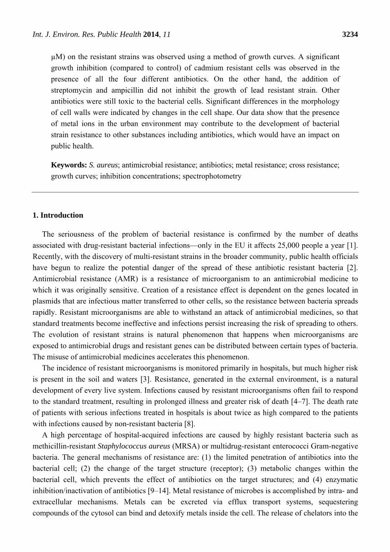
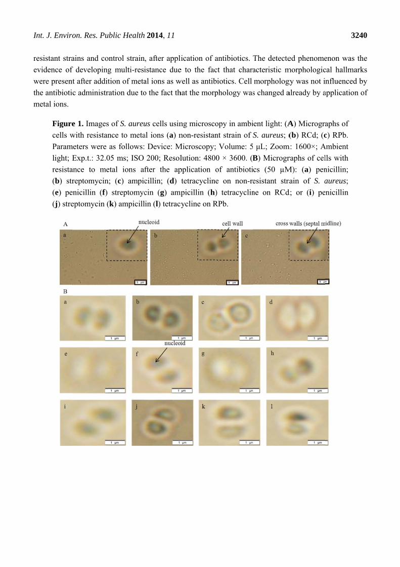
3.1.1. Morphological Characterization
Significant morphological changes were observed in the cells in terms of cell shapes. The presence
of so called cross walls (septal midline) was observed (Figure 1). These inner transverse walls were
formed due to the development of the resistance. Similar phenomenon is commonly observed in case
of MRSA strains [29–31]. The above mentioned morphological changes were observed in both, metal
I
r
e
w
th
m
Int. J. Enviro
esistant stra
evidence of
were presen
he antibioti
metal ions.
Figure
cells w
Param
light; E
resista
(b) str
(e) pe
(j) stre
on. Res. Pu
ains and co
f developing
nt after addit
c administr
e 1. Images
with resistan
meters were
Exp.t.: 32.0
ance to me
reptomycin
enicillin (f)
eptomycin (
ublic Health
ontrol strain
g multi-resi
tion of meta
ration due to
s of S. aureu
nce to meta
as follows:
05 ms; ISO
etal ions af
; (c) ampic
streptomyc
(k) ampicill
2014, 11
, after appli
istance due
al ions as w
o the fact th
us cells usin
al ions (a) n
Device: M
200; Resol
fter the ap
cillin; (d)
cin (g) amp
in (l) tetracy
ication of a
e to the fac
well as antib
hat the morp
ng microsco
non-resistan
Microscopy;
lution: 4800
pplication o
tetracycline
mpicillin (h)
ycline on R
antibiotics.
ct that chara
iotics. Cell
phology was
opy in ambi
nt strain of
Volume: 5
0 × 3600. (B
of antibioti
e on non-re
) tetracyclin
RPb.
The detecte
acteristic m
morpholog
s changed a
ent light: (A
S. aureus; (
μL; Zoom
B) Microgr
cs (50 µM
esistant stra
ne on RCd
ed phenome
morphologic
gy was not i
already by a
A) Microgra
(b) RCd; (c
m: 1600×; A
raphs of cel
M): (a) pen
ain of S. a
d; or (i) pe
324
enon was th
cal hallmark
nfluenced b
application o
aphs of
c) RPb.
Ambient
lls with
nicillin;
aureus;
nicillin
40
he
ks
by
of
Int. J. Environ. Res. Public Health 2014, 11 3241
3.1.2. Determination of Antimicrobial Activity
The mechanism of metal toxicity in the cell is determined by the interaction of the specific metals
with a specific biological species [32]. Some metals like cobalt, copper, nickel and zinc are essential
for many cellular processes in bacteria. However, higher concentrations of these can be cytotoxic.
Other heavy metals, including lead, cadmium, mercury, silver and chromium have unknown beneficial
effects on bacterial cells and are toxic even at low concentrations [33]. Metals enter into the cells in
two ways [34]. The first way is mediated through non-specific transporters. The second way, substrate
specific transport, is slower and often uses ATP hydrolysis as the energy source [35]. The heavy metal
resistance in bacteria is connected with various possible mechanisms as follows: (a) exclusion of
the metal by a permeability barrier; (b) exclusion by active export of the metal from the cell;
(c) intracellular physical sequestration of metal binding proteins or other ligands to prevent it from
damaging the metal-sensitive cellular targets; (d) extracellular sequestration and (e) transformation and
detoxification [36].
Cadmium resistance is supported mainly by efflux mechanisms. S. aureus had cadmium resistance
through an efflux mechanism consisting of the P-type ATPase transport system [34]. P-type ATPases
are a metal transporting group of proteins involved in the transport of heavy metals across biological
membranes. P-type ATPase consists of a single, large catalytic monomer of 70–200 kDa. The energy
released by the removal of the γ-phosphate from ATP is coupled to the translocation of an ion across
biological membranes. The direction of transport of P-type ATPases is mostly to the periplasm without
further transport from the periplasm to the outside. These metal transporters prevent the over
accumulation of highly toxic and reactive ions. The substrates in vivo are likely metal-thiolate
complexes rather than the free metals. P-type ATPases can be divided into two subgroups:
(I) Cu(I)/Ag(I)-translocating ATPases, e.g., copA in Enterococcus hirae, Helicobacter pylori,
Escherichia coli and (II) Zn(II)/Cd(II)/Pb(II)-translocating ATPases, e.g., zntA in E. coli and cadA in
S. aureus plasmid, pI258 [34,37].
Resistance to lead in S. aureus is probably mainly caused by the same cadA gene which encodes
multipurpose P-type ATPase [38,39], but in addition to the membrane transport pumps (that remove
metal ions from the bacterial cell), binding factors (detoxifying metals by sequestration) are also
involved in tolerance to heavy metal ions in some bacteria. Intracellular bioaccumulation is usually
connected with metallothioneins which play an important role in immobilization of toxic heavy metals,
thereby protecting bacterial metabolic processes catalysed by enzymes [35]. Sequestration is also a
detoxification mechanism for lead ions. S. aureus uses intra- and extracellular binding of lead to avoid
toxicity of free lead ions by precipitating this element as a phosphate salt [40].
The synergic effect of antibiotics on RCd, RPb and control strain was investigated in this study.
Antibiotics are one of the most commonly used drugs causing the resistance of the organism.
Significant impact of the drug on the bacterial cell was observed after exposition of the cells with the
existing heavy metal ions, resistance to the antibiotics. It can be expected that the mechanism of
antibiotic action differs from metal ions and they do not overlap [10,41].
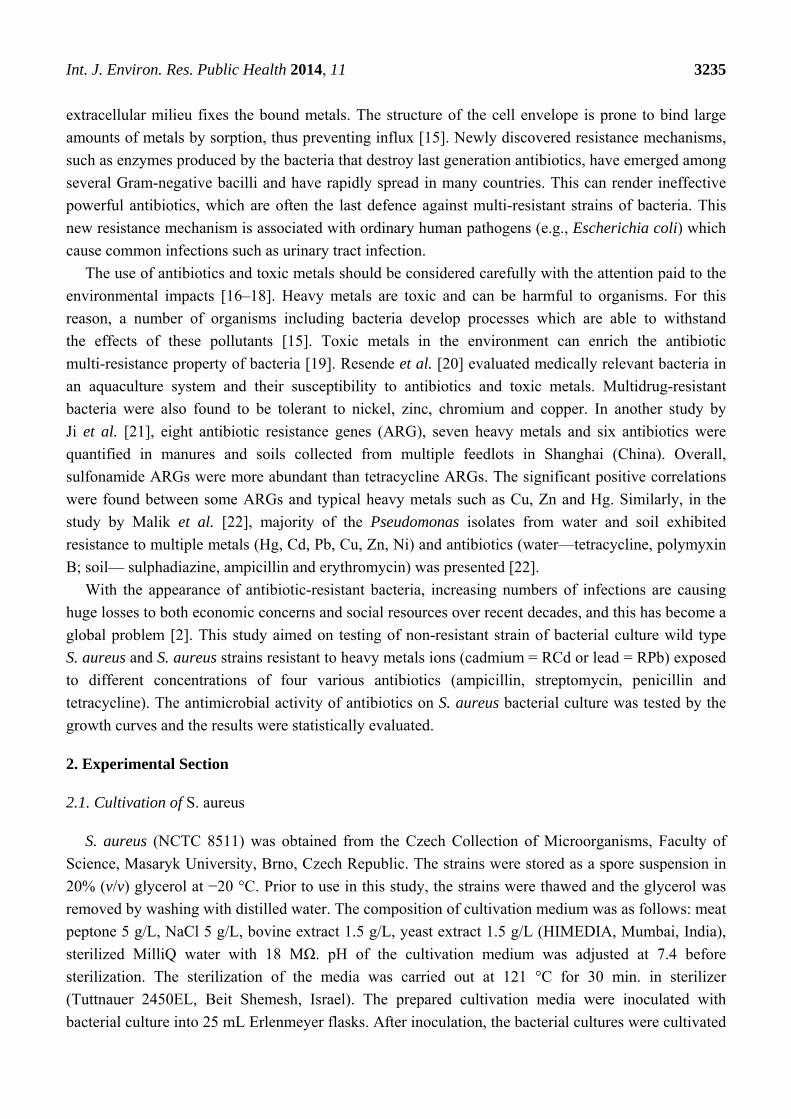
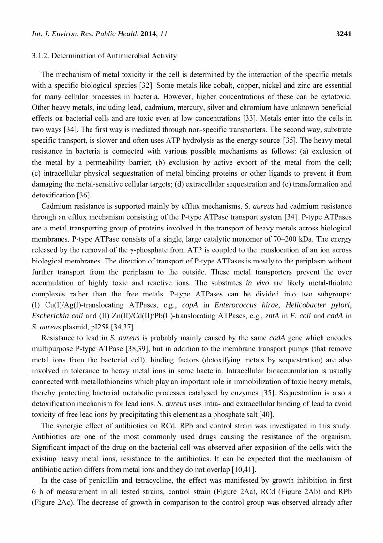
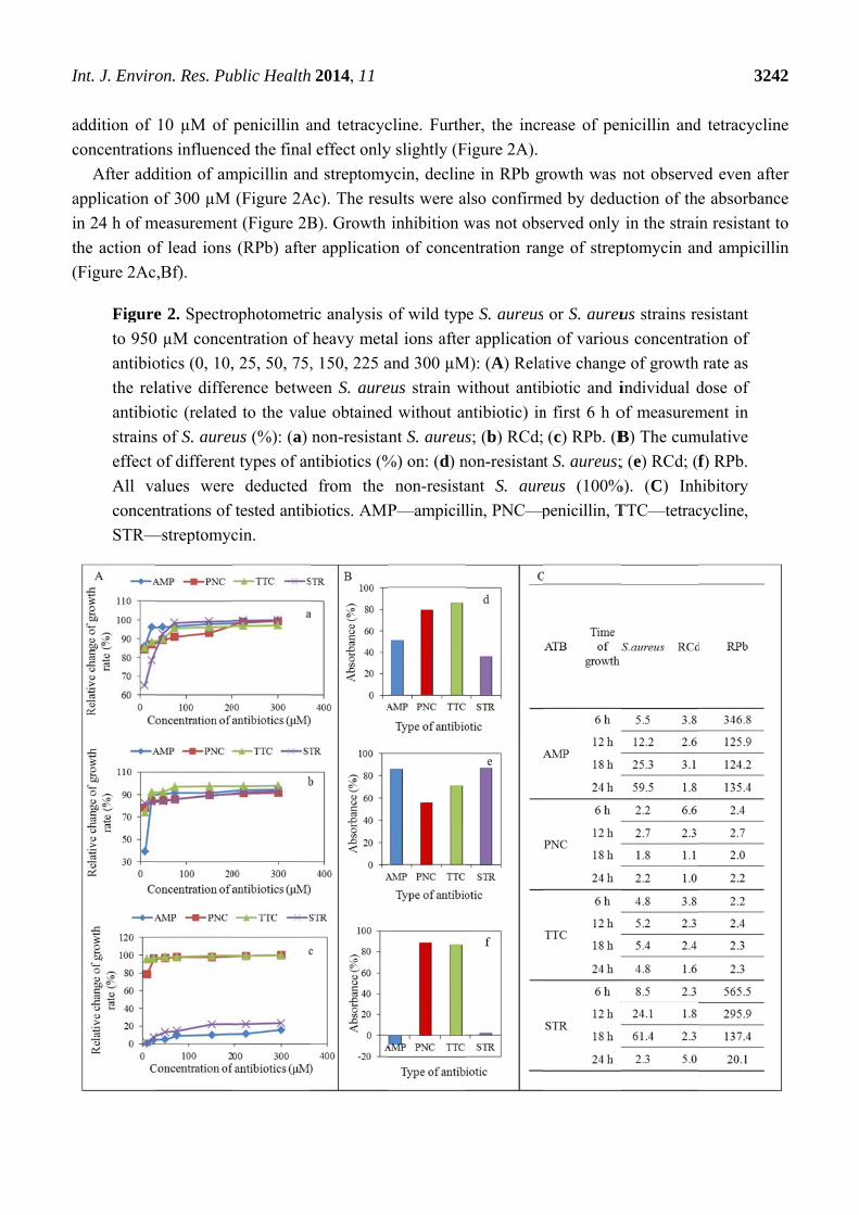
In the case of penicillin and tetracycline, the effect was manifested by growth inhibition in first
6 h of measurement in all tested strains, control strain (Figure 2Aa), RCd (Figure 2Ab) and RPb
(Figure 2Ac). The decrease of growth in comparison to the control group was observed already after
I
a
c
a
in
th
(
Int. J. Enviro
addition of
concentratio
After add
application o
n 24 h of m
he action o
Figure 2Ac
Figure
to 950
antibio
the rel
antibio
strains
effect
All va
concen
STR—
on. Res. Pu
10 µM of
ons influenc
dition of am
of 300 µM
measurement
of lead ions
c,Bf).
e 2. Spectro
0 µM conce
otics (0, 10,
lative differ
otic (related
s of S. aureu
of different
alues were
ntrations of
—streptomyc
ublic Health
penicillin a
ced the final
mpicillin and
(Figure 2A
t (Figure 2B
(RPb) afte
ophotometri
entration of
, 25, 50, 75,
rence betwe
d to the valu
us (%): (a)
t types of an
e deducted
tested antib
cin.
2014, 11
and tetracyc
l effect only
d streptomy
Ac). The res
B). Growth
r applicatio
ic analysis
heavy meta
, 150, 225 a
een S. aure
ue obtained
non-resistan
ntibiotics (%
from the
biotics. AMP
cline. Furth
y slightly (F
ycin, declin
sults were a
inhibition w
on of conce
of wild typ
al ions after
and 300 µM
eus strain w
d without an
ant S. aureus
%) on: (d) n
non-resista
P—ampicill
her, the incr
Figure 2A).
ne in RPb g
also confirm
was not obs
ntration ran
e S. aureus
r applicatio
M): (A) Rela
without antib
ntibiotic) in
s; (b) RCd;
non-resistan
ant S. aur
lin, PNC—p
rease of pen
rowth was
med by dedu
served only
nge of strep
or S. aureu
n of variou
ative change
biotic and i
n first 6 h o
(c) RPb. (B
t S. aureus;
eus (100%
penicillin, T
nicillin and
not observe
uction of the
in the strai
ptomycin an
us strains re
us concentra
e of growth
individual d
of measurem
B) The cum
; (e) RCd; (
%). (C) Inh
TTC—tetrac
324
d tetracyclin
ed even afte
e absorbanc
n resistant t
nd ampicilli
esistant
ation of
rate as
dose of
ment in
mulative
f) RPb.
hibitory
cycline,
42
ne
er
ce
to
in
I
e
v
n
a
w
T
w
e
d
p
e
c
g
a
p
g
a
Int. J. Enviro
The inhib
effects of te
value is exp
non-resistan
all strains te
was determi
The highest
when diame
effect of the
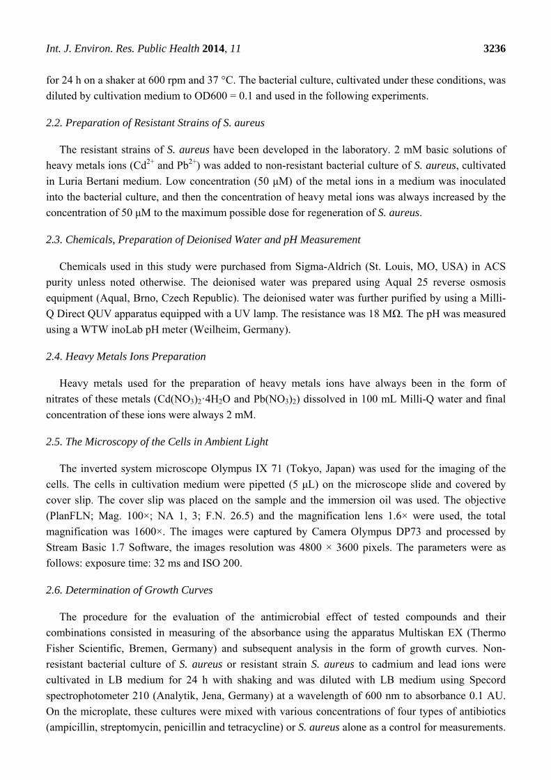
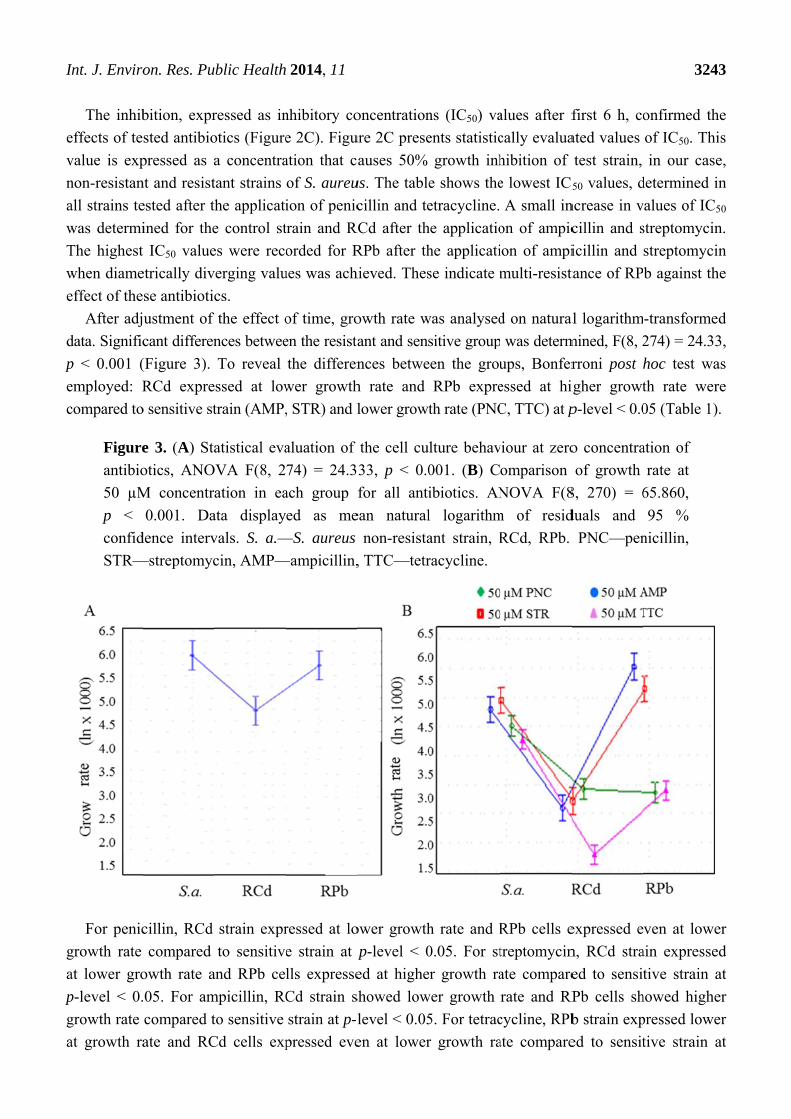
After adj
data. Signific
p < 0.001 (
employed: R
compared to
Figure
antibio
50 µM
p <
confid
STR—
For penic
growth rate
at lower gro
p-level < 0.
growth rate
at growth ra
on. Res. Pu
bition, expr
sted antibio
pressed as a
nt and resist
ested after t
ined for the
t IC50 value
etrically div
se antibiotic
ustment of
cant differen
(Figure 3).
RCd expre
o sensitive st
e 3. (A) Sta
otics, ANO
M concentr
0.001. Da
dence interv
—streptomyc
cillin, RCd
compared
owth rate a
.05. For am
compared t
ate and RC
ublic Health
ressed as in
otics (Figure
a concentra
tant strains
he applicati
e control st
es were reco
verging valu
cs.
the effect o
nces betwee
To reveal
essed at low
train (AMP,
atistical eva
OVA F(8, 2
ation in ea
ata display
vals. S. a.—
cin, AMP—
strain expr
to sensitive
and RPb ce
mpicillin, RC
to sensitive
Cd cells exp
2014, 11
nhibitory co
e 2C). Figu
ation that c
of S. aureu
ion of penic
train and R
orded for R
ues was ach
of time, gro
en the resista
the differen
wer growth
, STR) and l
aluation of
74) = 24.3
ach group
ed as me
—S. aureus
—ampicillin,
ressed at lo
e strain at p
lls expresse
Cd strain s
strain at p-l
pressed eve
oncentration
ure 2C prese
auses 50%
us. The table
cillin and te
RCd after th
RPb after th
hieved. Thes
owth rate w
ant and sens
nces betwe
h rate and
lower growt
the cell cu
33, p < 0.0
for all anti
ean natural
non-resista
, TTC—tetr
ower growth
p-level < 0
ed at highe
showed low
level < 0.05
en at lower
ns (IC50) va
ents statistic
growth inh
e shows the
etracycline.
he applicatio
he applicati
se indicate m
was analysed
sitive group
een the grou
RPb expre
th rate (PNC
lture behav
001. (B) C
ibiotics. AN
l logarithm
ant strain, R
racycline.
h rate and R
0.05. For st
er growth ra
wer growth
5. For tetrac
r growth ra
alues after f
cally evalua
hibition of
e lowest IC5
A small in
on of ampic
ion of ampi
multi-resist
d on natural
was determ
ups, Bonfer
essed at hi
C, TTC) at p
viour at zero
omparison
NOVA F(8
m of resid
RCd, RPb.
RPb cells e
treptomycin
ate compare
rate and RP
cycline, RPb
ate compare
first 6 h, co
ated values
test strain,
50 values, d
ncrease in v
cillin and s
icillin and
tance of RP
al logarithm
mined, F(8, 2
rroni post h
igher growt
p-level < 0.0
o concentra
of growth
8, 270) = 6
duals and
PNC—pen
expressed e
n, RCd stra
ed to sensit
Pb cells sh
b strain exp
ed to sensit
324
onfirmed th
of IC50. Th
in our cas
determined i
values of IC
streptomycin
streptomyci
b against th
m-transforme
274) = 24.3
hoc test wa
th rate wer
05 (Table 1)
ation of
rate at
65.860,
95 %
nicillin,
ven at lowe
ain expresse
tive strain
howed highe
pressed lowe
tive strain
43
he
his
e,
in
C50
n.
in
he
ed
3,
as
re
).
er
ed
at
er
er
at
Int. J. Environ. Res. Public Health 2014, 11 3244
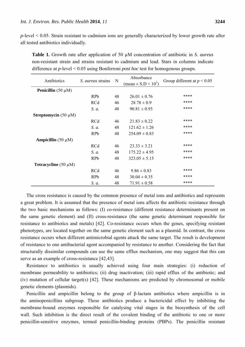
p-level < 0.05. Strain resistant to cadmium ions are generally characterized by lower growth rate after
all tested antibiotics individually.
Table 1. Growth rate after application of 50 µM concentration of antibiotic in S. aureus
non-resistant strain and strains resistant to cadmium and lead. Stars in columns indicate
difference at p-level < 0.05 using Bonferroni post hoc test for homogenous groups.
Antibiotics S. aureus strains N Absorbance
(mean ± S.D × 103)Group different at p < 0.05
Penicillin (50 µM)
RPb 48 26.01 ± 0.76 **** RCd 46 28.78 ± 0.9 **** S. a. 48 90.81 ± 0.93 ****
Streptomycin (50 µM)
RCd 46 21.83 ± 0.22 **** S. a. 48 121.62 ± 1.26 **** RPb 48 254.09 ± 0.83 ****
Ampicillin (50 µM)
RCd 46 23.33 ± 3.21 **** S. a. 48 175.22 ± 4.95 **** RPb 48 323.05 ± 5.15 ****
Tetracycline (50 µM)
RCd 46 9.86 ± 0.83 **** RPb 48 30.04 ± 0.35 **** S. a. 48 71.91 ± 0.58 ****
The cross resistance is caused by the common presence of metal ions and antibiotics and represents
a great problem. It is assumed that the presence of metal ions affects the antibiotic resistance through
the two basic mechanisms as follows: (I) co-resistance (different resistance determinants present on
the same genetic element) and (II) cross-resistance (the same genetic determinant responsible for
resistance to antibiotics and metals) [42]. Co-resistance occurs when the genes, specifying resistant
phenotypes, are located together on the same genetic element such as a plasmid. In contrast, the cross
resistance occurs when different antimicrobial agents attack the same target. The result is development
of resistance to one antibacterial agent accompanied by resistance to another. Considering the fact that
structurally dissimilar compounds can use the same efflux mechanism, one may suggest that this can
serve as an example of cross-resistance [42,43].
Resistance to antibiotics is usually achieved using four main strategies: (i) reduction of
membrane permeability to antibiotics; (ii) drug inactivation; (iii) rapid efflux of the antibiotic; and
(iv) mutation of cellular target(s) [42]. These mechanisms are predicted by chromosomal or mobile
genetic elements (plasmids).
Penicillin and ampicillin belong to the group of β-lactam antibiotics where ampicillin is in
the aminopenicillins subgroup. These antibiotics produce a bactericidal effect by inhibiting the
membrane-bound enzymes responsible for catalysing vital stages in the biosynthesis of the cell
wall. Such inhibition is the direct result of the covalent binding of the antibiotic to one or more
penicillin-sensitive enzymes, termed penicillin-binding proteins (PBPs). The penicillin resistant
Int. J. Environ. Res. Public Health 2014, 11 3245
bacteria produce an extracellular β-lactamase which inactivates antibiotics through hydrolysis of the
β-lactam ring [44,45].
The β-lactamase structural gene (blaZ) is present on Tn552-like transposons [46]. These elements
are located on β-lactamase plasmids which exhibit resistance to other antimicrobial agents, in
particular heavy metal ions. There are four subgroups of these lactamase. The most famous and well
described plasmids types are pI524 (encodes resistance to inorganic ions and organomercurials in
addition to β-lactamase production), pI258 (except metal ion resistance carries the erythromycin
resistance transposon Tn551) [46], and pSK23 which encodes metal ion resistance too. Tn552-like
elements have been found at various sites in naturally occurring plasmids such as pSK4 and pSK1. The
other locations of Tn552-like transposons are chromosomes [45].
Tetracycline inhibits protein synthesis by binding to the 30S ribosomal subunit and preventing
association of aminoacyl-tRNA with its acceptor site [47]. Two mechanisms of tetracycline resistance
have been identified in S. aureus; active efflux via tetA(K) and tetA(L) and ribosomal protection via
tetA(M) [45,47]. The closely related TetA(K) and TetA(L) efflux proteins belong to the major
facilitator superfamily. In most cases, tetracycline efflux in S. aureus strains is mediated by tetA(K),
which is commonly carried by plasmid pT181. This plasmid could be integrated into Type III SCCmec
elements of chromosome and thus chromosomally encoded resistance. Resistance to tetracycline can
also be mediated by mutations that cause increased expression of various chromosomally encoded
efflux pumps, such as Tet38 [45,47].
Streptomycin exhibited resistance due to chromosomal mutations, affecting ribosome affinity [48].
Low-level resistance was usually indicative of small plasmids, such as pS194, which carries
streptomycin adenyltransferase-encoding gene str. Chromosomal segment Tn5405 is responsible for
streptomycin resistance through gene aphC [45,49].
From the presented summary of individual antibiotics resistance mechanisms, it is obvious that
these mechanisms are predicted by the chromosomal and mobile genetic elements (plasmids,
transposons). The above presented results show that the lead resistant S. aureus is resistant to
ampicillin and streptomycin also. On the other hand, the cadmium resistant S. aureus shows no
resistance to ampicillin and streptomycin. When these results were compared to above descripted
mechanisms of antibiotic resistance, it seems that especially the role of transposons would be critical.
The resistance of penicillin and tetracycline is related, among other things, to the transposons. In
contrast, the streptomycin resistance is not connected with transposons. Similar results were published
elsewhere [44]. The linkage of the metal ions (cadmium and lead) and antibiotic resistance is obvious
at β-lactam antibiotics where the creation of co-resistance is offered due to the same location of genes
related to various antimicrobial agents. The genes responsible for metal and antibiotic resistance
located on plasmid pI258 are probably the main reason of cross resistance formation. But the
behaviour of ampicillin is unknown. Its resistance, according to our theory, should not be related with
transposons. But the general theory in literature for β-lactam antibiotics claims the contrary. No
specific study according to the ampicillin resistance in S. aureus has been published yet. Comparison
of the resulted data from different studies of toxic metal tolerance among bacteria is very complicated
because of lack of technical standards in these experimental designs.
Int. J. Environ. Res. Public Health 2014, 11 3246
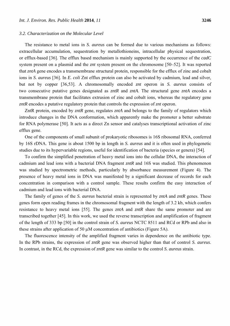
3.2. Characterization on the Molecular Level
The resistance to metal ions in S. aureus can be formed due to various mechanisms as follows:
extracellular accumulation, sequestration by metallothioneins, intracellular physical sequestration,
or efflux-based [36]. The efflux based mechanism is mainly supported by the occurrence of the cadC
system present on a plasmid and the znt system present on the chromosome [50–52]. It was reported
that zntA gene encodes a transmembrane structural protein, responsible for the efflux of zinc and cobalt
ions in S. aureus [36]. In E. coli Znt efflux protein can also be activated by cadmium, lead and silver,
but not by copper [36,53]. A chromosomally encoded znt operon in S. aureus consists of
two consecutive putative genes designated as zntR and zntA. The structural gene zntA encodes a
transmembrane protein that facilitates extrusion of zinc and cobalt ions, whereas the regulatory gene
zntR encodes a putative regulatory protein that controls the expression of znt operon.
ZntR protein, encoded by zntR gene, regulates zntA and belongs to the family of regulators which
introduce changes in the DNA conformation, which apparently make the promoter a better substrate
for RNA polymerase [50]. It acts as a direct Zn sensor and catalyses transcriptional activation of zinc
efflux gene.
One of the components of small subunit of prokaryotic ribosomes is 16S ribosomal RNA, conferred
by 16S rDNA. This gene is about 1500 bp in length in S. aureus and it is often used in phylogenetic
studies due to its hypervariable regions, useful for identification of bacteria (species or genera) [54].
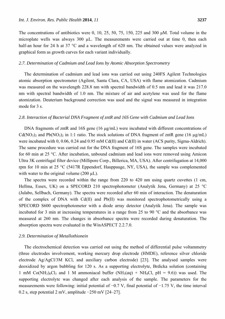
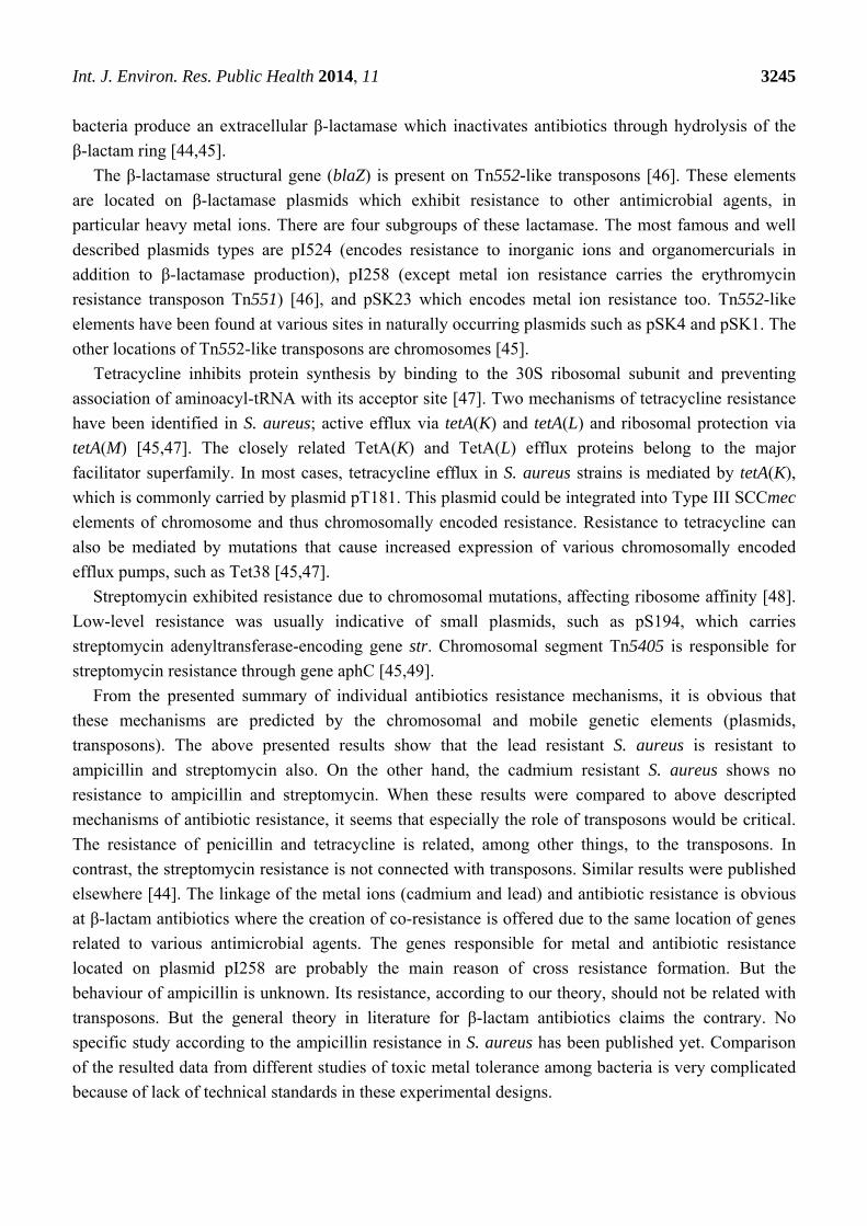
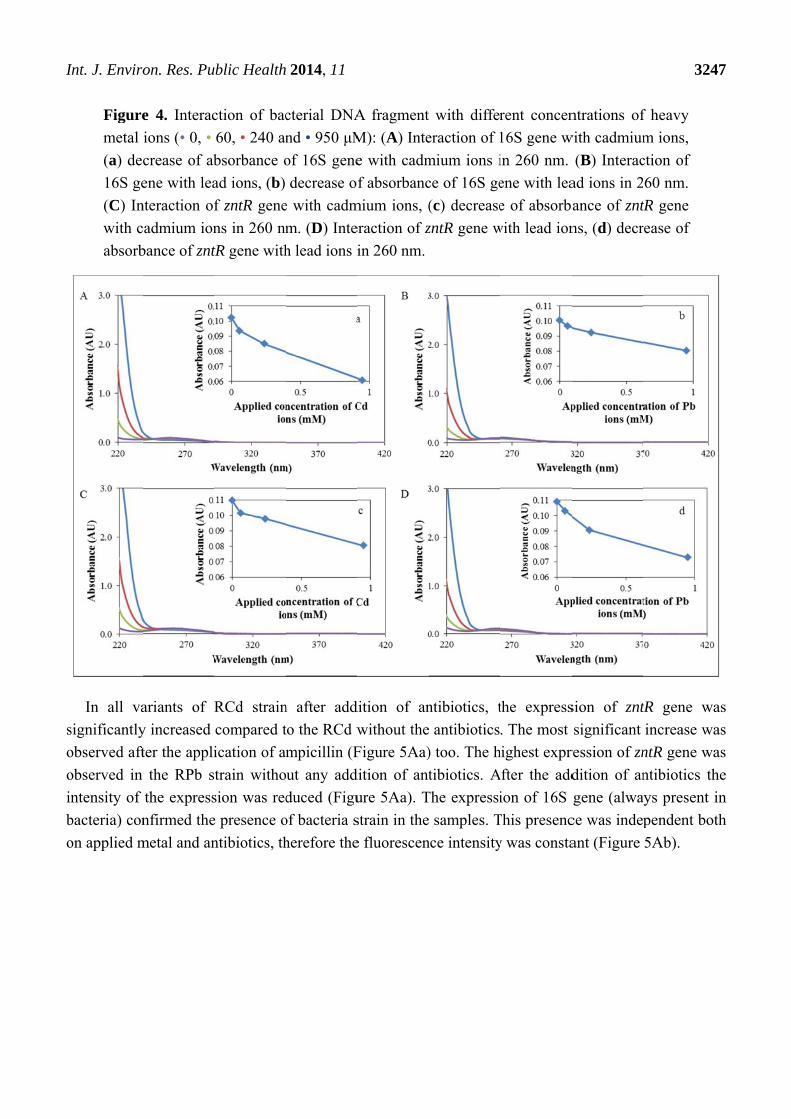
To confirm the simplified penetration of heavy metal ions into the cellular DNA, the interaction of
cadmium and lead ions with a bacterial DNA fragment zntR and 16S was studied. This phenomenon
was studied by spectrometric methods, particularly by absorbance measurement (Figure 4). The
presence of heavy metal ions in DNA was manifested by a significant decrease of records for each
concentration in comparison with a control sample. These results confirm the easy interaction of
cadmium and lead ions with bacterial DNA.
The family of genes of the S. aureus bacterial strain is represented by zntA and zntR genes. These
genes form open reading frames in the chromosomal fragment with the length of 3.2 kb, which confers
resistance to heavy metal ions [55]. The genes zntA and zntR share the same promoter and are
transcribed together [45]. In this work, we used the reverse transcription and amplification of fragment
of the length of 333 bp [50] in the control strain of S. aureus NCTC 8511 and RCd or RPb and also in
these strains after application of 50 µM concentration of antibiotics (Figure 5A).
The fluorescence intensity of the amplified fragment varies in dependence on the antibiotic type.
In the RPb strains, the expression of zntR gene was observed higher than that of control S. aureus.
In contrast, in the RCd, the expression of zntR gene was similar to the control S. aureus strain.
I
s
o
o
in
b
o
Int. J. Enviro
Figure
metal
(a) dec
16S ge
(C) In
with c
absorb
In all va
significantly
observed aft
observed in
ntensity of
bacteria) con
on applied m
on. Res. Pu
e 4. Interac
ions (• 0, •
crease of ab
ene with lea
nteraction of
cadmium ion
bance of znt
ariants of R
y increased
ter the appli
the RPb s
the express
nfirmed the
metal and an
ublic Health
ction of bac
60, • 240 an
bsorbance o
ad ions, (b)
f zntR gene
ns in 260 nm
tR gene with
RCd strain
compared t
ication of am
train witho
sion was red
e presence o
ntibiotics, th
2014, 11
cterial DNA
nd • 950 μM
of 16S gene
decrease of
e with cadm
m. (D) Inte
h lead ions i
n after add
to the RCd w
mpicillin (F
ut any add
duced (Figu
of bacteria s
herefore the
A fragment
M): (A) Inte
e with cadm
f absorbanc
mium ions, (
eraction of z
in 260 nm.
dition of an
without the
Figure 5Aa)
dition of ant
ure 5Aa). T
strain in the
e fluorescen
with differ
eraction of 1
mium ions i
ce of 16S ge
(c) decrease
zntR gene w
ntibiotics, t
antibiotics
) too. The h
tibiotics. A
The expressi
samples. T
nce intensity
rent concen
16S gene w
in 260 nm.
ene with lea
e of absorba
with lead ion
the express
. The most
highest expr
After the add
ion of 16S
This presenc
y was consta
ntrations of
with cadmium
(B) Interac
ad ions in 2
ance of znt
ns, (d) decr
sion of znt
significant
ression of zn
dition of an
gene (alwa
ce was indep
ant (Figure
324
f heavy
m ions,
ction of
60 nm.
tR gene
rease of
tR gene wa
increase wa
ntR gene wa
ntibiotics th
ys present i
pendent bot
5Ab).
47
as
as
as
he
in
th
I
Int. J. Enviro
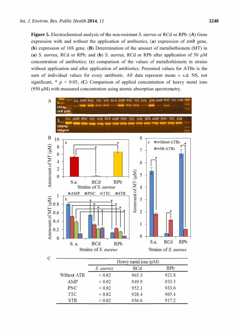
Figure
expres
(b) ex
(a) S.
concen
withou
sum o
signifi
(950 µ
on. Res. Pu
e 5. Electro
ssion with a
xpression of
aureus, RC
ntration of
ut applicatio
of individua
icant, * p <
µM) with m
ublic Health
ochemical an
and withou
f 16S gene.
Cd or RPb;
antibiotics;
on and afte
al values fo
< 0.05, (C)
easured con
2014, 11
nalysis of th
ut the applic
(B) Determ
; and (b) S
; (c) compa
er applicatio
or every an
) Comparis
ncentration
he non-resis
cation of a
mination of
S. aureus, R
arison of th
on of antibi
ntibiotic. A
son of appl
using atomi
stant S. aure
antibiotics, (
f the amoun
RCd or RPb
he values o
iotics. Prese
All data rep
lied concen
ic absorptio
eus or RCd
(a) express
nt of metallo
b after appl
of metalloth
ented value
present mea
ntration of
on spectrom
or RPb: (A
sion of zntR
othionein (M
lication of
thionein in
es for ATBs
an ± s.d. N
heavy met
metry.
324
A) Gene
R gene,
MT) in
50 µM
strains
s is the
NS, not
al ions
48
Int. J. Environ. Res. Public Health 2014, 11 3249
Bacterial genome contains genes coding different proteins providing the tolerance to the heavy
metal: metallo-regulatory genes [56]. These genes are usually from the family ArsR/SmtB. The
sequences of these genes are usually homologous between the members of this family, but may differ
in their active sites for binding metal [57]. Based on limited sequence homology, SmtB appears to be a
member of a family of bacterial metalloregulatory proteins, including the ArsR proteins that are
repressors of the arsenic-resistance operons and the CadC proteins which are the cadmium responsive
repressors of expression of the cadmium efflux ATPase [57]. The SmtB, ArsR and CadC proteins
contain conserved cysteine residues associated with the N-terminal extremity of putative DNA-binding
helix-turn-helix motifs [58]. It has been predicted that these cysteine residues bind to metals
via formation of metal-thiolate bonds and thus inhibit binding via the adjacent helix-turn-helix
region [57,58]. Unlike ArsR/SmtB metalloregulatory proteins, above mentioned ZntR does not contain
any cysteine residue and lacks the characteristic metal-binding elements [50]. However, it has two
histidine-rich regions, one at the C-terminus and the other near the N-terminus. Similar histidine-rich
regions have been reported for zinc and cobalt transporters which are thought to be domains for
zinc-binding ions [36].
Metallothioneins (MTs) are known as proteins containing thiol groups in their structure, especially
cysteine, which is the cause of affinity to metals—such as Cd, Pb, Hg, Cu or Zn [59–61]. The
metallothionein gene, smtA, is controlled by the SmtB repressor [62], which also regulates a
zinc-transporting P-type ATPase [56,57]. The primary function of MT is detoxification of heavy
metals in living organisms, which became the subject of a number of studies [63–65]. Occurrence of
metallothionein was observed in a variety of organisms and microorganisms such as bacteria,
invertebrates or vertebrates [60,66–69].
In this study the presence of metallothionein in strains of S. aureus with resistance to the effects of
heavy metals was determined. Much higher concentration of metallothionein (more than 6 µM) in
RPb in comparison to the control has been reported and the other resistant strain RCd, where the
concentration of metallothionein was much lower in comparison to the control S. aureus, values
around 0.5 µM (Figure 5Ba). These results show possible variation in the metal resistance origin for
cadmium and lead. While cadmium is mainly removed through the CadA transport system, lead is
mainly removed after bounding to metallothioneins. CadA transport system was proofed as lead and
cadmium transporter too [39]. Probably all the zinc/cadmium translocating P-type ATPases are also
effective in lead ions export [39]. After the application of antibiotics the values of determined
metallothionein in both resistant strains decreased, especially in RPb (Figure 5Bb). Only the
application of streptomycin caused the growth of MT level in RCd compared to the control (S. aureus).
Here, the presented results confirm the trends which are obvious in the evaluation of spectrophotometric
analysis (Section 3.1.2.) and suggest that the mechanisms of cross resistance at ampicillin and
streptomycin will be different because of various MT level in RCd. As it was mentioned above
(Section 3.1.2.) the ampicillin and streptomycin caused cross resistance in RPb and here it is the
confirmation of such deduction in the lowest MT levels for these combinations. It is necessary to
remember that the presented results are related to the MT level inside of cells, therefore the higher
change of MT level indicates the highest removal of complex MT-metal ions. The summarized view
(Figure 5Bc) then shows a significant effect of antibiotic application, especially on RPb where the
decrease of metallothionein is greatest. On the other hand, the application of the antibiotic caused an
Int. J. Environ. Res. Public Health 2014, 11 3250
increase in the amount of metallothionein in RCd (Figure 5Bc). These results confirm the results
obtained from the expression of zntR gene (Figure 5A) and thus confirm further the direct relationship
between zntR gene and metallothionein expression. The great question is about mechanism of this
relationship because till now no explanation was published about the influence of zntR gene expression
on metallothionein level. Because of the fact that metallothionein production is closely connected with
zinc level (zinc-sensing transcriptional repressor SmtB) [62], and zntR gene expression too [36], and
both genes are chromosomally located, we only assumed the creation of similar effect which was
called in the cross resistance formation as co-resistance. The second possibility, and it is important to
highlight that it is only in hypothesis, is the reaction of zntR receptor for the presence of other metal
ions (lead and cadmium) but this assumption was not proofed elsewhere.
The content of heavy metals in the samples was determined by atomic absorption spectrometry
measurements. The determined content of metal ions is presented in Figure 5C. The concentrations of
cadmium and lead ions (950 µM) in both samples were detected in almost total applied concentration
(940 µM). The same concentration was measured in samples after the application of antibiotics.
In control strain, the concentration of metals was below the detection limit (Figure 5C).
The influence of metallothioneins in the metal resistance was tested in some bacteria strains [70].
It must be said that the lead and cadmium resistance (in combination with antibiotics) in point of view
of various resistance mechanisms (efflux, metallothioneins) are very seldom tested and there is no
integrated output model of S. aureus resistance to these agents.
4. Conclusions
The reported experiments were performed to study the effects of antibiotic drugs (ampicillin,
streptomycin, penicillin and tetracycline) on non-resistant strains of S. aureus and S. aureus strains
resistant to the effects of heavy metal ions (cadmium or lead). Our results pointed to a significant
antimicrobial effect of penicillin and tetracycline in both the control strain and the strains resistant to
heavy metal ions. Microscopic methods only confirmed the morphological changes of resistant strains
in comparison with the control, independently of the application of the tested antibiotic drug. On the
other hand, the lead resistant S. aureus strain showed resistance to the effect of ampicillin and
streptomycin. Cross resistance was thus observed only in RPb after the application of these two
antibiotics. The obtained results can be used for further experiments with bacterial strains in terms of a
deeper understanding of bacterial resistance caused by environmental factors.
Conflicts of Interest
The authors declare no conflict of interest.
Authors Contributions
Dagmar Chudobova cultivated strains resistant to heavy metal ions with antibiotics and prepared
samples for other analysis. Simona Dostalova participated on the data from molecular biology
including isolation, amplification of genes. Iva Blazkova observed cells by microscopy in ambient
light. Petr Michalek participated on expression of genes. Branislav Ruttkay-Nedecky participated on
Int. J. Environ. Res. Public Health 2014, 11 3251
the preparation of the manuscript and in the design of spectrophotometric experiment. Matej Sklenar
participated on the data about determination of antimicrobial activity. Lukas Nejdl performed the
analysis of the interaction of bacterial DNA fragment of zntR gene with heavy metal ions. Jiri Kudr
performed the analysis of the interaction of bacterial DNA fragment of 16S gene with heavy metal
ions. Jaromir Gumulec made the statistical analysis. Katerina Tmejova participated on determination
of metallothionein. Marie Konecna performed the analysis of the data and participated in preparation
of the manuscript. Marketa Vaculovicova treated molecular-biological data and participated in the
preparation of the manuscript. David Hynek treated electrochemical data and participated in the
preparation of the manuscript. Michal Masarik participated in preparation of the manuscript and in the
design of microbiological experiment. Jindrich Kynicky participated in preparation of the manuscript
and optimized the samples preparation. Rene Kizek participated in design and coordination of the
study. Vojtech Adam conceived of the study, and participated in its design and drafted manuscript.
Acknowledgments
Financial support by VSKE project, NanoCeva TA CR TA01010088 and SIX CZ.1.05/2.1.00/03.0072
is highly acknowledged.
References
1. Levy, S.B.; Marshall, B. Antibacterial resistance worldwide: Causes, challenges and responses.
Nat. Med. 2004, 10, S122–S129.
2. Zhou, F.; Wang, Y. Characteristics of antibiotic resistance of airborne Staphylococcus isolated
from metro stations. Int. J. Environ. Res. Public Health 2013, 10, 2412–2426.
3. Gatica, J.; Cytryn, E. Impact of treated wastewater irrigation on antibiotic resistance in the soil
microbiome. Environ. Sci. Pollut. Res. 2013, 20, 3529–3538.
4. Stone, N.D.; Lewis, D.R.; Lowery, H.K.; Darrow, L.A.; Kroll, C.M.; Gaynes, R.P.; Jernigan, J.A.;
McGowan, J.E.; Tenover, F.C.; Richards, C.L. Importance of bacterial burden among
methicillin-resistant Staphylococcus aureus carriers in a long-term care facility. Infect. Control
Hosp. Epidemiol. 2008, 29, 143–148.
5. Bastug, A.; Yilmaz, G.R.; Kayaaslan, B.; Akinci, E.; Bodur, H. Risk factors for mortality
in patients with nosocomial Staphylococcus aureus bacteremia. Turk. J. Med. Sci. 2012, 42,
1222–1229.
6. Tacconelli, E.; Pop-Vicas, A.E.; D’Agata, E.M.C. Increased mortality among elderly patients with
meticillin-resistant Staphylococcus aureus bacteraemia. J. Hosp. Infect. 2006, 64, 251–256.
7. Cosgrove, S.E.; Qi, Y.L.; Kaye, K.S.; Harbarth, S.; Karchmer, A.W.; Carmeli, Y. The impact of
methicillin-resistance in Staphylococcus aureus bacteremia on patient outcomes: Mortality, length
of stay, and hospital charges. Infect. Control Hosp. Epidemiol. 2005, 26, 166–174.
8. Ammerlaan, H.S.M.; Harbarth, S.; Buiting, A.G.M.; Crook, D.W.; Fitzpatrick, F.; Hanberger, H.;
Herwaldt, L.A.; van Keulen, P.H.J.; Kluytmans, J.; Kola, A.; et al. Secular trends in nosocomial
bloodstream infections: Antibiotic-resistant bacteria increase the total burden of infection.
Clin. Infect. Dis. 2013, 56, 798–805.
Int. J. Environ. Res. Public Health 2014, 11 3252
9. Ye, Y.; Li, S.L.; Li, Y.J.; Ren, T.S.; Liu, K.G. Mycoplasma pneumoniae 23S rRNA Gene
Mutations and Mechanisms of Macrolide Resistance. Labmedicine 2013, 44, 63–68.
10. Aktan, Y.; Tan, S.; Icgen, B. Characterization of lead-resistant river isolate Enterococcus faecalis
and assessment of its multiple metal and antibiotic resistance. Environ. Monit. Assess. 2013, 185,
5285–5293.
11. Hellweger, F.L. Simple model of tetracycline antibiotic resistance in aquatic environment:
Accounting for metal coselection. J. Environ. Eng.-ASCE 2013, 139, 913–921.
12. My, N.H.; Hirao, H.; Van, D.U.; Morokuma, K. Computational studies of bacterial resistance to
beta-lactam antibiotics: Mechanism of covalent inhibition of the penicillin-binding protein 2a
(PBP2a). J. Chem. Inf. Model. 2011, 51, 3226–3234.
13. Yuan, W.C.; Hu, Q.W.; Cheng, H.; Shang, W.L.; Liu, N.; Hua, Z.Y.; Zhu, J.M.; Hu, Z.;
Yuan, J.Z.; Zhang, X.; et al. Cell wall thickening is associated with adaptive resistance to
amikacin in methicillin-resistant Staphylococcus aureus clinical isolates. J. Antimicrob. Chemother.
2013, 68, 1089–1096.
14. Anaya-Lopez, J.L.; Lopez-Meza, J.E.; Ochoa-Zarzosa, A. Bacterial resistance to cationic
antimicrobial peptides. Crit. Rev. Microbiol. 2013, 39, 180–195.
15. Majzlik, P.; Strasky, A.; Adam, V.; Nemec, M.; Trnkova, L.; Zehnalek, J.; Hubalek, J.; Provaznik, I.;
Kizek, R. Influence of zinc(II) and copper(II) ions on Streptomyces bacteria revealed by
electrochemistry. Int. J. Electrochem. Sci. 2011, 6, 2171–2191.
16. Sobrova, P.; Zehnalek, J.; Adam, V.; Beklova, M.; Kizek, R. The effects on soil/water/plant/animal
systems by platinum group elements. Cent. Eur. J. Chem. 2012, 10, 1369–1382.
17. Krizkova, S.; Huska, D.; Beklova, M.; Hubalek, J.; Adam, V.; Trnkova, L.; Kizek, R.
Protein-based electrochemical biosensor for detection of silver(I) ions. Environ. Toxicol. Chem.
2010, 29, 492–496.
18. Pereira, P.M.; Filipe, S.R.; Tomasz, A.; Pinho, M.G. Fluorescence ratio imaging microscopy
shows decreased access of vancomycin to cell wall synthetic sites in vancomycin-resistant
Staphylococcus aureus. Antimicrob. Agents Chemother. 2007, 51, 3627–3633.
19. Summers, A.O. Genetic linkage and horizontal gene transfer, the roots of the antibiotic
multi-resistance problem. Anim. Biotechnol. 2006, 17, 125–135.
20. Resende, J.A.; Silva, V.L.; Fontes, C.O.; Souza, J.A.; de Oliveira, T.L.R.; Coelho, C.M.;
Cesar, D.E.; Diniz, C.G. Multidrug-resistance and toxic metal tolerance of medically important
bacteria isolated from an aquaculture system. Microbes Environ. 2012, 27, 449–455.
21. Ji, X.L.; Shen, Q.H.; Liu, F.; Ma, J.; Xu, G.; Wang, Y.L.; Wu, M.H. Antibiotic resistance gene
abundances associated with antibiotics and heavy metals in animal manures and agricultural soils
adjacent to feedlots in Shanghai, China. J. Hazard. Mater. 2012, 235, 178–185.
22. Malik, A.; Aleem, A. Incidence of metal and antibiotic resistance in Pseudomonas spp. from the
river water, agricultural soil irrigated with wastewater and groundwater. Environ. Monit. Assess.
2011, 178, 293–308.
23. Adam, V.; Fabrik, I.; Kohoutkova, V.; Babula, P.; Hubalek, J.; Vrba, R.; Trnkova, L.; Kizek, R.
Automated electrochemical analyzer as a new tool for detection of thiols. Int. J. Electrochem. Sci.
2010, 5, 429–447.
Int. J. Environ. Res. Public Health 2014, 11 3253
24. Adam, V.; Baloun, J.; Fabrik, I.; Trnkova, L.; Kizek, R. An electrochemical detection of
metallothioneins at the zeptomole level in nanolitre volumes. Sensors 2008, 8, 2293–2305.
25. Adam, V.; Blastik, O.; Krizkova, S.; Lubal, P.; Kukacka, J.; Prusa, R.; Kizek, R. Application of
the Brdicka reaction in determination of metallothionein in patients with tumours. Chem. Listy
2008, 102, 51–58.
26. Maret, W. Fluorescent probes for the structure and function of metallothionein. J. Chromatogr. B
2009, 877, 3378–3383.
27. Sobrova, P.; Vyslouzilova, L.; Stepankova, O.; Ryvolova, M.; Anyz, J.; Trnkova, L.; Adam, V.;
Hubalek, J.; Kizek, R. Tissue specific electrochemical fingerprinting. PLoS One 2012, 7, e49654,
doi:10.1371/journal.pone.0049654.
28. Brissonnoel, A.; Trieucuot, P.; Courvalin, P. Mechanisms of action of spiramycin and other
macrolides. J. Antimicrob. Chemother. 1988, 22, 13–23.
29. Jenkins, R.; Burton, N.; Cooper, R. Manuka honey inhibits cell division in methicillin-resistant
Staphylococcus aureus. J. Antimicrob. Chemother. 2011, 66, 2536–2542.
30. Belley, A.; Harris, R.; Beveridge, T.; Parr, T.; Moeck, G. Ultrastructural effects of oritavancin
on methicillin-resistant Staphylococcus aureus and vancomycin-resistant Enterococcus.
Antimicrob. Agents Chemother. 2009, 53, 800–804.
31. Webster, D.; Rennie, R.P.; Brosnikoff, C.L.; Chui, L.; Brown, C. Methicillin-resistant
Staphylococcus aureus with reduced susceptibility to vancomycin in Canada. Diagn. Microbiol.
Infect. Dis. 2007, 57, 177–181.
32. Rouch, D.A.; Lee, B.T.O.; Morby, A.P. Understanding cellular-responses to toxic agents—A
model for mechanism-choice in bacterial metal resistance. J. Ind. Microbiol. 1995, 14, 132–141.
33. Trajanovska, S.; Britz, M.L.; Bhave, M. Detection of heavy metal ion resistance genes in
Gram-positive and Gram-negative bacteria isolated from a lead-contaminated site. Biodegradation
1997, 8, 113–124.
34. Nies, D.H.; Silver, S. Ion efflux systems involved in bacterial metal resistances. J. Ind. Microbiol.
1995, 14, 186–199.
35. Nies, D.H. Microbial heavy-metal resistance. Appl. Microbiol. Biotechnol. 1999, 51, 730–750.
36. Choudhury, R.; Srivastava, S. Zinc resistance mechanisms in bacteria. Curr. Sci. 2001, 81,
768–775.
37. Naik, M.M.; Dubey, S.K.; Khanolkar, D.; D’Costa, B. P-type ATPase and MdrL efflux pump-
mediated lead and multi-drug resistence in estuarine bacterial isolates. Curr. Sci., 2013, 105,
1366–1372.
38. Blaszak, M.; Bienkowska, D. Effect of soil pollution on bacterial resistance to lead ions:
An experimental approach. Pol. J. Ecol. 2009, 57, 555–560.
39. Rensing, C.; Sun, Y.; Mitra, B.; Rosen, B.P. Pb(II)-translocating P-type ATPases. J. Biol. Chem.
1998, 273, 32614–32617.
40. Levinson, H.S.; Mahler, I.; Blackwelder, P.; Hood, T. Lead resistance and sensitivity in
Staphylococcus aureus. FEMS Microbiol. Lett. 1996, 145, 421–425.
41. Deb, S.; Ahmed, S.F.; Basu, M. Metal accumulation in cell wall: A possible mechanism of
cadmium resistance by Pseudomonas stutzeri. Bull. Environ. Contam. Toxicol. 2013, 90,
323–328.
Int. J. Environ. Res. Public Health 2014, 11 3254
42. Baker-Austin, C.; Wright, M.S.; Stepanauskas, R.; McArthur, J.V. Co-selection of antibiotic and
metal resistance. Trends Microbiol. 2006, 14, 176–182.
43. Chapman, J.S. Disinfectant resistance mechanisms, cross-resistance, and co-resistance.
Int. Biodeterior. Biodegrad. 2003, 51, 271–276.
44. Jensen, S.O.; Kwong, S.M.; Lyon, B.R.; Firth, N. Evolution of multiple drug resistance in
Staphylococci. Microbiol. Aust. 2008, 29, 120–123.
45. Jensen, S.O.; Lyon, B.R. Genetics of antimicrobial resistance in Staphylococcus aureus.
Future Microbiol. 2009, 4, 565–582.
46. Novick, R.P.; Roth, C. Plasmid-linked resistance to inorganic salts in Staphylococcus aureus.
J. Bacteriol. 1968, 95, 1335–1342.
47. Chopra, I.; Roberts, M. Tetracycline antibiotics: Mode of action, applications, molecular biology,
and epidemiology of bacterial resistance. Microbiol. Mol. Biol. Rev. 2001, 65, 232–260.
48. Lacey, R.W.; Chopra, I. Evidence for mutation to streptomycin resistance in clinical strains of
Staphylococcus aureus. J. Gen. Microbiol. 1972, 73, 175–180.
49. Derbise, A.; de Cespedes, G.; el Solh, N. Nucleotide sequence of the Staphylococcus aureus
transposon, Tn5405, carrying aminoglycosides resistance genes. J. Basic Microbiol. 1997, 37,
379–384.
50. Singh, V.K.; Xiong, A.M.; Usgaard, T.R.; Chakrabarti, S.; Deora, R.; Misra, T.K.; Jayaswal, R.K.
ZntR is an autoregulatory protein and negatively regulates the chromosomal zinc resistance
operon znt of Staphylococcus aureus. Mol. Microbiol. 1999, 33, 200–207.
51. Yoon, K.P.; Misra, T.K.; Silver, S. Regulation of the cadA cadmium resistance determinant of
Staphylococcus aureus plasmid PI258. J. Bacteriol. 1991, 173, 7643–7649.
52. Yoon, K.P.; Silver, S. A 2nd gene in the Staphylococcus aureus cadA cadmium resistance
determinant of plasmid-PI258. J. Bacteriol. 1991, 173, 7636–7642.
53. Okkeri, J.; Haltia, T. Expression and mutagenesis of ZntA, a zinc-transporting P-type ATPase
from Escherichia coli. Biochemistry 1999, 38, 14109–14116.
54. Stomeo, F.; Valverde, A.; Pointing, S.B.; McKay, C.P.; Warren-Rhodes, K.A.; Tuffin, M.I.;
Seely, M.; Cowan, D.A. Hypolithic and soil microbial community assembly along an aridity
gradient in the Namib Desert. Extremophiles 2013, 17, 329–337.
55. Xiong, A.M.; Jayaswal, R.K. Molecular characterization of a chromosomal determinant
conferring resistance to zinc and cobalt ions in Staphylococcus aureus. J. Bacteriol. 1998, 180,
4024–4029.
56. Thelwell, C.; Robinson, N.J.; Turner-Cavet, J.S. An SmtB-like repressor from Synechocystis PCC
6803 regulates a zinc exporter. Proc. Natl. Acad. Sci. USA 1998, 95, 10728–10733.
57. Cook, W.J.; Kar, S.R.; Taylor, K.B.; Hall, L.M. Crystal structure of the cyanobacterial
metallothionein repressor SmtB: A model for metalloregulatory proteins. J. Mol. Biol. 1998, 275,
337–346.
58. Shi, W.P.; Dong, J.; Scott, R.A.; Ksenzenko, M.Y.; Rosen, B.P. The role of arsenic thiol
interactions in metalloregulation of the ars operon. J. Biol. Chem. 1996, 271, 9291–9297.
59. Prusa, R.; Kizek, R.; Trnkova, L.; Vacek, J.; Zehnalek, J. Study of relationship between
metallothionein and heavy metals by CPSA method. Clin. Chem. 2004, 50, A28–A29.
Int. J. Environ. Res. Public Health 2014, 11 3255
60. Vasak, M. Advances in metallothionein structure and functions. J. Trace Elem. Med. Biol. 2005,
19, 13–17.
61. Skalickova, S.; Zitka, O.; Nejdl, L.; Krizkova, S.; Sochor, J.; Janu, L.; Ryvolova, M.; Hynek, D.;
Zidkova, J.; Zidek, V.; et al. Study of interaction between metallothionein and CdTe quantum
dots. Chromatographia 2013, 76, 345–353.
62. Huckle, J.W.; Morby, A.P.; Turner, J.S.; Robinson, N.J. Isolation of a prokaryotic metallothionein
locus and analysis of transcriptional control by trace-metal ions. Mol. Microbiol. 1993, 7, 177–187.
63. Klaassen, C.D.; Liu, J.; Diwan, B.A. Metallothionein protection of cadmium toxicity.
Toxicol. Appl. Pharmacol. 2009, 238, 215–220.
64. Templeton, D.M.; Cherian, M.G. Toxicological significance of metallothionein. Method Enzymol.
1991, 205, 11–24.
65. Sochor, J.; Pohanka, M.; Ruttkay-Nedecky, B.; Zitka, O.; Hynek, D.; Mares, P.; Zeman, L.;
Adam, V.; Kizek, R. Effect of selenium in organic and inorganic form on liver, kidney, brain and
muscle of Wistar rats. Cent. Eur. J. Chem. 2012, 10, 1442–1451.
66. Coyle, P.; Philcox, J.C.; Carey, L.C.; Rofe, A.M. Metallothionein: The multipurpose protein.
Cell. Mol. Life Sci. 2002, 59, 627–647.
67. Henkel, G.; Krebs, B. Metallothioneins: Zinc, cadmium, mercury, and copper thiolates and
selenolates mimicking protein active site features—Structural aspects and biological implications.
Chem. Rev. 2004, 104, 801–824.
68. Adam, V.; Fabrik, I.; Eckschlager, T.; Stiborova, M.; Trnkova, L.; Kizek, R. Vertebrate
metallothioneins as target molecules for analytical techniques. TRAC-Trends Anal. Chem. 2010,
29, 409–418.
69. Krizkova, S.; Ryvolova, M.; Hrabeta, J.; Adam, V.; Stiborova, M.; Eckschlager, T.; Kizek, R.
Metallothioneins and zinc in cancer diagnosis and therapy. Drug Metab. Rev. 2012, 44, 287–301.
70. Naik, M.M.; Shamim, K.; Dubey, S.K. Biological characterization of lead-resistant bacteria to
explore role of bacterial metallothionein in lead resistance. Curr. Sci. 2012, 103, 426–429.
© 2014 by the authors; licensee MDPI, Basel, Switzerland. This article is an open access article
distributed under the terms and conditions of the Creative Commons Attribution license
(http://creativecommons.org/licenses/by/3.0/).