Embed Size (px)
Citation preview
Grand Valley State UniversityScholarWorks@GVSU
Honors Projects Undergraduate Research and Creative Practice
2012
Effect of Adopting Proper Running FormTechniques on Hip Strength in Healthy FemalesPatrick J. LawrenceGrand Valley State University
Heather Gulgin Ph.D.Grand Valley State University
Jason Burgess
Follow this and additional works at: http://scholarworks.gvsu.edu/honorsprojects
This Open Access is brought to you for free and open access by the Undergraduate Research and Creative Practice at ScholarWorks@GVSU. It hasbeen accepted for inclusion in Honors Projects by an authorized administrator of ScholarWorks@GVSU. For more information, please [email protected].
Recommended CitationLawrence, Patrick J.; Gulgin, Heather Ph.D.; and Burgess, Jason, "Effect of Adopting Proper Running Form Techniques on HipStrength in Healthy Females" (2012). Honors Projects. 133.http://scholarworks.gvsu.edu/honorsprojects/133

Running head: RUNNING FORM & HIP STRENGTH
Effect of Adopting Proper Running Form Techniques on Hip Strength in
Healthy Females
Patrick J. Lawrence; Heather Gulgin, Ph.D.; Jason Burgess
Grand Valley State University

RUNNING FORM & HIP STRENGTH 2
ABSTRACT
The purpose of this study is to determine if instruction and practice in “proper” running form
techniques strengthens the hip abductor and hip external rotator muscles and thereby reduce the
risk of certain knee injuries such as patellofemoral pain syndrome and iliotibial band syndrome.
Four healthy, college-aged female recreational runners completed this study. Subjects were
randomly placed into a control and experimental group. Both groups ran within a controlled
range of 12-16 miles per week on a treadmill for six weeks, and were measured for hip strength
at the first week, third week, and sixth week of the running protocol. Isometric hip abduction and
hip external rotation strengths were measured with a hand-held dynamometer. The experimental
group received 3-sessions of proper running form instruction. Six separate two-way ANOVA
tests were performed to identify changes in hip abductor and hip external rotator strength over
time and intervention. Due to the small sample size, no statistically significant results were
found, but there was an observed trend in increased hip abduction strength and increased hip
muscle strength symmetry in the experimental group. This suggests a need for future studies with
a larger sample size.

RUNNING FORM & HIP STRENGTH 3
INTRODUCTION
Running, as it continues to grow as a popular exercise mode, also grows as a topic of
interest among researchers and physical rehabilitators. It is predicted that over 50% of
recreational runners sustain a running-related injury per year, with half of those injuries
occurring at the knee (van Gent et al., 2007; Taunton et al., 2002). Consequently, the large
volume of injured persons per year indicates a need for research in determining the etiology of
these injuries and methods to prevent and rehabilitate from them.
Of the many running-related knee injuries, the two most prevalent are patellofemoral pain
syndrome (PFP) and iliotibial band syndrome (ITBS). Recent research has shown strong
evidence correlating hip muscular weakness and incidence of PFP and ITBS, among other knee
injuries (Ferber et al., 2011; Fredericson et al., 2000; Ireland et al., 2003). Research has found
that both males and females suffering from PFP were significantly weaker in hip abduction and
hip external rotation than non-symptomatic individuals, and those suffering from ITBS
demonstrated weaker hip abduction in the affected limb versus the unaffected limb (Ferber et al.,
2011; Ireland et al., 2003; Fredericson, 2000). It is theorized that weakness in the muscles that
control for these hip motions results in excessive internal rotation of the femur, which strains the
iliotibial band and tibiofemoral joint (Ireland et al., 2003). This hypothesis is supported by Souza
and Powers (2009), who also found significantly higher femoral internal rotation in females with
PFP than a control group when performing step-down and running activities.
Additionally, Fredericson et al. (2000) and Ferber et al. (2011) found that 90% of
individuals with PFP and ITBS that underwent a 3- to 6-week resistance training protocol for the
gluteus medius and minimus experienced partial or complete alleviation in PFP and ITBS pain.
Earl and Hoch (2011) found similar results in females with PFP who underwent an 8-week

RUNNING FORM & HIP STRENGTH 4
strengthening protocol for the proximal muscle groups; of 19 subjects, 17 improved in symptoms
post-rehabilitation and 13 reported a maintained improvement six months later. These studies
indicate clinical application for strengthening routines of the hip muscle groups. Therefore, the
discovery of various and effective methods of hip strengthening is essential for a widespread
reduction in running-related injuries.
One recent method claimed to reduce injury risk is adopting a “proper” running form.
The long-standing notion that an individual should run the way most natural-feeling, regardless
of technique, is being challenged by the theory that a proper technique exists for running as
much as a proper technique exists for golfing and swimming. Numerous methodologies of
running form now exist, including barefoot or “minimalist” running, Chi running, Pose running,
and the Playmaker’s Good Form Running (GFR®) method. Many of these strategies share
common techniques that have become characteristic of proper running form: striking the ground
with the midfoot or forefoot, landing with a flexed knee, maintaining a straight posture that leans
forward slightly from the ankles, swinging bent arms back-and-forth in a strict sagittal plane
motion, maintaining a short stride length, and running with a stride rate of at least 180 steps per
minute. These techniques are believed to be more biomechanically efficient than the traditional
heel-strike running form and claimed to reduce one’s risk of sustaining running-related injuries
(Playmakers, 2011; Pose Tech Corp., 2009; Dreyer, 2009).
Despite these injury-prevention claims, scientific research linking running form
techniques and injury rate is still preliminary and remains inconclusive. To date, most research
on running form has mainly focused on the effects of individual, isolated techniques (i.e.
footstrike) on the lower leg biomechanics. Research thus far has shown landing on the midfoot or
forefoot, rather than the heel, decreases the impulse of ground impact forces on the foot, which

RUNNING FORM & HIP STRENGTH 5
may reduce the risk of impact-related injuries like stress fractures (Divert et al., 2005; Lieberman
et al., 2010). Other research found that increasing one’s stride rate by 5 to 10% at a preferred
speed decreases the amount of energy absorption in the ankle, knee, and hip joints, which may be
linked to a reduced risk of injury (Heiderscheit et al., 2011). Heidersheit et al. (2011) also
reported a reduction in peak hip adduction and hip internal rotation, which suggests that altering
one’s running form to a higher cadence and shorter stride may help prevent knee injuries related
to those hip motions. Another study found that a 6-week hip strengthening protocol resulted in
lower extremity kinematic changes during running, including reduced hip internal rotation
(Snyder et al., 2009). The studies of Heidersheit et al. (2011) and Snyder et al. (2009) suggest
that a relationship may exist between hip musculature and running form kinematics.
More evidence is still needed to validate the clinical application of proper running form
techniques for injury-prevention and recovery, especially concerning the effect of form
alterations on hip musculature and kinematics. To the best of our knowledge, no research has
been conducted that observes the effect of altering one’s running form on hip muscle strength. If
a positive correlation exists between proper running form techniques and hip muscle strength, it
would help validate the application of a proper running form as a method for injury prevention
and rehabilitation.
Females are reported to have a higher incidence of PFP, ITBS, and other gluteus medius
injuries than males, (Taunton et al., 2002). In addition, females display a stronger correlation
between hip abductor strength and landing kinematics than males (Jacobs et al., 2007). For these
reasons, healthy females were selected as the target population for this study.
RUNNING FORM & HIP STRENGTH 6
Purpose of Research
The purpose of this study is to determine whether instruction and practice in “proper”
running form techniques has the potential to decrease the risk of lower extremity injuries, such as
PFP and ITBS, by strengthening the hip abductor and hip external rotator muscles. It is
hypothesized that female recreational runners who receive instruction and practice proper
running techniques during 6 weeks of consistent, controlled mileage running will show a greater
increase in hip abductor and hip external rotation strength than a control group of female runners
that do not receive instruction. It is also hypothesized that the experimental group will display an
increased symmetry in hip muscle strength between the left and right sides.
MATERIALS AND METHODS
Participants
Five college-aged female recreational runners were recruited for this study (mean ±
standard deviation: age = 19.8 ± 0.8 years; height = 163.4 ± 4.3 cm; weight = 60.8 ± 7.6 kg).
Recruitment methods included emails to Grand Valley State University students, flyers on
campus, and through word-of-mouth. To be included in the study, volunteers had to meet the
inclusion criteria as evaluated by an electronic questionnaire (Table 1). All qualified participants
signed an informed consent form outlining the purpose, procedures, risks, and potential benefits
of the study. Participants were randomly assigned to an experimental (E) group and a control
(C) group based on their order of signing for the study. Group demographics are given in Table
3 in the results section.

RUNNING FORM & HIP STRENGTH 7
Table 1. Inclusion criteria met for the study
Criteria Rationale
Running 10-20 miles per week for at least two
weeks immediately prior to the study
Recruiting volunteers already running within the
desired range for the study reduces the effects of
training or detraining caused by a change in mileage;
it minimizes the risk of injury as participants will
continue running at an accustomed mileage
Free from all lower extremity and core injuries
at least one month prior to the study
Reduces risk of injury onset during the study and
eliminates uncontrolled variables related to form-
impairing injuries.
Received no official instruction from a coach,
clinician, or professional about Good Form
Running, Chi Running, and other “proper” form
methodologies
Helps ensure that changes in hip strength are caused
by practicing “novel” techniques of “proper” form
Be willing to learn and practice a running form
different from one’s habitual form
All participants must be motivated to practice a new
form if placed in the E group
Be willing to abstain from all other consistent (2
or more times per week) physical activities
involving the core and legs
Reduces the possibility of results being influenced by
uncontrolled variables
Running Routine
Both the E and C groups followed a protocol of running 12 to 16 miles per week on a
treadmill for six weeks. Though a runner’s form in treadmill running has been shown to differ
from overland running (Elliott & Blanksby, 1976), using treadmill running for this study had
several advantages: 1) it kept the protocol homogenous for all participants and eliminated the
partially uncontrollable variables of course surface type, condition, and elevation; 2) it allowed
for easy measurement of mileage, pace, and time; 3) it allowed for consistent training and data
recording during the winter months; 4) and allowed for easy video recording to use as visual
feedback in running form instruction. These methods are consistent with running store and health
clinic methods, many of which evaluate a client’s running form using a treadmill.
Pace was self-selected to allow for a normative running experience. Each participant was
instructed to record details of each run (time, distance, pace, comments on physical status) in an
electronic running journal. The running journal was emailed to the researchers at the end of each

RUNNING FORM & HIP STRENGTH 8
week for consistent tracking of mileage. Additionally, the researchers were attentive to physical
status comments to identify early signs of injury. In cases where early signs of injury became
present, the primary researcher contacted the participant and advised her to temporarily reduce
daily mileage and provided stretching instructions to alleviate painful symptoms.
The weekly mileage range of 12-16 miles was chosen because it is achievable for most
recreational runners and promotes a running frequency of 3 to 5 days per week, which is a high
enough frequency to observe physical adaptations. Participants were discouraged from running
more than five miles in a single day in order to maintain a consistent, homogenous running
routine between all participants.
All participants were discouraged from engaging consistently (defined as two or more
times per week) in other modes of physical activity that could affect hip musculature, including
lower body and core resistance training, outdoor running (including races), yoga, intramural
sports, hiking, and outdoor games involving running. However, participants were permitted to
warm-up with running or any other mode of cardio exercise if the warm-up run was five minutes
or less.
Strength Testing
Participants underwent isometric strength testing for hip abduction and hip external
rotation at baseline (the week starting the running routine), midline (third week of the routine),
and at endline (week after routine completion). All data collection was performed by the same
two testers, who showed an inter-rater reliability of 0.71 for hip abduction and 0.95 for external
rotation during pilot testing. Participants met the testers in an assessment room in the campus
recreation center.

RUNNING FORM & HIP STRENGTH 9
The strength test procedures were adopted with slight modifications from the procedures
of Ireland et al. (2003) and Earl and Hoch (2011). These procedures of isometric strength testing
are reported to be reliable as they eliminate the effect of tester strength on the hand-held manual
muscle tester (MMT). The MMT used to collect data on force output was a MicroFET 2 of
Hoggan Health Industries.
To measure isometric hip abduction strength, participants laid in a sidelying position on a
plinth table. Pillows and towels were used to abduct the hips into a neutral hip position as
determined by an inclinometer near the knee joint. Participants were secured to the table with a
buckled lap belt running underneath the table and over the pelvis of the participant (Figure 1).
Participants grabbed the edge of the table with one hand for further stabilization. The MMT was
placed over a mark 5 cm proximal to the lateral femoral epicondyle, and then secured in place
using another buckled lap belt running underneath the table and through the strap of the MMT.
A tester placed a hand on the MMT to prevent lateral movement and then instructed
participants to abduct the leg upward at maximal contraction for 3 seconds. Force outputs were
measured in pounds and converted to kilograms before statistical analysis. Participants
performed one practice trial and three experimental trials with 30 seconds of rest between trials.
These procedures will be repeated for the opposite hip and leg.
For isometric external rotation strength testing, participants sat up on the table with hips
and knees flexed 90 degrees and feet off the floor (Figure 2). A buckled lap belt stabilized the
thigh of the tested leg to prevent hip flexion, and a rolled towel was placed between the
participants’ knees to prevent excessive hip adduction motion. Participants sat on their hands and
kept a straight posture. Sitting on the hands, rather than grabbing the edge of the table, was found
to be a better option since grabbing the edge of the table enabled recruitment of the upper body
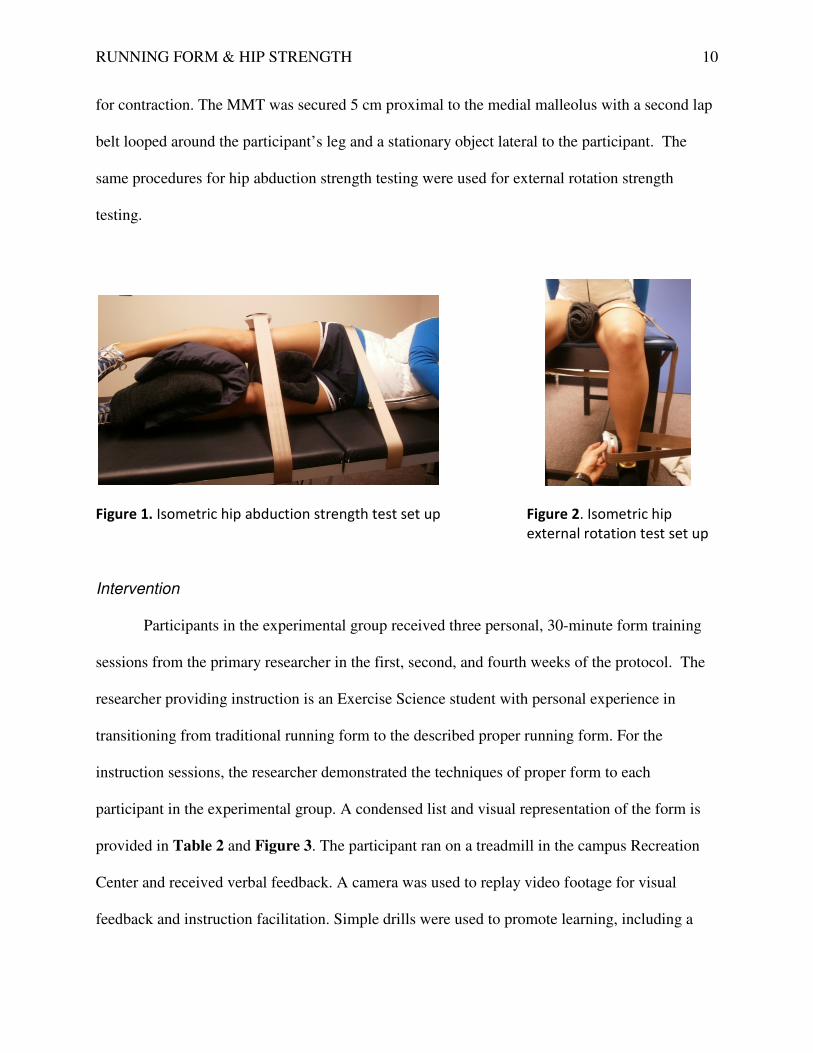
RUNNING FORM & HIP STRENGTH 10
for contraction. The MMT was secured 5 cm proximal to the medial malleolus with a second lap
belt looped around the participant’s leg and a stationary object lateral to the participant. The
same procedures for hip abduction strength testing were used for external rotation strength
testing.
Figure 1. Isometric hip abduction strength test set up Figure 2. Isometric hip
external rotation test set up
Intervention
Participants in the experimental group received three personal, 30-minute form training
sessions from the primary researcher in the first, second, and fourth weeks of the protocol. The
researcher providing instruction is an Exercise Science student with personal experience in
transitioning from traditional running form to the described proper running form. For the
instruction sessions, the researcher demonstrated the techniques of proper form to each
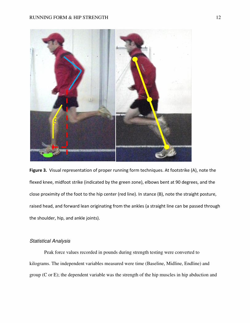
participant in the experimental group. A condensed list and visual representation of the form is
provided in Table 2 and Figure 3. The participant ran on a treadmill in the campus Recreation
Center and received verbal feedback. A camera was used to replay video footage for visual
feedback and instruction facilitation. Simple drills were used to promote learning, including a

RUNNING FORM & HIP STRENGTH 11
drill that consisted of leaning against a wall and lifting the knees to better comprehend the
forward lean and knee lifting motion. A checklist of form techniques and useful stretches was
given as a reference for each E group participant. Furthermore, the participants in the E group
were advised to work on the form gradually, slowly increasing how much of each run was spent
practicing the form. It was aimed for each E group participant to be running their full runs in the
proper form within the third or fourth week. The distance and time ran during these sessions
were not counted towards a participant’s weekly mileage because the running was limited and
intermittent.
Meanwhile, participants in the C group followed the protocol without receiving any
comment or instruction concerning their running form. However, those in the C group were
offered the same running form instruction after the completion of the study.
Table 2. A condensed list of the proper running form techniques taught to the E group
• Straight posture with a slight forward lean from the ankles, which utilizes the force of gravity to
pull the runner forward
• Head up and looking forward
• Shoulders relaxed and dropped down
• Arms relaxed at the sides, with elbows held at 90 degrees. When running, arms swing forward
and back, never crossing the midline of the body.
• Landing on a bent/flexed knee rather than a straight knee
• Short strides with a high cadence (stride rate) of at least 180 steps per minute
• Landing near one’s center of gravity (underneath the hips).
• Aiming to land on the middle (midfoot) or front (forefoot) of the foot rather than at the heel
(heel strike).
• Lower leg is relaxed
RUNNING FORM & HIP STRENGTH 12
Figure 3. Visual representation of proper running form techniques. At footstrike (A), note the
flexed knee, midfoot strike (indicated by the green zone), elbows bent at 90 degrees, and the
close proximity of the foot to the hip center (red line). In stance (B), note the straight posture,
raised head, and forward lean originating from the ankles (a straight line can be passed through
the shoulder, hip, and ankle joints).
Statistical Analysis
Peak force values recorded in pounds during strength testing were converted to
kilograms. The independent variables measured were time (Baseline, Midline, Endline) and
group (C or E); the dependent variable was the strength of the hip muscles in hip abduction and

RUNNING FORM & HIP STRENGTH 13
hip external rotation. Six separate (one for each side and strength test) two-way repeated measure
ANOVA tests were performed. A confidence value of 0.95 was set to assess significance.
RESULTS
Participants
Four of the five participants completed the protocol; one participant was dismissed in the
fifth week due to excessive calf muscle soreness that hindered her from running the target
mileage for two consecutive weeks. The demographics of the C and E group were similar (Table
3). The mild difference in average weekly mileage is not believed to have affected results.
Table 3. Participant demographics (Mean ± SD)
Group Age (years) Height (cm) Weight (kg) Average Weekly
Mileage (miles)
C (n = 2) 20.5 ± 0.7 162.3 ± 3.9 64.6 ± 12.2 12.8 ± 1.1
E (n = 2) 19.0 ± 0 166.8 ± 3.9 59.4 ± 5.2 14.8 ± 0.4
As there were only four participants who completed the study, the statistical power of the results
was low and yielded no significant differences between groups. However, trends were present
that both followed and contradicted the hypotheses.
Hip Abduction: The E group showed an initial decrease in mean force output on both sides from
baseline to midline, followed by an increase from midline to endline. A higher force output
occurred at endline than at the baseline (Table 2). The C group showed a general decrease in
force output from baseline to endline. This result is consistent with the hypothesis that the E
group would exhibit a greater overall increase in hip strength than the C group.

RUNNING FORM & HIP STRENGTH 14
Table 2. Force output (Mean ± SD) of isometric hip abduction over time per side (R = right, L =
left) and group (E = experimental, C = control)
Abduction Force Output (kg) Significance
Between
Groups Side Group Baseline Midline Endline
R E 16.1 ± 5.8 13.8 ± 4.5 17.9 ± 2.8
0.522 C 16.6 ± 2.6 14.1 ± 3.2 13.3 ± 1.1
L E 14.0 ± 1.5 12.6 ± 1.5 17.3 ± 1.2
0.372 C 14.9 ± 2.4 14.3 ± 3.0 11.8 ± 2.3
External Rotation: Contrary to the hypothesis, the E group showed a small decrease in mean
force output from start to finish (Table 3). The C group showed an initial decrease on the right
and an initial increase on the left, followed by a return towards baseline values for either side.
Thus, the C group remained relatively unchanged overall.
Table 3. Force output (Mean ± SD) of isometric hip external rotation over time per side (R =
right, L = left) and group (E = experimental, C = control)
External Rotation Force Output (kg) Significance
Between
Groups Side Group Baseline Midline Endline
R E 6.4 ± 2.1 6.4 ± 3.1 5.7 ± 1.6
0.931 C 6.5 ± 0.7 5.7 ± 2.6 6.1 ± 3.0
L E 6.1 ± 2.3 6.0 ± 2.1 5.6 ± 0.6
0.386 C 4.4 ± 1.1 5.2 ± 2.9 4.8 ± 3.8
Symmetry of Hip Strength: Consistent with the hypothesis, the E group showed a decrease in
both abduction and external rotation force output difference between sides, indicating a
progression towards improved symmetry (Table 4). The C group’s difference in the abduction
and external rotation tests was unchanged between baseline and endline, despite changes seen at
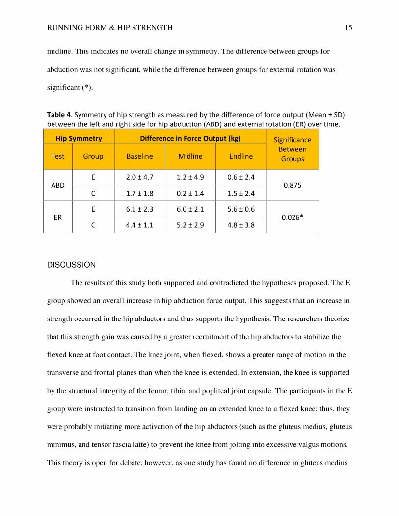
RUNNING FORM & HIP STRENGTH 15
midline. This indicates no overall change in symmetry. The difference between groups for
abduction was not significant, while the difference between groups for external rotation was
significant (*).
Table 4. Symmetry of hip strength as measured by the difference of force output (Mean ± SD)
between the left and right side for hip abduction (ABD) and external rotation (ER) over time.
Hip Symmetry Difference in Force Output (kg) Significance
Between
Groups Test Group Baseline Midline Endline
ABD E 2.0 ± 4.7 1.2 ± 4.9 0.6 ± 2.4
0.875 C 1.7 ± 1.8 0.2 ± 1.4 1.5 ± 2.4
ER E 6.1 ± 2.3 6.0 ± 2.1 5.6 ± 0.6
0.026* C 4.4 ± 1.1 5.2 ± 2.9 4.8 ± 3.8
DISCUSSION
The results of this study both supported and contradicted the hypotheses proposed. The E
group showed an overall increase in hip abduction force output. This suggests that an increase in
strength occurred in the hip abductors and thus supports the hypothesis. The researchers theorize
that this strength gain was caused by a greater recruitment of the hip abductors to stabilize the
flexed knee at foot contact. The knee joint, when flexed, shows a greater range of motion in the
transverse and frontal planes than when the knee is extended. In extension, the knee is supported
by the structural integrity of the femur, tibia, and popliteal joint capsule. The participants in the E
group were instructed to transition from landing on an extended knee to a flexed knee; thus, they
were probably initiating more activation of the hip abductors (such as the gluteus medius, gluteus
minimus, and tensor fascia latte) to prevent the knee from jolting into excessive valgus motions.
This theory is open for debate, however, as one study has found no difference in gluteus medius

RUNNING FORM & HIP STRENGTH 16
activation between subjects despite a significantly higher occurrence of knee valgus in half the
subjects during a single leg jump test (Russell, Palmieri, Zinder, & Ingersoll, 2006).
The initial decrease in force output from baseline to midline in the E group could be
related to an initial performance drop linked to new skill acquisition; adopting the proper running
form is a skill acquisition that requires the retraining of certain musculature. Other explanations
may be liable, however, since neuromuscular strength gains are typically observed two weeks
into the acquisition of a new skill. Participant motivation to perform the strength tests was
probably lowest at midline, as it did not have the “excitement” of beginning or ending the
protocol. In addition, running before midline strength tests was not controlled, so participants
may have ran before the test and been experiencing some degree of muscular fatigue.
The overall decrease in strength in the C group is also intriguing. They should have
experienced no change in hip abduction strength as they had no intervention and little change in
running volume. The results suggest that unforeseen changes in the participants’ exercise
routines may have occurred. One possibility is those that experienced a drop in force output had
switched to treadmill running from circular track running, where the turning may have recruited
the hip abductors more heavily.
The observed decrease in external rotation strength in the E group may be due to the
shortening of the subjects’ stride length. Longer strides exhibit more transverse movement of the
hip in order to extend the leg in front of the body. If the pelvis rotates, then the femur needs to
counter-rotate in the external direction to keep the leg upright, forward, and ahead. Shorter
strides would exhibit less transverse movement at the hips, and consquently the external rotators
would be less activated to rotate the femur. The C group showed no overall change, which is
expected as there was little change in their running routine.

RUNNING FORM & HIP STRENGTH 17
The symmetry of hip strength was also focused on in this study because some research
suggests that hip strength asymmetry, not just overall muscle weakness, is a factor in knee
injuries such as PFP (Robinson & Nee, 2007). The E group showed an increase in symmetry
while the C group remained unchanged, possibly suggesting that the form alterations promoted
more balanced muscle recruitment. In addition, the group difference for external rotation was
found to be significant, suggesting that a difference in symmetry seen between the E and C
groups actually existed. Due to the low number of subjects, a post-hoc analysis was not possible
to perform between the groups, so it cannot be concluded where the significance occurred; it is
possible that the groups were significantly different to start and were not influenced by the
intervention.
Many limitations existed in this study. The mileage was self-reported by the subjects,
which opens the possibility for uncontrolled error. Other uncontrolled variables may have
affected results, including sleep status, motivation, and time of testing. There was also no
standardized system to evaluate subjects’ running form before and after the protocol, so a
subject’s level of form adoption was not taken into account for the results. Future studies should
utilize a standardized point system that can quantitatively assess a runner’s form for comparisons
before and after interventions as well as between subjects. This would allow researchers to better
attribute proper running form adoption to any kinetic, kinematic, and neuromuscular changes.
Other studies should also test external rotation on a long axis with the hip extended, as it better
tests the strength of the gluteus maximus, the primary external rotator during running. The
external rotation test used in this study measured external rotation strength with the hip in a
flexed position, which recruits the smaller external rotators of the hip instead of the gluteus
maximus.
RUNNING FORM & HIP STRENGTH 18
Regardless of the results, this study proposes a novel method of testing proper running
form over a multi-week period and as a set of combined techniques rather separate techniques
(i.e. focusing only on midfoot strike). It is the researchers’ hope that this study will provide a
model for future studies to modify, perfect, and find conclusive evidence regarding proper
running form and its influence on injury risk.
CONCLUSIONS
No statistically significant changes in hip abduction strength and hip external rotation
strength were found after a 6-week running and proper form training protocol due to the small
sample size. However, there was an observed trend for female recreational runners who received
proper form training to have mild strength gains in hip abduction and an increase in hip strength
symmetry between the left and right sides. This suggests more research should be conducted
with a larger sample size to explore these trends and draw conclusions about proper running
form’s effect on hip strength.

RUNNING FORM & HIP STRENGTH 19
REFERENCES
Divert, C., Mornieux, G., Baur, H., Mayer, F., & Belli, A. (2005). Mechanical comparison of
barefoot and shod running. International Journal of Sports Medicine, 26(7), 593-598.
doi:10.1055/s-2004-821327
Dreyer, D. (2009). Chi running: A revolutionary approach to effortless, injury-free running.
New York: Fireside
Earl, J. E., & Hoch, A. Z. (2011). A proximal strengthening program improves pain, function,
and biomechanics in women with patellofemoral pain syndrome. American Journal of
Sports Medicince, 39(1), 154-163. doi: 10.1177/0363546510379967
Elliott, B. C., & Blanksby, B. A. (1976) A cinematographic analysis of overground and treadmill
running by males and females. Medicine and Science in Sports, 8(2), 84-87. Retrieved
from:http://ukpmc.ac.uk/abstract/MED/957936/reload=0;jsessionid=EZDGgZSqwOVuP
MzM4U03.138
Ferber, R., Kendall, K. D., Farr, L. (2011). Changes in knee biomechanics after a hip-abductor
strengthening protocol for runners with patellofemoral pain syndrome. Journal of Athletic
Training, 46(2), 142-149. Retrieved from http://www.mendeley.com/research/changes-
knee-biomechanics-after-hipabductor-strengthening-protocol-runners-patellofemoral-
pain-syndrome/
Fredericson, M., Cookingham, C. L., Chaudhari, A. M., Dowdell, B. C., Oestreicher, N., &
Sahrmann, S. A. (2000). Hip abductor weakness in distance runners with iliotibial band
syndrome. Clinical Journal of Sport Medicine, 10, 169-175. Retrieved from
http://www.udel.edu/PT/davis/Hip_strength_PFP.pdf

RUNNING FORM & HIP STRENGTH 20
Heiderscheit, B. C., Chumanov, E. S., Michalski, M. P., Wille, C. M., & Ryan, M. B. (2011).
Effects of step rate manipulation on joint mechanics during running. Medicine and
science in sports and exercise, 43(2). doi:10.1249/MSS.0b013e3181ebedf4
Ireland, M. L., Willson, J. D., Ballantyne, B. T., & Davis, I. M. (2003). Hip strength in females
with and without patellofemoral pain. Journal of Orthopedic and Sports Physical
Therapy, 33(11), 671-676. Retrieved from
http://www.udel.edu/PT/davis/Hip_strength_PFP.pdf
Jacobs, C. A., Uhl, T. L., Mattacola, C. G., Shapiro, R., Rayens, W. S. (2007). Hip abductor
function and lower extremity landing kinematics: Sex differences. Journal of Athletic
Training,42(1), 76-83. Retrieved from:
http://www.ncbi.nlm.nih.gov.ezproxy.gvsu.edu/pmc/articles/PMC1896084/pdf/i1062-
6050-42-1-76.pdf
Lieberman, D. E., Venkadesan, M., Werbel, W. A., Daoud, A. I., D'Andrea, S., Davis, I. S.,
Mang'Eni, R.O., & Pitsiladis, Y. (2010). Foot strike patterns and collision forces in
habitually barefoot versus shod runners. Nature, 463(28). doi:10.1038/nature08723
Playmakers. (2011). Good form running: Four simple steps to good form. Retrieved from
http://www.goodformrunning.com/
Pose Tech Corp. (2009). Pose method in running. Retrieved from
http://www.posetech.com/pose_method/pose-method-of-running-technique.html
Robinson, R., & Nee, R. (2007). Analysis of hip strength in females seeking physical therapy
treatment for unilateral patellofemoral pain syndrome. Journal of Orthopedic and Sports
Physical Therapy, 37(5). Doi:10.2519/jospt.2007.2439
RUNNING FORM & HIP STRENGTH 21
Russell, K. A., Palmieri, R. M., Zinder, S. M., & Ingersoll, C. D. (2006) Sex differences in
valgus knee angle during a single-leg drop jump. Journal of Athletic Training, 41(2),
166-171. Retrieved from: http://www.ncbi.nlm.nih.gov/pmc/articles/PMC1472649/
Snyder, K. R., Earl, J. E., O'Conner, K. M., Ebersole, K. T. (2009). Resistance training is
accompanied by increases in hip strength and changes in lower extremity biomechanics
during running. Clinical Biomechanics, 24. Retrieved from
http://www.clinbiomech.com/article/S0268-0033%2808%2900279-9/abstract
Souza, R. B. & Powers, C. M. (2009). Differences in hip kinematics, muscle strength, and
muscle activation between subjects with and without patellofemoral pain. Journal of
Orthopaedic and Sports Physical Therapy, 39(1), 12-19. Retrieved from
http://rehabeducation.com/main/wp-content/uploads/January2009-RR-
Souza%5B1%5D.pdf
Taunton, J. E., Ryan, M. B., Clement, D. B., McKenzie, D. C., Lloyd-Smith, D. R., & Zumbo, B.
D. (2002). A retrospective case-control analysis of 2002 running injuries. British Journal
of Sports Medicine, 36, 95-101. Retrieved from
http://www.ncbi.nlm.nih.gov/pmc/articles/PMC1724490/pdf/v036p00095.pdf
van Gent, R. N., van Middlekoop, M., van Os, A. G., Bierma-Zeinstra, S. M. A., & Koes, B. W.
(2007). Incidence and determinants of lower extremity running injuries in long distance
runners: A systematic review. British Journal of Sports Medicine, 41, 469-480. doi:
10.1136/bjsm.2006.033548

RUNNING FORM & HIP STRENGTH 22


![Hip, Hip, Hooray! - goodsamdayton.org1].pdf · right hip within the month, ... Hip, Hip, Hooray! ... to her new hip. H E A LT H TA L K| O RTHOPEDICS 6. Title: SHTK602-Sum06REVfin](https://img.pdfslide.us/doc/110x75/5ab989bf7f8b9ac1058dfdf4/hip-hip-hooray-1pdfright-hip-within-the-month-hip-hip-hooray-.jpg)

![Appendix 1 HIP Male and Female - University of East Anglia · App14.1!HIP!v3.2_02_05_2012!!!!!Health’Improvement’Profile[HIP]’ ’’’’’’’’’’’’’’’’’’’’’’’’’’’’(HIP)–’Male](https://img.pdfslide.us/doc/110x75/5f0af26b7e708231d42e1f1c/appendix-1-hip-male-and-female-university-of-east-anglia-app141hipv3202052012healthaimprovementaprofilehipa.jpg)