Embed Size (px)
Citation preview

Effect of Lidocaine Hydrochloride on Membrane
Conductance in Mammalian Cardiac Purkinje Fibers
MORTONF. ARNsDoRFand J. THOMASBicGER, JR.
From the Departments of Pharmacology and Medicine, College of Physiciansand Surgeons, Columbia University, NewYork 10032
A B S T R A C T Lidocaine depresses automaticity in car-diac Purkinje fibers by decreasing the slope of slow dia-stolic depolarization, but the mechanisms of this effectare poorly understood. To test the proposal that theantiautomatic effect of lidocaine might be mediated byan increase in membrane potassium conductance, trans-membrane voltage (Vm) was measured in Purkinjefibers perfused with sodium-deficient Tyrode containingcholine as the major cation. Vmwas varied by alteringthe external potassium concentration, [K]0, from 0.5 to150 mMbefore and after lidocaine, 2.14 X 10⁵ M, a con-centration considered equivalent to clinical plasma anti-arrhythmic levels. In Purkinje fibers, resting Vmvarieslinearly with EK]o plotted on a logarithmic scale from4 to 150 mm, approximately as predicted by the Nernstequation. At [K]o of 0.5-2.7 mm, resting Vm divergesfrom the predicted potassium equilibrium potential(VK) resulting in an increased driving force for the out-ward K+ current (Vm - VK). In choline Tyrode at[K]o of 2.7 mmor less, lidocaine caused a significantincrease in Vm, the change being a positive linear func-tion of (Vm - VK) with a P < 0.01. This effect wasmore striking in Purkinje fibers with a Vm reduced bystretch. These findings imply that lidocaine increasedmembrane chord conductance for the potassium ion(gK).
Current-voltage relationships using intracellular cur-rent pulses were performed in choline Tyrode at [K]0of 0.5, 2.0, and 4.0 mmand, at each [K]0, lidocaine wasfound to increase membrane slope conductance (GK).The increase in GKwas even more apparent when the
This work was presented in part at the spring meetingof The American Society for Pharmacology and Experi-mental Therapeutics in Chicago, Illinois, April, 1971.
Dr. Arnsdorf is a National Heart and Lung InstituteTrainee (Grant No. 5-T12-HE 05864).
Dr. Bigger is a Senior Investigator, New York HeartAssociation.
Received for publication 10 January 1972 and in revisedform 18 April 1972.
current-voltage relationships in long Purkinje fibers wascorrected for cable complications or when experimentswere done in short Purkinje fibers. To minimize com-plications due to membrane rectifier properties, GKwas measured using intracellular application of smallhyperpolarizing current pulses as Vm was decreasedfrom -90 to -60 mv by increasing the [K]0 from 3 to15 mMbefore and after lidocaine. Lidocaine increasedthe GKover this range of Vm.
These results suggest that lidocaine increases mem-brane potassium conductance within the range of Vmwhere the pacemaker potential is seen, an action whichcan account for its ability to suppress automaticity,and, in part, for its ability to prevent reentrantarrhythmias.
INTRODUCTION
The efficacy of lidocaine hydrochloride in the treatmentof cardiac ventricular arrhythmias after open heartsurgery, myocardial infarction, and digitalis has beenestablished and recently reviewed (1). Electrophysio-logic investigations have shown that in cardiac Purkinjefibers, lidocaine: (a) shortens the action potential dura-tion and decreases the effective refractory period, and(b) exerts an antiautomatic effect by decreasing theslope of slow diastolic depolarization without affectingthe maximum diastolic transmembrane voltage (2-4).These observations led Bigger and Mandel to postulatethat lidocaine may increase potassium conductance inmammalian cardiac Purkinje fibers (3). The presentexperiments were designed to test this hypothesis insheep Purkinje fibers using microelectrode techniques.The effects of lidocaine in a concentration equivalent toclinical plasma antiarrhythmic levels on the transmem-brane voltage and on current-voltage relationshipssuggest that lidocaine does increase potassium conduc-tance in Purkinje fibers.
2252 The Journal of Clinical Investigation Volume 51 September 1972

METHODS
Young adult female sheep were anesthetized with sodiumpentobarbital (30 mg/kg). The heart was rapidly removed andfalse tendon (Purkinje fiber) preparations were selected forstudy. Our standard techniques for stimulating and recordinghave been previously described (5,6). When current-voltagerelationships were studied, methods similar to those of Weid-mann (7) and Hall, Hutter, and Nobel (8) were employed.Fig. 1 summarizes the experimental arrangement. LongPurkinje fibers were employed which were either unbranchedor were of sufficient length to permit impalement with twomicroelectrodes at a distance of 8-10 mmfrom any branches.In some experiments, the Purkinje fiber was divided into seg-ments of less than 2 mmby ligation with 6-0 nylon thread-so-called "short" Purkinje fibers (9). A pair of extracellularsilver wire electrodes, insulated to the tip, was placed on thesurface of the fiber and used for stimulation. A constant cur-rent generator' provided rectangular anodal or cathodalpulses, the amplitude and duration of which could be varied.Constant current pulses of 100 msec duration were passedintracellularly through a glass microelectrode filled with either3 M KCl or 2.5 M potassium citrate. The current was led bya chloride-coated-silver wire placed in the tissue bath to anoperational amplifier.2 This amplifier amplified the currenta millionfold and kept the bath at virtual ground. The am-plified current pulse was displayed on a dual-beam cathoderay oscilloscope.3 Transmembrane voltage was recorded witha second glass microelectrode which was placed within 50-200 IAof the stimulating microelectrode. The transmembrane voltagewas amplified by a high input impedance and variable capacityneutralization amplifier4 and displayed on the dual-beam oscil-loscope. The oscilloscopic display of transmembrane voltageand current was photographed' and the enlarged imagesmeasured.
The Tyrode solution employed contained, in mmoles perliter: NaCl, 137; MgC12, 0.5; NaH2PO2H2O, 1.8; CaCl2, 1.8;1.8; NaHCO3, 12; dextrose, 5.5; potassium concentration wasvaried. Sodium-deficient Tyrode solution was prepared bysubstituting choline chloride for sodium chloride on a mole forbasis. The sodium-deficient Tyrode contained 13.8 mMsodium.Sodium-deficient Tyrode in which choline is the major cationis known to produce minimal changes in resting membraneconductance and to have little effect on the membrane current-voltage relationships; the findings in choline Tyrode are com-parable to those observed in a completely sodium-free solutionor those in normal Tyrode at subthreshold voltages (8). Insodium-deficient Tyrode, the effect of the sodium conductanceon transmembrane voltage (Vm)6 is minimized and the restingmembrane conductance is determined primarily by potassium
I Designed by Mr. S. Ross, Columbia University, NewYork.2 Philbrick/Nexus Research, Dedham, Mass. (P 25).3 Tektronix, Inc., Beaverton, Ore.4Bioelectric Instruments Div., General Microwave Corp.,
Farmingdale, N. Y. (NFl).6 Grass Instrument Co;, Quincy, Mass.IA bbreviations used in this paper: Cm, membrane capaci-
tance; GK, membrane slope conductance for potassium ion;GM, membrane slope conductance; gK, membrane chord con-ductance for potassium ion; gM, membrane chord conduc-tance; I, applied current; Ii, ionic current; IK, membranepotassium current; im, membrane current density; [K]i, intra-cellular potassium concentration; [K]0, external potassiumconcentration; PK, membrane permeability for potassium ion;VK, potassium equilibrium potential; Vm, transmembranevoltage; TD, membrane time constant determined with de-polarizing current pulses.
FIGURE 1 Experimental arrangement used for stimulatingand for recording both the applied intracellular current andresultant transmembrane voltage change during the study ofcurrent-voltage relationships in Purkinje fibers. See Methods.
conductance with other ions playing a minor role (10-14).When high external potassium concentrations [K]0 of 10-120mMwere used, the solutions were made isomolar by decreasingcholine.
The Tyrode solution was equilibrated with 95 %oxygen and5 %carbon dioxide in a reservoir and infused into the tissuebath at a constant rate of 8 ml/min. In the tissue bath tem-perature was maintained between 35.5 and 36.50C and the pHwas 7.36.
Lidocaine hydrocholoride was used in a concentration of2.14X10-5 M(5 ,ug/ml). In cardiac Purkinje fibers, this con-centration both abbreviates the action potential duration andexerts a strong antiautomatic effect by decreasing the slopeof slow diastolic depolarization (2-4). For the reasons discussedby Bigger and Mandel, this concentration is considered equiv-alent to clinical antiarrhythmic plasma levels (3).
The statistical significance of changes before and, afterlidocaine was determined by the t test for paired samples(15). Correlation coefficients and levels of statistical signifi-cance were determined by standard methods (15).
RESULTS
Effect of lidocaine on the transmembrane voltage charac-teristics of the Purkinje fiber in a sodium-deficient Tyrodesolution at various external potassium concentrations. Inour experiments, the mole for mole substitution ofcholine chloride for sodium chloride produced either nochange or a small increase in the resting transmembranevoltage (Vm) similar to that seen in previous investiga-tions (8, 16). Spontaneous diastolic depolarization didnot occur in the sodium-deficient Tyrode solution norcould electrically induced regenerative depolarizationbe elicited in the Purkinje fibers studied.
Lidocaine on Membrane Conductance 2253

TABLE I
Effect of Lidocaine on Resting Transmembrane VoltageCharacteristics* and Potassium Permeability in Sodium-
Deficient Tyrode at Various [K]0
Control Lidocaine
PKlidocaine/[K]0 n Resting Vm Resting Vm P PK0ontrol
mM mv mv
0.5 4 -51.7:16.17 -64.818.08 <0.001 1.891.0 4 -64.4 ±4.18 -75.6-14.93 <0.005 1.651.5 4 -73.8±8.30 -84.2 48.30 <0.001 1.682.0 4 -78.04±5.45 -84.8 ±5.82 <0.005 1.452.7 4 -87.8±-4.04 -90.04±2.35 <0.010 1.27
No significant change in Vmwas noted in 12 experiments at [K]) of 4.0,10.0, 60.0, 90.0, and 120.0 mM.* Mean ASD n = number of preparations; resting Vm = resting trans-membrane voltage (mv); P = probability based on t test.for paired sam-ples; PK00ntr1 = potassium permeability, control; PKjidOCi,. = potassiumpermeability after lidocaine.
In each experiment, multiple impalements were em-ployed to determine the resting Vm before and afterthe application of lidocaine. Results obtained in 32different preparations appear in Table I. In Fig. 2,the mean resting Vm from these experiments is plotted
as a function of [K]o on a logarithmic scale. Undercontrol conditions (circles), the resting Vm was deter-mined at [K]o of 10, 60, 90, and 120 mmto calculatefrom experimental observations a regression line (theheavy line). Extrapolation of the regression line inter-sects the abscissa at [K]0 of 150 mmgiving an estimateof the intracellular potassium concentration (13). Thethin line represents the potassium equilibrium potential(VK) as predicted from the Nernst equation were themembrane of the Purkinje fiber acting as a perfect potas-sium electrode, assuming [K]i to be 150 mm. The regres-sion line was also extrapolated towards the ordinate andrepresents the relationship between Vm and [K]0plotted on a logarithmic scale were the membraneconductance constant over the entire range of [K]oemployed in these experiments.
According to the Nernst equation, at a temperatureof 360C, a 61 mv change in resting Vmis expected forevery decade change in [K]0. The regression line de-rived from our experimental observations showed a 51mv change in resting Vm for every decade change in[K]0. Approximately the same divergence between thepredictions of the Nernst equation and the experi-mental observations has been previously noted in nor-mal saline Tyrode (17). In the present experiments, theapproximation of VK as derived from the experimentalobservations has been employed in the calculations.The interpretation of our results is unaffected whetherone or the other Vmis used in the calculations.
Since a sodium-deficient Tyrode solution was em-ployed and chloride ions are known to play only aminor role in determining the resting ionic current (Ii)of the resting Purkinje fiber (10, 11), resting Ii can beexpressed as:
Ii IK = gK(Vm - VK),
mM
FIGURE 2 The resting transmembrane voltage (Vm) insodium-deficient Tyrode during the control period (circles)and after lidocaine (triangles) is plotted as a function of[K]0 on a logarithmic scale. The arrows show the direction ofchange. The heavy line is a regression line determined fromexperimental results at [K]0 of 10, 60, 90, and 120 mmole/literwhich has been extrapolated both to the abscissa and towardsthe ordinate. The thin line represents VK as calculated fromthe Nernst equation. Significant hyperpolarization occursafter the application of lidocaine at [K]o of 2.7 mMand less.See text for discussion.
(1)where IK is potassium current and gK is membranechord conductance for potassium ion. Note that undercontrol conditions in Fig. 2, Vm approximates VKabove a [K]0 of 4.0 mm. As [K]0 is further lowered,increasing divergence is noted between Vm and VE
-due to a decrease in the membrane conductance tothe outward flow of potassium ions (8, 17). Theseresults agree well with the radioactive potassium effluxstudies of Carmeliet which were conducted over a widerange of [K]0 (10, 11). From Fig. 2, it is clear that thedriving force for the potassium ion (Vm - VK) in-creases as [K]0 is progressively lowered below 4.0 mm(18).
After the application of lidocaine (Table I and Fig.2), statistically significant hyperpolarization occurredwhen (Vm - VK) was significantly greater than zero,i.e., at [K]0 of 2.7 mmor less. The magnitude of thehyperpolarization induced by lidocaine was positivelycorrelated with the magnitude of the driving force
2254 M. F. Arnsdorf and J. T. Bigger, Jr.

30 r
20 FVm
%Ao
10
0 a a I I
0 20 40 60 80
l I a I I
2.7 2.0 1.5 1.0
(Vm - VK)mv
[K mM0.5
FIGURE 3 The per cent change in resting transmembrane voltage (Vm) caused byapplication of lidocaine [(Vmlidocaine - Vmcontrol) X 100] is plotted as a function ofthe outward driving force for the potassium ion (Vmcontrol - VK). [K]0 is also in-dicated on the abscissa. A strong linear positive correlation exists between the percent change in Vmand (Vm - VK) (r = 0.99, P < 0.01).
(Vm - VK) for the potassium ion. This is seen in Fig.3 which shows the mean per cent change in resting Vmtobe a linear function of (Vm- VK) (r = 0.99, P < 0.01).As predicted from equation 1, no significant change inthe resting Vmwas noted after the application of lido-caine (Table I and Fig. 2) when (Vm- VK) approachedzero at [K]0 of 4.0 mmor above.
It is possible to calculate the permeability for thepotassium ion (PK) of the resting membrane with theGoldman equation using the change in Vm caused bylidocaine at [K]0 of 2.7 mmor less (19-21). These cal-culations showed clearly that PK increased after theapplication of lidocaine (Table I).
Effect of lidocaine on transmembrane voltage charac-teristics of stretched Purkinje fibers. Lidocaine hada remarkable restorative effect on Vm of fibers with adecreased resting or maximum diastolic transmembranevoltage due to stretch. In Fig. 4, panel A shows a spon-taneous action potential of a stretched fiber in normalTyrode solution at a [K]0 of 2.7 mm. The preparationhad been stimulated electrically through external elec-trodes at a cycle length of 800 msec for 1 hr after whichspontaneous depolarization was allowed to stabilize foran additional 30 min. The maximum diastolic trans-membrane voltage was -70 mv. Panel B of Fig. 4 wasrecorded after 20 min of quiescence caused by the sub-stitution of sodium-deficient Tyrode for normal Tyrodeat the same [K]o. At this time, Vm was -61 mv, es-sentially unchanged from the maximum diastolictransmembrane voltage in normal Tyrode. Panel C ofFig. 4 was recorded in sodium-deficient Tyrode at the
same [K]0 30 min after the application of lidocaine atwhich time the resting Vm was -93 mv. Similarly,lidocaine produced a significant hyperpolarization infive other fibers which showed a decreased resting Vmormaximum diastolic transmembrane voltage due tostretch; two of these experiments were performed at
A 100 msec
50mV
B C
-61 mV
FIGURE 4 The effect of lidocaine on a sheep Purkinje fiberinjured by stretching ([Kh. = 2.7 mmin each panel).
(A) Transmembrane voltage (Vm) recorded in normalTyrode shows markedly reduced maximum diastolic Vmandautomaticity. (B) Shows the resting transmembrane voltagerecorded in sodium-deficient Tyrode. Spontaneous activityceased when sodium was reduced. The figure shows withdrawalof the microelectrode to determine resting Vm. The restingtransmembrane voltage was -61 mv. (C) 30 min after addinglidocaine the resting transmembrane voltage was -93 mva 32 mv hyperpolarization.
Lidocaine on Membrane Conductance 2255

B
-1.2 -0.8 -0.4
-20
[i]0.5 IBM
-40
-60;
-80-
Vm
mv -Loo-
04 0.8 1.2 L6
im
[K+]. 0.5 mM
[K+]. 2.0 mM
Vm
mv
FIGURE 5 Current-voltage relationships in long Purkinje fibers perfused with sodium-deficientTyrode at [K]j of 0.5 mm(upper curves, experiment 25, Table II) and 2.0 mm(lower curves,
experiment 20, Table II) before (circles) and after (triangles) the application of lidocaine. In panelA, the observed transmembrane voltage (V.) is plotted as a function of the applied intracellularcurrent (I) in ;a. In Panel B, the membrane current density (im) was calculated to correct for cablecomplications; the observed Vmis plotted as a function of im in arbitrary units. Slope conductance(GM) was found to increase after lidocaine for both depolarizing and hyperpolarizing pulses overthe entire range of the curves at both values of [K]0. For depolarizing currents, the increased GMwas manifested by the curve becoming less steep and shifting to the right after lidocaine. Sincethe GMfor both depolarizing and hyperpolarizing pulses increases after lidocaine, the curvesmight be expected to cross-over (23) but this does not occur due to the increase in resting Vm.Chord conductance (gM) was estimated from tangents drawn to the curves in panel B (the inter-rupted lines) at im = 0. The ratio of resting membrane conductance after lidocaine as compared tothe control value was 3.79 at [K]0 = 0.5 mMand 1.30 at [K]0 = 2.0 mm. See text for furtherdiscussion.
[K]0 of 2.7 mmand one each at [K]0 of 0.5, 1.5, and4.0 mm. These results suggest that stretch decreasesmembrane potassium conductance thus increasing or
creating a difference between Vmand VK. Lidocaine, byincreasing membrane potassium conductance, causes
Vm to approach VK and hyperpolarizes the membrane.
Current-voltage relationships in long Purkinje fibers.Membrane current-voltage relationships of long Pur-
kinje fibers were measured in sodium-deficient Tyrodebefore and after the application in a total of nine ex-
periments. Three experiments were performed at each
of three [K]0: 0.5, 1.5, and 4.0 mm. Representativeexperiments of the effect of lidocaine on membranecurrent-voltage relationships are shown in Fig. 5([K]o = 0.5 and 2.0 mM) and Fig. 6 ([K]O = 4.0 mM).In panel A of Figs. 5 and 6, the observed Vmis plottedas a function of the amplitude of the applied current(I). In panel B of Figs. 5 and 6, the fiber -is consideredan infinite cable and the relationship suggested by Coleand Curtis (22) to correct for cable complications was
used to estimate the membrane current density (im):
(2)
2256 M. F. Arnsdorf and J. T. Bigger, Jr.
A
0 CONTROLA LIDOCAINE
i. ~:- Is- (dl/dV).

I was measured directly and tangents were constructo the curves in panel A of Figs. 5 and 6 to obtain dI/these two values were used to solve equation 2 forThe observed Vm was then plotted as a function of(panels B of Figs. 5 and 6). The cable properties ofPurkinje fiber tend to "smooth-out" nonlinearitiesthe membrane current-voltage relationship, butmathematically correcting for cable complications usequation 2, one can approximate the relationshipa uniformly depolarized membrane which accentuathe curvatures in the membrane current-voltage r(tionship (8, 22).
The membrane slope conductance (GM) can befined by the following:
Panel A of Figs. 5 and 6 GM= dI/dV,
Panel B of Figs. 5 and 6 GM= dim/dV.
GMwas determined by constructing tangents tocurves in Figs. 5 and 6. Lidocaine was found to increGMover the entire curve at each of the three []studied. For depolarizing currents, the curve becaless steep and shifted to the right after lidocaine inexperiments. Since the GMfor both depolarizinghyperpolarizing pulses increases after lidocaine,curves might be expected to "cross-over" (23), aindeed, this occurs at a [K]0 of 4.0 mmwhere the restVm is unaffected (Fig. 6). Crossing-over did not oc
A
-50O
-70
at either a [K]0 of 0.5 or 2.0 mmdue to-the increase inresting Vm after lidocaine (Fig. 5). The increase in Vmafter the application of lidocaine at [K]0 of 0.5 and 2.0mmmight in itself produce a voltage-dependent increasein membrane conductance (23). Further experiments,to be discussed below, were designed to determinewhether the increase in GMwas due to lidocaine or tothe increased Vmper se.
The steady-state membrane current-voltage relation-ship gives an estimate of the slope conductance (GM)rather than the chord conductance (gM) of the restingmembrane. The relationship between GMand GKcanbe described by the following when equation 1 pertains:
(3) GM z GK= dIK/dV = gK + (Vm-VK) dV' (5)
when I or i. = 0, GKapproximates gK.Table II lists the gK at I or i. = 0, the polarization
resistance, and the ratio of gK after lidocaine as com-pared to the control for three experiments each at [K]oof 0.5, 2.0, and 4.0 mm. In every experiment, the gKincreased and the polarization resistance decreasedafter the application of lidocaine.
Current-voltage relationships in short Purkinje fibers.In two experiments, current-voltage relationships weredetermined in short Purkinje fibers in sodium-deficientTyrode at a [K]o of 4.0 mm, The fibers were shortened
B
Vmmv
[K+]0 40mmM
* CONTROLA LIDOCAINE
Vmmv
FIGURE 6 Current-voltage relationships in a long Purkinje fiber at[K]. = 4.0 mm(experiment 30A, Table II). The symbols and calcula-tions are as in Figure 7. The slope conductance (GM) for both depolariz-ing and hyperpolarizing intracellular currents increased after lidocaine,and since no shift in the resting Vmoccurred, crossing-over of the curves(23) was noted at I or i. = 0. For depolarizing currents, the slope be-came less steep and the curve was shifted to the right. The ratio of mem-brane conductance at the resting Vmafter lidocaine as compared to thecontrol was- 1.74. See text for futrher discussion.
Lidocaine on Membrane Conductance 2257

TABLE I IEffect of Lidocaine on Membrane Conductance and Polarization Resistance as De-
termined from Current- Voltage Relationships in Long and Short Sheep PurkinjeFibers at Various [K]0 in Sodium-Deficient Tyrode
Control Lidocaine
Polarization Polarization gKlidooaine/[K]0 Exp. gK resistance gK resistance gKoontroi
pmho kR2 pmho k2
Long Purkinje fibers0.5 50 2.08 480 2.65 377 1.27
24 4.35 230 5.26 190 1.2125 2.74 364 10.20 98 3.72
2.0 16 1.89 530 1.96 510 1.0420 3.52 284 4.59 218 1.3014 2.00 500 6.58 152 3.2937 3.21 312 3.93 254 1.2240 4.18 239 5.37 186 1.2830A 5.95 168 7.46 134 1.25
Short Purkinje fibers*4.0 51A 1.77 567 2.77 361 1.56
51B 2.12 471 2.99 334 1.41
gK = membrane potassium chord conductance from GKat I = 0 or for smallhyperpolarizing pulses (see text and equations 8 and 9); gKlid.,ize/gKotroj= ratio of membrane conductance after lidocaine to that of the control.
* Segments less than 2 mmin length.
by a ligature to lengths of less than 2.0 mM. In the shortpreparation, cable complications are minimized andapplied current produces fairly uniform membranepolarization throughout the preparation (9). In thisexperimental arrangement, the intracellularly appliedcurrent (I) approximates the membrane current density(im) which had to be calculated in experiments on longPurkinje fibers (see equation 2 and the discussion ofim above). Fig. 7 shows the current-voltage relationshipobtained in a short fiber before and after the applica-tion of lidocaine. Resting Vm was unchanged and thepolarization resistance of the preparation decreasedafter lidocaine (Table II). Over the entire current-voltage relationship, the slope became less steep and thecurve was shifted to the right after lidocaine, i.e.,crossing-over (23) of the curves occurred. These changesall indicate an increase in GM(GK). Both GMand gKas determined from tangents constructed to the curvesin Fig. 7 were increased. The magnitude of change isequivalent to that noted in long Purkinje fibers anddirectly confirms the validity of mathematically cor-recting for cable properties. Lidocaine, therefore, in-creases membrane gK at a [K]0 of 4.0 mm, a normalvalue for plasma or extracellular fluid K+.
Membrane time constant in long Purkinje fibers. Themembrane time constant was calculated from the volt-age decay curve after offset of depolarizing current
pulses (TD). Further:
TD = Cm/GM, (6)where Cm represents the membrane capacitance andGMthe membrane slope conductance. Since membranecapacitance is thought to remain constant (24, 25),it would be anticipated that if lidocaine increased GM,TD should decrease. It is clear from Table III thatlidocaine did cause a significant decrease in TD at [K]0of 0.5, 2.0, and 4.0 mm. Time constants calculated fromhyperpolarizing pulses, showed essentially the samechange as TD.
Membrane conductance changes before and after lido-caine while varying the resting Vmby altering [K]o. Inthree experiments, the resting Vm of the Purkinjefiber was gradually decreased by increasing the [K]0from 3.0 to 15.0 mmin sodium-deficient Tyrode. Studiesin this [K]o range obviate the voltage-dependent con-ductance changes caused by a lidocaine-induced shiftin resting Vm as occurred in experiments at a [K]o of2.7 mmor less. Depolarization of the membrane by in-creasing [K]0 increases membrane potassium conduc-tance (8, 25, 26) unlike depolarization by applied cur-rent pulses which results in decreased membrane potas-sium conductance (8, 23, 26-30).
Membrane slope conductance (GM) was calculatedfrom small intracellular hyperpolarizing pulses of con-
2258 M. F. Arnsdorf and J. T. Bigger, Jr.

001 002
[K]0 =4O.mM
* ControlA Lidocane (2./4XIO-M)
-100.
Iua
Vmmv
FIGURE 7 Current-voltage relationships determined in a short Purkinjefiber (1.6 mm) at [K]0 = 4.0 mmbefore (circles) and after the applica-tion of lidocaine (triangles). The use of a short fiber minimizes cablecomplications, and much less applied current is required to producea change in the transmembrane voltage (Vm) than in long Purkinjefibers (see Fig. 8). After lidocaine, depolarizing currents result in a lesssteep curve which is shifted to the right, and with no change in the rest-ing Vm, hyperpolarizing pulses result in crossing-over at I = 0. Theseresults indicate that lidocaine increased membrane slope conductancefor both depolarizing and hyperpolarizing pulses. At I = 0, i.e., at theresting Vm, the ratio of membrane conductance after lidocaine as com-pared to the control was 1.56. See text for further discussion.
stant amplitude which resulted in small membranevoltage changes (< - 7 mv). For the reasons discussedabove, GMin sodium-deficient Tyrode is determinedprimarily by GK; GM measured by small hyper-
TABLE IIIMembrane Time Constant in Long Purkinje Fibers
EK]O TD~ontrol TDlidocaine %A TDonto I/TDlidoesine
mm msec msec0.5 42.0 13.0 66.7 3.23
24.6 14.8 39.8 1.78
2.0 16.6 12.4 25.3 1.3427.0 21.1 22.0 1.2824.4 18.4 24.6 1.32
4.0 22.7 14.9 34.4 1.5620.5 13.7 33.2 1.4918.0 15.3 15.0 1.18
rDconUV = membrane time constant, control; IrDlid,,i= membrane time constant after lidocaine; %A= per centdecrease in TD after lidocaine.
polarizing pulses approximates resting membranechord conductance (gK).
In Fig. 8, GMis plotted as a function of the restingVmas the Vmwas gradually decreased by increasing the[K]o from 3.0 to 15.0 mm. Both the control data andthe data after lidocaine showed a good fit (least squaresmethod) for the exponential regression of GM onVm(P < 0.01). The exponential character of the curvemay be accounted for by the exponential relationshipbetween Vm and [K]o (see Fig. 2). Clearly, lidocaineproduces an increase in GMover the range of V.studied. The ratio of GMlidocaine/GMcontrol varied be-tween 1.14 and 1.20 which is very similar to the ratiosfound in both long and short Purkinje fibers as calcu-lated from current-voltage relationships (Table II) andto the permeability ratios calculated with the Goldmanequation from the change in Vmlidocaine was appliedto fibers perfused with [K]0 < 2.7 mm.
DISCUSSION
Previous electrophysiologic investigations have shownthat lidocaine in concentrations equivalent to clinicalantiarrhythmic plasma levels shortens the action po-
Lidocaine on Membrane Conductance 2259
003

* CONTROLA LIDOCAINE
A
A
11.0
10.0
GM
/mho9.0
8.0
7.0
-90 -80 -70 -60Vm( mV)
FIGURE 8 Membrane conductance (GM) in ,umho calculatedfrom changes in transmembrane voltage produced by smallhyperpolarizing constant current pulses plotted as a functionof the resting Vmwhich was gradually decreased by increasing[K-1 from 3 to 15 mM. Control data is represented by thecircles; data after lidocaine by the triangles. The arrows showthe direction of change. GMis increased after lidocaine overthe range of Vmand [K]0 studied.
tential duration and effective refractory period in bothPurkinje and ventricular muscle fibers, and exerts anantiautomatic effect in Purkinje, pacemaker cells bydecreasing the slope of slow diastolic depolarizationwithout affecting the maximum diastolic transmem-brane voltage at [K]o of 2.7-3.0 mm (2-3). On thebasis of these observations, Bigger and Mandel postu-lated that lidocaine may increase potassium conduc-tance in cardiac Purkinje fibers (3).
The present study has shown by a variety of tech-niques that lidocaine increases membrane conductance,by increasing membrane potassium conductance, overa wide range of [K]o (0.5-15 mM). The increase inmembrane potassium slope (GK) or chord (gK) con-ductance as determined from current-voltage relation-ships is of comparable magnitude at the various [K]0employed and is of the same magnitude as the increasein potassium permeability (PK) calculated from thechange in resting Vm in the presence of a significantoutward driving force for the potassium ion (Tables Iand II). The current-voltage relationships also demon-strate that lidocaine increases membrane potassiumconductance over the range of the pacemaker potentialat external potassium concentrations seen clinically.
Kabela has recently shown that lidocaine enhancesthe effilux of 42K from canine Purkinje fibers at a [K]0
of 5.0 mmwhich he attributes to an increased membranepotassium conductance.7 He further noted that lido-caine did not have this effect in atrial tissues. It isperhaps significant that lidocaine is efficacious in thetreatment of ventricular arrhythmias but-is rather in-effective in the treatment of atrial arrhythmias (1, 31,32). Lidocaine has been thought to have minimal in-fluence on the electrophysiologic characteristics of theatrial fiber (33, 34).
In considering our findings, it seems appropriate todiscuss the possible role of ions other than K+. Restingmembrane conductance in normal saline Tyrode solu-tion is determined primarily by potassium conductancewith other ions having been shown to play a minorrole (8-14, 35). The accepted ratio of resting gNa to gKis 1/19 (9). The contribution of the sodium ion wasfurther minimized in our study by the use of sodium-deficient Tyrode in which the external and internalsodium concentrations were essentially the same, thusthe effect of a change in gNa on resting Vm must beminimal. In normal saline Tyrode solution, an increasedmembrane conductance due to an increase in eithergNa or gCa would result in depolarization of the restingmembrane rather than in the observed hyperpolariza-tion, and would increase rather than decrease the slopeof slow diastolic depolarization (36). It is difficult,therefore, to assign a major role to the sodium, chloride,or calcium ion which in the presence of an increasedmembrane conductance could account for the effect oflidocaine on slow diastolic depolarization. If the in-creased GMafter lidocaine does result in part from anincreased sodium, chloride, or calcium conductance, orif it occurs despite an actual decrease in sodium con-ductance, the influence of these conductance changes inthe range of the pacemaker potential must be maskedby a more profound increase in potassium conductance.
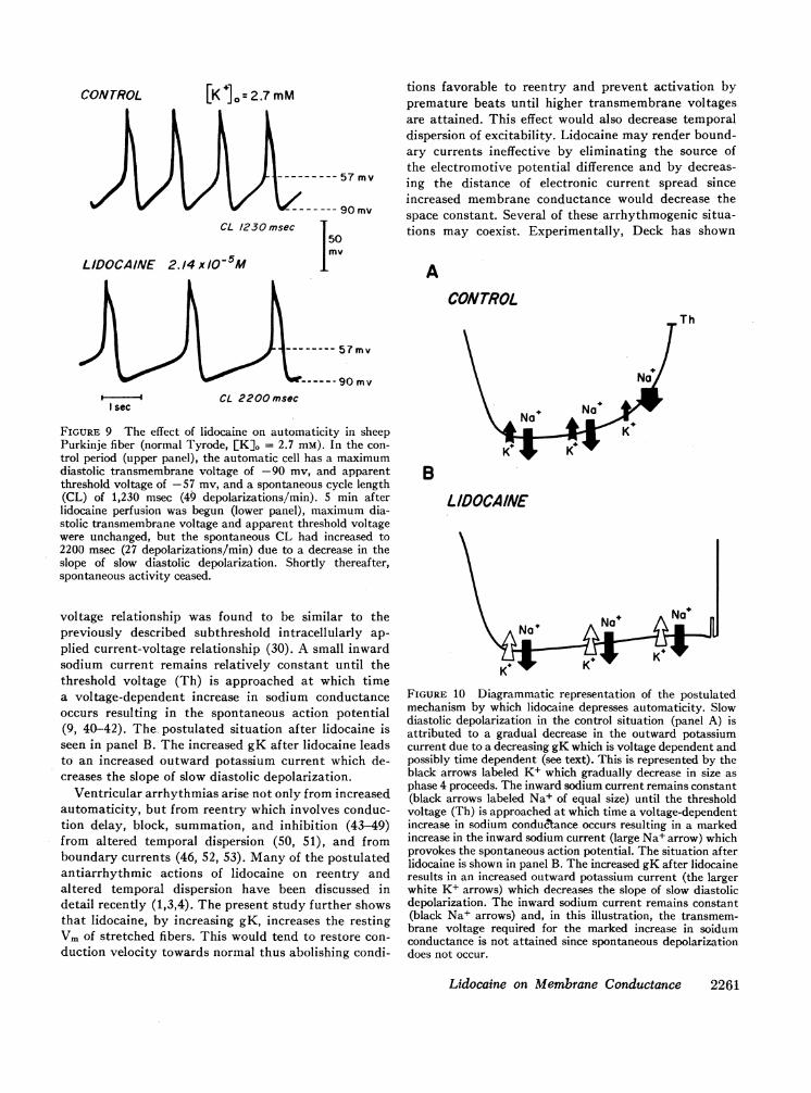
The effect of lidocaine on an automatic Purkinjepacemaker cell in normal Tyrode at a [K]o of 2.7 mmis seen in Fig. 9. The decreased spontaneous rate canclearly be attributed to a decrease in the slope of slowdiastolic depolarization since neither the maximumdiastolic transmembrane voltage nor the apparentthreshold voltage had changed. The probably mecha-nism is illustrated in Fig. 10. Before lidocaine (panelA), slow diastolic depolarization is present. The causeof diastolic depolarization in Purkinje fibers is thoughtto be a gradual decrease in the outward potassiumcurrent due to a decreasing gK which is voltage andtime dependent (23, 37-39). This view is supported bythe radioactive tracer studies of Haas and Kern whodetermined potassium fluxes in voltage-clamped Pur-kinje fibers (30). The subthreshold potassium current-
7Kabela, E. L. 1972. The effects of lidocaine on potassiumefflux from various tissues of dog heart. Manuscript submittedfor publication.
2260 M. F. Arnsdorf and J. T. Bigger, Jr.
[K lo=2.7 mM
CL 1230 msec
50m\
LIDOCAINE 2.14 xl/O-5M
tions favorable to reentry and prevent activation bypremature beats until higher transmembrane voltagesare attained. This effect would also decrease temporaldispersion of excitability. Lidocaine may render bound-ary currents ineffective by eliminating the source ofthe electromotive potential difference and by decreas-ing the distance of electronic current spread sinceincreased membrane conductance would decrease thespace constant. Several of these arrhythmogenic situa-tions may coexist. Experimentally, Deck has shown
ACONTROL
,Th
s-c CL 2200 msecI sec
FIGURE 9 The effect of lidocaine on automaticity in sheepPurkinje fiber (normal Tyrode, [K]0 = 2.7 mM). In the con-
trol period (upper panel), the automatic cell has a maximumdiastolic transmembrane voltage of -90 mv, and apparentthreshold voltage of -57 mv, and a spontaneous cycle length(CL) of 1,230 msec (49 depolarizations/min). 5 min afterlidocaine perfusion was begun (lower panel), maximum dia-stolic transmembrane voltage and apparent threshold voltagewere unchanged, but the spontaneous CL had increased to2200 msec (27 depolarizations/min) due to a decrease in theslope of slow diastolic depolarization. Shortly thereafter,spontaneous activity ceased.
voltage relationship was found to be similar to thepreviously described subthreshold intracellularly ap-
plied current-voltage relationship (30). A small inwardsodium current remains relatively constant until thethreshold voltage (Th) is approached at which timea voltage-dependent increase in sodium conductanceoccurs resulting in the spontaneous action potential(9, 40-42). The- postulated situation after lidocaine isseen in panel B. The increased gK after lidocaine leadsto an increased outward potassium current which de-creases the slope of slow diastolic depolarization.
Ventricular arrhythmias arise not only from increasedautomaticity, but from reentry which involves conduc-tion delay, block, summation, and inhibition (43-49)from altered temporal dispersion (50, 51), and fromboundary currents (46, 52, 53). Many of the postulatedantiarrhythmic actions of lidocaine on reentry andaltered temporal dispersion have been discussed indetail recently (1,3,4). The present study further showsthat lidocaine, by increasing gK, increases the restingVmof stretched fibers. This would tend to restore con-
duction velocity towards normal thus abolishing condi-
No
B
LIDOCAINE
K
FIGURE 10 Diagrammatic representation of the postulatedmechanism by which lidocaine depresses automaticity. Slowdiastolic depolarization in the control situation (panel A) isattributed to a gradual decrease in the outward potassiumcurrent due to a decreasing gK which is voltage dependent andpossibly time dependent (see text). This is represented by theblack arrows labeled K+ which gradually decrease in size as
phase 4 proceeds. The inward sodium current remains constant(black arrows labeled Na+ of equal size) until the thresholdvoltage (Th) is approached at which time a voltage-dependentincrease in sodium conductance occurs resulting in a markedincrease in the inward sodium current (large Na+ arrow) whichprovokes the spontaneous action potential. The situation afterlidocaine is shown in panel B. The increased gK after lidocaineresults in an increased outward potassium current (the largerwhite K+ arrows) which decreases the slope of slow diastolicdepolarization. The inward sodium current remains constant(black Na+ arrows) and, in this illustration, the transmem-brane voltage required for the marked increase in soidumconductance is not attained since spontaneous depolarizationdoes not occur.
Lidocaine on Membrane Conductance 2261
CONTROL

that in Purkinje fibers, a modest stretch results ina decreased maximum diastolic transmembrane voltagewith an increased space constant and specific membraneresistance, as well as, in some preparations, an increasein spontaneous depolarization (54, 55). Trautwein andKassebaum demonstrated that in the sheep Purkinjefiber, the application of weak depolarizing currents,which would be comparable to boundary currents, in-creases the slope of slow diastolic depolarization result-ing in the production or enhancement of spontaneousrhythmicity (56). Enhanced automaticity due to anincrease in slow diastolic depolarization will not onlyin itself produce cardiac arrhythmias, but may also de-crease the membrane activation voltage thus decreas-ing conductivity. ~in the Purkinje fiber, a situation con-ducive to the development of reentrant arrhythmias(57). These phenomena are presumed to underly manyarrhythmias encountered clinically; in each, the abilityof lidocaine to increase gK would have an antiarrhyth-mic effect.
ACKNOWLEDGMENTSThis work was supported in part by United States PublicHealth Service Grants HE 05741 and HE 12738 and in partby a Grant-in-Aid from the New York Heart Association.
REFERENCES1. Bigger, J. T., Jr., and R. H. Heissenbuttel, 1969. The use
of procaine amide and lidocaine in the treatment of cardiacarrhythmias. Prog. Cardiovasc. Dis. 11: 515.
2. Davis, L. D., and J. V. Temte. 1969. Electrophysiologicalactions of lidocaine on canine ventricular muscle andPurkinje fibers. Circ. Res. 24: 639.
3. Bigger, J. T., Jr., and W. J. Mandel. 1970. The effect oflidocaine on the electrophysiological properties of ven-tricular muscle and Purkinje fibers. J. Clin. Invest. 49: 63.
4. Bigger, J. T., Jr., and W. J. Mandel, 1970. Effect of lido-caine on conduction in canine Purkinje fibers and at theventricular muscle-Purkinje fiber junction. J. Pharmacol.Exp. Ther. 172: 239.
5. Bigger, J. T., Jr., A. L. Bassett, and B. F. Hoffman. 1968.Electrophysiological effects of diphenylhydantoin oncanine Purkinje fibers. Circ. Res. 22: 221.
6. Strauss, H. C., J. T. Bigger, Jr., A. L. Bassett, and B. F.Hoffman. 1968. Action of diphenylhydantoin on the elec-trical properties of isolated rabbit and canine atria. Circ.Res. 23: 463.
7. Weidmann, S. 1951. Effect of current flow on the mem-brane potential of cardiac muscle. J. Physiol. (Lond.).115: 227.
8. Hall, A. E., 0. F. Hutter, and-D. Noble. 1963. Current-voltage relations of Purkinje fibers in sodium-deficientsolutions. J. Physiol. (Lond.) 166: 225.
9. Deck, K. A., and W. Trautwein. 1964. Ionic currents incardiac excitation. Pfluegers Arch. Gesamte Physiol.Menschen Tiere. 280: 63.
10. Carmeliet, E. E. 1961. Chloride ions and the membranepotential of Purkinje fibers. J. Physiol. (Lond.). 156: 375.
11. Carmeliet, E. E. 1961. Chloride and potassium permeabil-ity in cardiac Purkinje fibers. Presses AcademiquesEuropeennes, S. C. Brussels.
12. Hutter, 0. F., and D. Nobel. 1961. Anion conductance ofcardiac muscle. J. Physiol. (Lond.). 157: 335.
13. Dudel, J., K. Peper, R. Rudel, and W. Trautwein. 1967.The dynamic chloride component of membrane current inPurkinje fibres. Pfluegers Arch. Gesamte Physiol. MenschenTiere. 295: 197.
14. Dudel, J. K. Peper, R. Rudel, and W. Trautwein. 1967.The potassium component of membrane current inPurkinje fibres. Pfluegers Arch. Gesamte Physiol. Men-schen There. 296: 308.
15. Snedecor, G. W., and W. G. Cochran. 1967. StatisticalMethods. Iowa State University Press, Ames. 6th edition.
16. Draper, M. H., and S. Weidmann, 1951. Cardiac restingand action potentials recorded with an intracellular elec-trode. J. Physiol. (Lond.). 115: 74.
17. Weidmann, S. 1956. Electrophysiologie der Herzmuskel-fazer. Hans Huber, Bern.
18. Adrian, R. H. 1956. The effect of internal and externalpotassium concentration on the membrane potential offrog muscle. J. Physiol. (Lond.). 133: 631.
19. Goldman, D. E. 1943. Potential, impedance, and rectifi-cation in membranes. J. Gen. Physiol. 27: 37.
20. Hodgkin, A. L., and B. Katz. 1949. The effect of sodiumions on the electrical activity of the giant axon of thesquid. J. Physiol. (Lond.). 108: 37.
21. Leonard, E., and Hajdu. 1962. Action of electrolytes anddrugs on the contractile mechanism of the cardiac musclecell. Handb. Physiol. 2: 158.
22. Cole, K. S., and H. J. Curtis. 1941. Membrane potentialof the squid giant axon during current flow period. J. Gen.Physiol. 24: 551.
23. Noble, D. 1965. Electrical properties of cardiac muscleattributable to inward-going (anomalous) rectification.J. Cell. Comp. Physiol. 66 (Suppl 2): 127.
24. Weidmann, S. 1952. The electrical constants of Purkinjefibres. J. Physiol. (Lond.). 118: 348.
25. Dominguez, G., and H. A. Fozzard. 1970. Influence ofextracellular K+ concentration on cable properties andexcitability of sheep cardiac Purkinje fibers. Circ. Res.26: 565.
26. Vassalle, M. 1966. Analysis of cardiac pacemaker potentialusing a "voltage-clamp" technique. Am. J. Physiol. 210:1335.
27. Weidmann, S. 1955. Rectifier properties of Purkinjefibers. Amer. J. Physiol. 183: 671. (Abstr.)
28. Hutter, 0. F., and D. Noble. 1960. Rectifying propertiesof heart muscle. Nature (Lond.). 188: 495.
29. Noble, D. 1962. The voltage dependence of the cardiacmembrane conductance. Biophys. J. 2: 381.
30. Haas, H. G., and R. Kern. 1966. Potassium fluxes involtage clamped Purkinje fibers. Pfluegers Arch. GesamtePhysiol. Menschen Tiere. 291: 69.
31. Grossman, J. I., L. A. Lubow, J. Frieden, and I. L.Rubin. 1968. Lidocaine in cardiac arrhythmias. Arch.Intern. Med. 121: 396.
32. Spracklen, F. H. N., J. J. Kimerling, E. M. M. Besterman,and J. W. Litchfield. 1968. Use of lignocaine in treatmentof cardiac arrhythmias. Br. Med. J. 1: 89.
33. Mandel, W. J., and J. T. Bigger, Jr. 1971. Electrophysio-logic effects of lidocaine on isolated canine and rabbit atrialtissue. J. Pharmacol. Exp. Ther. 178: 81.
34. Bigger, J. T., Jr. 1971. Electrophysiological effects oflidocaine on mammalian heart muscle. In Lidocaine in theTreatment of Ventricular Arrhythmias. D. B. Scott andD. G. Julian, editors. E. & S. Livingstone Ltd., Edinburgh.43.
2262 M. F. Arnsdorf and J. T. Bigger, Jr.

35. Carmeliet, E. E. 1960 L'influence de la concentrationextracellulaire du K sur la permeabilite de la membranedes fibres de Purkinje de mouton pour les ions "aK. Helv.Physiol. Pharmacol. Acta 18: C15.
36. Temte, J. V., and L. D. Davis. 1967. Effect of calciumconcentration on the transmembrane potentials ofPurkinje fibers. Circ. Res. 20: 32.
37. Dudel, J., K. Peper R., Rudel, and W. Trautwein. 1967.The effect of tetrodotoxin on the -membrane current incardiac muscle (Purkinje fibers). Pfluegers Arch. GesamtePhysiol. Menschen Tiere. 295: 213.
38. Noble, D. 1962. A modification of the Hodgkin-Huxleyequations applicable to Purkinje fibre action and pace-maker potentials. J. Physiol. (Lond.). 160: 317.
39. McAllister, R. E., and D. Noble. 1966. The time andvoltage dependence of the slow outward current in thecardiac Purkinje fibers. J. Physiol. (Lond.). 186: 632.
40. Dudel, J., K. Peper, R. Rudel, and W. Trautwein. 1966.Excitatory membrane current in heart msucle (Purkinjefibers). Pfluegers Arch. Gesamte Physiol. Menschen Tiere.292: 255.
41. Weidmann, S. 1955. Effects of calcium ions and localanesthetics on electrical properties of Purkinje fibers. J.Physiol. (Lond.). 129: 568.
42. Weidmann, S. 1955. The effect of the cardiac membranepotential on the rapid availability of the sodium-carryingsystem. J. Physiol. (Lond.). 127: 213.
43. Kao, C. Y., and B. F. Hoffman. 1958. Graded and decre-mental response in heart muscle fibers. Am. J. Physiol.194: 187.
44. Hoffman, B. F., and P. F. Cranefield. 1964. The physio-logical basis of cardiac arrhythmias. Am. J. Med. 37: 670.
45. Hoffman, B. F. 1966. The genesis of cardiac arrhythmias.Prog. Cardiovasc. Dis. 8: 319.
46. Singer, D. H., and R. E. Ten Eick. 1969. Pharmacologyof cardiac arrhythmias. Prog. Cardiovasc. Dis. 11: 488.
47. Cranefield, P. F., H. 0. Klein, and B. F. Hoffman. 1971.Conduction of the cardiac impulse. I. Delay, block, andone-way block in depressed Purkinje fibers. Circ. Res. 28:199.
48. Cranefield, P. F., and B. F. Hoffman, 1971. Conduction ofthe cardiac impulse. II. Summation and inhibition. Circ.Res. 28: 220.
49. Langendorf, R., A. Pick, and M. Winternitz. 1955.Mechanisms of intermittent ventricular bigeminy. I. Ap-pearance of ectopic beats dependent upon length of theventricular cycle, the "rule of bigeminy." Circulation. 11:22.
50. Han, J., and G. K. Moe. 1964. Nonuniform recovery ofexcitability in ventricular muscle. Circ. Res. 14: 44.
51. Han. J., P. Garcia de Jalon, and G. K. Moe. 1964. Adren-ergic effects on ventricular vulnerability. Circ. Res. 14:516.
52. Harris, A. S., and A. Guevara Rojas. 1943. The initiationof ventricular fibrillation due to coronary occlusion.Exp. Med. Surg. 1:105.
53. Harris, A. S., and W. P. Matlock. 1947. The effects ofanoxemic anoxia on the excitability, conduction, andrefractoriness of mammalian cardiac muscle. Am. J.Physiol. 150: 493.
54. Deck, K. A. 1964. Anderungen des Ruhepotentials undder Kabeleigenschaften von Purkinje-Faden bei derDehnung. Pfluegers Arch. Gesamte Physiol. MenschenTiere. 280: 131.
55. Deck, K. A. 1964. Dehnungseffekte am spontanschlagen-den, isolierten Sinusknoten. Pjiuegers Arch. GesamtePhysiol. Menschen Tiere. 280: 120.
56. Trautwein, W., and D. G. Kassebaum. 1961. On themechanism of spontaneous impulse generation in thepacemaker of the heart. J. Gen. Physiol. 45: 317.
57. Singer, D. H., R. Lazzara, and B. F. Hoffman. 1967.Interrelationships between automaticity and conductionin Purkinje fibers. Circ. Res. 21: 537.
Lidocaine on Membrane Conductance 2263