Embed Size (px)
Citation preview
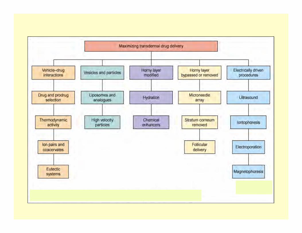
Challenges in Topical & TransdermalDrug Delivery:
Overcoming the Stratum Corneum Barrier
Bozena B. Michniak-Kohn, Ph.D.Ernest Mario School of Pharmacy, Rutgers-The State University
of NJLaboratory for Drug Delivery, NJ Center for Biomaterials
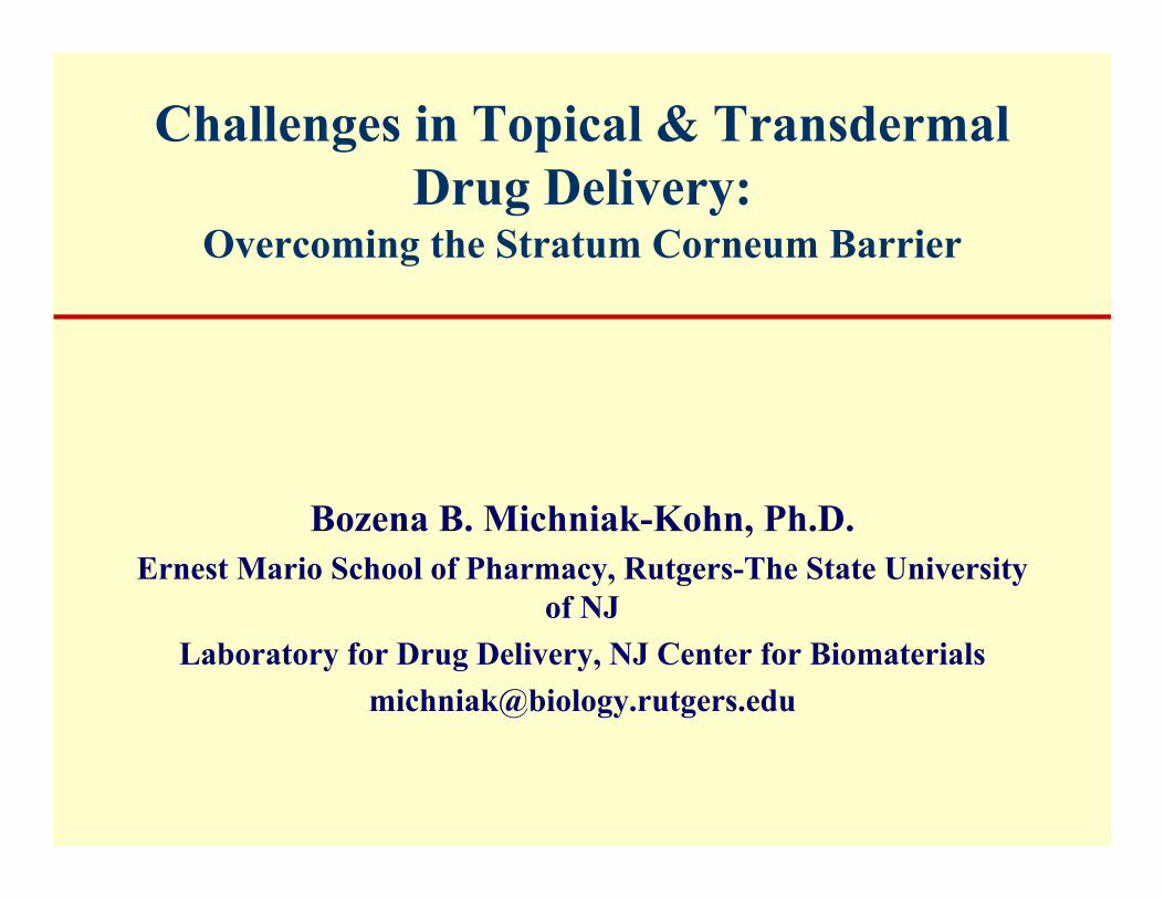
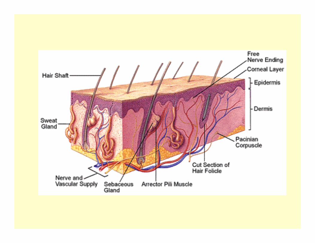
Laboratory for Drug Delivery
• Optimization of transdermal and topical drugdelivery: formulations, carriers (nanospheres)
• Skin constructs /models for testing drugpermeability & irritation
• Drug permeability enhancement: physical &chemical approaches (chemical enhancers/retardants)
• Investigation of drug permeation pathways• In vivo animal pharmacokinetics• Computational modeling and mathematical
predictions
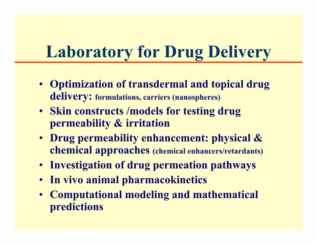
Screening Systems
• Human cadaver/surgical biopsy skin• Human buccal, uterine membrane models• Skin constructs /models for testing drug
permeability & irritation• Normal & transformed human keratinocytes &
fibroblasts for cell toxicity testing• Animal skins: hairless mouse etc.• Polymeric membranes
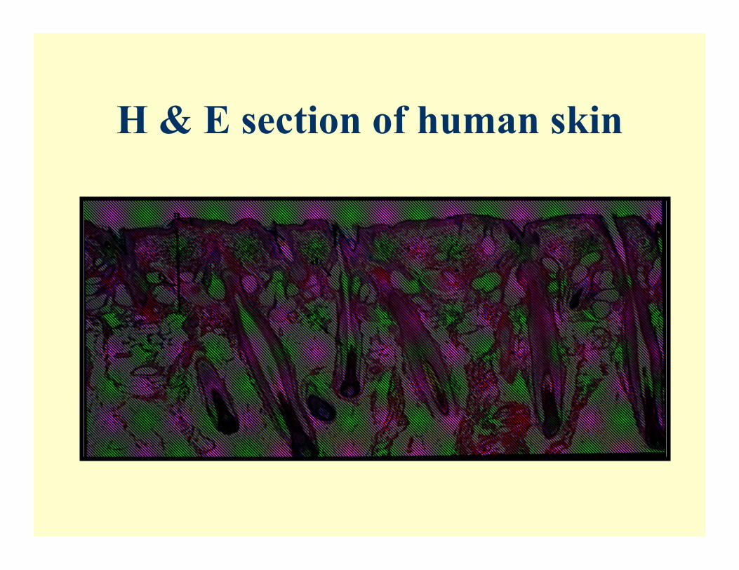
H & E section of human skin
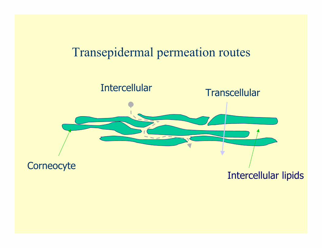
Corneocyte
Intercellular Transcellular
Intercellular lipids
Transepidermal permeation routes
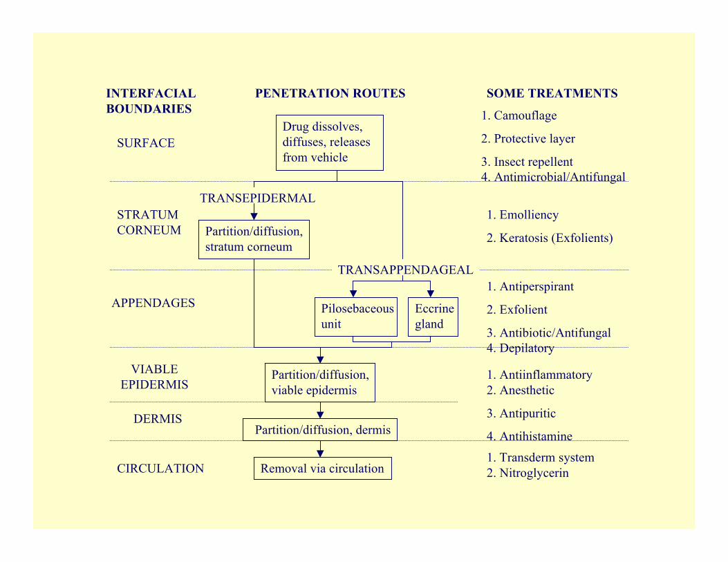
1. Camouflage
2. Protective layer
3. Insect repellent4. Antimicrobial/Antifungal
Drug dissolves,diffuses, releasesfrom vehicle
Partition/diffusion,stratum corneum
1. Emolliency
2. Keratosis (Exfolients)
TRANSEPIDERMAL
1. Antiperspirant
2. Exfolient
3. Antibiotic/Antifungal4. Depilatory
Pilosebaceousunit
Eccrinegland
Partition/diffusion,viable epidermis
Partition/diffusion, dermis
Removal via circulation
1. Antiinflammatory2. Anesthetic
3. Antipuritic
4. Antihistamine
TRANSAPPENDAGEAL
INTERFACIALBOUNDARIES
PENETRATION ROUTES SOME TREATMENTS
SURFACE
STRATUMCORNEUM
APPENDAGES
VIABLEEPIDERMIS
DERMIS
CIRCULATION1. Transderm system2. Nitroglycerin
I. Development of a tissueengineered human skin model
(HSE)
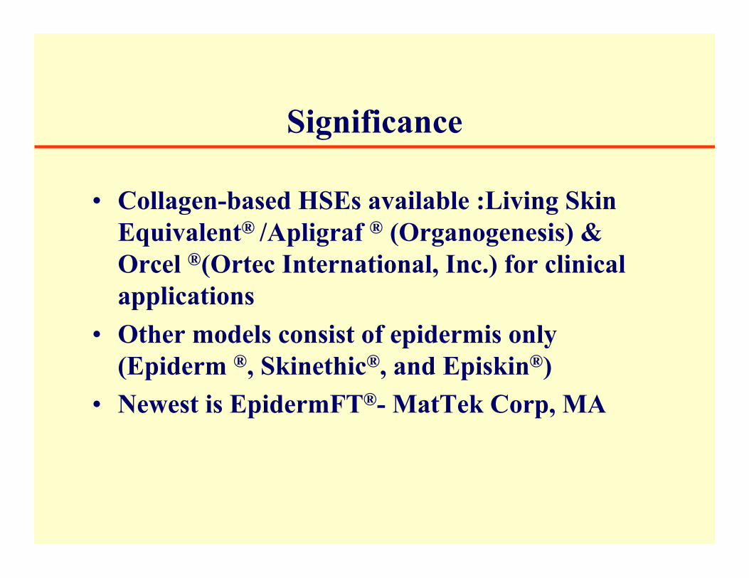
Significance
• Collagen-based HSEs available :Living SkinEquivalent® /Apligraf ® (Organogenesis) &Orcel ®(Ortec International, Inc.) for clinicalapplications
• Other models consist of epidermis only(Epiderm ®, Skinethic®, and Episkin®)
• Newest is EpidermFT®- MatTek Corp, MA
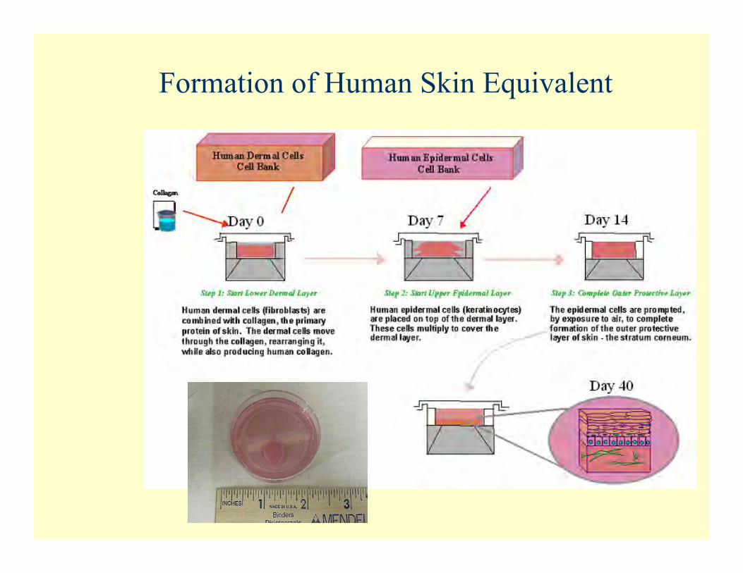
Formation of Human Skin Equivalent
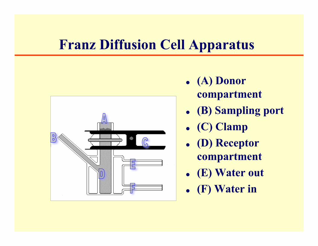
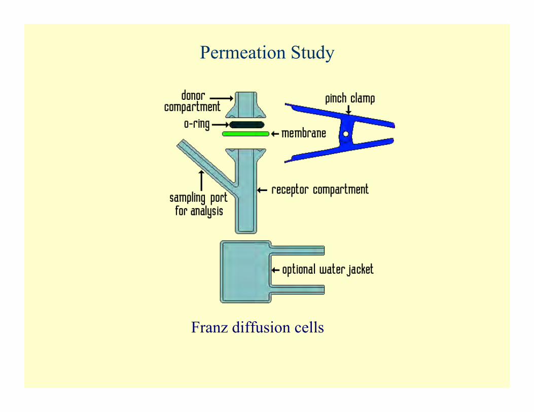
Franz Diffusion Cell Apparatus
(A) Donorcompartment
(B) Sampling port (C) Clamp (D) Receptor
compartment (E) Water out (F) Water in
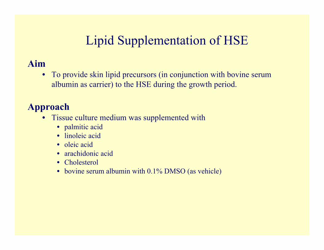
Lipid Supplementation of HSE
Aim• To provide skin lipid precursors (in conjunction with bovine serum
albumin as carrier) to the HSE during the growth period.
Approach• Tissue culture medium was supplemented with
• palmitic acid• linoleic acid• oleic acid• arachidonic acid• Cholesterol• bovine serum albumin with 0.1% DMSO (as vehicle)
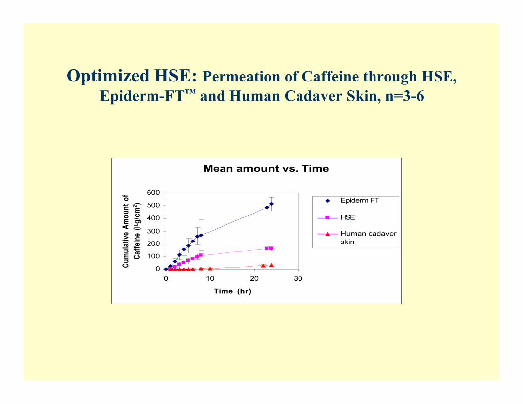
Optimized HSE: Permeation of Caffeine through HSE,Epiderm-FT™ and Human Cadaver Skin, n=3-6
Mean amount vs. Time
0
100
200
300
400
500
600
0 10 20 30
Time (hr)
Cu
mu
lati
ve A
mo
un
t o
f
Caf
fein
e (µ
g/c
m2 ) Epiderm FT
HSE
Human cadaver
skin
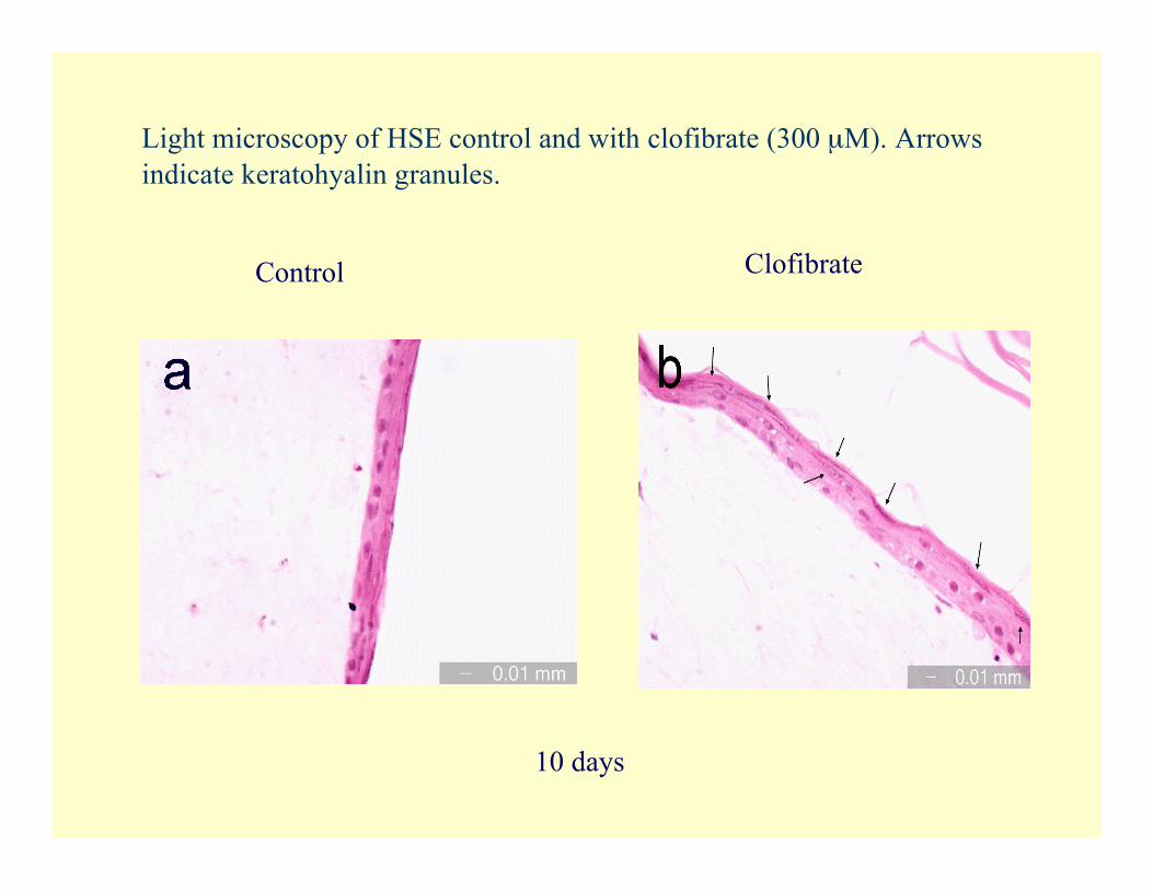
Light microscopy of HSE control and with clofibrate (300 µM). Arrowsindicate keratohyalin granules.
Control
10 days
Clofibrate
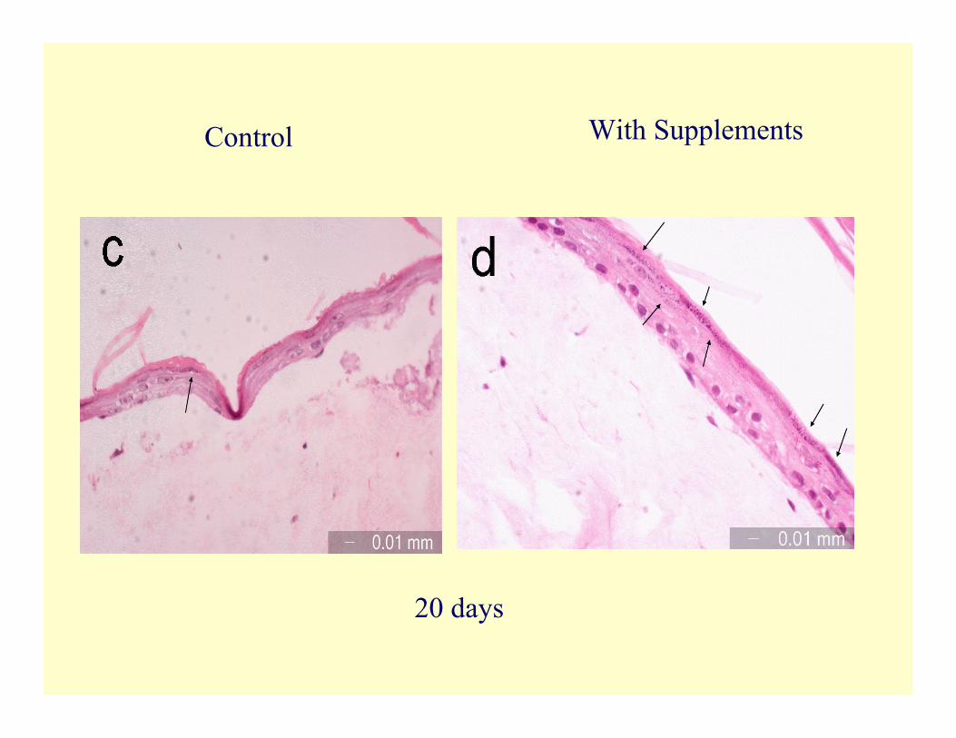
Control With Supplements
20 days
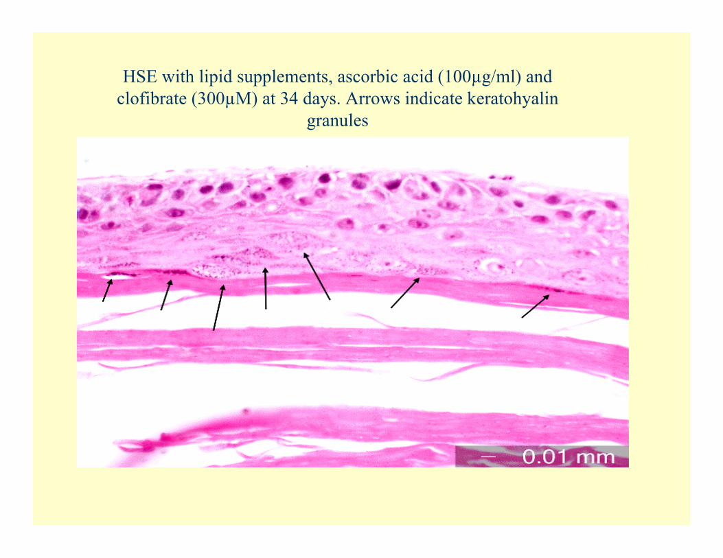
HSE with lipid supplements, ascorbic acid (100µg/ml) andclofibrate (300µM) at 34 days. Arrows indicate keratohyalin
granules
Improving mechanical strength ofHSE model
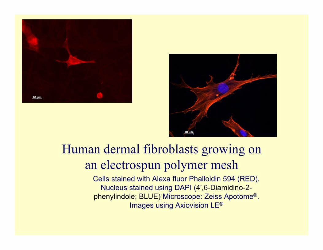
Human dermal fibroblasts growing onan electrospun polymer mesh
Cells stained with Alexa fluor Phalloidin 594 (RED).Nucleus stained using DAPI (4',6-Diamidino-2-
phenylindole; BLUE) Microscope: Zeiss Apotome®.Images using Axiovision LE®
II. Chemical enhancement
Site of Penetration EnhancerActivity
• Site A - Polar head groups of lipids• Site B - Solvent action (water, propylene glycol, ethanol)• Site C - Lipid packing in the stratum corneum
Structures of the Compounds
F
N-
O
S+
CH3
CH3
Br
N-
O
S+
CH3
CH3
I
N-
O
S+
CH3
CH3
N-
O
S+
CH3
CH3
CH3
N-
O
S+
CH3
CH3
CH3
QH1
QH2
QH3
QH4
QH5
Experimental Methods
• Drug : Hydrocortisone (HC)• Vehicle: Propylene glycol (PG)• Enhancers: QH1, QH2, QH3, QH4, QH5• Diffusion cells: Vertical Franz diffusion cells (5.1 ml, 0.64 cm2)• Skin: Human Cadaver Skin
Permeation Study
Franz diffusion cells
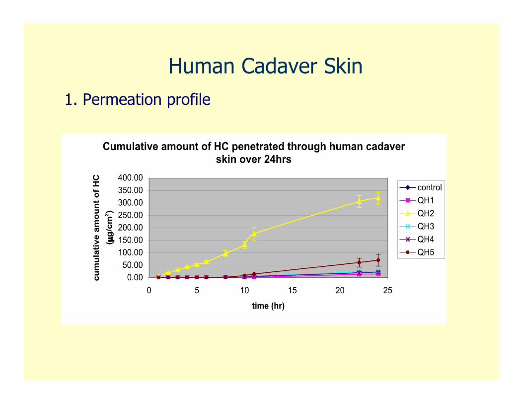
Human Cadaver Skin1. Permeation profile
Cumulative amount of HC penetrated through human cadaver skin over 24hrs
0.00
50.00
100.00
150.00
200.00
250.00
300.00
350.00
400.00
0 5 10 15 20 25
time (hr)
cu
mu
lati
ve
am
ou
nt
of
HC
( µg
/cm
2)
control
QH1
QH2
QH3
QH4
QH5
Permeation Study for QH2
0
1
2
3
4
5
6
7
8
9
10
control 0.1M 0.2M
QH2
En
han
cem
en
t ra
tio
ERQ24
ERFlux
ERTlag

III. Cell Toxicity Studies
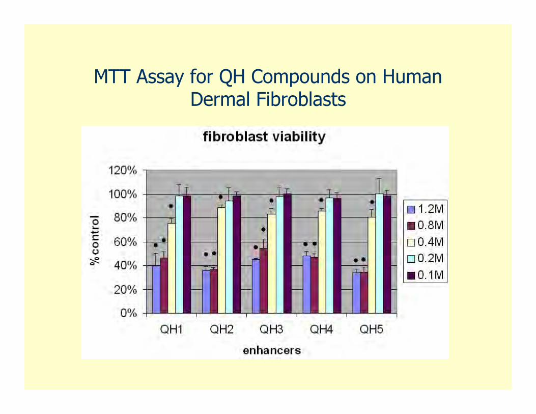
MTT Assay for QH Compounds on HumanDermal Fibroblasts
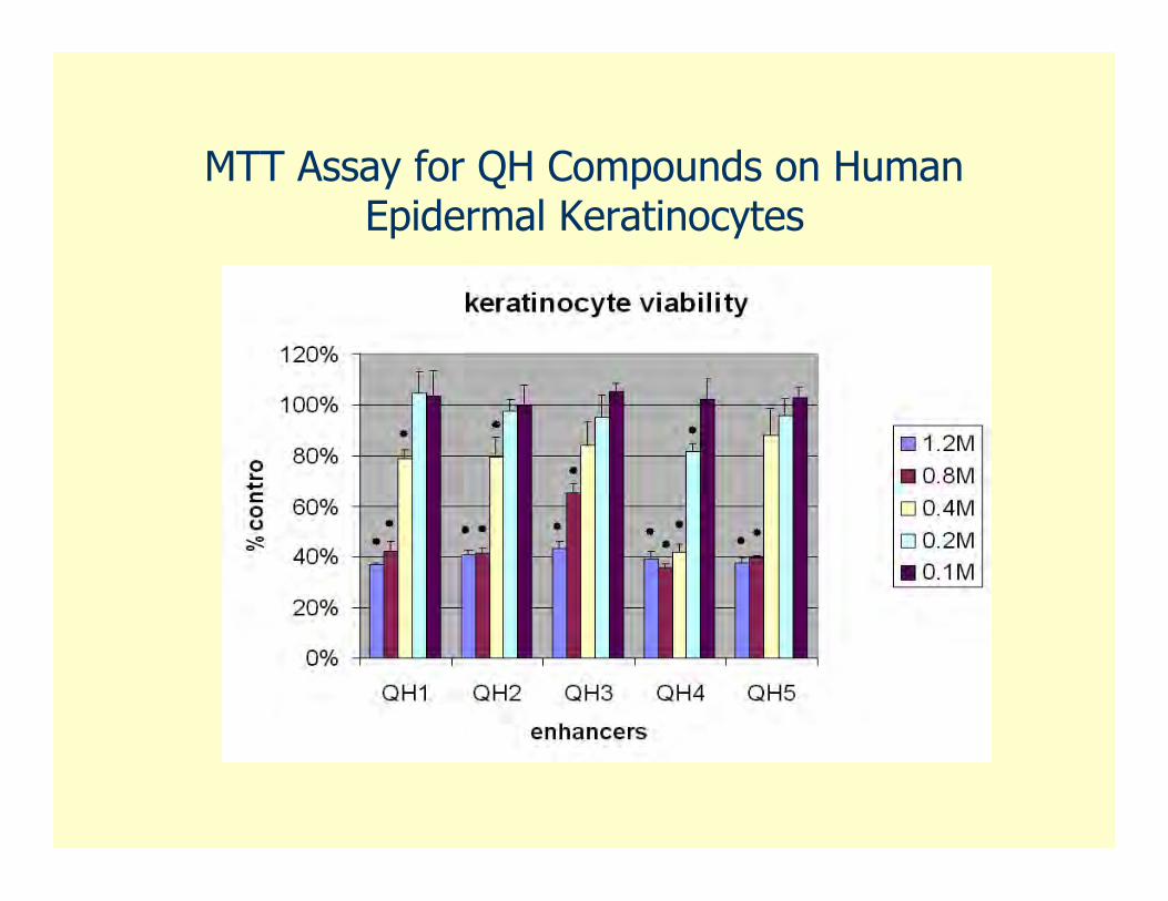
MTT Assay for QH Compounds on HumanEpidermal Keratinocytes
IV. Physical Enhancement
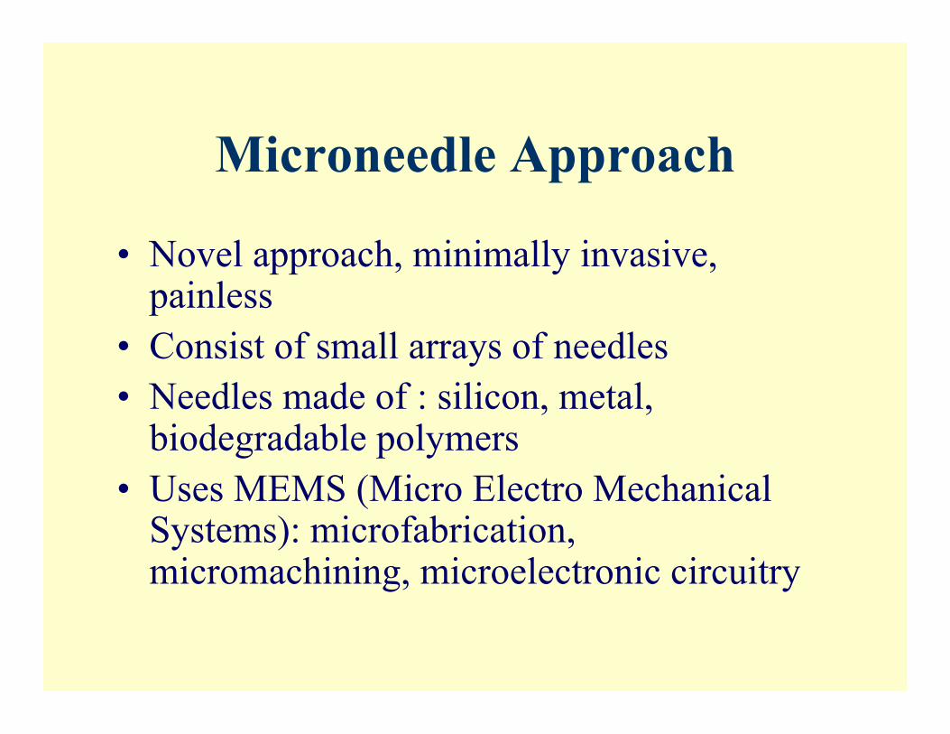
Microneedle Approach
• Novel approach, minimally invasive,painless
• Consist of small arrays of needles• Needles made of : silicon, metal,
biodegradable polymers• Uses MEMS (Micro Electro Mechanical
Systems): microfabrication,micromachining, microelectronic circuitry
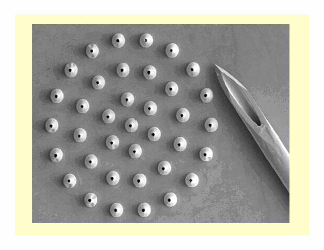
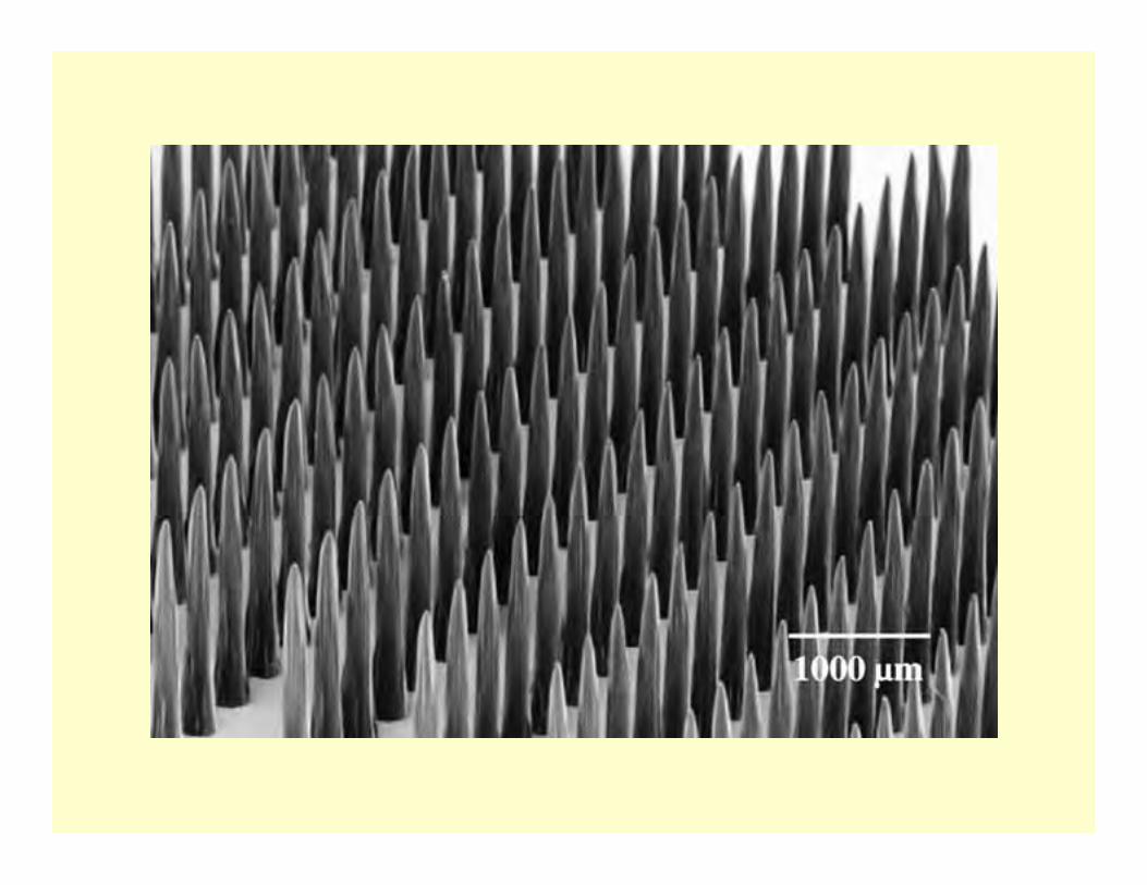
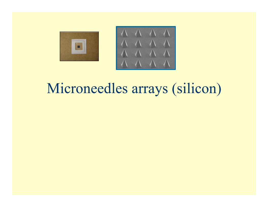
Microneedles arrays (silicon)
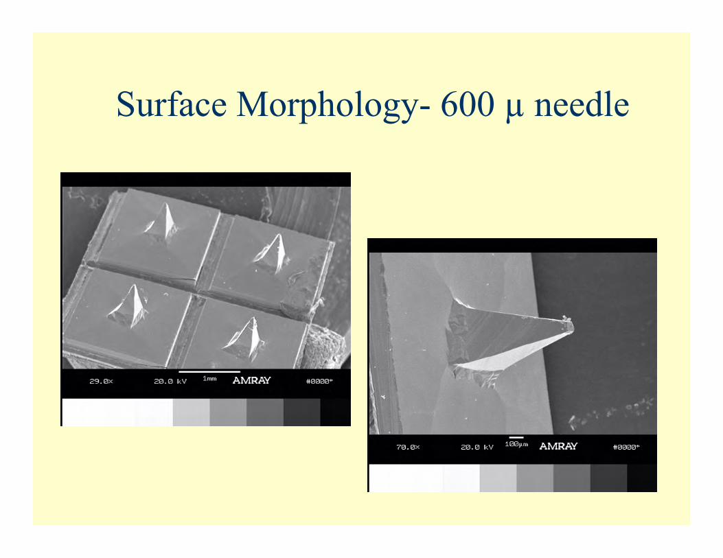
Surface Morphology- 600 µ needle
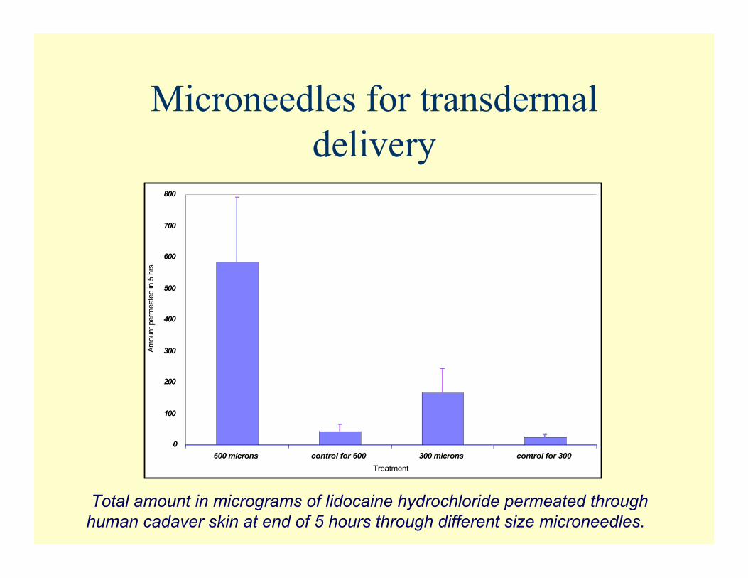
Microneedles for transdermaldelivery
Total amount in micrograms of lidocaine hydrochloride permeated throughhuman cadaver skin at end of 5 hours through different size microneedles.
0
100
200
300
400
500
600
700
800
600 microns control for 600 300 microns control for 300
Treatment
Am
ount perm
eate
d in
5 h
rs
V
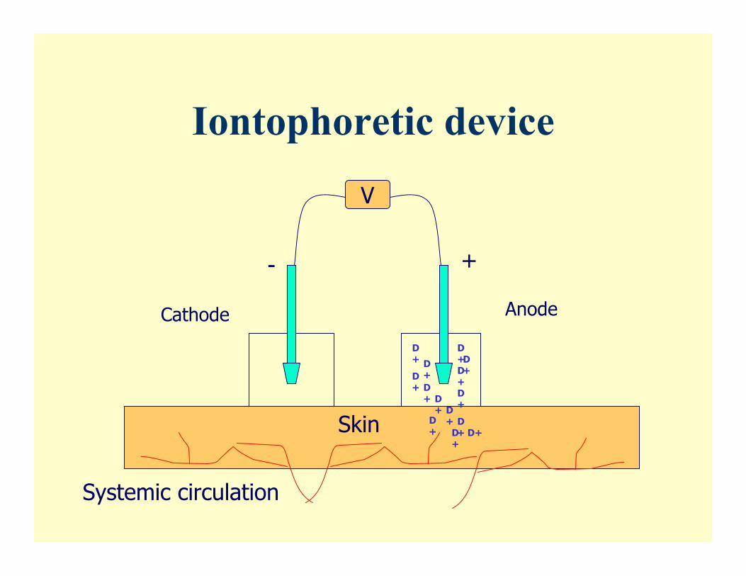
+-
D+ D+ D+
D+
D+
D+
D+
D+
D+
D+ D+ D+D+
D+
AnodeCathode
Skin
Systemic circulation
Iontophoretic device
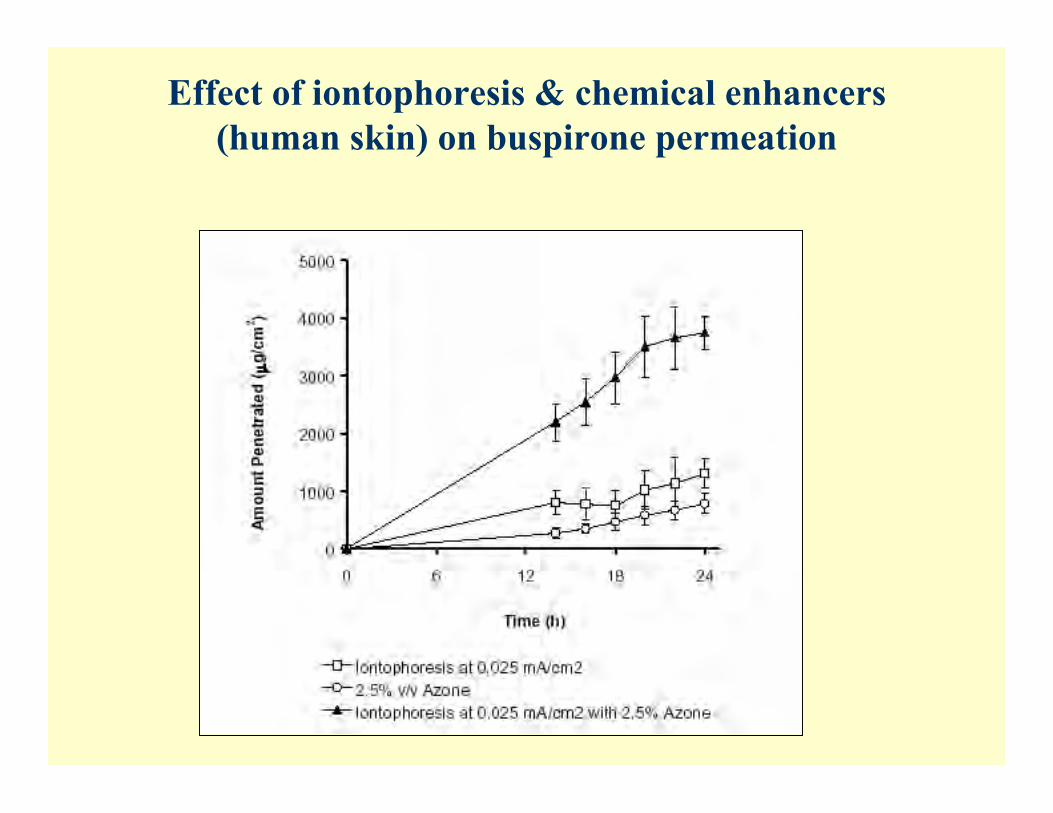
Effect of iontophoresis & chemical enhancers(human skin) on buspirone permeation
V. EM and FTIR Studies
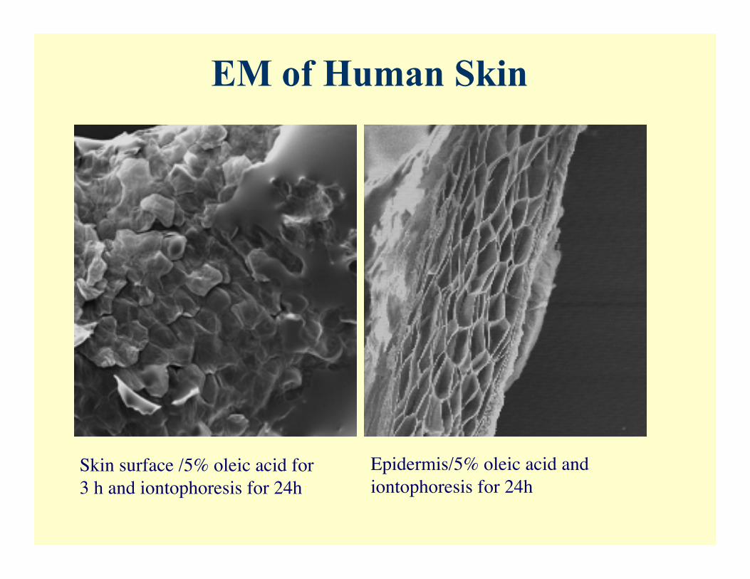
EM of Human Skin
Skin surface /5% oleic acid for3 h and iontophoresis for 24h
Epidermis/5% oleic acid andiontophoresis for 24h
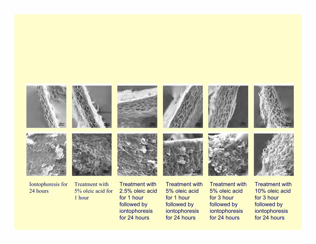
Treatment with10% oleic acidfor 3 hourfollowed byiontophoresisfor 24 hours
Treatment with5% oleic acidfor 3 hourfollowed byiontophoresisfor 24 hours
Treatment with5% oleic acidfor 1 hourfollowed byiontophoresisfor 24 hours
Treatment with2.5% oleic acidfor 1 hourfollowed byiontophoresisfor 24 hours
Treatment with5% oleic acid for1 hour
Iontophoresis for24 hours
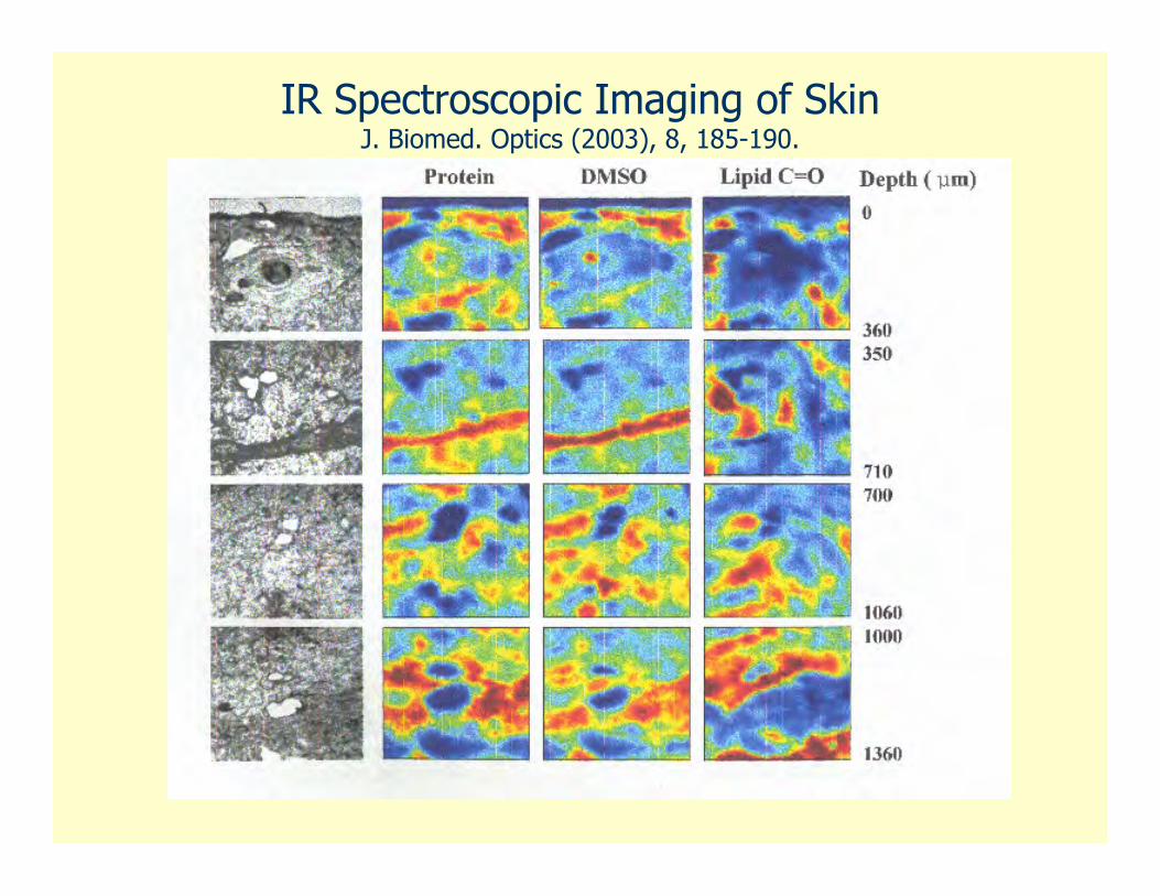
IR Spectroscopic Imaging of SkinJ. Biomed. Optics (2003), 8, 185-190.
Top, from left: Loreto Valenzuela, Peter Zhang, Sonali Bose, Lawrence Hu, LarisaSheihet
Bottom, from left: Rashmi Thakur, Prafulla Chandra, Diksha Kaushik, Priya Batheja
Acknowledgements• Ph.D. students:Rashmi ThakurPriya BathejaDiksha KaushikLawrence HuYiping WangYifan SongQuixi FanMohammad Al-KhaliliHui Chen Chen
• Post Doctoral/FacultyR. Mendelssohn, Ph.D.V. Meidan, Ph.D. (U.K.)J. Kohn, Ph.D.P. Chandra, Ph.D.J. Chapman, Ph.D. (USC)P. Wertz, Ph.D. (Iowa)Funding:NJ Center for Biomaterials, US
Army Contract # W81XWH-04-2-003, Coulter Foundation,Apogee Technologies, Inc. ,Norwood, MA.

Thank you


![Review Article TRANSDERMAL DRUG DELIVERY SYSTEM: A REVIEW · 2015-06-17 · Transdermal drug delivery systems have following benefits:[11,12,13,14,16,28] 1. Transdermal medication](https://img.pdfslide.us/doc/110x75/5ed608e452ff8c0277343f0d/review-article-transdermal-drug-delivery-system-a-review-2015-06-17-transdermal.jpg)


![Transdermal Drug Delivery System [TDDS]](https://img.pdfslide.us/doc/110x75/587c9c9c1a28abfa5e8b7b4f/transdermal-drug-delivery-system-tdds.jpg)