Embed Size (px)
Citation preview
ECG Analysis for
Resting 12-lead ECG
Physician’s Guide For use with Office Medic™ and CardioView ™ v4.4 and higher,
Pocket Medic™ v3.2 and higher and PocketView ™ v1.2 and higher
CAUTION: The computerized interpretation provided is only valid when
used in conjunction with clinical findings. All computer generated tracings
and interpretations must be confirmed by a qualified physician.
Q R S D I A G N O S T I C
6901 East Fish Lake Road, Suite 188
Maple Grove, MN 55369 USA
www.QRSdiagnostic.com
Email: [email protected]
Tel (763) 559-8492
Fax (763) 559-2961

Copyright Notice
Copyright © 2011 by QRS Diagnostic All rights reserved.
No part of the documentation may be reproduced, stored in a retrieval system or
transmitted in any form, or by any means, electronic, mechanical, photocopy,
recording or otherwise, without the prior consent of QRS Diagnostic
Information in this manual is subject to change without notice and does not
represent a commitment on the part of QRS Diagnostic
License Agreement
The software described in this manual is supplied under a license agreement
and may only be used in accordance with the terms of that agreement.
Trademarks
All brands and product names are trademarks or registered trademarks of their
respective holders and are hereby acknowledged
6000-4158 Rev H
(07/2011)
CAUTION: Federal law restricts this device to the sale by or on the order of a
physician.
TABLE OF CONTENTS
CHAPTER 1 – INTRODUCTION .................................................. 4
About the Analysis Module ................................................................ 4
Conventions ....................................................................................... 4
CHAPTER 2 – RHYTHM CRITERIA ............................................ 5
Rhythm Statements ............................................................................ 5
CHAPTER 3 - MORPHOLOGY CRITERIA ................................ 14
Lead Reversal / Dextrocardia ......................................................... 14
Atrial Hypertrophy .......................................................................... 14 Left Atrial Hypertrophy ............................................................... 14
Right Atrial Hypertrophy............................................................. 15
Biatrial Hypertrophy ................................................................... 15
Ventricular Pre-Excitation .............................................................. 15
QRS Abnormalities .......................................................................... 17 Abnormal QRS Amplitude ......................................................... 17
Abnormal QRS Duration ............................................................ 18
Abnormal Axis ............................................................................ 18
Positional Variance Of The Precordials ………………………...18
Bundle Branch Blocks ..................................................................... 19
Ventricular Hypertrophy ................................................................. 23 Right Ventricular Hypertrophy ................................................... 23
Left Ventricular Hypertrophy ...................................................... 25
Biventricular Hypertrophy .......................................................... 26
Infarction ......................................................................................... 27 Anterior Infarction ....................................................................... 31
Lateral Infarction ........................................................................ 32
Possible Lateral Infarction ......................................................... 33
Atypical Q Wave ........................................................................ 35
Repolarisation Troubles .................................................................. 35
CHAPTER 4 - ECG ANALYSIS PERFORMANCE AND ACCURACY ............................................................................... 39
Performance Results ....................................................................... 39 A. CSE Database ....................................................................... 39
B.Cardionics/UCLMorphologyDatabase ................................... 40 C. Cardionics/UCL Rhythm Database....................................... 42
Mapping Morphology Statements to CSE Codes ............................ 43
Mapping of Rhythm Statements to Rhythm Test Codes .................. 48

4 CHAPTER 1 – Introduction
CHAPTER 1 – INTRODUCTION
About the Analysis Module The ECG Analysis Program is a software component that provides analysis and
interpretation of 12 channel ECGs. The ECG analysis program was developed
and tested by Cardionics SA in conjunction with the Université Catholique de
LOUVAIN (UCL). The ECG analysis program has also been independently
evaluated by the Common Standards for Quantitative Electrocardiography
(CSE) Coordinating Center.
Conventions
R* means the R wave duration.
Q* means the Q wave duration.
R'* means the R' wave duration.
I represents Lead I.
R(I) represent the R wave amplitude in Lead I (in mV).
R*(I) represent the R wave duration in Lead I (in ms).
HR = Heart Rate
An index representing the mass of the patient is calculated according to the sex,
height and weight of the patient:
Index = Weight / (Height x Height) kg/m2
If this Index is < 18 in a man and < 17 in a woman, the patient is considered
lightweight.
If this Index is > 28 in a man and > 27 in a woman, the patient is considered
overweight.
If neither height nor weight data are available, the Index is set at 20 (normal
patient).
This Index is of particular usefulness to set the detection thresholds of left
ventricular hypertrophies.
Analysis Statements in this Guide are produced by the software program after
interpretation of the ECG file. Statements are followed by a number in
paranthesis. This number is the code for the statement and is only used only by
the software. Any reference within the criteria to a number is a reference to a
Statement Code only. The descriptions and calculations below the Statements
are intended to provide the interpreting physician with an understanding of how
the software determines each possible statement.
5 CHAPTER 2 – Rhythm Criteria
CHAPTER 2 – RHYTHM CRITERIA
Rhythm Statements
Pacemaker rhythm (001)
Spike is present before the QRS complex.
Regular rhythm (002)
a. Regular rhythm.
AND b. Atrial ectopic beats.
AND c. P wave negative in V1.
Flutter cannot be ruled out (003)
a. Flutter detected with low probability.
AND b. Regular rhythm.
Normal sinus rhythm (004)
a. Regular rhythm.
AND b. P wave positive in V1.
AND c. 60 < HR < 100 bpm.
Sinus bradycardia (005)
a. Regular rhythm and HR < 60 bpm.
OR b. Irregular rhythm and HR < 60 bpm.
Marked sinus bradycardia (006)
a. Regular rhythm.
AND b. P wave positive in V1.
AND c. HR < 45 bpm.
Sinus tachycardia (007)
a. Regular rhythm and HR > 100 bpm.
OR b. Irregular rhythm and HR > 100 bpm.
6 CHAPTER 2 – Rhythm Criteria
Sinus rhythm with 1st degree AV block (008)
a. True if following conditions are met:
1. One QRS class.
AND 2. Regular rhythm.
AND 3. 60 < HR < 100 bpm.
AND 4. PR interval > 200 ms.
OR b. True if following conditions are met:
1. Two QRS classes.
AND 2. Regular rhythm for class 1.
AND 3. 60 < HR < 100 bpm.
AND 4. PR interval > 200 ms.
Sinus bradycardia with 1st degree AV block (009)
a. True if the following conditions are met:
1. One QRS class.
AND 2. Regular rhythm.
AND 3. HR < 60 bpm.
AND 4. PR interval > 200 ms.
OR b. True if the following conditions are met:
1. Two QRS classes.
AND 2. Irregular rhythm but regular rhythm for class 1.
AND 3. HR < 60 bpm.
AND 4. PR interval > 200 ms.
Sinus tachycardia with 1st degree AV block (010)
a. True if the following conditions are met:
1. One QRS class.
AND 2. Regular rhythm.
AND 3. HR > 100 bpm.
AND 4. PR interval > 200 ms.
OR b. True if the following conditions are met:
1. Two QRS classes.
AND 2. Irregular rhythm for class 2 but regular rhythm for class 1.
AND 3. HR > 100 bpm.
AND 4. PR interval > 200 ms.

7 CHAPTER 2 – Rhythm Criteria
Slow atrial rhythm (011)
a. One QRS class.
AND b. Regular rhythm.
AND c. 60 < HR < 100 bpm.
AND d. PR < 100 ms.
AND e. P wave detected in only one or two QRS complexes over the
10 second period.
Junctional rhythm, atrial fibrillation with AV block cannot be ruled
out (013)
a. One QRS class.
AND b. Regular rhythm.
AND c. PR interval null.
AND d. HR < 60 bpm.
AND e. Low probability of atrial fibrillation.
Accelerated junctional rhythm (014)
a. One QRS class.
AND b. Regular rhythm.
AND c. PR interval null.
AND d. 60 < HR < 100 bpm.
AND e. Low probability of atrial fibrillation.
Junctional tachycardia (015)
a. True if the following conditions are met:
1. One QRS class.
AND 2. Regular rhythm.
AND 3. PR interval null.
AND 4. 100 < HR < 220 bpm.
AND 5. Low probability of atrial fibrillation.
OR b. True if the following conditions are met:
1. Two QRS classes.
AND 2. Irregular rhythm in class 2 but regular for class 1.
AND 3. PR interval null.
AND 4. HR > 100 bpm.
Accelerated idioventricular rhythm (017)
Ventricular run or triplets detected with a rate < 100 bpm.
8 CHAPTER 2 – Rhythm Criteria
Ventricular or supraventricular tachycardia with aberrant
conduction (018)
a. One QRS class.
AND b. Regular rhythm.
AND c. QRS duration > 124 ms.
AND d. HR > 100 bpm.
AND e. No P wave detected.
Sinus rhythm (019)
a. Atrial bigeminy, trigeminy or quadrigeminy detected.
AND b. One QRS class.
AND c. Regular rhythm.
AND d. 60 < HR < 100 bpm.
Atrial flutter with 1:1 conduction (020)
a. Flutter detected.
AND b. Regular rhythm.
AND c. HR > 175 bpm.
Atrial flutter with 2:1 conduction (021)
a. Flutter detected.
AND b. Regular rhythm.
AND c. 125 < HR < 175 bpm.
Atrial flutter with 3:1 conduction (022)
a. Flutter detected.
AND b. Regular rhythm.
AND c. 80 < HR < 125 bpm.
Atrial flutter with 4:1 conduction (023)
a. Flutter detected.
AND b. Regular rhythm.
AND c. 60 < HR < 80 bpm.
Atrial flutter with 5:1 conduction (024)
a. Flutter detected.
AND b. Regular rhythm.
AND c. HR < 60 bpm.
9 CHAPTER 2 – Rhythm Criteria
Atrial flutter with variable AV block (025)
a. Flutter detected.
AND b. Irregular rhythm.
Atrial fibrillation (026)
a. QRS detected, no P wave, and irregular rhythm.
OR b. One QRS class, PR interval not defined, and weak P wave.
OR c. Atrial bigeminy (031), atrial trigeminy (032) or atrial
quadrigeminy (083) detected.
OR d. Two QRS classes, PR interval not defined, and irregular rhythm.
OR e. One QRS class, regular rhythm, PR interval null, HR > 60
bpm, and high probability of atrial fibrillation.
OR f. Two QRS classes, irregular rhythm, PR interval null, HR > 60
bpm, and high probability of atrial fibrillation.
Irregular rhythm with atrial extrasystole(s) (029)
Irregular rhythm with at least two short R intervals.
Sinus arrhythmia (030)
Irregular rhythm.
Atrial bigeminy (031)
a. One QRS class and bigeminy detected.
OR b. Two QRS classes, no wide complex, and intermittent bigeminy
detected.
Atrial trigeminy (032)
a. One QRS class and trigeminy detected.
OR b. Two QRS classes, no wide complex, and intermittent
trigeminy detected.
Intermittent atrial bigeminy (033)
a. One QRS class and intermittent bigeminy detected.
OR b. Two QRS classes, no wide complex, and intermittent
trigeminy detected.
(Poorly defined P waves on averaged complexes) (034)
PR interval not defined.
10 CHAPTER 2 – Rhythm Criteria
Atrial fibrillation cannot be ruled out (035)
a. PR interval not defined and low probability of atrial fibrillation.
OR b. Atrial bigeminy (031) or atrial trigeminy (032) detected, PR
interval not defined, and low probability of atrial fibrillation.
OR c. Mobitz (039), (040), (041), (042) or (043) detected, irregular
rhythm, PR interval not defined, and low probability of atrial
fibrillation.
OR d. One QRS class, regular rhythm, PR interval null, and low
probability of atrial fibrillation.
(Low voltage P waves on averaged complexes) (036)
The P wave on averaged beat is weak or undetectable.
Atrial ectopic beats (037)
a. Regular rhythm.
AND b. Atrial ectopic beats detected.
AND c. P wave negative in V1.
Atrial-ventricular dissociation between : (039)
Mobitz type I detected and HR > 45 bpm.
AV block (Mobitz type II) with 2:1 conduction (040)
Mobitz type II with 2:1 conduction detected.
AV block (Mobitz type I) Wenckebach phenomenon (041)
Mobitz type I detected and number of prolonged intervals > 0.
AV block (Mobitz type II) (042)
Mobitz type II detected.
AV block type III (043)
Mobitz type III detected and HR > 45 bpm.
Ventricular bigeminy (044)
a. Two QRS classes; one wide class and one narrow class.
AND b. Bigeminy detected.

11 CHAPTER 2 – Rhythm Criteria
Ventricular trigeminy (045)
a. Two QRS classes; one wide class and one narrow class.
AND b. Irregular rhythm and trigeminy detected.
Intermittent ventricular bigeminy (046)
a. Intermittent bigeminy detected, two QRS classes, and irregular
rhythm.
OR b. Two QRS classes; one wide class and one narrow class, and
bigeminy detected.
(Wandering baseline!) (047)
The baseline is not flat enough.
Ventricular extrasystole(s) with bundle branch block (050)
a. Irregular rhythm.
AND b. Two QRS classes, of which one is wide.
AND c. Two short RR intervals detected.
Unifocal ventricular extrasystole(s) (052)
Two QRS classes and extrasystole(s) detected with the same morphology.
Multifocal ventricular extrasystole(s) (053)
Three QRS classes and extrasystole(s) detected with multiple
morphology.
Atrial extrasystole(s) (055)
One QRS class several long intervals, and no run detected.
Ventricular extrasystole(s) (056)
a. Several QRS classes without bigeminy or trigeminy.
OR b. Ventricular extrasystoles detected in bigeminy or trigeminy.
OR c. Two QRS classes and extrasystole(s) detected.
Interpolated ventricular extrasystole(s) (057)
Two QRS classes, extrasystoles detected, and normal interval before and
after the extrasystoles.
12 CHAPTER 2 – Rhythm Criteria
Ventricular extrasystole(s) with full compensation (058)
Two QRS classes, extrasystoles detected, and long interval after the
extrasystoles.
Ventricular extrasystole(s) without full compensation (059)
Two QRS classes, extrasystoles detected, and normal interval after the
extrasystoles.
Supraventricular extrasystole(s) (060)
One QRS class, extrasystoles detected, several long intervals, and
supraventricular extrasystoles detected between the RR intervals.
Ventricular couplets (068)
Two QRS classes, several long intervals, and couplet detected (two
ectopic beats in succession).
Ventricular triplets (069)
Two QRS classes, several long intervals, and triplet detected (three
ectopic beats in succession).
Run of ventricular extrasystoles (070)
Two QRS classes, several long intervals, and run detected (more than
three ectopic beats in succession).
Atrial couplets (071)
One QRS class, several long intervals, and couplet detected (two ectopic
beats in succession).
Atrial triplets (072)
One QRS class, several long intervals, and triplet detected (three ectopic
beats in succession).
Run of atrial extrasystoles (073)
One QRS class, several long intervals, and run detected (more than three
ectopic beats in succession).
Atrial flutter with a variable block cannot be ruled out (075)
Flutter detected with low risk and irregular rhythm.

13 CHAPTER 2 – Rhythm Criteria
(False beat excluded) (076)
An incorrect beat has been detected caused probably by noise. This beat
has been rejected for the analysis.
Very low flutter probability (078)
Possible flutter detected with very low probability.
Atrial pacing (079)
Spikes are present before each QRS.
Sinus arrhythmia with 1st degree AV block (080)
Irregular rhythm and PR interval > 100 ms.
Pacing??? (082)
Spikes are present but the software does not recognize the type of
pacemaker.
Atrial quadrigeminy (083)
a. One QRS class and quadrigeminy detected.
OR b. Two QRS classes, not too wide, and quadrigeminy detected.
Ventricular quadrigeminy (084)
Two QRS classes, one wide class, and quadrigeminy detected.
AV block type I (085)
Regular rhythm and PR interval > 100 ms.
Extreme bradycardia (086)
Regular rhythm and HR < 45 bpm.
Idioventricular rhythm (087)
a. One QRS class.
AND b. Regular rhythm.
AND c. QRS duration > 124 ms.
AND d. HR < 100 bpm.
AND e. No P wave detected.
14 CHAPTER 3 - Morphology Critera
CHAPTER 3 - MORPHOLOGY CRITERIA
Lead Reversal / Dextrocardia Note: If one of these conditions is detected no further analysis is performed.
Inverted limb electrodes (089)
True if the following conditions are met:
a. Q wave present in I (Q*(I) > 12 ms.).
AND b. QRS duration < 110 ms.
AND c. P wave negative in Lead I.
AND d. S(I) > (R(I) + 0.2 mV).
AND e. R(V1) or R(V2) > (S(V1) + 0.3 mV) or (S(V2) + 0.3 mV).
AND f. R(V1) or R(V2) > R(V5).
Congenital dextrocardia (090)
True if the following conditions are met:
a. Q wave present in I (Q*(I) > 12 ms.).
AND b. QRS duration < 110 ms.
AND c. P wave negative in Lead I.
AND d. S(I) > (R(I) + 0.2 mV).
Atrial Hypertrophy
Left Atrial Hypertrophy
Possible left atrial hypertrophy (092)
a. Terminal surface of P in V1 > 0.04 mm/s
AND b. P(V1) < 1 mm.
Left atrial hypertrophy (091)
a. Terminal surface of P in V1 > 0.04 mm/s
AND b. P axis -15°
AND c. P duration > 110 ms
15 CHAPTER 3 - Morphology Critera
Right Atrial Hypertrophy
Possible right atrial hypertrophy (096)
True if one of the following conditions is met:
a. P(II) > 0.25 mV or P’(II) > 0.25 mV.
OR b. P(III) > 0.25 mV or P’(III) > 0.25 mV.
OR c. P(aVF) > 0.25 mV or P’(aVF) > 0.25 mV.
OR d. P axis > 75°.
Right atrial hypertrophy (095)
True if any three of the following conditions are met:
a. P(II) > 0.25 mV or P’(II) > 0.25 mV.
b. P(III) > 0.25 mV or P’(III) > 0.25 mV.
c. P(aVF) > 0.25 mV or P’(aVF) > 0.25 mV.
d. P axis > 75°.
Biatrial Hypertrophy
Possible biatrial hypertrophy (099)
a. Possible left atrial hypertrophy (092).
AND b. Possible right atrial hypertrophy (096).
Biatrial hypertrophy (098)
a. Right atrial hypertrophy (095) or left atrial hypertrophy (091).
AND b. Possible right atrial hypertrophy (096) or Possible left atrial
hypertrophy (092).
Ventricular Pre-Excitation Note: If ventricular pre-excitation is detected no further analysis is performed.
Wolff-Parkinson-White syndrome (104)
Delta waves in at least three of the 12 leads.
Wolff-Parkinson-White syndrome (type A) (102)
a. R(V1) > S(V1).
AND b. Delta waves in at least three leads.
AND c. P-R interval < 120 ms.
16 CHAPTER 3 - Morphology Critera
AND d. No atrial fibrillation.
17 CHAPTER 3 - Morphology Critera
Wolff-Parkinson-White syndrome (type B) (103)
a. R(V1) < S(V1).
AND b. Delta waves in at least three leads.
AND c. P-R interval < 120 ms.
AND d. No atrial fibrillation.
Ventricular pre-excitation syndrome cannot be ruled out (105)
a. Delta waves in at least two leads.
AND b. P-R interval < 100 ms.
QRS Abnormalities
Abnormal QRS Amplitude
Low QRS voltages (109)
a. Maximum amplitude of < 0.5 mV. in two of the limb leads.
AND b. Maximum amplitude of < 1 mV in four of the precordial leads.
Voltage beyond the criteria for left ventricular hypertrophy, may be
normal variant by the weight (093)
a. R(aVL) >= 1.2mV.
OR b. R(I) or R(II) >= 1.5mV.
OR c. (R(III) > 2mV) or (S(III) > 1.2mV).
OR d. R(aVF) > 2mV.
OR e. (S(V1) > 2.2mV) or (S(V2) > 2.5mV).
OR f. (R(V3) > 2mV) or (S(V3) > 2.2mV).
OR g. (R(V4) > 2mV) or (S(V4) > 1.7mV).
OR h. (R(V5) > 2.2mV) or (R(V6) > 2mV).
OR i. (R(V5) + S(V1)) > 3.5mV for women or > 4mV for men.
OR j. (R(V6) + S(V1)) > 3.5mV for women or > 4mV for men.
Note: If one of the above conditions occurs in a proportion of at least 90%, this
criteria applies; if a LBBB is present this criteria does not apply.
18 CHAPTER 3 - Morphology Critera
Abnormal QRS Duration Note: The following statements are calculated only in the absence of LBBB or
RBBB.
Non systematic minor intraventricular block (118)
115 ms < QRS duration 130 ms.
Non systematic major intraventricular block (117)
QRS duration > 130 ms.
Abnormal Axis
Right QRS axis deviation (123)
QRS Axis 130°
Left QRS axis deviation (124)
QRS Axis -30°
Positional Variance Of The Precordials
Premature QRS transition in right precordials, positional variance
(128)
a. R/S > 1 in V1 or V2.
AND b. No posterior myocardial infarction and no inferior myocardial
infarction or presence of right ventricular hypertrophy.
Late QRS transition in left precordials, positional variance (130)
a. R/S < 1 in V4 or V5.
AND b. Neither infarction nor left ventricular hypertrophy are present.
19 CHAPTER 3 - Morphology Critera
Bundle Branch Blocks
Incomplete left bundle branch block (112)
True if QRS duration > 100 ms and any four of the following
conditions are met:
a. S(V1) wave > 3 x R(V1) wave and S(V2) wave > 3 x R(V2) wave.
b. Two of the following conditions are met:
1. R*(I) > 80 ms.
2. R*(V5) > 80 ms.
3. R*(V6) > 80 ms.
c. Two of the following conditions are met:
1. Q(I) < 1 mm.
2. Q(V5) < 1 mm.
3. Q(V6) < 1 mm.
d. T(V5) < -1 mm or T(V6) < -1 mm.
e. Delayed intrinsicoid deflection in V5 and V6.
Complete left bundle branch block (110)
True if QRS duration > 120 ms and any four of the following
conditions are met:
a. (S(V1) > 3 x R(V1)) and (S(V2) > 3 x R(V2)).
b. True if two of the following conditions are met:
1. R*(I) > 80 ms.
2. R*(V5) > 80 ms.
3. R*(V6) > 80 ms.
c. True if two of the following conditions are met:
1. Q(I) < 1 mm.
2. Q(V5) < 1 mm.
3. Q(V6) < 1 mm.
d. T(V5) < -1 mm or T(V6) < -1 mm.
e. Delayed intrinsicoid deflection in V5 and V6.
20 CHAPTER 3 - Morphology Critera
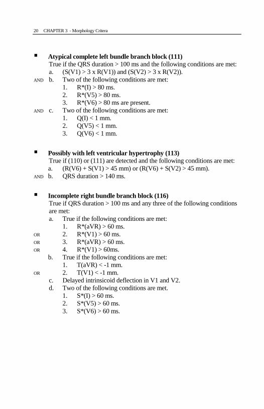
Atypical complete left bundle branch block (111)
True if the QRS duration > 100 ms and the following conditions are met:
a. (S(V1) > 3 x R(V1)) and (S(V2) > 3 x R(V2)).
AND b. Two of the following conditions are met:
1. R*(I) > 80 ms.
2. R*(V5) > 80 ms.
3. R*(V6) > 80 ms are present.
AND c. Two of the following conditions are met:
1. Q(I) < 1 mm.
2. Q(V5) < 1 mm.
3. Q(V6) < 1 mm.
Possibly with left ventricular hypertrophy (113)
True if (110) or (111) are detected and the following conditions are met:
a. (R(V6) + S(V1) > 45 mm) or (R(V6) + S(V2) > 45 mm).
AND b. QRS duration > 140 ms.
Incomplete right bundle branch block (116)
True if QRS duration > 100 ms and any three of the following conditions
are met:
a. True if the following conditions are met:
1. R*(aVR) > 60 ms.
OR 2. R*(V1) > 60 ms.
OR 3. R*(aVR) > 60 ms.
OR 4. R*(V1) > 60ms.
b. True if the following conditions are met:
1. T(aVR) < -1 mm.
OR 2. T(V1) < -1 mm.
c. Delayed intrinsicoid deflection in V1 and V2.
d. Two of the following conditions are met.
1. S*(I) > 60 ms.
2. S*(V5) > 60 ms.
3. S*(V6) > 60 ms.

21 CHAPTER 3 - Morphology Critera
Complete right bundle branch block (114)
True if QRS duration > 120 ms and any three of the following conditions
are met:
a. True if the following conditions are met:
1. R*(aVR) > 60 ms.
OR 2. R*(V1) > 60 ms.
OR 3. R*(aVR) > 60 ms.
OR 4. R*(V1) > 60ms.
b. True if the following conditions are met:
1. T(aVR) < -1 mm.
OR 2. T(V1) <-1 mm.
c. Delayed intrinsicoid deflection in V1 and V2.
d. True if two of the following conditions are met:
1. S*(I) > 60 ms.
2. S*(V5) > 60 ms.
3. S*(V6) > 60 ms.
Compatible with a bundle branch block (131)
True if the following conditions are met:
a. QRS duration > 95 ms.
AND b. No left bundle branch block.
AND c. (R(V1) > 1 mm) and (R’(V1) > 1 mm).
Atypical complete right bundle branch block (115)
True if the following conditions are met:
a. QRS duration > 120 ms.
AND b. True if any two of the following conditions are met:
1. S*(I) > 60 ms.
2. S*(V5) > 60 ms.
3. S*(V6) > 60 ms.
RSR' in V1, could be normal (280)
True if the following conditions are met:
a. QRS duration > 90 ms.
AND b. No left bundle branch block.
AND c. True if any one of the following conditions is met:
1. (R(aVR) > 1 mm) and (R’(aVR) > 1 mm).
OR 2. (R(V1) > 1 mm) and (R’(V1) > 1 mm).
22 CHAPTER 3 - Morphology Critera
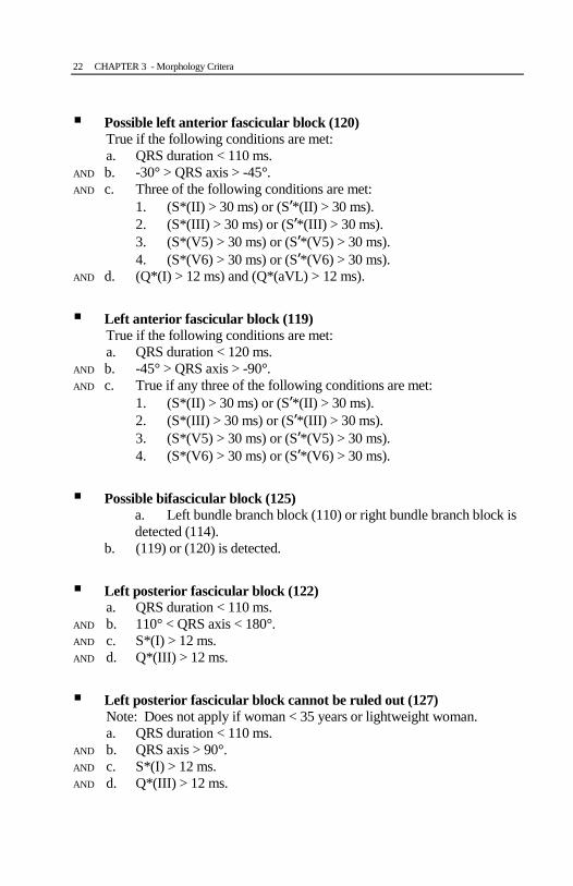
Possible left anterior fascicular block (120)
True if the following conditions are met:
a. QRS duration < 110 ms.
AND b. -30° > QRS axis > -45°.
AND c. Three of the following conditions are met:
1. (S*(II) > 30 ms) or (S’*(II) > 30 ms).
2. (S*(III) > 30 ms) or (S’*(III) > 30 ms).
3. (S*(V5) > 30 ms) or (S’*(V5) > 30 ms).
4. (S*(V6) > 30 ms) or (S’*(V6) > 30 ms).
AND d. (Q*(I) > 12 ms) and (Q*(aVL) > 12 ms).
Left anterior fascicular block (119)
True if the following conditions are met:
a. QRS duration < 120 ms.
AND b. -45° > QRS axis > -90°.
AND c. True if any three of the following conditions are met:
1. (S*(II) > 30 ms) or (S’*(II) > 30 ms).
2. (S*(III) > 30 ms) or (S’*(III) > 30 ms).
3. (S*(V5) > 30 ms) or (S’*(V5) > 30 ms).
4. (S*(V6) > 30 ms) or (S’*(V6) > 30 ms).
Possible bifascicular block (125)
a. Left bundle branch block (110) or right bundle branch block is
detected (114).
b. (119) or (120) is detected.
Left posterior fascicular block (122)
a. QRS duration < 110 ms.
AND b. 110° < QRS axis < 180°.
AND c. S*(I) > 12 ms.
AND d. Q*(III) > 12 ms.
Left posterior fascicular block cannot be ruled out (127)
Note: Does not apply if woman < 35 years or lightweight woman.
a. QRS duration < 110 ms.
AND b. QRS axis > 90°.
AND c. S*(I) > 12 ms.
AND d. Q*(III) > 12 ms.
23 CHAPTER 3 - Morphology Critera
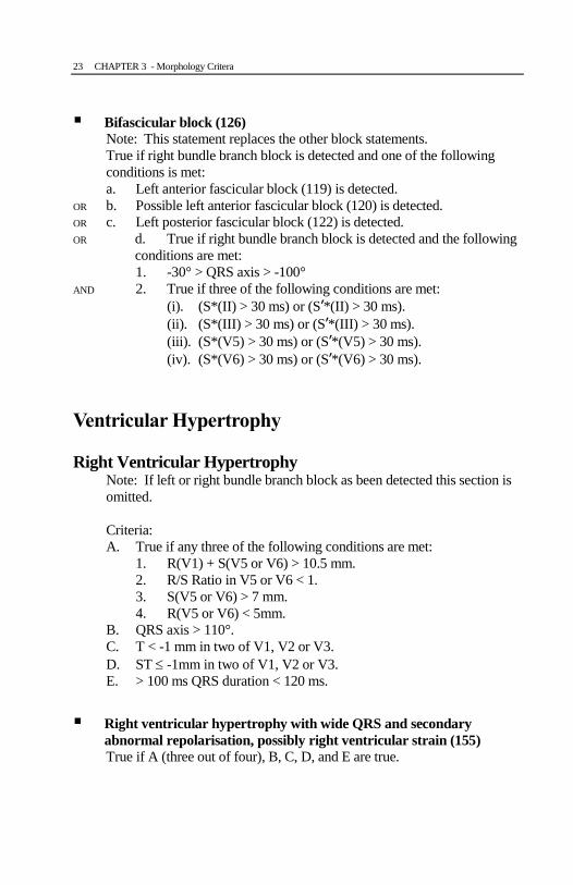
Bifascicular block (126)
Note: This statement replaces the other block statements.
True if right bundle branch block is detected and one of the following
conditions is met:
a. Left anterior fascicular block (119) is detected.
OR b. Possible left anterior fascicular block (120) is detected.
OR c. Left posterior fascicular block (122) is detected.
OR d. True if right bundle branch block is detected and the following
conditions are met:
1. -30° > QRS axis > -100°
AND 2. True if three of the following conditions are met:
(i). (S*(II) > 30 ms) or (S’*(II) > 30 ms).
(ii). (S*(III) > 30 ms) or (S’*(III) > 30 ms).
(iii). (S*(V5) > 30 ms) or (S’*(V5) > 30 ms).
(iv). (S*(V6) > 30 ms) or (S’*(V6) > 30 ms).
Ventricular Hypertrophy
Right Ventricular Hypertrophy Note: If left or right bundle branch block as been detected this section is
omitted.
Criteria:
A. True if any three of the following conditions are met:
1. R(V1) + S(V5 or V6) > 10.5 mm.
2. R/S Ratio in V5 or V6 < 1.
3. S(V5 or V6) > 7 mm.
4. R(V5 or V6) < 5mm.
B. QRS axis > 110°.
C. T < -1 mm in two of V1, V2 or V3.
D. ST -1mm in two of V1, V2 or V3.
E. > 100 ms QRS duration < 120 ms.
Right ventricular hypertrophy with wide QRS and secondary
abnormal repolarisation, possibly right ventricular strain (155)
True if A (three out of four), B, C, D, and E are true.
24 CHAPTER 3 - Morphology Critera
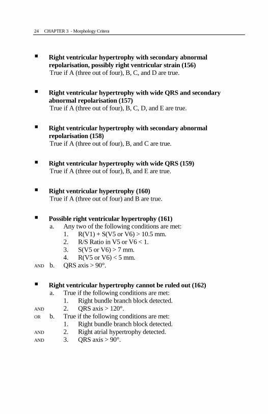
Right ventricular hypertrophy with secondary abnormal
repolarisation, possibly right ventricular strain (156)
True if A (three out of four), B, C, and D are true.
Right ventricular hypertrophy with wide QRS and secondary
abnormal repolarisation (157)
True if A (three out of four), B, C, D, and E are true.
Right ventricular hypertrophy with secondary abnormal
repolarisation (158)
True if A (three out of four), B, and C are true.
Right ventricular hypertrophy with wide QRS (159)
True if A (three out of four), B, and E are true.
Right ventricular hypertrophy (160)
True if A (three out of four) and B are true.
Possible right ventricular hypertrophy (161)
a. Any two of the following conditions are met:
1. R(V1) + S(V5 or V6) > 10.5 mm.
2. R/S Ratio in V5 or V6 < 1.
3. S(V5 or V6) > 7 mm.
4. R(V5 or V6) < 5 mm.
AND b. QRS axis > 90°.
Right ventricular hypertrophy cannot be ruled out (162)
a. True if the following conditions are met:
1. Right bundle branch block detected.
AND 2. QRS axis > 120°.
OR b. True if the following conditions are met:
1. Right bundle branch block detected.
AND 2. Right atrial hypertrophy detected.
AND 3. QRS axis > 90°.
25 CHAPTER 3 - Morphology Critera
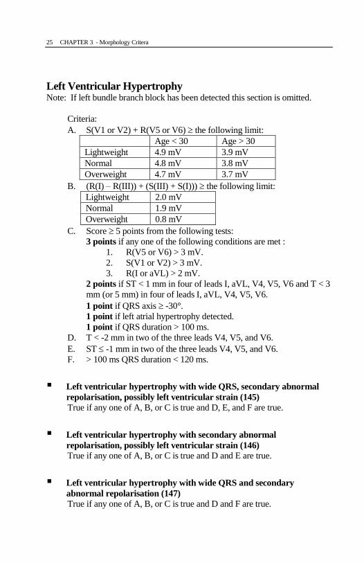
Left Ventricular Hypertrophy Note: If left bundle branch block has been detected this section is omitted.
Criteria:
A. S(V1 or V2) + R(V5 or V6) the following limit:
Age < 30 Age > 30
Lightweight 4.9 mV 3.9 mV
Normal 4.8 mV 3.8 mV
Overweight 4.7 mV 3.7 mV
B. (R(I) – R(III)) + (S(III) + S(I))) the following limit:
Lightweight 2.0 mV
Normal 1.9 mV
Overweight 0.8 mV
C. Score 5 points from the following tests:
3 points if any one of the following conditions are met :
1. R(V5 or V6) > 3 mV.
2. S(V1 or V2) > 3 mV.
3. R(I or aVL) > 2 mV.
2 points if ST < 1 mm in four of leads I, aVL, V4, V5, V6 and T < 3
mm (or 5 mm) in four of leads I, aVL, V4, V5, V6.
1 point if QRS axis -30°.
1 point if left atrial hypertrophy detected.
1 point if QRS duration > 100 ms.
D. T < -2 mm in two of the three leads V4, V5, and V6.
E. ST -1 mm in two of the three leads V4, V5, and V6.
F. > 100 ms QRS duration < 120 ms.
Left ventricular hypertrophy with wide QRS, secondary abnormal
repolarisation, possibly left ventricular strain (145)
True if any one of A, B, or C is true and D, E, and F are true.
Left ventricular hypertrophy with secondary abnormal
repolarisation, possibly left ventricular strain (146)
True if any one of A, B, or C is true and D and E are true.
Left ventricular hypertrophy with wide QRS and secondary
abnormal repolarisation (147)
True if any one of A, B, or C is true and D and F are true.
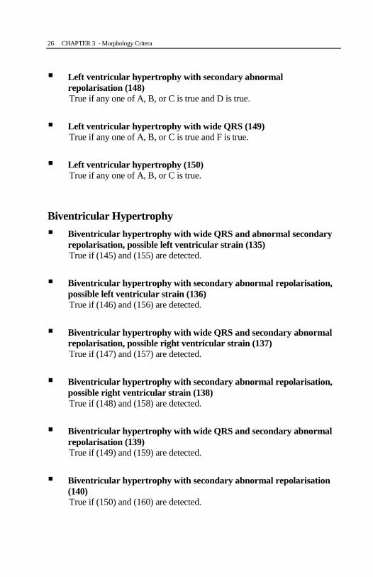
26 CHAPTER 3 - Morphology Critera
Left ventricular hypertrophy with secondary abnormal
repolarisation (148)
True if any one of A, B, or C is true and D is true.
Left ventricular hypertrophy with wide QRS (149)
True if any one of A, B, or C is true and F is true.
Left ventricular hypertrophy (150)
True if any one of A, B, or C is true.
Biventricular Hypertrophy
Biventricular hypertrophy with wide QRS and abnormal secondary
repolarisation, possible left ventricular strain (135)
True if (145) and (155) are detected.
Biventricular hypertrophy with secondary abnormal repolarisation,
possible left ventricular strain (136)
True if (146) and (156) are detected.
Biventricular hypertrophy with wide QRS and secondary abnormal
repolarisation, possible right ventricular strain (137)
True if (147) and (157) are detected.
Biventricular hypertrophy with secondary abnormal repolarisation,
possible right ventricular strain (138)
True if (148) and (158) are detected.
Biventricular hypertrophy with wide QRS and secondary abnormal
repolarisation (139)
True if (149) and (159) are detected.
Biventricular hypertrophy with secondary abnormal repolarisation
(140)
True if (150) and (160) are detected.

27 CHAPTER 3 - Morphology Critera
Biventricular hypertrophy with wide QRS (141)
True if (151) and (161) are detected.
Biventricular hypertrophy (142)
True if (152) and (162) are detected.
Infarction
Test 1: Q wave detected if:
1. Q length >= 40ms.
2. (3 x amplitude of Q) > amplitude R, and amplitude of Q >= 0.150
mV.
3. The following three conditions are met (QS aspect):
a. Length Q < 15 ms and amplitude of Q < 0.07 mV.
AND b. Length R < 15 ms and amplitude of R < 0.07 mV.
AND c. Length S > 40 ms and length S’ < 15 ms.
Specific test for the aVL lead:
1. Q length >= 40ms.
2. (2 x amplitude of Q) > amplitude R, and amplitude of Q >= 0.150
mV.
3. Amplitude of Q > 0.150 mV.
Specific test for the aVF lead:
1. Q length >= 40ms.
2. (3 x amplitude of Q) > amplitude R, and amplitude of Q >= 0.150
mV.
3. If (Q amplitude + R amplitude) < 0.200 mV and no Q wave
detected.
Test 2 for aVF:
1. 12 ms < Q(aVF) < 30 ms.
2. Q(aVF) > 0.150 mV.
3. ((3 x Q(aVF)) > (R(aVF)) and ((4 x Q(aVF)) < R(aVF)).
28 CHAPTER 3 - Morphology Critera
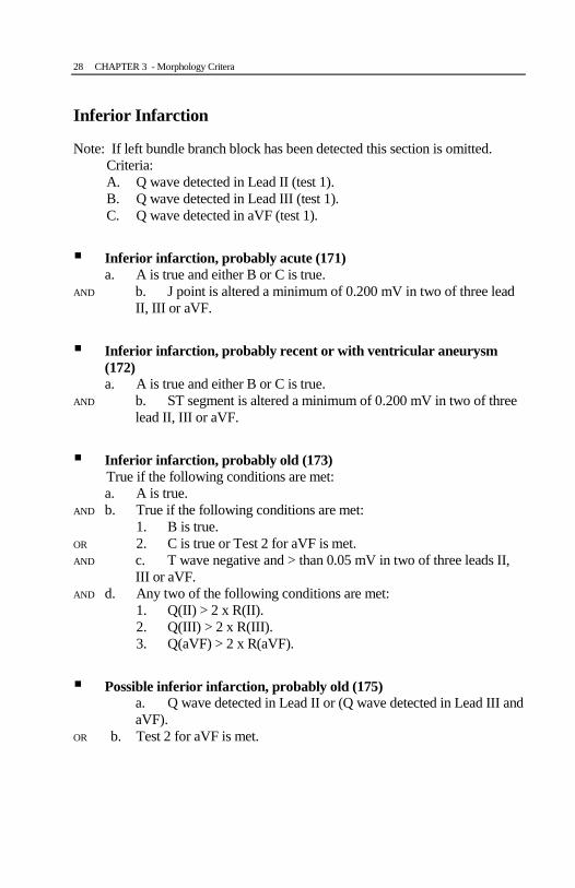
Inferior Infarction
Note: If left bundle branch block has been detected this section is omitted.
Criteria:
A. Q wave detected in Lead II (test 1).
B. Q wave detected in Lead III (test 1).
C. Q wave detected in aVF (test 1).
Inferior infarction, probably acute (171)
a. A is true and either B or C is true.
AND b. J point is altered a minimum of 0.200 mV in two of three lead
II, III or aVF.
Inferior infarction, probably recent or with ventricular aneurysm
(172)
a. A is true and either B or C is true.
AND b. ST segment is altered a minimum of 0.200 mV in two of three
lead II, III or aVF.
Inferior infarction, probably old (173)
True if the following conditions are met:
a. A is true.
AND b. True if the following conditions are met:
1. B is true.
OR 2. C is true or Test 2 for aVF is met.
AND c. T wave negative and > than 0.05 mV in two of three leads II,
III or aVF.
AND d. Any two of the following conditions are met:
1. Q(II) > 2 x R(II).
2. Q(III) > 2 x R(III).
3. Q(aVF) > 2 x R(aVF).
Possible inferior infarction, probably old (175)
a. Q wave detected in Lead II or (Q wave detected in Lead III and
aVF).
OR b. Test 2 for aVF is met.
29 CHAPTER 3 - Morphology Critera
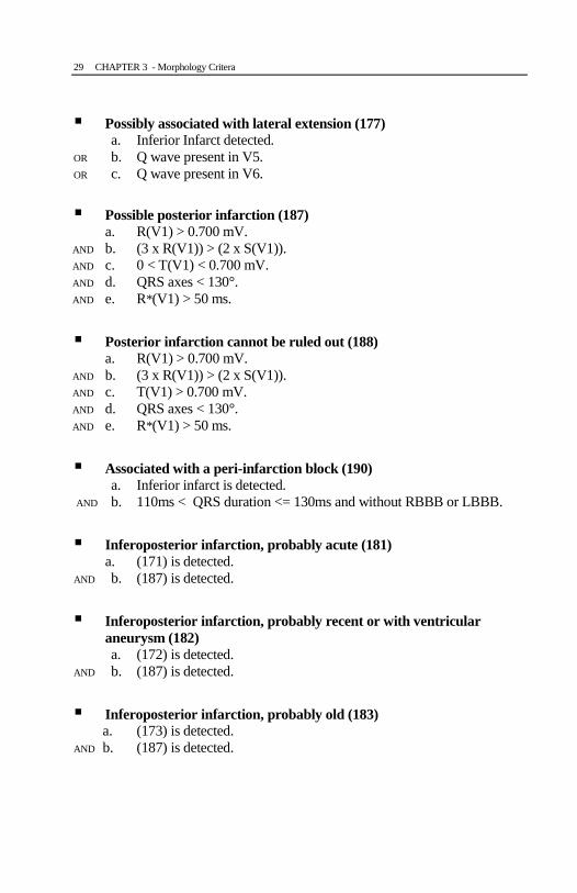
Possibly associated with lateral extension (177)
a. Inferior Infarct detected.
OR b. Q wave present in V5.
OR c. Q wave present in V6.
Possible posterior infarction (187)
a. R(V1) > 0.700 mV.
AND b. (3 x R(V1)) > (2 x S(V1)).
AND c. 0 < T(V1) < 0.700 mV.
AND d. QRS axes < 130°.
AND e. R*(V1) > 50 ms.
Posterior infarction cannot be ruled out (188)
a. R(V1) > 0.700 mV.
AND b. (3 x R(V1)) > (2 x S(V1)).
AND c. T(V1) > 0.700 mV.
AND d. QRS axes < 130°.
AND e. R*(V1) > 50 ms.
Associated with a peri-infarction block (190)
a. Inferior infarct is detected.
AND b. 110ms < QRS duration <= 130ms and without RBBB or LBBB.
Inferoposterior infarction, probably acute (181)
a. (171) is detected.
AND b. (187) is detected.
Inferoposterior infarction, probably recent or with ventricular
aneurysm (182)
a. (172) is detected.
AND b. (187) is detected.
Inferoposterior infarction, probably old (183)
a. (173) is detected.
AND b. (187) is detected.
30 CHAPTER 3 - Morphology Critera
Possible inferoposterior infarction, probably old (184)
a. (173) is detected.
AND b. (188) is detected.
Inferoposterior infarction cannot be ruled out (185)
a. (175) is detected.
AND b. (188) is detected.
Anteroseptal infarction, probably acute (191)
True if J point is altered a minimum of 0.200 mV in lead V1 or V2 and the
first two conditions are met or the third:
a. Q wave detected in V1 (test 1).
b. Q wave detected in V2 (test 1).
c. Q wave detected in V2 (test 1) and (2 x R(V1) < S(V1)) (RS
aspect).
Anteroseptal infarction, probably recent or with ventricular
aneurysm (192)
True if ST segment is altered in lead V1 or V2 and the first two conditions
are met or the third:
a. Q wave detected in V1 (test 1).
b. Q wave detected in V2 (test 1).
c. Q wave detected in V2 (test 1) and (2 x R(V1) < S(V1)) (RS
aspect).
Anteroseptal infarction, probably old (193)
True if T wave is negative more than 0.05 mV in lead V1 or V2 and the
first two conditions are met or the third:
a. Q wave detected in V1 (test 1).
b. Q wave detected in V2 (test 1).
c. Q wave detected in V2 (test 1) and (2 x R(V1) < S(V1)) (RS
aspect).

31 CHAPTER 3 - Morphology Critera
Possible anteroseptal infarction, probably old (195)
True if no repolarisation troubles in lead V1 or V2 and the first two
conditions are met or the third:
a. Q wave detected in V1 (test 1).
b. Q wave detected in V2 (test 1).
c. Q wave detected in V2 (test 1) and (2 x R(V1) < S(V1)) (RS
aspect).
Anterior Infarction
Anterior infarction, probably acute (201)
True if J point is altered a minimum of 0.200 mV in two of V2, V3 or V4
and two of following conditions are met:
a. Q wave detected in V2 (test 1).
b. Q wave detected in V3 (test 1).
c. Q wave detected in V4 (test 1).
Anterior infarction, probably recent or with ventricular aneurysm
(202)
True if ST segment is altered in two of V2, V3 or V4 and two of following
conditions are met:
a. Q wave detected in V2 (test 1).
b. Q wave detected in V3 (test 1).
c. Q wave detected in V4 (test 1).
Anterior infarction, probably old (203)
True if T wave is negative > 0.05 mV in two of V2, V3 or V4 and two of
following conditions are met:
a. Q wave detected in V2 (test 1).
b. Q wave detected in V3 (test 1).
c. Q wave detected in V4 (test 1).
Possible anterior infarction, probably old (205)
True if no repolarisation troubles in V2, V3 or V4 and two of following
conditions are met:
a. Q wave detected in V2 (test 1).
b. Q wave detected in V3 (test 1).
c. Q wave detected in V4 (test 1).
32 CHAPTER 3 - Morphology Critera
Lateral Infarction Criteria:
A. Q wave present in lead I:
1. Q duration > 40 ms.
2. Q amplitude x 2 > R amplitude.
3. Q amplitude > 150 mV.
B. Q wave present in aVL:
1. Q amplitude < 10 mV and R amplitude < 70 mV
and S’ amplitude < 10 mV
and S* > 40 ms.
2. Q* > 40 ms.
3. Q amplitude > 150 mV and (Q amplitude x 3 > R amplitude).
4. Q* < 15 ms and Q amplitude < 70 mV
and R* < 15 ms
and R amplitude < 70 mV
and S’ amplitude < 10 mV
and S* > 40 ms.
Lateral infarction, probably acute (211)
a. Three conditions of A are met.
AND b. One condition of B is met.
AND c. ST elevation > 250 mV in lead I or aVL.
Lateral infarction, probably recent or with ventricular aneurysm
(212)
a. Three conditions of A are met.
AND b. One condition of B is met.
AND c. ST elevation > 100 mV in lead I or aVL.
Lateral infarction, probably old (213)
a. Three conditions of A are met.
AND b. One condition of B is met.
33 CHAPTER 3 - Morphology Critera
Possible Lateral Infarction Criteria:
A. Q wave present in lead I:
1. Q duration > 20 ms.
2. Q amplitude x 3 > R amplitude.
3. Q amplitude > 150 mV.
B. Q wave present in aVL:
1. Q duration > 20 ms.
2. Q amplitude x 3 > R amplitude.
3. Q amplitude > 150 mV.
4. R amplitude < 1200 mV.
5. S Amplitude < 1200 mV.
Possible lateral infarction, probably recent or with ventricular
aneurysm (214)
a. Three conditions of A are met.
AND b. Five conditions of B are met.
AND c. ST elevation > 100 mV in lead I or aVL.
Possible lateral infarction, probably old (215)
a. Three conditions of A are met.
AND b. Five conditions of B are met.
Lateral infarction cannot be ruled out (216)
True if the conditions of A and B are met.
Widespread anterior infarction, probably old (223)
True if the following conditions are met:
a. One of (191), (193) or (195) is detected.
AND b. One of (201), (202), (203) or (205) is detected.
AND c. One of (211), (212), (213) or (215) is detected or Q*(V5) > 15ms.
34 CHAPTER 3 - Morphology Critera
Widespread anterior infarction, probably acute (221)
True if the following conditions are met:
a. True if the following conditions are met:
1. (223) is true.
AND 2. ST segment elevation > 0.2 mV in V2, V3, V4 or V5.
OR b. True if the following conditions are met:
1. (201) is detected.
AND 2. Q wave detected in lead I.
AND 3. Q wave detected in aVL.
Widespread anterior infarction, probably recent or with ventricular
aneurysm (222)
True if the following conditions are met:
a. True if the following conditions are met:
1. (223) is detected.
AND 2. ST segment elevation > 0.1 mV in V2, V3, V4 or V5.
OR b. True if the following conditions are met:
1. (201) is detected.
AND 2. Q wave detected in lead I.
AND 3. Q wave detected in aVL.
Anterolateral infarction, probably old (219)
True if the following conditions are met:
a. True if the following conditions are met:
1. One of (203) or (205) is detected
OR 2. Q(V3) > 0.15 mV.
AND b. One of (213), (215) or (216) is detected.
Anterolateral infarction, probably acute (217)
a. (219) is detected.
AND b. ST segment elevation > 0.2 mV in V3, V4, V5 or V6.
Anterolateral infarction, probably recent or with ventricular
aneureysm (218)
a. (219) is true.
AND b. ST segment elevation > 0.1 mV in V3, V4, V5 or V6.

35 CHAPTER 3 - Morphology Critera
Atypical Q Wave
Note: These criteria are only applicable if no infarction is detected.
Criteria:
A. Q wave in lead III > 0.35 mV.
B. Q wave in lead II > 0.2 mV.
C. Q wave in aVF > 0.3 mV.
Inferior infarction cannot be ruled out (176)
True if Q wave detected in lead III and A, B, and C are true.
Atypical Q wave in lead III (169)
True if Q wave detected in lead III and A is true.
Insignificant Q wave in high lateral (168)
a. Q wave in lead I > 0.2 mV.
AND b. Q wave in aVL > 0.2 mV.
Repolarisation Troubles Criteria:
The detection tests of the following repolarisation troubles are not realized
if:
A. A right or left bundle branch block is already detected.
OR B. hypertrophy with secondary repolarisation troubles is already
detected.
Ischemic ST-T changes compatible with epicardial injury in inferior
leads (231)
a. ST elevation > 0.1 mV in two of leads II, III or aVF.
AND b. A and B are true.
Ischemic ST-T changes compatible with epicardial injury in lateral
leads (232)
a. ST elevation > 0.1 mV in three of leads I, aVL, V5 or V6.
AND b. A and B are true.
36 CHAPTER 3 - Morphology Critera
Ischemic ST-T changes compatible with epicardial injury in anterior
leads (233)
a. ST elevation > 0.1 mV in three of V1, V2, V3 or V4.
AND b. A and B are true.
Ischemic ST-T changes compatible with epicardial injury in
anterolateral leads (229)
a. (232) and (233) are detected.
AND b. A and B are true.
Ischemic ST-T changes in posterior leads (239)
Negative T wave > -0.1 mV in V1 and V2.
Ischemic ST-T changes in inferior leads (241)
Negative T wave > -0.1 mV in two of leads II, III or aVF.
Ischemic ST-T changes in lateral leads (242)
Negative T wave > -0.1 mV in three of leads I, aVL, V5 or V6.
Ischemic ST-T changes in anterior leads (243)
Negative T wave > -0.1 mV in three of V1, V2, V3 or V4.
Ischemic ST-T changes in anterolateral leads (244)
True if (242) and (243) are detected.
Ischemic ST-T changes posterolateral leads (236)
True if (239) and (242) are detected.
Ischemic ST-T changes in inferoposterolateral leads (237)
True if (239), (241), and (242) are detected.
Ischemic ST-T changes in inferoposterior leads (238)
True if (239) and (241) are detected.
37 CHAPTER 3 - Morphology Critera
Ischemic ST-T changes compatible with subendocardial injury in
inferoapical leads (261)
True if horizontal or negative slope ST segment depression > -0.1 mV in
two of leads II, III or aVF.
Ischemic ST-T changes compatible with subendocardial injury in
lateral leads (262)
True if horizontal or negative slope ST segment depression > -0.1 mV in
two of leads I, aVL, V5 or V6.
Ischemic ST-T changes compatible with subendocardial injury in
anterior leads (263)
True if horizontal or negative slope ST segment depression > -0.1 mV in
two of V1, V2, V3 or V4.
Ischemic ST-T changes compatible with subendocardial injury in
anterolateral leads (264)
True if (262) and (263) are detected.
Abnormal repolarisation, may be due to digitalis effect (271)
True if ST segment depression with negative slope with a negative T-
wave in five or more leads.
Widespread abnormal repolarisation, pericarditis cannot be ruled
out (272)
True if ST elevation > 0.1 mV with a negative T wave or terminal point
of T wave < -0.05 mV in six or more leads.
Abnormal repolarisation, may be electrolytic unbalance (273)
True if T wave amplitude > 0.5 mV in eight or more leads.
Abnormal repolarisation, possibly non-specific (274)
a. Dominant R or R’ in the QRS complex and a negative T wave
of < 0.1 mV in four or more leads.
AND b. No infarction or ventricular hypertrophy or other abnormal ST-
T detected.
Abnormal repolarisation, possible coronaric ischemia (275)
38 CHAPTER 3 - Morphology Critera
a. Dominant R or R’ in the QRS complex and a negative T wave
of < 0.1 mV in four or more leads.
AND b. no infarction or ventricular hypertrophy or other abnormal ST-
T detected in men age 50 or women age 55.
Normal Trace
Poor R Progression in right precordial leads (279)
a. No abnormalities detected.
AND b. R(V1) < 0.2 mV.
AND c. R(V1) > R(V2) and R(V2) > R(V3). OR
a. No abnormalities detected.
AND b. R(V1) < 0.2 mV.
AND c. R(V1) < R(V2) and R(V2) > R(V3).
RSR' in V1 could be normal (280)
RSR' with R' > R.
Prolonged QT interval (284)
QTc > 450 ms.
Short PR interval (106)
PR < 120 ms.
QRS within the normal limits (282)
True if the following conditions are true:
a. One of (91), (92), (95), (96), (97), (98), (99), (106), (109),
(118), (123), (124), (128), (130), (276) or (284) is detected.
AND b. No other abnormalities are detected.
Normal morphology (283)
No abnormalities are detected.
39 CHAPTER 4 - ECG Analysis Performance and Accuracy
CHAPTER 4 - ECG ANALYSIS PERFORMANCE AND
ACCURACY
The ECG analysis program is a software component that provides analysis and
interpretation of 12 lead ECGs. The ECG analysis program was developed and
tested by Cardionics SA in conjunction with the Université Catholique de
LOUVAIN (UCL). The ECG analysis program has also been independently
evaluated by the Common Standards for Quantitative Electrocardiography
(CSE) Coordinating Centre.
Standard formulas in which TP represents a true positive result, FN a false
negative result, TN a true negative result, and FP a false positive result, were
used to calculate sensitivity (TP/[TP+FN]), specificity (TN/[TN+FP]), positive
predictive value (TP/[TP+FP]), and negative predictive value (TN/[TN+FN]).
Prevalence is defined as the ratio of the number of occurrences of a particular
condition to the total number of cases in the database.
Note: Modifications may be made to this interpretive program from time to
time which could affect these results.
Performance Results
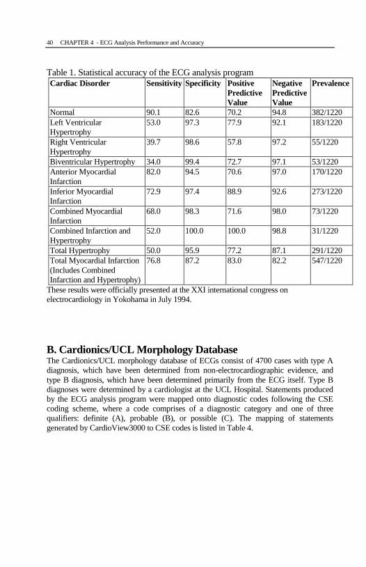
A. CSE Database The CSE database contains 1220 clinically validated cases with type A diagnosis, which
have been determined from non-electrocardiographic evidence. The following table
represents the statistical accuracy of the ECG analysis program.
40 CHAPTER 4 - ECG Analysis Performance and Accuracy
Table 1. Statistical accuracy of the ECG analysis program
Cardiac Disorder Sensitivity Specificity Positive
Predictive
Value
Negative
Predictive
Value
Prevalence
Normal 90.1 82.6 70.2 94.8 382/1220
Left Ventricular
Hypertrophy
53.0 97.3 77.9 92.1 183/1220
Right Ventricular
Hypertrophy
39.7 98.6 57.8 97.2 55/1220
Biventricular Hypertrophy 34.0 99.4 72.7 97.1 53/1220
Anterior Myocardial
Infarction
82.0 94.5 70.6 97.0 170/1220
Inferior Myocardial
Infarction
72.9 97.4 88.9 92.6 273/1220
Combined Myocardial
Infarction
68.0 98.3 71.6 98.0 73/1220
Combined Infarction and
Hypertrophy
52.0 100.0 100.0 98.8 31/1220
Total Hypertrophy 50.0 95.9 77.2 87.1 291/1220
Total Myocardial Infarction
(Includes Combined
Infarction and Hypertrophy)
76.8 87.2 83.0 82.2 547/1220
These results were officially presented at the XXI international congress on
electrocardiology in Yokohama in July 1994.
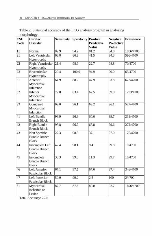
B. Cardionics/UCL Morphology Database The Cardionics/UCL morphology database of ECGs consist of 4700 cases with type A
diagnosis, which have been determined from non-electrocardiographic evidence, and
type B diagnosis, which have been determined primarily from the ECG itself. Type B
diagnoses were determined by a cardiologist at the UCL Hospital. Statements produced
by the ECG analysis program were mapped onto diagnostic codes following the CSE
coding scheme, where a code comprises of a diagnostic category and one of three
qualifiers: definite (A), probable (B), or possible (C). The mapping of statements
generated by CardioView3000 to CSE codes is listed in Table 4.
41 CHAPTER 4 - ECG Analysis Performance and Accuracy
Table 2. Statistical accuracy of the ECG analysis program in analysing
morphology.
CSE
Code
Cardiac
Disorder
Sensitivity Specificity Positive
Predictive
Value
Negative
Predictive
Value
Prevalence
11 Normal 82.9 94.2 81.2 94.8 1056/4700
21 Left Ventricular
Hypertrophy
63.8 86.9 41.5 94.3 596/4700
22 Right Ventricular
Hypertrophy
21.4 98.9 22.7 98.8 70/4700
23 Biventricular
Hypertrophy
29.4 100.0 94.9 99.0 63/4700
31 Anterior
Myocardial
Infarction
64.9 88.2 47.9 93.8 673/4700
32 Inferior
Myocardial
Infarction
72.8 83.4 62.5 89.0 1293/4700
33 Combined
Myocardial
Infarction
69.0 96.1 69.2 96.1 527/4700
41 Left Bundle
Branch Block
93.9 96.8 60.6 99.7 231/4700
42 Right Bundle
Branch Block
93.8 96.7 63.8 99.6 272/4700
43 Non Specific
Bundle Branch
Block
22.3 98.5 37.1 97.0 175/4700
44 Incomplete Left
Bundle Branch
Block
47.4 98.1 9.4 99.8 19/4700
45 Incomplete
Bundle Branch
Block
33.3 99.0 11.3 99.7 18/4700
46 Left Anterior
Fascicular Block
67.1 97.5 67.6 97.4 346/4700
47 Left Posterior
Fascicular Block
50.0 99.2 2.5 100 2/4700
81 Myocardial
Ischemia or
Lesion
87.7 87.6 80.0 92.7 1696/4700
Total Accuracy: 75.0
42 CHAPTER 4 - ECG Analysis Performance and Accuracy
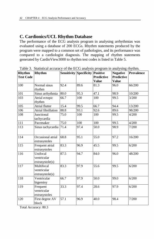
C. Cardionics/UCL Rhythm Database The performance of the ECG analysis program in analysing arrhythmias was
evaluated using a database of 200 ECGs. Rhythm statements produced by the
program were mapped to a common set of pathologies, and its performance was
compared to a cardiologist diagnosis. The mapping of rhythm statements
generated by CardioView3000 to rhythm test codes is listed in Table 5.
Table 3. Statistical accuracy of the ECG analysis program in analysing rhythm.
Rhythm
Test Code
Rhythm Sensitivity Specificity Positive
Predictive
Value
Negative
Predictive
Value
Prevalence
100 Normal sinus
rhythm
92.4 89.6 81.3 96.0 66/200
101 Sinus arrhythmia 80.0 95.3 47.1 98.9 10/200
103 Atrial ectopic
rhythm
66.7 100 100 99.5 3/200
105 Atrial flutter 15.4 99.5 66.7 94.4 13/200
106 Atrial fibrillation 88.8 93.1 92.6 89.6 98/200
108 Junctional
tachycardia
75.0 100 100 99.5 4/200
111 Pacemaker 75.0 100 100 99.5 4/200
113 Sinus tachycardia 71.4 97.4 50.0 98.9 7/200
114 Occasional atrial
extrasystoles
68.8 95.1 55.0 97.2 16/200
115 Frequent atrial
extrasystoles
83.3 96.9 45.5 99.5 6/200
116 Unifocal
ventricular
extrasystole(s)
87.5 94.7 84.0 96.0 48/200
117 Multifocal
ventricular
extrasystole(s)
83.3 97.9 55.6 99.5 6/200
118 Ventricular
bigeminy
66.7 97.9 50.0 99.0 6/200
119 Frequent
ventricular
extrasystoles
33.3 97.4 28.6 97.9 6/200
120 First degree AV
block
57.1 96.9 40.0 98.4 7/200
Total Accuracy: 80.3
43 CHAPTER 4 - ECG Analysis Performance and Accuracy
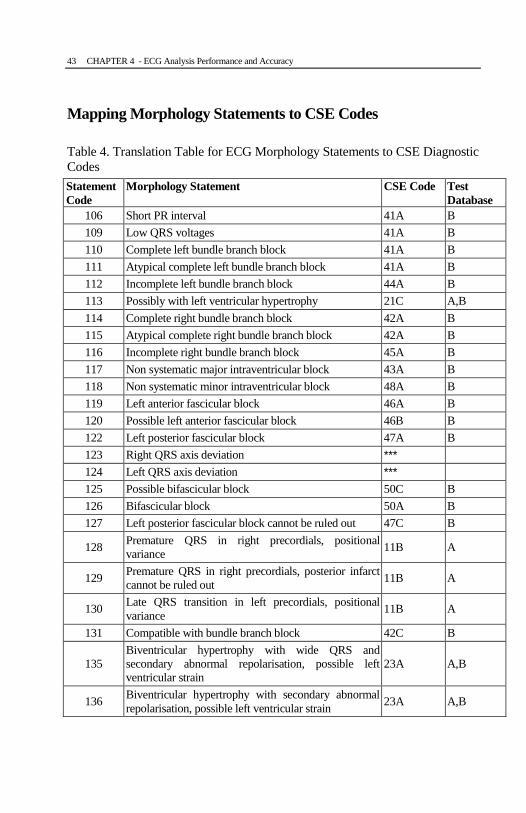
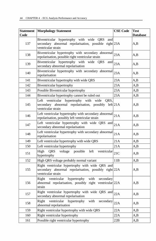
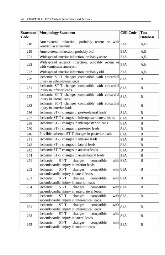
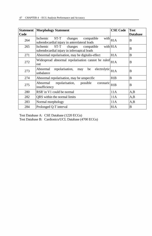
Mapping Morphology Statements to CSE Codes
Table 4. Translation Table for ECG Morphology Statements to CSE Diagnostic
Codes
Statement
Code
Morphology Statement CSE Code Test
Database
106 Short PR interval 41A B
109 Low QRS voltages 41A B
110 Complete left bundle branch block 41A B
111 Atypical complete left bundle branch block 41A B
112 Incomplete left bundle branch block 44A B
113 Possibly with left ventricular hypertrophy 21C A,B
114 Complete right bundle branch block 42A B
115 Atypical complete right bundle branch block 42A B
116 Incomplete right bundle branch block 45A B
117 Non systematic major intraventricular block 43A B
118 Non systematic minor intraventricular block 48A B
119 Left anterior fascicular block 46A B
120 Possible left anterior fascicular block 46B B
122 Left posterior fascicular block 47A B
123 Right QRS axis deviation ***
124 Left QRS axis deviation ***
125 Possible bifascicular block 50C B
126 Bifascicular block 50A B
127 Left posterior fascicular block cannot be ruled out 47C B
128 Premature QRS in right precordials, positional
variance 11B A
129 Premature QRS in right precordials, posterior infarct
cannot be ruled out 11B A
130 Late QRS transition in left precordials, positional
variance 11B A
131 Compatible with bundle branch block 42C B
135
Biventricular hypertrophy with wide QRS and
secondary abnormal repolarisation, possible left
ventricular strain
23A A,B
136 Biventricular hypertrophy with secondary abnormal
repolarisation, possible left ventricular strain 23A A,B
44 CHAPTER 4 - ECG Analysis Performance and Accuracy
Statement
Code
Morphology Statement CSE Code Test
Database
137
Biventricular hypertrophy with wide QRS and
secondary abnormal repolarisation, possible right
ventricular strain
23A A,B
138 Biventricular hypertrophy with secondary abnormal
repolarisation, possible right ventricular strain 23A A,B
139 Biventricular hypertrophy with wide QRS and
secondary abnormal repolarisation 23A A,B
140 Biventricular hypertrophy with secondary abnormal
repolarisation 23A A,B
141 Biventricular hypertrophy with wide QRS 23A A,B
142 Biventricular hypertrophy 23A A,B
143 Possible Biventricular hypertrophy 23A A,B
144 Biventricular hypertrophy cannot be ruled out 23A A,B
145
Left ventricular hypertrophy with wide QRS,
secondary abnormal repolarisation, possibly left
ventricular strain
21A A,B
146 Left ventricular hypertrophy with secondary abnormal
repolarisation, possibly left ventricular strain 21A A,B
147 Left ventricular hypertrophy with wide QRS and
secondary abnormal repolarisation 21A A,B
148 Left ventricular hypertrophy with secondary abnormal
repolarisation 21A A,B
149 Left ventricular hypertrophy with wide QRS 21A A,B
150 Left ventricular hypertrophy 21A A,B
151 High QRS voltage possible left ventricular
hypertrophy 21C A,B
152 High QRS voltage probably normal variant 11B A,B
155
Right ventricular hypertrophy with wide QRS and
secondary abnormal repolarisation, possibly right
ventricular strain
22A A,B
156
Right ventricular hypertrophy with secondary
abnormal repolarisation, possibly right ventricular
strain
22A A,B
157 Right ventricular hypertrophy with wide QRS and
secondary abnormal repolarisation 22A A,B
158 Right ventricular hypertrophy with secondary
abnormal repolarisation 22A A,B
159 Right ventricular hypertrophy with wide QRS 22A A,B
160 Right ventricular hypertrophy 22A A,B
161 Possible right ventricular hypertrophy 22B A,B

45 CHAPTER 4 - ECG Analysis Performance and Accuracy
Statement
Code
Morphology Statement CSE Code Test
Database
162 Right ventricular hypertrophy cannot be ruled out 22C A,B
169 Atypical Q wave in lead III 11B A
171 Inferior infarction, probably acute 32A A,B
172 Inferior infarction, probably recent or with ventricular
aneurysm 32A A,B
173 Inferior infarction, probably old 32A A,B
175 Possible inferior infarction, probably old 32B A,B
176 Inferior infarction cannot be ruled out 32C A,B
177 Possibly associated with lateral extension. 31C A,B
181 Inferoposterior infarction, probably acute 32A A,B
182 Inferoposterior infarction, probably recent or with
ventricular aneurysm 32A A,B
183 Inferoposterior infarction, probably old 32A A,B
184 Possible inferoposterior infarction, probably old 32B A,B
185 Inferoposterior infarction cannot be ruled out 32C A,B
186 Possible posterolateral infarction 33B A,B
187 Possible posterior infarction 32B A,B
188 Posterior infarction cannot be ruled out 32C A,B
190 Associated with a peri-infarction block (190) ***
191 Anteroseptal infarction, probably acute 31A A,B
192 Anteroseptal infarction, probably recent or with
ventricular aneurysm 31A A,B
193 Anteroseptal infarction, probably old 31A A,B
195 Possible anteroseptal infarction, probably old 31B A,B
201 Anterior infarction, probably acute 31A A,B
202 Anterior infarction, probably recent or with ventricular
aneurysm 31A A,B
203 Anterior infarction, probably old 31A A,B
205 Possible anterior infarction, probably old 31B A,B
211 Lateral infarction, probably acute 31A A,B
212 Lateral infarction, probably recent or with ventricular
aneurysm 31A A,B
213 Lateral infarction, probably old 31A A,B
214 Possible lateral infarction, probably recent or with
ventricular aneurysm 31B A,B
215 Possible lateral infarction, probably old 31B A,B
216 Lateral infarction cannot be ruled out 31C A,B
217 Anterolateral infarction, probably acute 31A A,B
46 CHAPTER 4 - ECG Analysis Performance and Accuracy
Statement
Code
Morphology Statement CSE Code Test
Database
218 Anterolateral infarction, probably recent or with
ventricular aneurysm 31A A,B
219 Anterolateral infarction, probably old 31A A,B
221 Widespread anterior infarction, probably acute 31A A,B
222 Widespread anterior infarction, probably recent or
with ventricular aneurysm 31A A,B
223 Widespread anterior infarction, probably old 31A A,B
229 Ischemic ST-T changes compatible with epicardial
injury in anterolateral leads 81A B
231 Ischemic ST-T changes compatible with epicardial
injury in inferior leads 81A B
232 Ischemic ST-T changes compatible with epicardial
injury in lateral leads 81A B
233 Ischemic ST-T changes compatible with epicardial
injury in anterior leads 81A B
236 Ischemic ST-T changes in posterolateral leads 81A B
237 Ischemic ST-T changes in inferoposterolateral leads 81A B
238 Ischemic ST-T changes in inferoposterior leads 81A B
239 Ischemic ST-T changes in posterior leads 81A B
240 Possible ischemic ST-T changes in posterior leads 81A B
241 Ischemic ST-T changes in inferior leads 81A B
242 Ischemic ST-T changes in lateral leads 81A B
243 Ischemic ST-T changes in anterior leads 81A B
244 Ischemic ST-T changes in anterolateral leads 81A B
251 Ischemic ST-T changes compatible with
subendocardial injury in inferior leads
81A B
252 Ischemic ST-T changes compatible with
subendocardial injury in lateral leads
81A B
253 Ischemic ST-T changes compatible with
subendocardial injury in anterior leads
81A B
254 Ischemic ST-T changes compatible with
subendocardial injury in anterolateral leads
81A B
255 Ischemic ST-T changes compatible with
subendocardial injury in inferoapical leads
81A B
261 Ischemic ST-T changes compatible with
subendocardial injury in inferoapical leads 81A B
262 Ischemic ST-T changes compatible with
subendocardial injury in lateral leads 81A B
263 Ischemic ST-T changes compatible with
subendocardial injury in anterior leads 81A B
47 CHAPTER 4 - ECG Analysis Performance and Accuracy
Statement
Code
Morphology Statement CSE Code Test
Database
264 Ischemic ST-T changes compatible with
subendocardial injury in anterolateral leads 81A B
265 Ischemic ST-T changes compatible with
subendocardial injury in inferoapical leads
81A B
271 Abnormal repolarisation, may be digitalis-effect 81A B
272 Widespread abnormal repolarisation cannot be ruled
out 81A B
273 Abnormal repolarisation, may be electrolytic
unbalance 81A B
274 Abnormal repolarisation, may be unspecific 81B B
275 Abnormal repolarisation, possible coronaric
insufficiency 81B B
280 RSR' in V1 could be normal 11A A,B
282 QRS within the normal limits 11A A,B
283 Normal morphology 11A A,B
284 Prolonged Q-T interval 81A B
Test Database A: CSE Database (1220 ECGs)
Test Database B: Cardionics/UCL Database (4700 ECGs)
48 CHAPTER 4 - ECG Analysis Performance and Accuracy
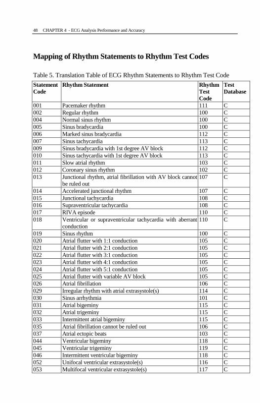
Mapping of Rhythm Statements to Rhythm Test Codes
Table 5. Translation Table of ECG Rhythm Statements to Rhythm Test Code
Statement
Code
Rhythm Statement Rhythm
Test
Code
Test
Database
001 Pacemaker rhythm 111 C
002 Regular rhythm 100 C
004 Normal sinus rhythm 100 C
005 Sinus bradycardia 100 C
006 Marked sinus bradycardia 112 C
007 Sinus tachycardia 113 C
009 Sinus bradycardia with 1st degree AV block 112 C
010 Sinus tachycardia with 1st degree AV block 113 C
011 Slow atrial rhythm 103 C
012 Coronary sinus rhythm 102 C
013 Junctional rhythm, atrial fibrillation with AV block cannot
be ruled out
107 C
014 Accelerated junctional rhythm 107 C
015 Junctional tachycardia 108 C
016 Supraventricular tachycardia 108 C
017 RIVA episode 110 C
018 Ventricular or supraventricular tachycardia with aberrant
conduction
110 C
019 Sinus rhythm 100 C
020 Atrial flutter with 1:1 conduction 105 C
021 Atrial flutter with 2:1 conduction 105 C
022 Atrial flutter with 3:1 conduction 105 C
023 Atrial flutter with 4:1 conduction 105 C
024 Atrial flutter with 5:1 conduction 105 C
025 Atrial flutter with variable AV block 105 C
026 Atrial fibrillation 106 C
029 Irregular rhythm with atrial extrasystole(s) 114 C
030 Sinus arrhythmia 101 C
031 Atrial bigeminy 115 C
032 Atrial trigeminy 115 C
033 Intermittent atrial bigeminy 115 C
035 Atrial fibrillation cannot be ruled out 106 C
037 Atrial ectopic beats 103 C
044 Ventricular bigeminy 118 C
045 Ventricular trigeminy 119 C
046 Intermittent ventricular bigeminy 118 C
052 Unifocal ventricular extrasystole(s) 116 C
053 Multifocal ventricular extrasystole(s) 117 C
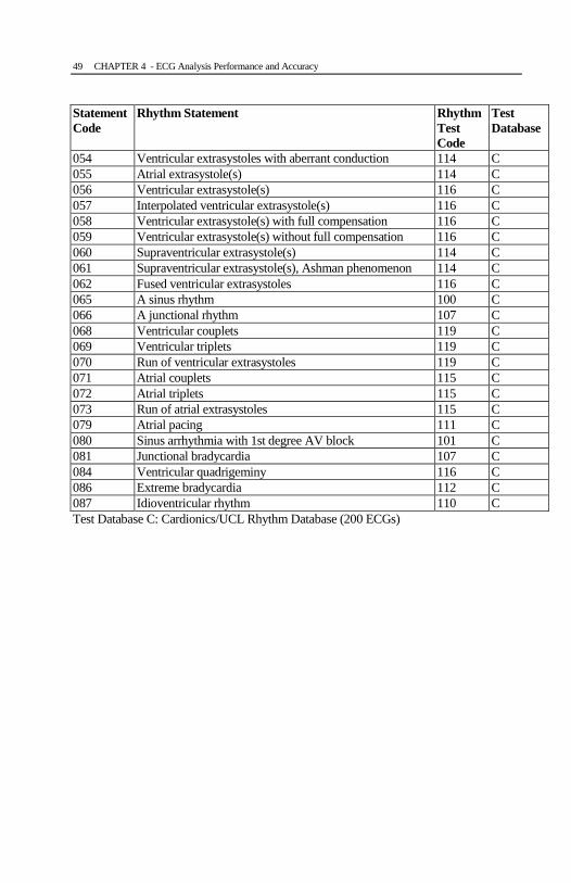
49 CHAPTER 4 - ECG Analysis Performance and Accuracy
Statement
Code
Rhythm Statement Rhythm
Test
Code
Test
Database
054 Ventricular extrasystoles with aberrant conduction 114 C
055 Atrial extrasystole(s) 114 C
056 Ventricular extrasystole(s) 116 C
057 Interpolated ventricular extrasystole(s) 116 C
058 Ventricular extrasystole(s) with full compensation 116 C
059 Ventricular extrasystole(s) without full compensation 116 C
060 Supraventricular extrasystole(s) 114 C
061 Supraventricular extrasystole(s), Ashman phenomenon 114 C
062 Fused ventricular extrasystoles 116 C
065 A sinus rhythm 100 C
066 A junctional rhythm 107 C
068 Ventricular couplets 119 C
069 Ventricular triplets 119 C
070 Run of ventricular extrasystoles 119 C
071 Atrial couplets 115 C
072 Atrial triplets 115 C
073 Run of atrial extrasystoles 115 C
079 Atrial pacing 111 C
080 Sinus arrhythmia with 1st degree AV block 101 C
081 Junctional bradycardia 107 C
084 Ventricular quadrigeminy 116 C
086 Extreme bradycardia 112 C
087 Idioventricular rhythm 110 C
Test Database C: Cardionics/UCL Rhythm Database (200 ECGs)


![Electrocardiography Series Singapore Med J 2011; 52(3) : 146 · of the QRS complex on the surface ECG.(1) A narrow complex (QRS width < 120 milliseconds [ms]) reflects rapid activation](https://img.pdfslide.us/doc/110x75/5ec19bbde544aa0a7e74b446/electrocardiography-series-singapore-med-j-2011-523-146-of-the-qrs-complex.jpg)


![Energy Efficient Fetal ECG Telemonitoring Using Wearable ... · [4] G. Da Poian, R. Bernardini, R. Rinaldo, “ Sparse Representation for Fetal QRS Detection in Abdominal ECG Recordings,”](https://img.pdfslide.us/doc/110x75/5f87061a7372046e385a4c42/energy-efficient-fetal-ecg-telemonitoring-using-wearable-4-g-da-poian-r.jpg)