Embed Size (px)
Citation preview
1
A Pmk1-interacting gene is involved in appressorium differentiation and plant infection 1
in Magnaporthe oryzae 2
3
Haifeng Zhang1,2
, Chaoyang Xue1�, Lingan Kong
1, Guotian Li
1, and Jin-Rong Xu
1* 4
5
1Department of Botany and Plant Pathology, Purdue University, West Lafayette, IN 47907, 6
USA 7
2Department of Plant Pathology, College of Plant Protection, Nanjing Agricultural 8
University, and Key Laboratory of Monitoring and Management of Crop Diseases and Pest 9
Insects, Ministry of Agriculture, Nanjing 210095, China 10
���� current address: Public Health Research Institute, University of Medicine and Dentistry of 11
New Jersey, Newark, NJ 07103 12
13
* Corresponding author 14
Dr. Jin-Rong Xu 15
[email protected]; 765-496-6918 16
17
Running Title: Pmk1-interacting genes 18
19
Keywords: Appressorium formation, PMK1, plant penetration, surface recognition 20
21
Copyright © 2011, American Society for Microbiology and/or the Listed Authors/Institutions. All Rights Reserved.Eukaryotic Cell doi:10.1128/EC.00007-11 EC Accepts, published online ahead of print on 3 June 2011
on April 26, 2019 by guest
http://ec.asm.org/
Dow
nloaded from
2
ABSTRACT 22
23
In the rice blast fungus Magnaporthe oryzae, the PMK1 MAP kinase gene regulates 24
appressorium formation and infectious growth. Its homologs in many other fungi also play 25
critical roles in fungal development and pathogenicity. However, the targets of this important 26
MAP kinase and its interacting genes are not well characterized. In this study, we 27
constructed two yeast two-hybrid libraries of M. oryzae and screened for Pmk1-interacting 28
proteins. Among the nine Pmk1-interacting clones (PICs) identified, two of them, PIC1 and 29
PIC5, were selected for further characterization. Pic1 has one putative nuclear localization 30
signal and one putative MAP kinase phosphorylation site. Pic5 contains one transmembrane 31
domain and two functionally unknown CTNS motifs. The interaction of Pmk1 with Pic1 or 32
Pic5 was confirmed by co-immunoprecipitation assays. Targeted gene deletion of PIC1 had 33
no apparent effects on vegetative growth and pathogenicity but resulted in a significant 34
reduction in conidiation and abnormal germ tube differentiation on onion epidermal cells. 35
Deletion of PIC5 led to a reduction in conidiation and hyphal growth. Autolysis of aerial 36
hyphae became visible in cultures older than 4 days. The pic5 mutant was defective in germ 37
tube growth and appressorium differentiation. It was reduced in appressorial penetration and 38
virulence on plant. Both PIC1 and PIC5 are conserved in filamentous ascomycetes but none 39
of their orthologs have been functionally characterized. Our data indicate that PIC5 is a 40
novel virulence factor involved in appressorium differentiation and pathogenesis in M. 41
oryzae. 42
on April 26, 2019 by guest
http://ec.asm.org/
Dow
nloaded from
3
43
INTRODUCTION 44
Rice blast caused by the heterothallic ascomycete Magnaporthe oryzae (anamorph 45
Pyricularia grisea) is one of the most severe fungal diseases of rice throughout the world (36, 46
41). The fungus uses the enormous turgor pressure generated inside a highly specialized 47
infection structure known as appressorium for plant penetration (5, 11). After penetration, 48
the bulbous invasive hyphae grow biotrophically inside plant cells (31). At late stages, blast 49
lesions are developed on rice plants and the pathogen produces numerous conidia to reinitiate 50
the infection cycle. 51
In the past two decades, several signal transduction pathways involved in surface 52
recognition, appressorium formation, and infectious growth have been characterized in M. 53
oryzae (6, 36, 42). The cAMP signaling pathway is known to be important for surface 54
recognition and appressorium turgor generation (25, 37, 45). The PMK1 MAP kinase gene 55
plays an important role in appressorium development and infectious growth. PMK1 is 56
orthologous to the FUS3 and KSS1 genes in Saccharomyces cerevisiae. The pmk1 deletion 57
mutant is defective in appressorium formation and infectious growth but it still recognizes 58
hydrophobic surfaces and respond to exogenous cAMP (43). Another MAPK gene essential 59
for plant infection in M. oryzae is MPS1 (44), an ortholog of S. cerevisiae SLT2. The mps1 60
mutant is non-pathogenic and defective in appressorial penetration (44). Deletion of MCK1, 61
a MEK kinase gene functioning upstream from Mps1, resulted in similar defects with the 62
mps1 mutant (13). Studies in other plant pathogenic fungi, including Fusarium 63
graminearum, F. oxysporum, Ustilago maydis, Cochliobolus heterostrophus, Claviceps 64
on April 26, 2019 by guest
http://ec.asm.org/
Dow
nloaded from
4
purpurea, and Colletotrichum lagenarium, also have shown the importance of the cAMP 65
signaling and MAP kinase pathways in regulating different plant infection processes (1, 7, 12, 66
17, 22, 24, 26, 32, 35, 40, 53). 67
In M. oryzae, a number of upstream components involved in the activation of Pmk1 68
MAP kinase have been identified, including the MST50, MST11, MST7, MGB1, and RAS2 69
genes (27, 29, 50). Mst50 functions as an adaptor protein that binds with both the Mst7 MEK 70
and Mst11 MEK kinase. Mst7 also directly interacts with Mst11 in yeast two-hybrid and co-71
immunoprecipitation (co-IP) assays (50). However, the interaction of Pmk1 with Mst7, 72
Mst11, or Mst50 was not detectable in yeast two-hybrid assays (50). In co-IP assays with 73
proteins isolated from appressoria, Mst7 and Pmk1 interact with each other via the MAP 74
kinase docking site and docking region. Therefore, it has been hypothesized that the Mst7-75
Pmk1 interaction is relatively transient and the weak interaction between Mst7 and Pmk1 76
may be stabilized or facilitated by additional components of the Pmk1 MAP kinase pathway 77
during appressorium formation (51). 78
For the downstream targets of Pmk1, to date Mst12 is the only transcription factor 79
that is known to weakly interact with Pmk1 in yeast two-hybrid assays (30). Unlike PMK1, 80
MST12 is dispensable for appressorium formation. However, appressoria formed by the 81
mst12 mutant fail to penetrate and infect rice seedlings. The mst12 mutant is defective in 82
appressorial penetration and cytoskeleton reorganization in mature appressoria (28). Studies 83
in C. lagenarium also indicate that CST12 is dispensable for appressorium formation but 84
essential for plant infection (38). In U. maydis, the Prf1 transcription factor gene functions 85
downstream from both the cAMP signaling and MAP kinase pathways (14). However, M. 86
on April 26, 2019 by guest
http://ec.asm.org/
Dow
nloaded from
5
oryzae and other filamentous ascomycetes lack a distinct homolog of Prf1. 87
To further characterize the PMK1 MAP kinase pathway, in this study we constructed 88
two yeast two-hybrid libraries and screened for genes that interacted with Pmk1. Two of the 89
Pmk1-interacting genes, PIC1 and PIC5, were selected for detailed characterization. Results 90
from this study indicate that some of these Pmk1-interacting clones may be involved in 91
surface attachment and appressorium morphogenesis in M. oryzae. 92
93
94
RESULTS 95
96
Construction of yeast two-hybrid libraries: RNA samples isolated from vegetative hyphae 97
grown under nitrogen starvation conditions and appressoria formed on hydrophobic surfaces 98
(36 h) were used to construct the yeast two-hybrid cDNA libraries. The nitrogen starvation (-99
N) and appressorium (AP) libraries consisted of 5.7x10⁹ and 1.25x10
10 pfu/ml primary 100
clones, respectively. The average insertion size was estimated to be 0.70-kb and 0.83-kb, 101
respectively. Both libraries were amplified and preserved at -80oC. 102
103
PIC1 and PIC5 interact with PMK1: For library screening, the PMK1 bait construct 104
pCX38 was co-transformed into yeast strain YRG-2 with plasmid DNA isolated from 105
amplified -N and AP cDNA libraries. A total of 9 Pmk1-interacting clones (named PICs; 106
Table 1) were identified and confirmed by re-transformation into yeast. Five of them encode 107
hypothetical proteins with no known functional domains. One of them, PIC8, encodes a 108
on April 26, 2019 by guest
http://ec.asm.org/
Dow
nloaded from
6
protein homologous to nmrA of Aspergillus nidulans that is a negative regulator of nitrogen 109
metabolism (33). PIC9 is similar to the ribosome organization protein gene BRX1 in S. 110
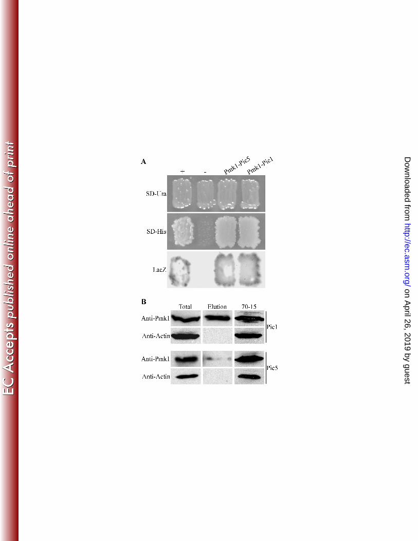
cerevisiae (9). 111
Two of them, PIC1 (MGG_11168.6) and PIC5 (MGG_08600.6), were selected for 112
further characterization in this study. In yeast two-hybrid assays, both PIC1 and PIC5 113
strongly interacted with PMK1 (Fig. 1A). PIC1 encodes an 832-amino acid protein that 114
contains one putative nuclear localization signal (NLS) and one putative MAP kinase 115
phosphorylation site. PIC5 encodes a 271-amino acid protein that has one transmembrane 116
(TM) domain, two putative CTNS (Cystinosin/ERS1p repeat) motifs (IPR006603). The 117
function of CTNS motif is not clear. It was defined by the repetitive motif present between 118
the transmembrane helices of the cystinosin protein encoded by the human CTNS gene. Both 119
PIC1 and PIC5 are conserved in filamentous ascomycetes but lack distinct homologs in the 120
budding or fission yeast. The Pic1 protein shares 19% and 21% identity with its homologs 121
from Neurospora crassa and F. graminearum, respectively. Compared with PIC1, PIC5 122
homologs are more conserved in filamentous ascomycetes. Its homologs from N. crassa and 123
F. graminearum share 70% and 65% identity with Pic5. 124
To confirm the interaction of Pmk1 with Pic1 and Pic5, the PIC1-3xFLAG and PIC5-125
3xFLAG constructs were generated and transformed into the wild-type strain 70-15. 126
Transformants HZA6 and HZB8 expressing the PIC1-3xFLAG and PIC5-3xFLAG 127
constructs, respectively, were identified by PCR and confirmed by western blot analysis with 128
an anti-FLAG antibody (data not shown). When detected with an anti-Pmk1 antiserum (2), 129
total proteins isolated from transformants HZA6 and HZB8 had a 42-kDa band of expected 130
on April 26, 2019 by guest
http://ec.asm.org/
Dow
nloaded from

7
Pmk1 size (Fig. 1B). In proteins eluted from anti-FLAG M2 beads, the same band was 131
detected with the anti-Pmk1 antibody but its intensity was much higher in transformant 132
HZA6 than in transformant HZB8 (Fig. 1B). Because the Pmk1 band had similar intensity in 133
total proteins isolated from HZB8 than HZA6, results from the co-IP assays indicate that 134
Pmk1 interacted with both Pic1 and Pic5 in vivo but the interaction between Pic5 and Pmk1 135
may be relatively weaker than the Pic1-Pmk1 interaction. 136
137
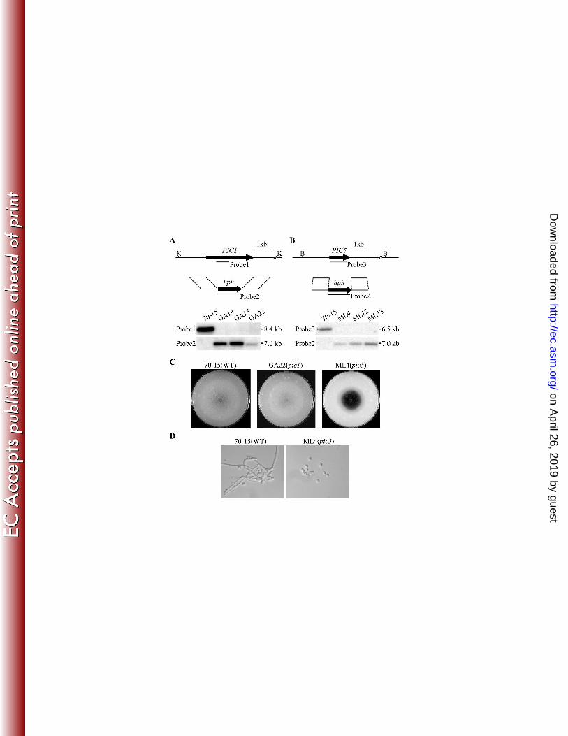
Targeted gene deletion of PIC1 and PIC5 in M. oryzae: To determine their functions in M. 138
oryzae, the PIC1 and PIC5 gene replacement constructs (Fig. 2A; 2B) were generated and 139
transformed into strain 70-15. Three putative deletion mutants of each gene (Table 2) were 140
identified and further confirmed by Southern blot analysis. When hybridized with a PIC1 141
fragment (probe 1), 70-15 had a single 8.4-kb KpnI band that was absent in the pic1 mutants 142
GA14, GA15, and GA22 (Fig. 2A). When hybridized with the hygromycin 143
phosphotransferase (hph) gene (probe 2), a 7.0-kb band was detected in transformants GA14, 144
GA15, and GA22 but not in 70-15 (Fig. 2A). 145
Similar approaches were used to identify and confirm the pic5 deletion mutants ML4, 146
ML12, and ML13 (Table 2). When Southern blots of genomic DNA samples digested with 147
BamHI were hybridized with a PIC5 fragment (probe 3), 70-15 had a 6.5-kb band that was 148
absent in the pic5 mutants ML4, ML12, and ML13 (Fig. 2B). When hybridized with the hph 149
gene as the probe, the pic5 mutants had a 7.0-kb BamHI band that was absent in 70-15 (Fig. 150
2B). 151
152
on April 26, 2019 by guest
http://ec.asm.org/
Dow
nloaded from
8
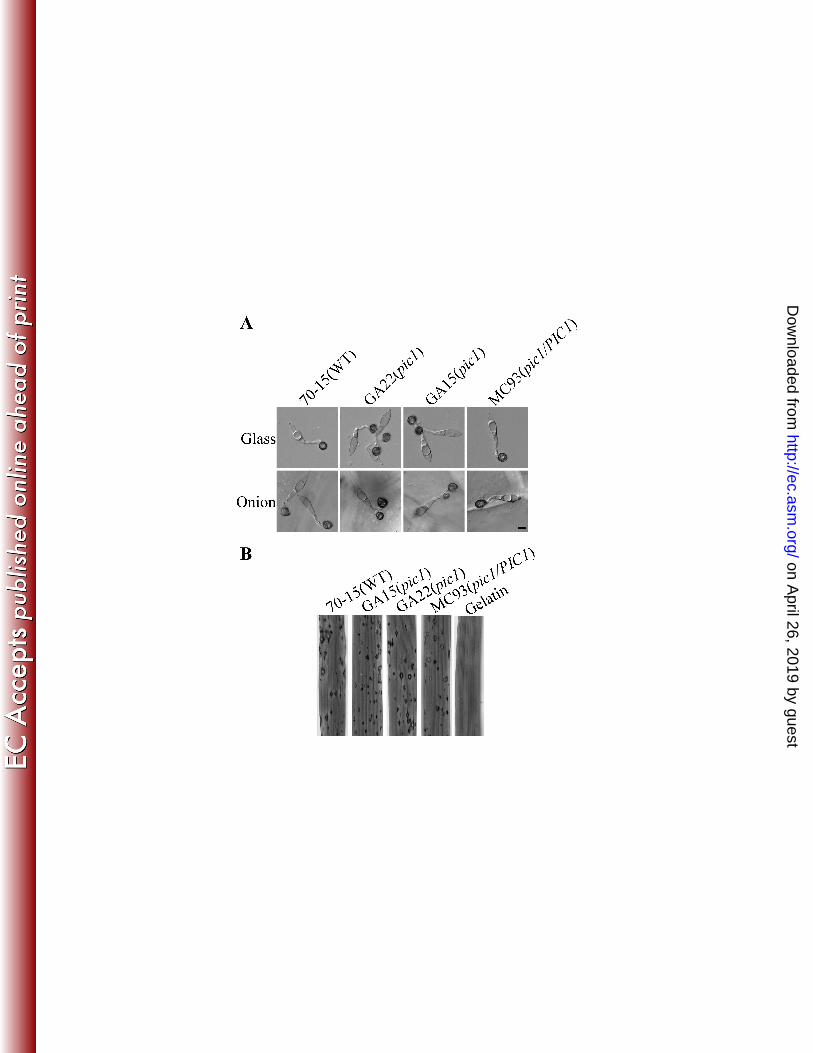
PIC1 is dispensable for growth and virulence but plays a role in conidiation: All three 153
pic1 mutants had similar phenotypes although only data for GA15 and GA22 were presented 154
below. The pic1 mutant had a normal growth rate and produced typical grayish colonies on 155
oatmeal agar plates (Fig. 2C). Conidium morphology was normal but conidiation was 156
reduced approximately 74% in the pic1 mutant compared with the wild type (Table 3). When 157
assayed for appressorium formation on artificial hydrophobic surfaces, mutant GA22 had no 158
defects in germination and formed normal appressoria. However, on onion epidermal cells, 159
many of the pic1 conidia produced more than one appressoria on either branched germ tubes 160
or multiple germ tubes emerged from the same conidium compartment (Fig. 3A). Under the 161
same conditions, this phenomenon was rarely observed in the wild type. Nevertheless, the 162
pic1 mutant had no obvious changes in virulence. On rice leaves sprayed with pic1 conidia, 163
similar amounts of blast lesions were observed 7 days post-inoculation (dpi) on leaves 164
sprayed with the wild type and pic1 mutant strains (Fig. 3B). 165
166
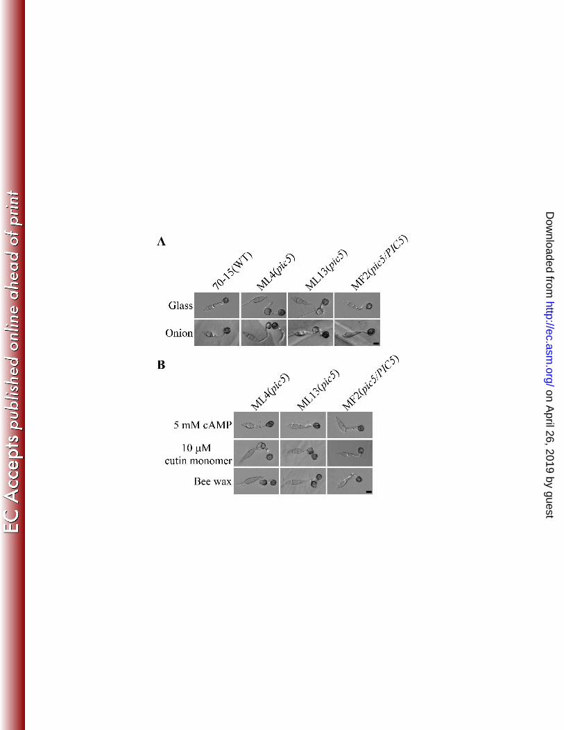
The pic5 mutant is defective in colony morphology and appressorium differentiation: 167
Unlike the pic1 mutant, the pic5 mutant had defects in colony morphology. Although only 168
data for ML4 were presented below, the three pic5 mutants (Table 1) generated in this study 169
had similar phenotypes. When cultured on oatmeal agar plates, the pic5 mutant had a 170
reduced growth rate (Table 3). It also was reduced in the production of aerial hyphae (Fig. 171
2C) and conidiation (Table 3). After 4 days, autolysis of aerial hyphae was observed in the 172
pic5 mutant (Fig. 2C), indicating that the pic5 mutant had defects in cell wall integrity. To 173
confirm this observation, we tested the sensitivity of vegetative hyphae of the pic5 mutant to 174
on April 26, 2019 by guest
http://ec.asm.org/
Dow
nloaded from
9
cell wall degrading enzymes. After digestion with 5 mg/ml lytic enzyme for 40 min, the pic5 175
mutant produced abunt spharoplasts. Hyphal fragments were rarely observed (Fig. 2D). 176
Under the same conditions, the wild type produced fewer spharoplasts and still had many 177
hyphae or hyphal fragments (Fig. 2D), indicating increased sensitivity to lytic enzyme in the 178
pic5 mutant. 179
When assayed for appressorium formation on glass cover slips, 79.3±7.0% of pic5 180
conidia were defective in appressorium formation (Fig. 4A). Chains of appressoria or 181
appressorium-like structures were formed on germ tubes, which may be derived from 182
appressoria formed on germinating appressoria. In the wild type, melanized appressoria were 183
formed apically on the un-branched germ tubes (Fig. 4A). Under the same conditions, no 184
further growth or germination from appressoria formed on hydrophobic surfaces were 185
observed in the wild type. Germ tube growth and appressorium formation also were 186
abnormal in the pic5 mutant on the surface of onion epidermal cells (Fig. 4A). We then 187
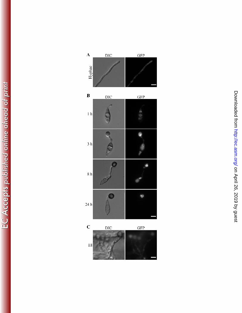
assayed appressorium formation on artificial surfaces treated with exogenous cAMP, a cutin 188
monomer, and bee waxes. Germ tube growth and appressorium formation became normal in 189
the pic5 mutant in the presence of 5 mM cAMP (Fig. 4B). Treatments with 10 µM 1,16 190
hexadecanediol and bee waxes had no obvious effects on the defects of the pic5 mutant in 191
appressorium formation (Fig. 4B). These results indicate that the pic5 mutant may be 192
defective in surface attachment and sensing for chemical and physical signals of plant 193
surface. This defect could be suppressed by cAMP treatment that may increase the 194
intracellular cAMP level and bypass the recognition for surface signals. 195
196
on April 26, 2019 by guest
http://ec.asm.org/
Dow
nloaded from
10
The pic5 mutant is reduced in virulence on plant: To determine whether PIC5 is involved 197
in plant infection, rice seedlings were sprayed with conidia from the pic5 mutant. On leaves 198
inoculated with conidia of the wild type 70-15 and complemented transformant MF2 199
(pic5/PIC5), similar numbers of typical rice blast lesions were formed 7 dpi (Fig. 5A). Under 200
the same conditions, only a few lesions were observed on rice leaves sprayed with conidia of 201
the pic5 mutant (Fig. 5A). To further confirm these observations, we repeated infection 202
assays with barley seedlings. While barley leaves sprayed with 70-15 and complemented 203
strain MF2 developed abundant lesions 6 dpi, only a few lesions were observed on barley 204
leaves inoculated with the pic5 mutant (Fig. 5B). Similar results were obtained in injection 205
infection assays. On leaves inoculated with the same amount of conidia, the wild type caused 206
more lesions than the pic5 mutant surrounding the wounding sites (Fig. 5C). These data 207
indicate that deletion of PIC5 results a significant reduction in virulence. 208
209
The PIC5 gene plays an important role in appressorium penetration: Because the pic5 210
mutant formed appressoria but had a reduced virulence, we assayed appressorial penetration 211
in the pic5 mutant. On the surface of rice leaf sheaths, melanized appressoria were formed by 212
the pic5 mutant. However, similar to what was observed on artificial surfaces, the pic5 213
mutant often formed chains of appressoria or appressorium-like structures (Fig. 6). While the 214
majority of the appressoria formed by the wild type 70-15 and complemented transformant 215
MF2 penetrated epidermal cells of rice leaf sheaths and formed branching invasive hyphae at 216
48 h (Fig. 6), successful penetration and development of invasive hyphae were rarely 217
observed in the pic5 mutant (Table 3; Fig. 6). Similar results were obtained in penetration 218
on April 26, 2019 by guest
http://ec.asm.org/
Dow
nloaded from
11
assays with onion epidermal cells (data not shown). Appressoria formed by the pic5 mutant 219
appeared to be defective in appressorial penetration and differentiation of invasive hyphae, 220
which may be related to the reduction in the virulence. 221
222
Expression and subcellular localization of PIC1-GFP and PIC5-GFP fusion proteins: To 223
determine their expression profiles, we generated PIC1-GFP and PIC5-GFP fusion constructs 224
and transformed them into the pic1 and pic5 knockout mutants GA22 and ML4, respectively. 225
On western blots of proteins isolated from transformants expression the PIC1- and PIC5-GFP 226
fusion constructs, a 118-kD and a 56-kD band of the predicted Pic1-GFP and Pic5-GFP 227
fusion proteins, respectively, were detected with an anti-GFP antibody (data not shown). 228
These results demonstrated that full-length Pic1-GFP and Pic5-GFP fusion proteins were 229
expressed in these transformants. In the PIC1-GFP transformant MC93 (Table 2), weak GFP 230
signals were observed in the vegetative hyphae, conidia, appressoria, and invasive hyphae 231
(data not shown). Transformant MC93 was normal in conidiation (Table 3) and appressorium 232
formation on onion epidermal cells (data not shown), indicating that expression of PIC1-GFP 233
complemented the defects of the pic1 mutant. However, no specific subcellular localization 234
pattern was observed for the Pic1-GFP fusion proteins. 235
In transformant MF2 (Table 2) expressing the PIC5-GFP construct, GFP signals were 236
detected in the cytoplasm of vegetative hyphae (Fig. 7A), germinating conidia, and young 237
germ tubes (Fig. 7B). On hydrophobic surfaces, single melanized appressoria were formed 238
apically on germ tubes of transformant MF2, indicating that expression of PIC5-GFP 239
complemented the defects of the pic5 mutant in appressorium formation. By 8 h, fluorescent 240
on April 26, 2019 by guest
http://ec.asm.org/
Dow
nloaded from
12
signals were still visible in the germ tubes and conidia but significantly enhanced in young 241
appressoria. By 24 h, faint GFP signals were still observed in the cytoplasm of mature 242
appressoria but not in the germ tubes or conidia (Fig. 7B). Unlike Pmk1 (2), nuclear 243
localization was not observed in the PIC1- and PIC5-GFP transformants MC93 and MF2. 244
We also examined GFP signals in invasive hyphae formed by transformant MF2 in onion 245
epidermal cells. No specific distribution patterns could be recognized although weak 246
fluorescent signals appeared to be in the cytoplasm of invasive hyphae (Fig. 7C). 247
248
249
250
DISCUSSION 251
In M. oryzae, the PMK1 MAP kinase pathway is important for appressorium formation and 252
plant infection (43, 49). PMK1 is constitutively expressed but its expression is increased 253
during appressorium formation and young conidium development (2). The Pmk1-GFP fusion 254
protein localizes to the nucleus in appressoria. To further characterize the Pmk1 pathway, in 255
this study we constructed yeast two-hybrid libraries and identified nine putative Pmk1-256
interacting genes. Whereas six of the PIC genes were isolated from the appressorium library, 257
two were identified in the nitrogen starvation library. PIC1 was the only one that that was 258
identified in both libraries. Four of them, PIC2, PIC4, PIC7, and PIC9, have one putative 259
MAP kinase docking site. Some of these proteins may be involved in stimulating or stabilize 260
the interaction Mst7-Pmk1 interaction during appressorium formation. In these screens, the 261
upstream MST7 MEK and downstream MST12 transcription factor were not identified as 262
on April 26, 2019 by guest
http://ec.asm.org/
Dow
nloaded from
13
Pmk1-interacting clones. It has been reported that the Pmk1-Mst7 interaction was not 263
detectable in yeast two-hybrid assays (50). For Mst12, its weak interaction with Pmk1 (30) 264
may be too transient to be detected in the yeast two-hybrid library screening. However, it is 265
also possible that Pmk1 only interacts with full-length Mst12. The average insert size of 266
these two yeast two-hybrid libraries is less than 0.9-kb, which is smaller than the 2,148-bp 267
MST12 ORF. 268
The difference in the phenotypes of the pmk1 and mst12 mutants has lead to the 269
hypothesis that additional transcription factors other than MST12 must exist in M. oryzae for 270
regulating appressorium formation. Unfortunately, no putative transcription factor genes 271
other than the nmrA ortholog were identified among the PIC genes. Pic1 is the only one with 272
a putative NLS but it lacks a DNA binding motif. For the nmrA ortholog, its function in 273
nitrogen metabolism and interaction with Pmk1 remain to be determined. Several 274
transcription factor genes have been reported to play critical roles in appressorium formation 275
in M. oryzae (Kim et al., 2009; Odenbach et al., 2007). However, none of them has been 276
shown to be functionally related with the Pmk1 pathway. Although the pth12 and con7 277
mutants were reported to fail to form appressoria (Kim et al., 2009; Odenbach et al., 2007), 278
deletion of PTH12 or CON7 in Guy11 or 70-15 in our lab resulted in a reduction in 279
appressorium formation and virulence but did not block appressorium formation (Xu, 280
unpublished). Transcription factors regulated by Pmk1 for appressorium formation may be 281
expressed at a relatively low level or their association with PMK1 is too transient to be 282
identified in yeast two-hybrid assays. 283
The PIC1 and PIC5 genes were selected for functional characterization because they 284
on April 26, 2019 by guest
http://ec.asm.org/
Dow
nloaded from
14
had the strongest interaction with PMK1 in yeast two-hybrid assays. In co-IP assays, the 285
interaction of Pmk1 with PIC1 and PIC5 was confirmed. Although their interaction with 286
Pmk1 was similar in yeast two-hybrid assays, PIC1 appeared to have stronger interaction 287
with Pmk1 than PIC5 in co-IP assays (Fig. 1B). Structurally, PIC1 and PIC5 lack any 288
common domains or motifs. While Pic1 has one putative NLS and one putative MAPK 289
phosphorylation site, Pic5 contains one transmembrane domain and two CTNS motifs. Both 290
PIC1 and PIC5 lack distinct homologous genes in the fission and budding yeasts but their 291
homologs are well conserved in filamentous ascomycetes that have been sequenced, 292
including N. crassa and F. graminearum (4, 10). However, none of the PIC1 and PIC5 293
homologs have been functionally characterized in fungi. PIC5 shares limited homology 294
(mainly in the PQ-loop repeats) with the mannose-P-dolichol utilization defect 1 protein of 295
mammalian cells (34). 296
Unlike the pmk1 mutant (43), the pic1 and pic5 mutants still produced melanized 297
appressoria. For the pic1 mutant, no obvious defects were observed in plant infection and 298
appressorium formation on artificial surfaces. Interestingly, germ tubes of the pic1 mutant 299
tended to branch and form multi-appressoria on onion epidermal cells. In the mst11∆RAD
300
mutant of M. oryzae, germ tubes formed appressoria often branch and form additional 301
appressoria (48). One hypothesis is that deletion of the RAS association domain (RAD) 302
domain may result in the defect of the feedback inhibition of germ tube branching and 303
formation of additional appressoria when one appressorium is formed on the germ tube tips. 304
The PIC1 gene may function downstream from Pmk1 and plays a role in this feedback 305
inhibition. However, the formation of branching germ tubes and multiple appressoria in the 306
on April 26, 2019 by guest
http://ec.asm.org/
Dow
nloaded from
15
mst11∆RAD
mutant (48) is not a surface-dependent event. The difference between artificial 307
hydrophobic surfaces and onion epidermal cells may be related to surface hydrophobicity, 308
hardness, and chemical signals that are known to play key roles in inducing appressorium 309
formation. Therefore, it will be interesting to further characterize the defects of the pic1 310
mutant in the production of multiple appressoria. 311
The pic5 mutant was defective in appressorium formation on both plant and artificial 312
surfaces (Fig. 4A). The vast majority (>70%) of appressorium-like structures appeared to 313
germinate and form another appressoria by 24 h (Fig. 4). To our knowledge, no mutants with 314
similar defects have been reported in M. oryzae. PIC5 may function in the terminal 315
commitment of deformed germ tubes to form appressoria that are arrested in the G1 stage. 316
Deletion of PIC5 may result in a defect in G1 arrest and failure to prevent the further growth 317
or germination of developing appressoria. However, it is also possible that appressoria 318
formed by the pic5 mutant failed to attach tightly to the surface. Appressorium formation 319
was aborted when the internal turgor lifts up the developing appressorium and resulted in the 320
further growth. Treatments with bee waxes and a cutin monomer had no effect on the defect 321
of the pic5 mutant in appressorium formation, further indicating that that PIC5 may be 322
involved in the surface attachment but not recognition of physical or chemical signals of the 323
surface. When treated with cAMP that is, in contrast to cutin monomers or waxes, an 324
intracellular messenger rather than an extracellular signal, appressorium formation became 325
normal in the pic5 mutant. The difference between cAMP and cutin monomers or waxes is 326
that the former is an intracellular messenger not extracellular signal molecules. Unlike the 327
pic5 mutant that formed chains of appressorium-like structures, the pmk1 mutant was blocked 328
on April 26, 2019 by guest
http://ec.asm.org/
Dow
nloaded from
16
in appressorium formation on artificial hydrophobic surfaces (43). However, the pmk1 329
mutant still responds to cAMP treatment. On hydrophilic surfaces, exogenous cAMP 330
stimulates the formation of subapical swollen bodies in the pmk1 mutant (43). In M. oryzae, 331
cAMP signaling may be involved in both surface attachment and surface sensing. 332
Unlike the pic1 mutant, the pic5 mutant displayed a significant reduction in plant 333
penetration and virulence. Appressorium penetration efficiency was reduced to 6.5% in the 334
pic5 mutant. In comparison with the wild type and complemented transformant, the pic5 335
mutant was reduced approximated 6-folds in the number of lesions formed on rice or barley 336
leaves. Because turgor generation is dependent on the carbon reserve in conidium 337
compartments, formation of chains of appressoria or appressorium-like structures will reduce 338
appressorium turgor and penetration efficiency. Therefore, the reduction of the pic5 mutant 339
in virulence may be directly related to its defects in appressorium formation and penetration. 340
However, the pic5 mutant has a reduced growth rate. It remains possible that PIC5 also plays 341
a role in the differentiation and growth of invasive hyphae after penetration. In M. oryzae, 342
both PMK1 and MST12 are known to be essential for infectious growth. 343
Colonies of the pic5 mutant produced less aerial hyphae and underwent autolysis in 344
cultures older than 10 days (Fig. 2C). Vegetative hyphae of the mutant also had increased 345
sensitivity to lytic enzyme (Fig. 2D), suggesting that deletion of PIC5 resulted in a defect in 346
cell wall integrity. In M. oryzae, the MPS1 MAP kinase cascade is known to be important for 347
regulating cell wall integrity and plant infection (13, 42). However, deletion of RLM1, one of 348
its downstream transcription factors, had no obvious cell wall defects (23). It is possible that 349
PIC5 is also functionally related to MPS1. Further characterization of the PIC5 gene may 350
on April 26, 2019 by guest
http://ec.asm.org/
Dow
nloaded from
17
lead to better understanding of possible cross talks between the PMK1 and MPS1 pathway. 351
In transformants expressing the PIC1- and PIC5-GFP fusion constructs, weak GFP 352
signals could be detected in vegetative hyphae, conidia, germ tubes, appressoria, and invasive 353
hyphae. PIC1 and PIC5 appeared to have similar expression patterns. However, PIC1 was 354
identified in both –N and AP libraries but PIC5 was isolated only in the AP library. The 355
expression level of PIC5 may be relatively low in vegetative hyphae. In M. oryzae, PMK1 is 356
constitutively expressed but its expression is increased during appressorium formation (2). 357
Pmk1-GFP fusion proteins localizes to the nucleus during appressorium formation. In PIC1- 358
and PIC5-GFP transformants, nuclear localization of GFP signals was not observed in 359
conidia, appressoria, vegetative or invasive hyphae. Pic5 has one putative transmembrane 360
domain. However, GFP signals were mainly observed in the cytoplasm in the pic5/PIC5-361
GFP transformant. The interaction between Pmk1 and Pic5 likely occurs in the cytoplasm, 362
which may interfere with the localization of Pmk1 to the nucleus. Unlike Pic5, Pic1 has a 363
putative nucleus localization signal. Our results indicate that the predicted NLS of Pic1 is not 364
functional or Pic1 only localizes to the nucleus at a specific stage. 365
Overall, although PIC1 and PIC5 are dispensable for the initiation and formation of 366
melanized appressoria, both of them appear to be involved in the late stages of appressorium 367
formation. PIC1 may be involved in the feedback inhibition of germ tube branching or 368
production of secondary germination from the same conidium compartment after the 369
formation of an appressorium apically under certain conditions. PIC5 is important for 370
arresting further growth of appressoria or preventing appressoria from germination. In 371
addition, PIC5 may be functionally related to the PMK1 MAP kinase in infectious growth 372
on April 26, 2019 by guest
http://ec.asm.org/
Dow
nloaded from

18
after penetration. These results indicate that isolating and characterizing the PMK1-373
interacting genes is a useful approach to identify genes important for appressorium formation 374
and function. It will be interesting to further characterize other PIC genes and determining 375
their functional relationship with Pmk1. 376
377
378
379
METHODS AND MATERIALS 380
381
Strains and culture conditions: All the wild-type and mutant strains of M. oryzae were 382
routinely cultured on complete medium (CM), 5xYEG (0.5% yeast extract and 2% glucose), 383
or oatmeal agar (OA) plates as described (19, 46). For DNA, RNA, and protein isolation, 384
vegetative hyphae were harvested from 2-day-old liquid 5xYEG or CM cultures. Growth rate 385
and conidiation were assayed with CM and OA cultures (18). For fungal transformation, 386
transformants were selected on TB3 medium (0.3% yeast extract, 0.3% casamino acids, and 387
20% glucose) with 200 µg/ml zeocin (Invitrogen, Carlsbad, CA) or 250 µg/ml hygromycin B 388
(Calbiochem, La Jolla, CA). For the cell wall integrity test, vegetative hyphae harvested 389
from 2-day old CM cultures were digested with 5mg/ml lytic enzyme (Sigma-Aldrich, St. 390
Louis, MO) for 40 min at 30oC as described (44). 391
392
Construction and screening of the yeast two-hybrid libraries: RNA samples used for 393
library construction were isolated from vegetative hyphae under nitrogen starvation and 394
on April 26, 2019 by guest
http://ec.asm.org/
Dow
nloaded from
19
appressoria formed on plastic surfaces for 36 h as described (8, 20). Total RNAs were 395
isolated with the TRIzol reagent (Invitrogen) and PolyA+ RNAs were purified with the 396
Oligotex mRNA isolation kit (Qiagen Inc., Valencia, CA) following the instructions provided 397
by the manufacturers. The cDNA libraries were constructed with the HybridZAP-2.1 XR 398
library construction kit (Stratagene, La Jolla, CA). The PMK1 gene was amplified with 399
primers MKBF (5’GATGAATTCATGTCTCGCGCCAATCC3’) and MKBR 400
(5’gatctcgagttaccgcataatttcctc3’) and cloned between the EcoRI and XhoI sites of pBD-GAL4 401
(Stratagene) as the bait construct. The yeast strain YRG-2 (ura3 trp1 leu2 his3) provided by 402
Stratagene was used for library screening. Trp+ Leu
+ colonies were collected and assayed for 403
growth on SD-Trp-Leu-His plates and LacZ activities as described (Ding et al., 2010; Zhao 404
and Xu, 2007). Yeast transformants expressing the MST11 bait-MST50 prey and PMK1 bait-405
MST50 prey constructs (Park et al., 2006; Zhao et al., 2005a) were used as the positive and 406
negative controls, respectively. 407
408
Co-immunoprecipitation (co-IP) and western blot analysis: The PIC1-3xFLAG and 409
PIC5-3xFLAG constructs were generated with the yeast gap repair approach (Bourett et al., 410
2002; Bruno et al., 2004) and confirmed by sequencing analysis. The resulting fusion 411
constructs were transformed into protoplasts of 70-15. Transformants expressing the PIC1-412
3xFLAG and PIC5-3xFLAG constructs were identified by PCR and confirmed by western 413
blot analysis with an anti-FLAG antibody (Sigma-Aldrich). For co-IP assays, total proteins 414
were isolated from vegetative hyphae as described (2) and incubated with anti-FLAG M2 415
beads (Sigma-Aldrich). Western blots of proteins eluted from the M2 beads were detected 416
on April 26, 2019 by guest
http://ec.asm.org/
Dow
nloaded from
20
with the anti-Pmk1 (2), anti-FLAG, and anti-actin (Sigma-Aldrich) antibodies with the ECL 417
Supersignal System (Pierce, Rockford, IL). 418
419
Appressorium formation, penetration, and plant infection assays: Conidia were 420
harvested from 10-day-old OA cultures with sterile distilled water and filtered through one 421
layer of Miracloth. For appressorium formation and penetration assays, freshly harvested 422
conidia were resuspended to 5×10⁴ conidia/ml in sterile water. Drops (30 µl) of conidium 423
suspension were placed on glass cover slips (Fisher Scientific Co., Pittsburgh, PA), onion 424
epidermal strips, or rice leaf sheaths. Appressorium formation and development of invasive 425
hyphae were examined after incubation in a moisture chamber for 24 or 48 h (15-16, 39). 426
Effects of treatments with 5 mM cAMP, 10 µM 1,16-hexadecanediol (Sigma-Aldrich), and 427
bee waxes were assayed as described (20). For plant infection assays, conidia were 428
resuspended to 5×10⁴ conidia/ml in 0.25% gelatin. Two-week-old seedlings of rice cultivar 429
Nipponbare and 8-day-old seedlings of barley cultivar Golden Promise were used for spray or 430
injection inoculation (Park et al., 2004; Tucker et al., 2004). 431
432
The PIC1 and PIC5 gene replacement constructs and mutants: The ligation PCR 433
approach (52) was used to generate the PIC1 and PIC5 gene replacement constructs. 434
Approximately 1-kb upstream and downstream flanking sequences of PIC1 were amplified 435
by PCR with primer pairs PIC1UF (5’GCTGGTTGTGTACCGATACTG3’)/PIC1UR 436
(5’TCAGGCGCGCCAAAGCGACTGAGCTGATCAC3’) and PIC1DF 437
(5’TCAGGCCGGCCGATGAGCCAAAGCTGGAGAAG3’)/PIC1DR 438
on April 26, 2019 by guest
http://ec.asm.org/
Dow
nloaded from
21
(5’GTAGCCCTGCTGCCAGATCCAAG3’), respectively. The flanking sequences of PIC5 439
were amplified with primer pairs PIC5UF (5’CCGACACCAAGCAGTTGATGT3’)/PIC5UR 440
(5’TCAGGCGCGCCCTTGTGTGTAATCGGCTGTAC3’) and PIC5DF 441
(5’TCAGGCCGGCCTCCTGACTGTAATAGAGTGCG3’)/PIC5DR 442
(5’CGAGATAGTCTTGGTAGTTTG3’). The resulting PCR products of PIC1 or PIC5 were 443
digested with FseI or AscI and ligated with the hph cassette released from pCX63 (52). The 444
PIC1and PIC5 gene replacement constructs were amplified from the ligation products and 445
transformed into protoplasts of 70-15 (3). Putative pic1 and pic5 mutants were identified by 446
PCR and further confirmed by Southern blot analyses. For complementation assays, the full-447
length PIC1 and PIC5 genes were amplified and cloned into the bleomycin-resistant vector 448
pYP1 (47) by the yeast in vivo recombination approach (2) and transformed into the pic1 and 449
pic5 mutants, respectively. The PIC1 and PIC5 genes were fused in frame with the GFP 450
reporter gene carried on vector pYP1 (47). 451
452
Generation of the PIC1- and PIC5-GFP fusion constructs: For generating the PIC1- and 453
PIC5-GFP fusion constructs, a 2.3-kb and a 4.4-kb fragments of the PIC1 and PIC5 genes 454
containing the promoter regions, respectively, were amplified and co-transformed with XhoI-455
digested pKB04 into S. cerevisiae strain XK1-25 (2). Plasmids pHZ10 and pHZ11 were 456
rescued from Trp+ yeast transformants and confirmed by sequencing analysis to contain the 457
in-frame PIC1- and PIC5-GFP fusion constructs. Zeocin-resistant transformants were 458
isolated after transformation of the pic1 and pic5 mutants with pHZ10 and pHZ11, 459
respectively. The resulting transformants were examined for GFP signals under an 460
on April 26, 2019 by guest
http://ec.asm.org/
Dow
nloaded from
22
epifluorescence microscope and the expression of full-length GFP fusion proteins with a 461
monoclonal anti-GFP antibody (Roche, Indianapolis, IN). 462
463
464
465
ACKNOWLEDGEMENT 466
We thank Drs. Larry Dunkle and Charles Woloshuk at Purdue University for critical reading 467
of this manuscript. This work was supported by grants to JX from the National Research 468
Initiative of the USDA CSREES (#2007-35319-102681) and the USDA National Institute of 469
Food and Agriculture (#2010-65110-20439). 470
471
on April 26, 2019 by guest
http://ec.asm.org/
Dow
nloaded from
23
472
REFERENCE 473
1. Brachmann, A., J. Schirawski, P. Muller, and R. Kahmann. 2003. An unusual MAP kinase is required for 474
efficient penetration of the plant surface by Ustilago maydis. EMBO J. 22:2199-2210. 475
2. Bruno, K. S., F. Tenjo, L. Li, J. E. Hamer, and J. R. Xu. 2004. Cellular localization and role of kinase 476
activity of PMK1 in Magnaporthe grisea. Eukaryot. Cell 3:1525-1532. 477
3. Chao, C. C. T., and A. H. Ellingboe. 1991. Selection for mating competence in Magnaporthe grisea 478
pathogenic to rice. Can. J. Bot. 69:2130-2134. 479
4. Cuomo, C. A., U. Gueldener, J. R. Xu, F. Trail, B. G. Turgeon, A. Di Pietro, J. D. Walton, L. J. Ma, S. E. 480
Baker, M. Rep, G. Adam, J. Antoniw, T. Baldwin, S. Calvo, Y. L. Chang, D. DeCaprio, L. R. Gale, S. 481
Gnerre, R. S. Goswami, K. Hammond-Kosack, L. J. Harris, K. Hilburn, J. C. Kennell, S. Kroken, J. K. 482
Magnuson, G. Mannhaupt, E. Mauceli, H. W. Mewes, R. Mitterbauer, G. Muehlbauer, M. 483
Munsterkotter, D. Nelson, K. O'Donnell, T. Ouellet, W. H. Qi, H. Quesneville, M. I. G. Roncero, K. Y. 484
Seong, I. V. Tetko, M. Urban, C. Waalwijk, T. J. Ward, J. Q. Yao, B. W. Birren, and H. C. Kistler. 2007. 485
The Fusarium graminearum genome reveals a link between localized polymorphism and pathogen 486
specialization. Science 317:1400-1402. 487
5. de Jong, J. C., B. J. McCormack, N. Smirnoff, and N. J. Talbot. 1997. Glycerol generates turgor in rice 488
blast. Nature 389:244-245. 489
6. Dean, R. A. 1997. Signal pathways and appressorium morphogenesis. Annu Rev Phytopathol 35:211-490
234. 491
7. Di Pietro, A., F. I. Garcia-MacEira, E. Meglecz, and M. I. Roncero. 2001. A MAP kinase of the vascular 492
wilt fungus Fusarium oxysporum is essential for root penetration and pathogenesis. Mol Microbiol 493
39:1140-1152. 494
8. Ebbole, D. J., Y. Jin, M. Thon, H. Q. Pan, E. Bhattarai, T. Thomas, and R. Dean. 2004. Gene discovery 495
and gene expression in the rice blast fungus, Magnaporthe grisea: Analysis of expressed sequence 496
tags. Mol. Plant-Microbe Interact. 17:1337-1347. 497
9. Eisenhaber, F., C. Wechselberger, and G. Kreil. 2001. The Brix domain protein family - a key to the 498
ribosomal biogenesis pathway? Trends in Biochemical Sciences 26:345-347. 499
10. Galagan, J. E., S. E. Calvo, K. A. Borkovich, E. U. Selker, N. D. Read, D. Jaffe, W. FitzHugh, L. J. Ma, S. 500
Smirnov, S. Purcell, B. Rehman, T. Elkins, R. Engels, S. G. Wang, C. B. Nielsen, J. Butler, M. Endrizzi, D. 501
Y. Qui, P. Ianakiev, D. B. Pedersen, M. A. Nelson, M. Werner-Washburne, C. P. Selitrennikoff, J. A. 502
Kinsey, E. L. Braun, A. Zelter, U. Schulte, G. O. Kothe, G. Jedd, W. Mewes, C. Staben, E. Marcotte, D. 503
Greenberg, A. Roy, K. Foley, J. Naylor, N. Stabge-Thomann, R. Barrett, S. Gnerre, M. Kamal, M. 504
Kamvysselis, E. Mauceli, C. Bielke, S. Rudd, D. Frishman, S. Krystofova, C. Rasmussen, R. L. 505
Metzenberg, D. D. Perkins, S. Kroken, C. Cogoni, G. Macino, D. Catcheside, W. X. Li, R. J. Pratt, S. A. 506
Osmani, C. P. C. DeSouza, L. Glass, M. J. Orbach, J. A. Berglund, R. Voelker, O. Yarden, M. Plamann, S. 507
Seiler, J. Dunlap, A. Radford, R. Aramayo, D. O. Natvig, L. A. Alex, G. Mannhaupt, D. J. Ebbole, M. 508
Freitag, I. Paulsen, M. S. Sachs, E. S. Lander, C. Nusbaum, and B. Birren. 2003. The genome sequence 509
of the filamentous fungus Neurospora crassa. Nature 422:859-868. 510
11. Howard, R. J., M. A. Ferrari, D. H. Roach, and N. P. Money. 1991. Penetration of hard substrates by a 511
fungus employing enormous turgor pressures. Proc. Natl. Acad. Sci. U.S.A. 88:11281-11284. 512
12. Jenczmionka, N. J., F. J. Maier, A. P. Losch, and W. Schafer. 2003. Mating, conidiation and 513
pathogenicity of Fusarium graminearum, the main causal agent of the head-blight disease of wheat, 514
are regulated by the MAP kinase GPMK1. Curr. Genet. 43:87-95. 515
on April 26, 2019 by guest
http://ec.asm.org/
Dow
nloaded from

24
13. Jeon, J., J. Goh, S. Yoo, M. H. Chi, J. Choi, H. S. Rho, J. Park, S. S. Han, B. R. Kim, S. Y. Park, S. Kim, and 516
Y. H. Lee. 2008. A putative MAP kinase kinase kinase, MCK1, is required for cell wall integrity and 517
pathogenicity of the rice blast fungus, Magnaporthe oryzae. Mol. Plant-Microbe Interact. 21:525-534. 518
14. Kaffarnik, F., P. Muller, M. Leibundgut, R. Kahmann, and M. Feldbrugge. 2003. PKA and MAPK 519
phosphorylation of Prf1 allows promoter discrimination in Ustilago maydis. Embo J 22:5817-5826. 520
15. Kankanala, P., K. Czymmek, and B. Valent. 2007. Roles for rice membrane dynamics and 521
plasmodesmata during biotrophic invasion by the blast fungus. Plant Cell 19:706-724. 522
16. Koga, H. 1994. Hypersensitive death, autofluorescence, and ultrastructural-changes in cells of leaf 523
sheaths of susceptible and resistant near-isogenic lines of rice (PI-Z(T)) in relation to penetration and 524
growth of Pycularia oryza. Can. J. Bot. 72:1463-1477. 525
17. Lev, S., A. Sharon, R. Hadar, H. Ma, and B. A. Horwitz. 1999. A mitogen-activated protein kinase of the 526
corn leaf pathogen Cochliobolus heterostrophus is involved in conidiation, appressorium formation, 527
and pathogenicity: Diverse roles for mitogen-activated protein kinase homologs in foliar pathogens. 528
Proc. Natl. Acad. Sci. U.S.A. 96:13542-13547. 529
18. Li, L., S. L. Ding, A. Sharon, M. Orbach, and J. R. Xu. 2007. Mir1 is highly upregulated and localized to 530
nuclei during infectious hyphal growth in the rice blast fungus. Mol. Plant-Microbe Interact. 20:448-531
458. 532
19. Li, L., C. Y. Xue, K. Bruno, M. Nishimura, and J. R. Xu. 2004. Two PAK kinase genes, CHM1 and MST20, 533
have distinct functions in Magnaporthe grisea. Mol. Plant-Microbe Interact. 17:547-556. 534
20. Liu, W., X. Zhou, G. Li, L. Li, L. Kong, C. Wang, H. Zhang, and J. R. Xu. 2011. Multiple plant surface 535
signals are sensed by different mechanisms in the rice blast fungus for appressorium formation. PLoS 536
Pathogens 7:e1001261. 537
21. Livak, K. J., and T. D. Schmittgen. 2001. Analysis of relative gene expression data using real-time 538
quantitative PCR and the 2(-Delta Delta C(T)) method. Methods 25:402-408. 539
22. Mayorga, M. E., and S. E. Gold. 1999. A MAP kinase encoded by the ubc3 gene of Ustilago maydis is 540
required for filamentous growth and full virulence. Mol. Microbiol. 34:485-497. 541
23. Mehrabi, R., S. Ding, and J. R. Xu. 2008. MADS-box transcription factor Mig1 is required for infectious 542
growth in Magnaporthe grisea. Eukaryot. Cell 7:791-799. 543
24. Mey, G., B. Oeser, M. H. Lebrun, and P. Tudzynski. 2002. The biotrophic, non-appressorium-forming 544
grass pathogen Claviceps purpurea needs a FUS3/PMK1 homologous mitogen-activated protein kinase 545
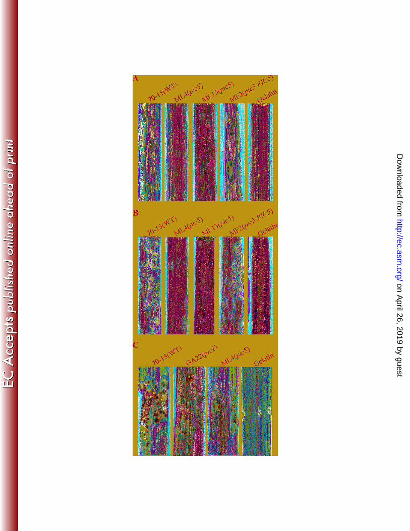
for colonization of rye ovarian tissue. Mol. Plant-Microbe Interact. 15:303-312. 546
25. Mitchell, T. K., and R. A. Dean. 1995. The cAMP-dependent protein kinase catalytic subunit is required 547
for appressorium formation and pathogenesis by the rice blast pathogen Magnaporthe grisea. Plant 548
Cell 7:1869-1878. 549
26. Muller, P., C. Aichinger, M. Feldbrugge, and R. Kahmann. 1999. The MAP kinase Kpp2 regulates 550
mating and pathogenic development in Ustilago maydis. Mol. Microbiol. 34:1007-1017. 551
27. Nishimura, M., G. Park, and J. R. Xu. 2003. The G-beta subunit MGB1 is involved in regulating multiple 552
steps of infection-related morphogenesis in Magnaporthe grisea. Mol. Microbiol. 50:231-243. 553
28. Park, G., K. S. Bruno, C. J. Staiger, N. J. Talbot, and J. R. Xu. 2004. Independent genetic mechanisms 554
mediate turgor generation and penetration peg formation during plant infection in the rice blast 555
fungus. Mol. Microbiol. 53:1695-1707. 556
29. Park, G., C. Xue, X. Zhao, Y. Kim, M. Orbach, and J. R. Xu. 2006. Multiple upstream signals converge 557
on an adaptor protein Mst50 to activate the PMK1 pathway in Magnaporthe grisea. Plant Cell 558
18:2822–2835. 559
on April 26, 2019 by guest
http://ec.asm.org/
Dow
nloaded from
25
30. Park, G., G. Y. Xue, L. Zheng, S. Lam, and J. R. Xu. 2002. MST12 regulates infectious growth but not 560
appressorium formation in the rice blast fungus Magnaporthe grisea. Mol. Plant-Microbe Interact. 561
15:183-192. 562
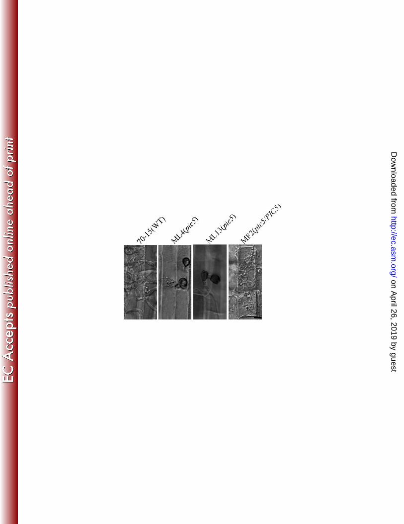
31. Perfect, S. E., and J. R. Green. 2001. Infection structures of biotrophic and hemibiotrophic fungal plant 563
pathogens. Mol Plant Pathol 2:101-108. 564
32. Ruiz-Roldan, M. C., F. J. Maier, and W. Schafer. 2001. PTK1, a mitogen-activated-protein kinase gene, 565
is required for conidiation, appressorium formation, and pathogenicity of Pyrenophora teres on barley. 566
Mol. Plant-Microbe Interact. 14:116-125. 567
33. Stammers, D. K., J. Ren, K. Leslie, C. E. Nichols, H. K. Lamb, S. Cocklin, A. Dodds, and A. R. Hawkins. 568
2001. The structure of the negative transcriptional regulator NmrA reveals a structural superfamily 569
which includes the short-chain dehydrogenase/reductases. Embo J 20:6619-6626. 570
34. Strausberg, R. L., E. A. Feingold, L. H. Grouse, J. G. Derge, R. D. Klausner, F. S. Collins, L. Wagner, C. 571
M. Shenmen, G. D. Schuler, S. F. Altschul, B. Zeeberg, K. H. Buetow, C. F. Schaefer, N. K. Bhat, R. F. 572
Hopkins, H. Jordan, T. Moore, S. I. Max, J. Wang, F. Hsieh, L. Diatchenko, K. Marusina, A. A. Farmer, 573
G. M. Rubin, L. Hong, M. Stapleton, M. B. Soares, M. F. Bonaldo, T. L. Casavant, T. E. Scheetz, M. J. 574
Brownstein, T. B. Usdin, S. Toshiyuki, P. Carninci, C. Prange, S. S. Raha, N. A. Loquellano, G. J. Peters, 575
R. D. Abramson, S. J. Mullahy, S. A. Bosak, P. J. McEwan, K. J. McKernan, J. A. Malek, P. H. 576
Gunaratne, S. Richards, K. C. Worley, S. Hale, A. M. Garcia, L. J. Gay, S. W. Hulyk, D. K. Villalon, D. M. 577
Muzny, E. J. Sodergren, X. H. Lu, R. A. Gibbs, J. Fahey, E. Helton, M. Ketteman, A. Madan, S. 578
Rodrigues, A. Sanchez, M. Whiting, A. Madan, A. C. Young, Y. Shevchenko, G. G. Bouffard, R. W. 579
Blakesley, J. W. Touchman, E. D. Green, M. C. Dickson, A. C. Rodriguez, J. Grimwood, J. Schmutz, R. 580
M. Myers, Y. S. N. Butterfield, M. I. Kryzywinski, U. Skalska, D. E. Smailus, A. Schnerch, J. E. Schein, S. 581
J. M. Jones, M. A. Marra, and M. G. C. M. Pro. 2002. Generation and initial analysis of more than 582
15,000 full-length human and mouse cDNA sequences. Proceedings of the National Academy of 583
Sciences of the United States of America 99:16899-16903. 584
35. Takano, Y., T. Kikuchi, Y. Kubo, J. E. Hamer, K. Mise, and I. Furusawa. 2000. The Colletotrichum 585
lagenarium MAP kinase gene CMK1 regulates diverse aspects of fungal pathogenesis. Mol. Plant-586
Microbe Interact. 13:374-383. 587
36. Talbot, N. J. 2003. On the trail of a cereal killer: Exploring the biology of Magnaporthe grisea. Annu. 588
Rev. Microbiol. 57:177-202. 589
37. Thines, E., R. W. S. Weber, and N. J. Talbot. 2000. MAP kinase and protein kinase A - dependent 590
mobilizationof triacylglycerol and glycogen during appressorium tugor generation by Magnaporthe 591
grisea. Plant Cell 12:1703-1718. 592
38. Tsuji, G., S. Fujii, S. Tsuge, T. Shiraishi, and Y. Kubo. 2003. The Colletotrichum lagenarium Ste12-like 593
gene CST1 is essential for appressorium penetration. Mol Plant Microbe In 16:315-325. 594
39. Tucker, S. L., C. R. Thornton, K. Tasker, C. Jacob, G. Giles, M. Egan, and N. J. Talbot. 2004. A fungal 595
metallothionein is required for pathogenicity of Magnaporthe grisea. Plant Cell 16:1575-1588. 596
40. Urban, M., E. Mott, T. Farley, and K. Hammond-Kosack. 2003. The Fusarium graminearum MAP1 gene 597
is essential for pathogenicity and development of perithecia. Mol. Plant Pathol. 4:347-359. 598
41. Valent, B., and F. G. Chumley. 1991. Molecular genetic analysis of the rice blast fungus Magnaporthe 599
grisea. Annu. Rev. Phytopathol. 29:443-467. 600
42. Xu, J. R. 2000. MAP kinases in fungal pathogens. Fungal Genet. Biol. 31:137-152. 601
43. Xu, J. R., and J. E. Hamer. 1996. MAP kinase and cAMP signaling regulate infection structure formation 602
and pathogenic growth in the rice blast fungus Magnaporthe grisea. Genes Dev. 10:2696-2706. 603
on April 26, 2019 by guest
http://ec.asm.org/
Dow
nloaded from
26
44. Xu, J. R., C. J. Staiger, and J. E. Hamer. 1998. Inactivation of the mitogen-activated protein kinase 604
Mps1 from the rice blast fungus prevents penetration of host cells but allows activation of plant 605
defense responses. Proc. Natl. Acad. Sci. USA. 95:12713-12718. 606
45. Xu, J. R., M. Urban, J. A. Sweigard, and J. E. Hamer. 1997. The CPKA gene of Magnaporthe grisea is 607
essential for appressorial penetration. Mol. Plant-Microbe Interact. 10:187-194. 608
46. Xue, C. Y., G. Park, W. B. Choi, L. Zheng, R. A. Dean, and J. R. Xu. 2002. Two novel fungal virulence 609
genes specifically expressed in appressoria of the rice blast fungus. Plant Cell 14:2107-2119. 610
47. Zhang, Y., Y. E. Choi, X. Zou, and J. R. Xu. 2010. The FvMK1 mitogen-activated protein kinase gene 611
regulates conidiation, pathogenesis, and fumonisin production in Fusarium verticillioides. Fungal 612
Genet Biol. 613
48. Zhao, X., Y. Kim, G. Park, and J. R. Xu. 2005. A mitogen-activated protein kinase cascade regulating 614
infection-related morphogenesis in Magnaporthe grisea. Plant Cell 17:1317-1329. 615
49. Zhao, X., R. Mehrabi, and J.-R. Xu. 2007. Mitogen-activated protein kinase pathways and fungal 616
pathogenesis. Eukaryot. Cell 6:1701-1714. 617
50. Zhao, X. H., Y. Kim, G. Park, and J. R. Xu. 2005. A mitogen-activated protein kinase cascade regulating 618
infection-related morphogenesis in Magnaporthe grisea. Plant Cell 17:1317-1329. 619
51. Zhao, X. H., and J. R. Xu. 2007. A highly conserved MAPK-docking site in Mst7 is essential for Pmk1 620
activation in Magnaporthe grisea. Mol. Microbiol. 63:881-894. 621
52. Zhao, X. H., C. Xue, Y. Kim, and J. R. Xu. 2004. A ligation-PCR approach for generating gene 622
replacement constructs in Magnaporthe grisea. Fungal Genet. Newsl. 51:17-18. 623
53. Zheng, L., M. Campbell, J. Murphy, S. Lam, and J. R. Xu. 2000. The BMP1 gene is essential for 624
pathogenicity in the gray mold fungus Botrytis cinerea. Mol. Plant-Microbe Interact. 13:724-732. 625
626
627
628
on April 26, 2019 by guest
http://ec.asm.org/
Dow
nloaded from
27
629
TABLES AND FIGURE LEGENDS 630
631
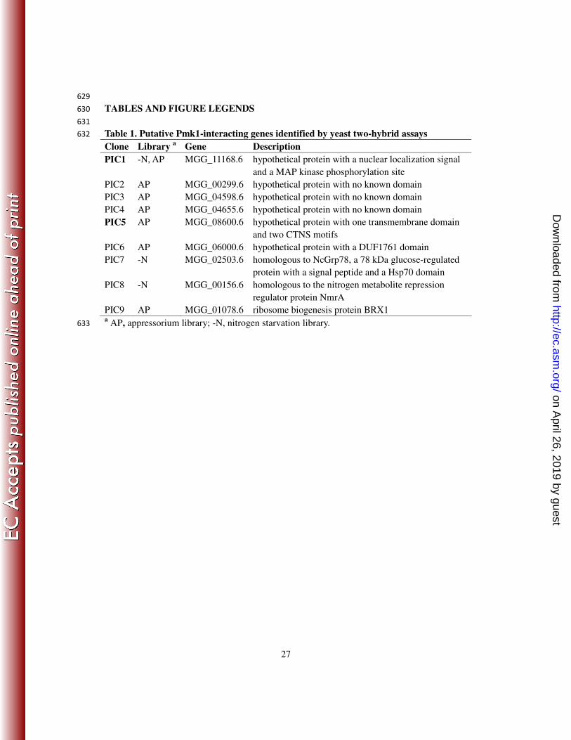
Table 1. Putative Pmk1-interacting genes identified by yeast two-hybrid assays 632
Clone Library a Gene Description
PIC1 -N, AP MGG_11168.6 hypothetical protein with a nuclear localization signal
and a MAP kinase phosphorylation site
PIC2 AP MGG_00299.6 hypothetical protein with no known domain
PIC3 AP MGG_04598.6 hypothetical protein with no known domain
PIC4 AP MGG_04655.6 hypothetical protein with no known domain
PIC5 AP MGG_08600.6 hypothetical protein with one transmembrane domain
and two CTNS motifs
PIC6 AP MGG_06000.6 hypothetical protein with a DUF1761 domain
PIC7 -N MGG_02503.6 homologous to NcGrp78, a 78 kDa glucose-regulated
protein with a signal peptide and a Hsp70 domain
PIC8 -N MGG_00156.6 homologous to the nitrogen metabolite repression
regulator protein NmrA
PIC9 AP MGG_01078.6 ribosome biogenesis protein BRX1 a AP, appressorium library; -N, nitrogen starvation library. 633
on April 26, 2019 by guest
http://ec.asm.org/
Dow
nloaded from
28
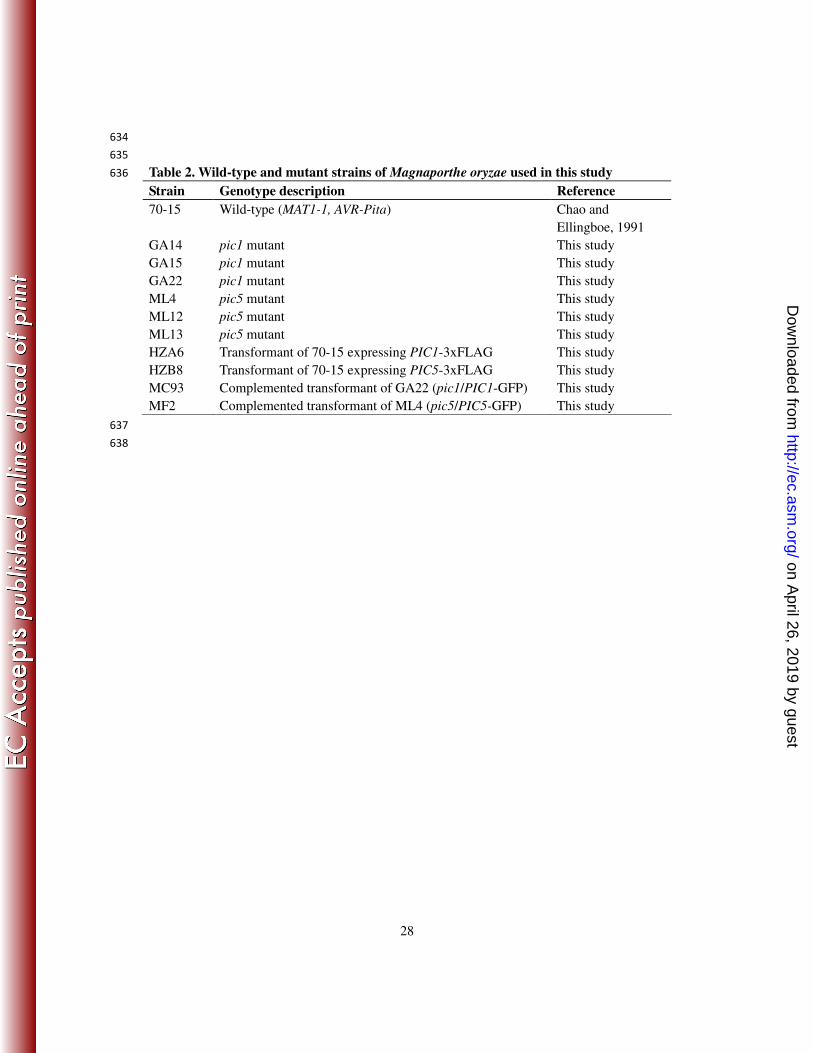
634
635
Table 2. Wild-type and mutant strains of Magnaporthe oryzae used in this study 636
Strain Genotype description Reference
70-15 Wild-type (MAT1-1, AVR-Pita) Chao and
Ellingboe, 1991
GA14 pic1 mutant This study
GA15 pic1 mutant This study
GA22 pic1 mutant This study
ML4 pic5 mutant This study
ML12 pic5 mutant This study
ML13 pic5 mutant This study
HZA6 Transformant of 70-15 expressing PIC1-3xFLAG This study
HZB8 Transformant of 70-15 expressing PIC5-3xFLAG This study
MC93 Complemented transformant of GA22 (pic1/PIC1-GFP) This study
MF2 Complemented transformant of ML4 (pic5/PIC5-GFP) This study
637
638
on April 26, 2019 by guest
http://ec.asm.org/
Dow
nloaded from
29
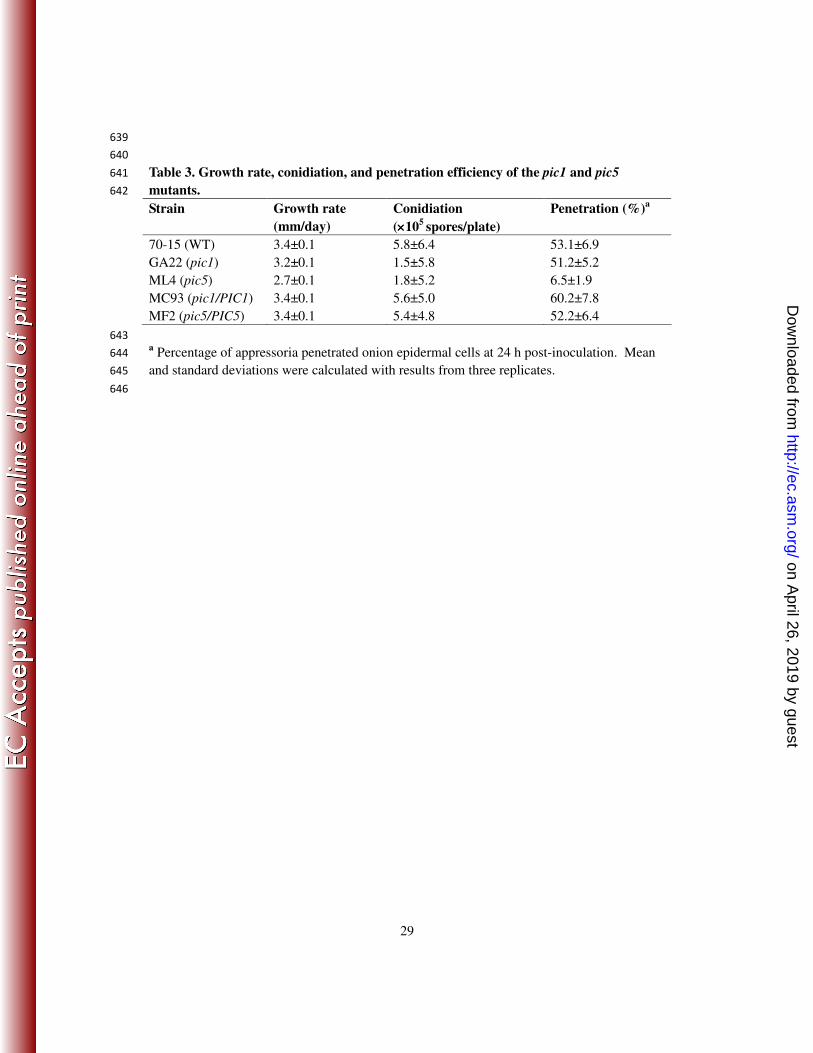
639
640
Table 3. Growth rate, conidiation, and penetration efficiency of the pic1 and pic5 641
mutants. 642
Strain Growth rate
(mm/day)
Conidiation
(××××105
spores/plate)
Penetration (%)a
70-15 (WT) 3.4±0.1 5.8±6.4 53.1±6.9
GA22 (pic1) 3.2±0.1 1.5±5.8 51.2±5.2
ML4 (pic5) 2.7±0.1 1.8±5.2 6.5±1.9
MC93 (pic1/PIC1) 3.4±0.1 5.6±5.0 60.2±7.8
MF2 (pic5/PIC5) 3.4±0.1 5.4±4.8 52.2±6.4
643 a Percentage of appressoria penetrated onion epidermal cells at 24 h post-inoculation. Mean 644
and standard deviations were calculated with results from three replicates. 645
646
on April 26, 2019 by guest
http://ec.asm.org/
Dow
nloaded from
30
647
648
FIGURE LEGENDS 649
650
Fig. 1. Yeast two-hybrid and co-IP assays for the interaction of PMK1 with PIC1 and PIC5. 651
A. Yeast transformants expressing the PMK1 bait and PIC1 or PIC5 prey constructs were 652
assayed for growth on SD-Leu-Trp-His (SD-His) plates and β-galactosidase (LacZ) activities. 653
Positive and negative controls were marked with + and -. B. Western blot analysis with total 654
proteins (Total) isolated from M. oryzae transformants expressing the PIC1-3xFLAG and 655
PIC5-3xFLAG constructs and proteins eluted from the anti-FLAG M2 beads (Elution). The 656
presence of Pmk1 was detected with an anti-Pmk1 antibody (2). Total proteins isolated from 657
the wild-type M. oryzae strain (70-15) and detection with an anti-actin antibody were 658
included as the controls. 659
660
Fig. 2. Targeted deletion of the PIC1 and PIC5 genes. A. Schematic diagram of the PIC1 661
gene and gene replacement construct and Southern blots of KpnI-digested genomic DNA of 662
the wild type (70-15) and pic1 mutants (GA14, GA15, and GA22) hybridized with probe 1 663
and probe 2. B. Schematic diagram of the PIC5 gene replacement constuct and Southern 664
blots of BamHI-digested DNA of 70-15 and pic5 mutants (ML4, ML12, and ML13) 665
hybridized with probe 2 and probe 3. C. Oatmeal agar cultures of 70-15, GA22, and ML4. 666
Photographs were taken after incubation for 10 days. The central part of the ML4 colony 667
underwent autolysis and became darkly pigmented. D. Hyphae harvested from 2-day-old CM 668
culturs of 70-15 and mutant ML4 were digested with 5 mg/ml lytic enzyme for 40 min. The 669
on April 26, 2019 by guest
http://ec.asm.org/
Dow
nloaded from
31
pic5 mutant produced abundant spharoplasts and had almost no hyphal fragments left. 670
671
Fig. 3. Appressorium formation and infection assays with the pic1 mutant. A. Conidia of the 672
wild type 70-15, pic1 mutant strains GA15 and GA22, and complemented transformant 673
MC93 were incubated on glass cover slips (upper panels) and onion epidermal cells (lower 674
panel) for 24 h. Bar=10 µm. B. Rice leaves sprayed with conidia from 70-15, pic1 mutants 675
GA15 and GA22, and complemented strain MC93. 676
677
Fig. 4. Appressorium formation assays with the pic5 mutant. A. Conidia from the wild type 678
70-15, pic5 mutants ML4 and ML13, and complemented transformant MF2 (pic5/PIC5) were 679
incubated on glass cover slips and onion epidermal cells for 24 h. Over 70% of the pic5 germ 680
tubes formed more than one appressoria on individual germ tubes. B. Appressoria formed by 681
the pic5 mutant strains ML4 and ML13 on glass cover slips in the presence of 5 mM cAMP, 682
10 µM cutin monomer 1,16-hexadecanediol, and bee waxes after incubation for 24 h. The 683
defect of the pic5 mutant in appressorium formation was suppressed by 5 mM cAMP. 684
Bar=10 µm. 685
686
Fig. 5. Infection assays with the pic5 mutant. Rice (A) and barley (B) leaves sprayed with 687
conidia from the wild type strain 70-15, pic5 mutant strains ML4 and ML13, and complement 688
transformant MF2 (pic5/PIC5). Inoculation with 0.25% gelatin solution was used as the 689
control. The pic5 mutant caused fewer lesions compared to the complemented transformant. 690
C. Injection infection assays. Rice leaves inoculated with 70-15 developed more lesions than 691
on April 26, 2019 by guest
http://ec.asm.org/
Dow
nloaded from

32
those inoculated with ML4. 692
693
Fig. 6. Penetration assays with rice leaf sheaths. Extensive invasive hyphae were developed 694
by the wild type (70-15) and pic5 complemented transformant (MF2) by 48 h after 695
inoculation. Most of the appressoria formed by the pic5 mutant strains ML4 and ML13 failed 696
to penetrate or had only limited growth of primary invasive hyphae. Bar=10 µm. 697
698
Fig.7. Expression and localization of PIC5-GFP. A. Vegetative hyphae of transformant MF2 699
expressing the PIC5-GFP fusion construct. B. Conidia from transformant MF2 were 700
incubated on glass cover slips at room temperature for 1, 3, 8, and 24 h. C. Invasive hyphae 701
formed by transformant MF2 in onion epidermal cells. The same field was examined under 702
DIC (left panels) and epifluorescence microscopy (right panels). Bar=10 µm. 703
704
705
706
on April 26, 2019 by guest
http://ec.asm.org/
Dow
nloaded from