Embed Size (px)
Citation preview


Learning points
• Clinical recognition of ectopic pregnancy
• Use of urine test to confirm pregnancy
• Radiology reports are influenced by clinical
details
• Junior doctors are not always to blame

** ? deaths from negative laparoscopies

Miss M
31 yr old female
Presented to GAU on 3/7/11
Presenting Complaint:
PV bleeding and lower abdominal pain for 5/7

History of PC
Saw GP 16/6 with 1 day of bleeding
LMP 2/5/11 (ie 6/40 wks?)
ßHCG 392
Scanned no evidence IUP ? Complete miscarriage
? Early IUP
? Ectopic
Further ßHCG: 20/6 1155
22/6 1445
Not rescanned

History continued..
GAU presentation on 3/7 after 5 days of:
Further PV bleeding, mod/heavy (changes pad every 2-3 hours, x1 clot)
Crampy lower abdominal pain
Not aware of passing POC
No discharge
Urine and BM normal

Gynae Hx
Menarche age 12
Regular periods since last pregnancy (2 ½ yrs ago)
Cycle = 4/28 regular
No menorrhagia or dysmenorrhea
No IMB or PCB
No hx of STIs or PID
Using condoms for contraception
Smear up to date (1 abnormal colposcopy d/c 3 yearly)

Obstetric History
G7 P3
3 normal vaginal births – heaviest baby 3.5kg, children now age 2 ½, 8, 12
2 miscarriages (early)
1 termination
Current pregnancy unplanned, unwanted

PMH
Medications
Carpal tunnel surgery
Cholecystectomy
Nil
Allergies
NKDA
Diabetes Type 2
Family Hx

Social Hx
Lives with partner and 3 children
Non smoker
Minimal alcohol
No recreational drug use
Review of systems
No concerns

On Examination
Alert, comfortable
High BMI
HR 75, BP 130/60, Afebrile, 97% O2 sats
Abdomen Soft, some lower midline tenderness
No guarding/rebound tenderness
BS +ve
Bimanual palpation Normal external genitalia
Closed os, no mass, non tender, no excitation
Speculum Minimal blood, os appears closed, no POC
x3 swabs taken

DDx
Complete miscarriage?
Ectopic pregnancy?

Plan
1. Analgesia (paracetamol/codeine)
2. Bloods CBC, U+Es, CRP, βHCG, ABO + Rh typing
3. USS scan

Results
Wbc 12.6 ↑ Neut 9.2 ↑
otherwise within normal range
Blood A+, -ve RBC antibodies
βHCG 7173
PV USS Empty uterus
L adnexal hyperechoic mass 60x40x24mm, separate from ovary consistent with ectopic
No free fluid, non tender

Management
Surgical Laparoscopy + L salpingectomy/removal
of ectopic pregnancy + tubal liagation R tube
* Risks • Bleeding, infection, DVT/PE
• Injure bladder, bowels, vessels, ureter
• Tubal ligation failure


Summary
Suspect when +ve pregnancy test, any pain or bleeding
Diagnosis:
beta HCG >1500
AND USS findings
Management
Surgical vs Medical

Early pregnancy case
• P1
• 23ish
• Bleeding PV 2/52
• Worsening abdo pain. Esp RIF

• Saw GP
• +ve pregnancy test
• Had used ECP before xmas
• Referred for USS

“No intrauterine pregnancy. Moderate
amount of free fluid in pelvis. Rt adnexal
mass 34 by 19 by 22 mm.
With a positive pregnancy test an ectopic
pregnancy needs to be excluded.
Correlation with serum B-HCG is also
recommended.”

• Referred to AGA by GP via O+G registrar.

Summary
• Pregnant
• Abdo Pain
• Abdo Tenderness
• Empty uterus
• Adnexal mass
• Free fluid in pelvis
• Arrived @ 1730
• HCG done
• Seen by HS approx 1830
Hx as above. Abdo tender
• For r/v when HCG available
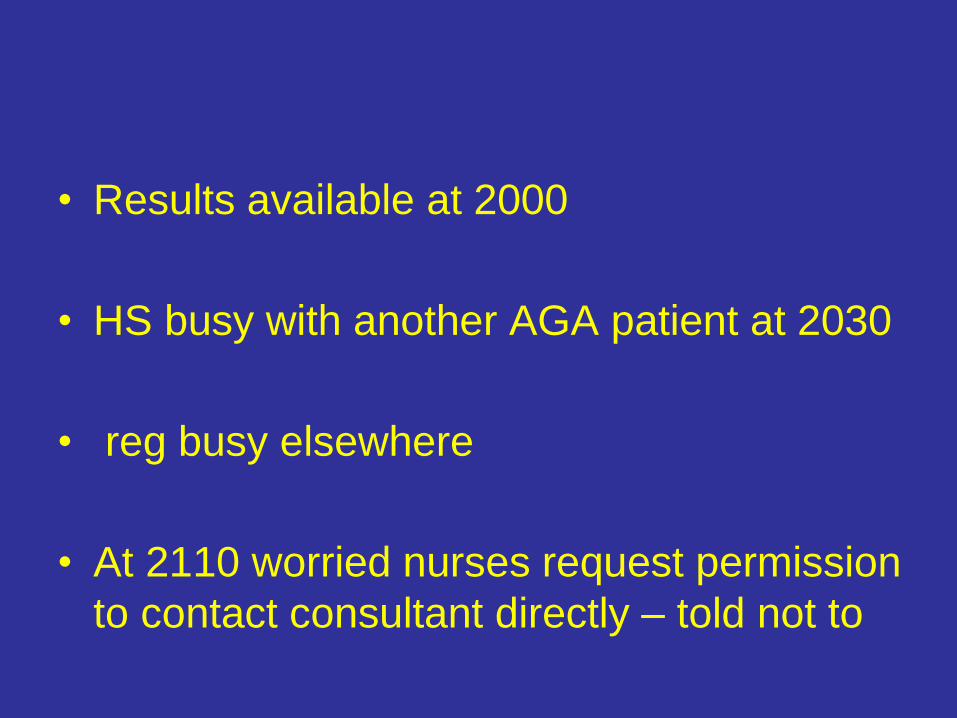
• Results available at 2000
• HS busy with another AGA patient at 2030
• reg busy elsewhere
• At 2110 worried nurses request permission
to contact consultant directly – told not to

• 2120 more experienced Dr comes to AGA
but decides to review a different patient
instead.
• 2135 patient reviewed
• 2155 a more experienced DR is sought

• 2200 seen by someone willing to make a
decision
• OT v.busy
• Surgery commences 0045 and finishes
0130.

Analysis
• Patient waited >4hrs in AGA before a
diagnosis and treatment plan made
• Diagnosis was obvious on arrival
• Root cause of delay was lack of
experienced input at beginning and
decision to await further results.

• Serum quantitative HCG is not a
compulsory test
• Doctors are still allowed to make clinical
decisions
• A negative laparoscopy is not a criminal
offence

• Radiologists are not clinicians and do not
know how to manage patients, therefore
what they recommend is irrelevant
• Should registrars review scans ?

• 1730 seen by Drs
• 1745 consent for surgery obtained
• 1930 laparoscopic salpingectomy
completed (before trauma victim arrives in
hospital and blocks theatres for the next 5
hours)

History:
• 44 year old
• Para 3
• Presented 21/11/07
• LMP 13/09/07
• Vomiting, diarrhoea, feinting 4-6/11/07
• PV bleed 11/11/07 ?passage of tissue
• Breast tenderness
• Continued lower abdo and RIF pain
• Prev tubal sterilisation

Examination:
• Cystic mass right adnexa
• Uterine enlargement
• Marked bimanual tenderness
• Investigations arranged
• Diagnosis uncertain. FU next day with results.
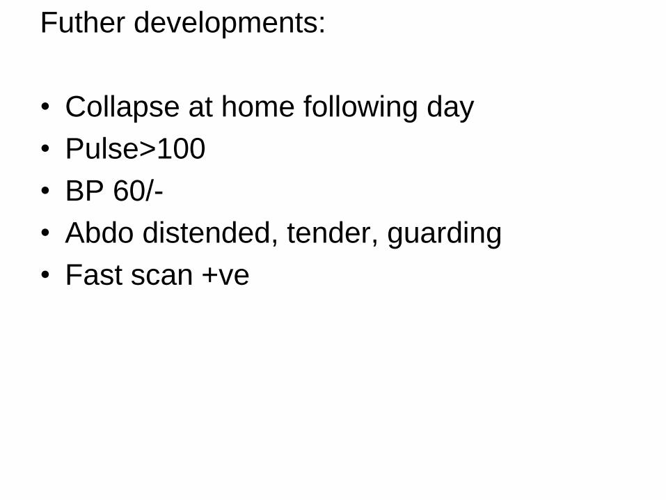
Futher developments:
• Collapse at home following day
• Pulse>100
• BP 60/-
• Abdo distended, tender, guarding
• Fast scan +ve

Diagnosis:
• Ruptured Ectopic pregnancy
Laparotomy findings:
• 2500ml haemoperitoneum

Other info:
• Scan report- uterus 113x59x60mm,
endometrium 6mm, right ovary significantly
enlarged 54x31x49mm heterogenous with
increased vascularity. The nature of this
change is uncertain but the appearances
are significantly abnormal
• Serum HCG 12400

• The scan appearances are typical of an ectopic pregnancy
• Provided clinical information was “amenorrhoea for 8 weeks with right adnexal mass”
• Pregnancy not considered likely at first assessment

Case Presentation
• 8/10/08
• Lower abdo pain
• PV bleeding
• Positive pregnancy test

Ultrasound scan
• Bulky retroverted uterus
• No evidence of intrauterine pregnancy
• Complex cystic/echogenic material
present in fundus, 35x24x43mm
• ?RPOC, ?molar pregnancy
• Normal adnexae
• No free fluid

Seen in AGA
• ? Molar pregnancy
• Booked for ERPOC
• HCG 20,884

ERPOC
• Straightforward procedure
• Products to histology
• Follow up in GTD clinic
GTD clinic
• Histology- decidua only

tests
• HCG: 17/10- 84
• 20/10- 6819
• Scan: same as before
• HCG: 23/10- 4215
• 28/10- 2509
• CXR: 3/11- normal
• HCG: 3/11- 1518

Scan- 3/11
• A transvaginal scan was performed.. Appearances are
similar to previous scans. Within the endometrium there
is a residual collapsed cystic space query old gestation
sac 32 mm x 22 mm with adjacent prominent decidual
reaction and colour Doppler flow. Deep to this, there are
several small cystic spaces of uncertain location, query
endometrium, query myometrial. There is a small soft
tissue mass posteriorly low in the body of the uterus
consistent with an incidental subserosal fibroid 18 mm x
21 mm. Both ovaries are normal in appearance. No
pelvic masses or fluid seen.

Scan 3/11
• Appearances are consistent with persisting retained
products of conception/molar change in the
endometrium, prominent vascular endometrium and
possibly some cystic change in the myometrium. The
interface between endometrium and myometrium is not
clearly seen and could be further assessed
preoperatively with an MRI scan
• Decision made for repeat erpoc/hysteroscopy

Repeat surgery - 6/11
• Empty uterus
• Defect in endometrium/myometrium at
fundus consistent with previous perforation
• Histology- minimal tissue. Mostly blood.
Weakly proliferative endometrium.

Repeat HCG
• “tumour HCG” - 937
• “pregnancy HCG” - 858

More HCG
• 13/11- 559
• 24/11- 267
• 1/12- 194 (*pregnancy, not tumour)
• 8/12- 160
• 15/12- 165

Another scan
• Persistent heterogeneous fundal mass,
with internal cystic areas. This may in fact
lie within the myometrium, ?cystic
degeneration of a fibroid. MRI is
suggested to further characterise this area,
and to assess for ongoing RPOC or
evidence of gestational trophoblastic
disease.

More HCG
• 22/12 -150
• 29/12 -125
• 5/1 -116
• 13/1 - for pelvic MRI
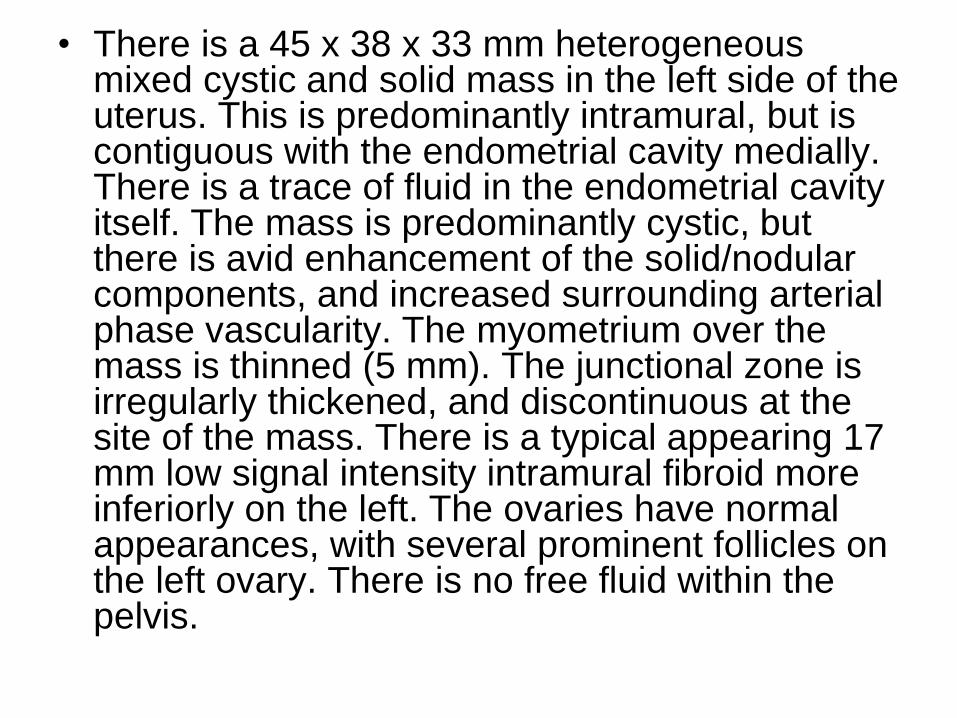
• There is a 45 x 38 x 33 mm heterogeneous mixed cystic and solid mass in the left side of the uterus. This is predominantly intramural, but is contiguous with the endometrial cavity medially. There is a trace of fluid in the endometrial cavity itself. The mass is predominantly cystic, but there is avid enhancement of the solid/nodular components, and increased surrounding arterial phase vascularity. The myometrium over the mass is thinned (5 mm). The junctional zone is irregularly thickened, and discontinuous at the site of the mass. There is a typical appearing 17 mm low signal intensity intramural fibroid more inferiorly on the left. The ovaries have normal appearances, with several prominent follicles on the left ovary. There is no free fluid within the pelvis.
MRI
• The cystic uterine mass could represent an atypical fibroid with cystic degeneration, and does appear to be centred within the myometrium. However given the persistent elevated beta-hCG, I cannot exclude gestational trophoblastic disease invading into the myometrium (potentially from a cornual site implantation). We will review the case at the gynae-oncology meeting.

• For repeat HCG
• Hysterectomy if remains elevated

• 27/1 HCG: 75
• 9/2 HCG: 48
• 16/2 HCG: 48
• 2/3 Hysterectomy
Histology
• Diagnosis:
• Uterus: Ectopic left cornual pregnancy
• (plus fibroids, plus adenomyosis)