Embed Size (px)
Citation preview

EANS/UEMS European examination in neurosurgery Part I (written) Variants of questions with answers (compilation - Vyacheslav S. Botev, Department of Neurosurgery, M.Gorky Donetsk National Medical University)
1. Which of the following cells are the most numerous in the CNS?
A. Neurons B. Astrocytes C. Microglial cells D. Ependymal cells E. Oligodendrocytes
2. After biopsy resection of a lymph node in his neck, a 27-year-old bank manager notices instability of his shoulder. Neurologic examination reveals winging of the scapula on the side of the surgery:
During surgery, he probably suffered damage to the A. Axillary nerve B. Deltoid muscle C. Long thoracic nerve D. Suprascapular nerve E. Serratus anterior muscle
3. Which of the following portions of the ventricular system does not contain choroid plexus? A. Third ventricle B. Fourth ventricle C. Lateral ventricle D. Cerebral aqueduct E. Interventricular foramen
4. Which of the following cells are responsible for the entry of human immunodeficiency virus (HIV) into the CNS?
A. Astrocytes B. Schwann cells C. Endothelial cells D. Oligodendrocytes E. Microglial macrophages
5. Which factor has the greatest impact on the development of a headache after lumbar puncture? A. Needle size B. Volume of spinal fluid removed C. Hydration after lumbar puncture D. Position of patient during the procedure E. Duration of bed rest after lumbar puncture
6. Who is most likely to develop a headache after lumbar puncture? A. A 6-year-old boy B. A 10-year-old girl C. A thin 25-year-old woman D. An overweight 50-year-old woman E. A 30-year-old man with a concussion
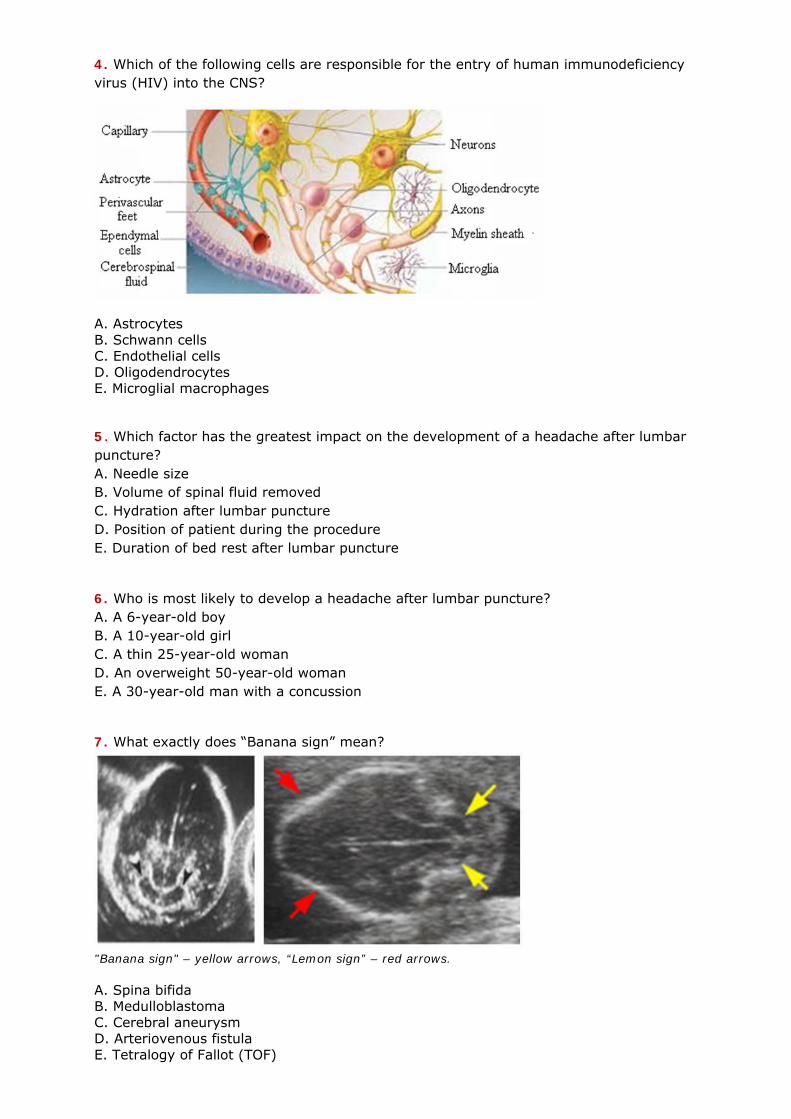
7. What exactly does “Banana sign” mean?
"Banana sign" – yellow arrows, “Lemon sign” – red arrows. A. Spina bifida B. Medulloblastoma C. Cerebral aneurysm D. Arteriovenous fistula Е. Tetralogy of Fallot (TOF)
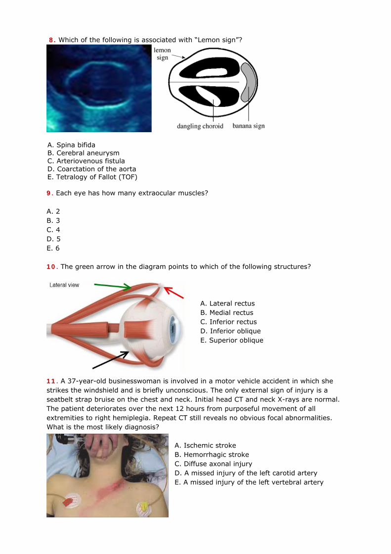
8. Which of the following is associated with “Lemon sign”?
A. Spina bifida B. Cerebral aneurysm C. Arteriovenous fistula D. Coarctation of the aorta Е. Tetralogy of Fallot (TOF)
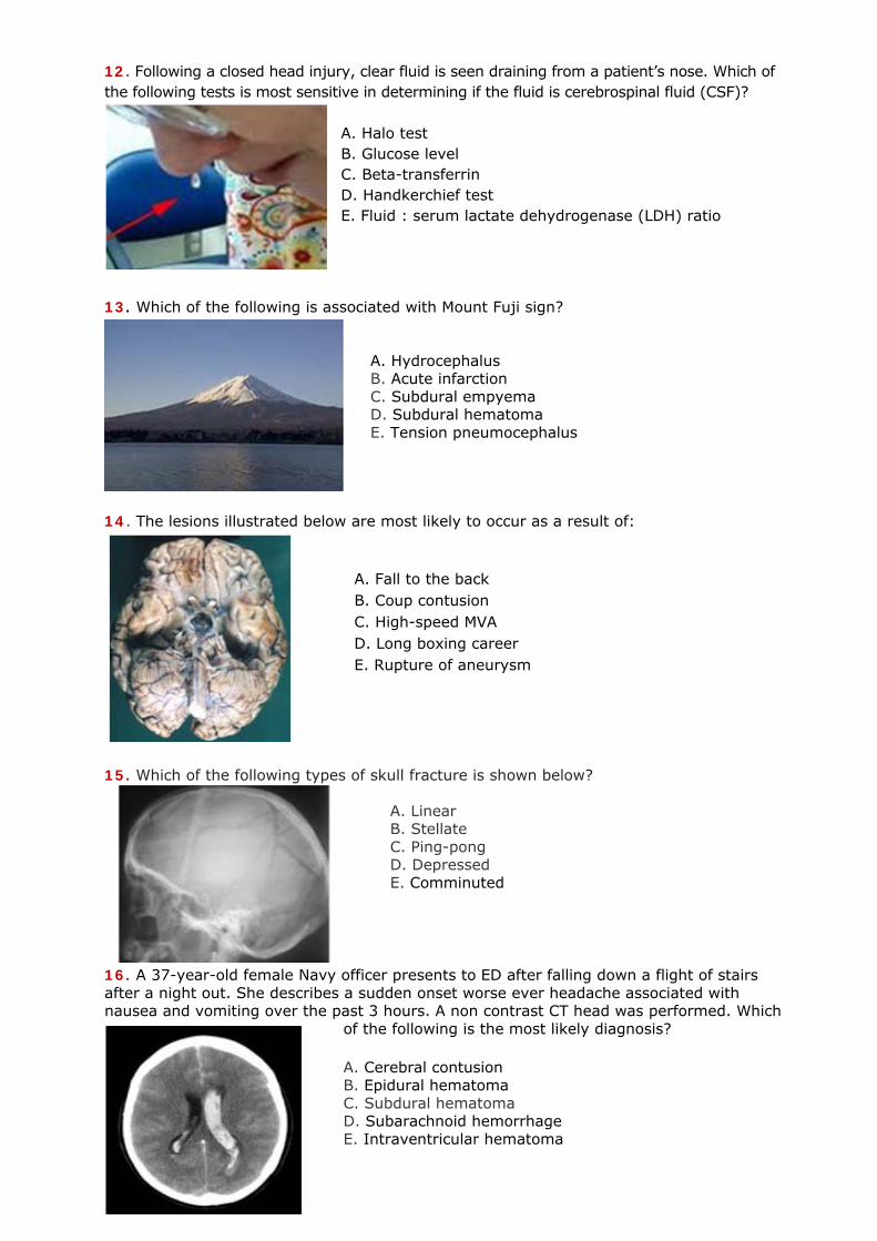
9. Each eye has how many extraocular muscles? A. 2 B. 3 C. 4 D. 5 E. 6 10. The green arrow in the diagram points to which of the following structures?
A. Lateral rectus B. Medial rectus C. Inferior rectus D. Inferior oblique E. Superior oblique
11. A 37-year-old businesswoman is involved in a motor vehicle accident in which she strikes the windshield and is briefly unconscious. The only external sign of injury is a seatbelt strap bruise on the chest and neck. Initial head CT and neck X-rays are normal. The patient deteriorates over the next 12 hours from purposeful movement of all extremities to right hemiplegia. Repeat CT still reveals no obvious focal abnormalities. What is the most likely diagnosis?
A. Ischemic stroke B. Hemorrhagic stroke C. Diffuse axonal injury D. A missed injury of the left carotid artery E. A missed injury of the left vertebral artery
12. Following a closed head injury, clear fluid is seen draining from a patient’s nose. Which of the following tests is most sensitive in determining if the fluid is cerebrospinal fluid (CSF)?
A. Halo test B. Glucose level C. Beta-transferrin D. Handkerchief test E. Fluid : serum lactate dehydrogenase (LDH) ratio
13. Which of the following is associated with Mount Fuji sign? A. Hydrocephalus B. Acute infarction C. Subdural empyema D. Subdural hematoma E. Tension pneumocephalus
14. The lesions illustrated below are most likely to occur as a result of:
A. Fall to the back B. Coup contusion C. High-speed MVA D. Long boxing career E. Rupture of aneurysm
15. Which of the following types of skull fracture is shown below?
A. Linear B. Stellate C. Ping-pong D. Depressed E. Comminuted
16. A 37-year-old female Navy officer presents to ED after falling down a flight of stairs after a night out. She describes a sudden onset worse ever headache associated with nausea and vomiting over the past 3 hours. A non contrast CT head was performed. Which
of the following is the most likely diagnosis?
A. Cerebral contusion B. Epidural hematoma C. Subdural hematoma D. Subarachnoid hemorrhage E. Intraventricular hematoma
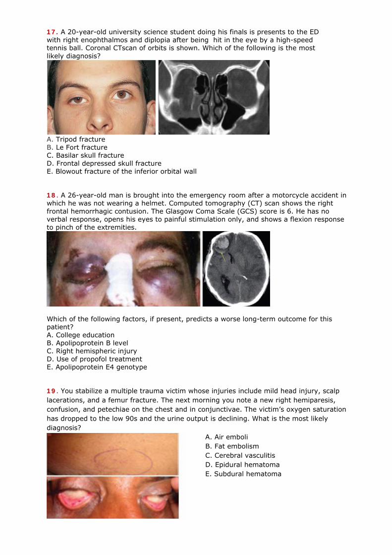
17. A 20-year-old university science student doing his finals is presents to the ED with right enophthalmos and diplopia after being hit in the eye by a high-speed tennis ball. Coronal CTscan of orbits is shown. Which of the following is the most likely diagnosis?
A. Tripod fracture B. Le Fort fracture C. Basilar skull fracture D. Frontal depressed skull fracture E. Blowout fracture of the inferior orbital wall
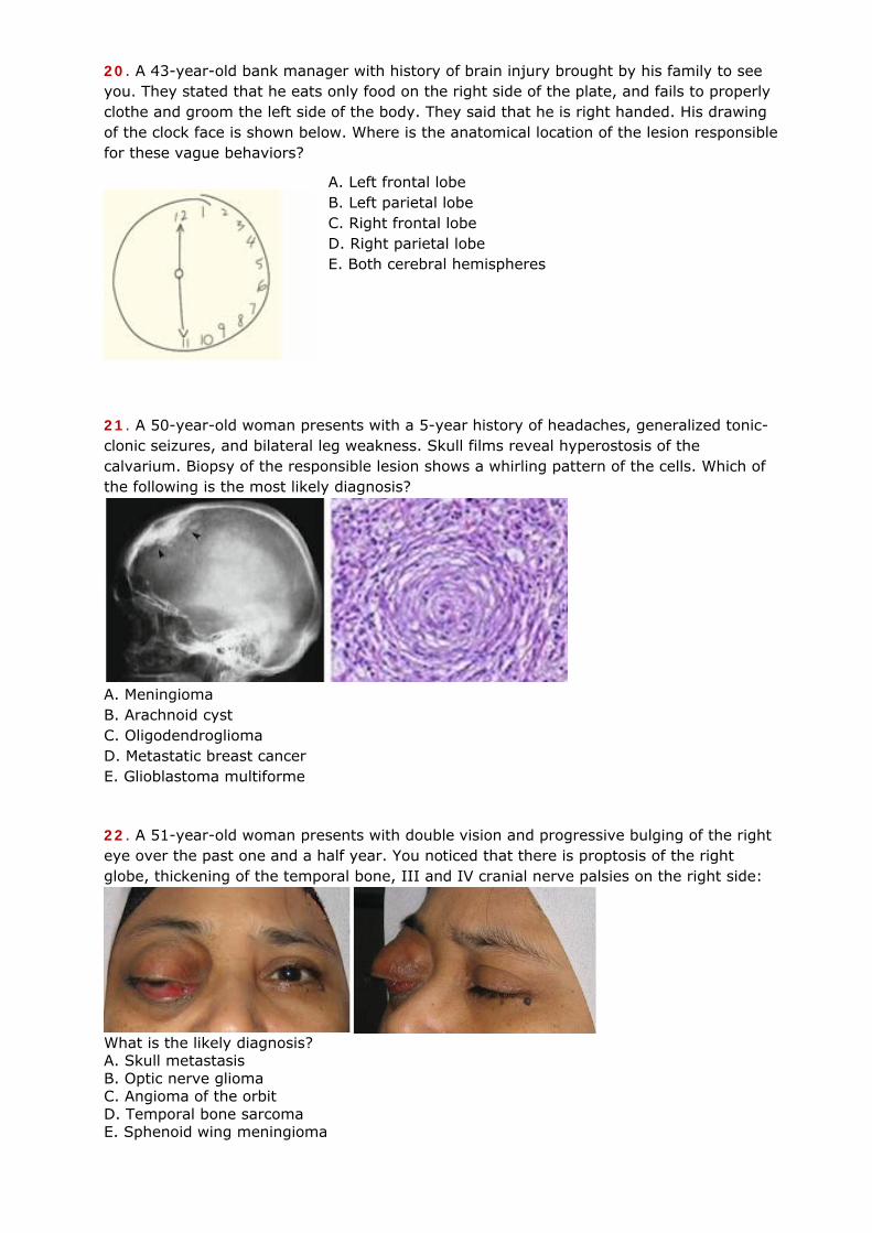
18. A 26-year-old man is brought into the emergency room after a motorcycle accident in which he was not wearing a helmet. Computed tomography (CT) scan shows the right frontal hemorrhagic contusion. The Glasgow Coma Scale (GCS) score is 6. He has no verbal response, opens his eyes to painful stimulation only, and shows a flexion response to pinch of the extremities.
Which of the following factors, if present, predicts a worse long-term outcome for this patient? A. College education B. Apolipoprotein B level C. Right hemispheric injury D. Use of propofol treatment E. Apolipoprotein E4 genotype
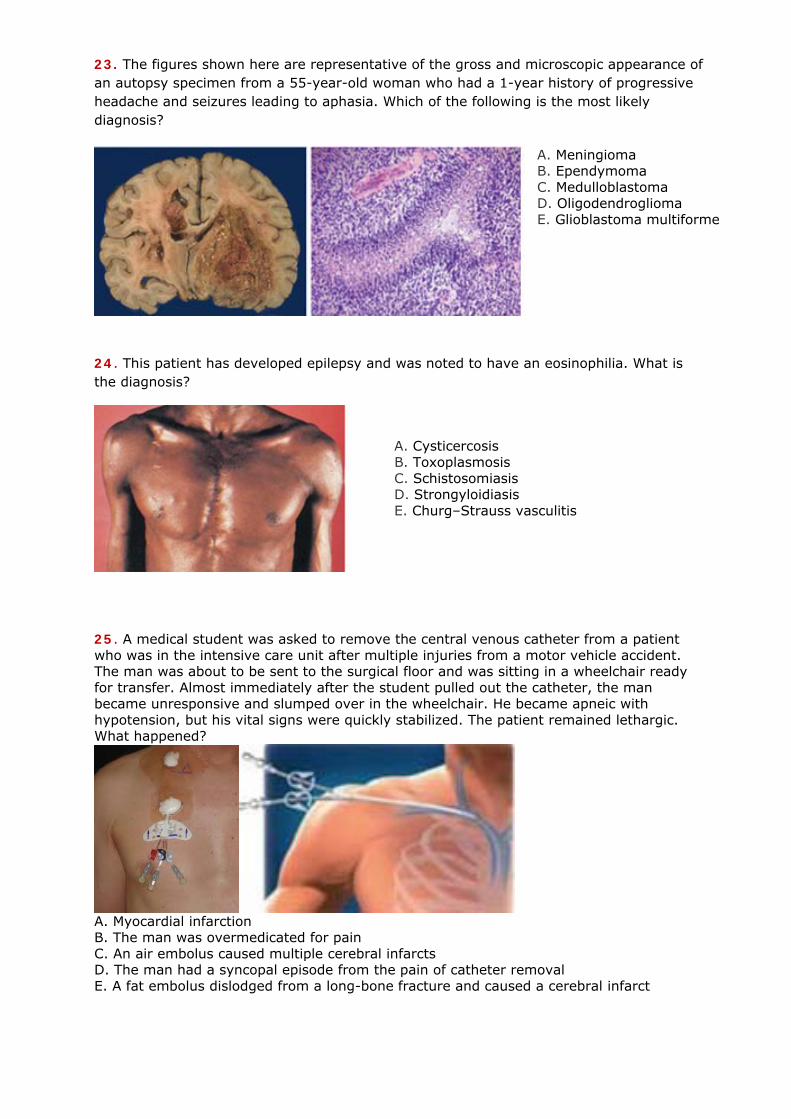
19. You stabilize a multiple trauma victim whose injuries include mild head injury, scalp lacerations, and a femur fracture. The next morning you note a new right hemiparesis, confusion, and petechiae on the chest and in conjunctivae. The victim’s oxygen saturation has dropped to the low 90s and the urine output is declining. What is the most likely diagnosis?
A. Air emboli B. Fat embolism C. Cerebral vasculitis D. Epidural hematoma E. Subdural hematoma
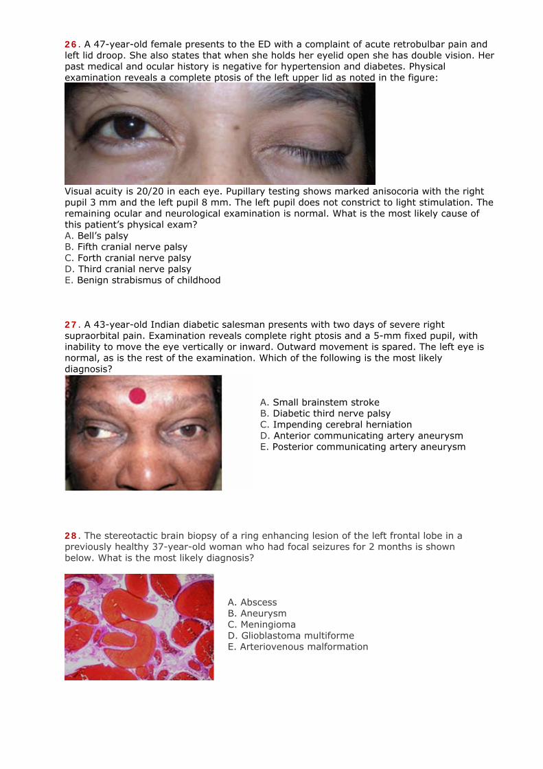
20. A 43-year-old bank manager with history of brain injury brought by his family to see you. They stated that he eats only food on the right side of the plate, and fails to properly clothe and groom the left side of the body. They said that he is right handed. His drawing of the clock face is shown below. Where is the anatomical location of the lesion responsible for these vague behaviors?
A. Left frontal lobe B. Left parietal lobe C. Right frontal lobe D. Right parietal lobe E. Both cerebral hemispheres
21. A 50-year-old woman presents with a 5-year history of headaches, generalized tonic-clonic seizures, and bilateral leg weakness. Skull films reveal hyperostosis of the calvarium. Biopsy of the responsible lesion shows a whirling pattern of the cells. Which of the following is the most likely diagnosis?
A. Meningioma B. Arachnoid cyst C. Oligodendroglioma D. Metastatic breast cancer E. Glioblastoma multiforme 22. A 51-year-old woman presents with double vision and progressive bulging of the right eye over the past one and a half year. You noticed that there is proptosis of the right globe, thickening of the temporal bone, III and IV cranial nerve palsies on the right side:
What is the likely diagnosis? A. Skull metastasis B. Optic nerve glioma C. Angioma of the orbit D. Temporal bone sarcoma E. Sphenoid wing meningioma
23. The figures shown here are representative of the gross and microscopic appearance of an autopsy specimen from a 55-year-old woman who had a 1-year history of progressive headache and seizures leading to aphasia. Which of the following is the most likely diagnosis?
A. Meningioma B. Ependymoma C. Medulloblastoma D. Oligodendroglioma E. Glioblastoma multiforme
24. This patient has developed epilepsy and was noted to have an eosinophilia. What is the diagnosis?
A. Cysticercosis B. Toxoplasmosis C. Schistosomiasis D. Strongyloidiasis E. Churg–Strauss vasculitis
25. A medical student was asked to remove the central venous catheter from a patient who was in the intensive care unit after multiple injuries from a motor vehicle accident. The man was about to be sent to the surgical floor and was sitting in a wheelchair ready for transfer. Almost immediately after the student pulled out the catheter, the man became unresponsive and slumped over in the wheelchair. He became apneic with hypotension, but his vital signs were quickly stabilized. The patient remained lethargic. What happened?
A. Myocardial infarction B. The man was overmedicated for pain C. An air embolus caused multiple cerebral infarcts D. The man had a syncopal episode from the pain of catheter removal Е. A fat embolus dislodged from a long-bone fracture and caused a cerebral infarct
26. A 47-year-old female presents to the ED with a complaint of acute retrobulbar pain and left lid droop. She also states that when she holds her eyelid open she has double vision. Her past medical and ocular history is negative for hypertension and diabetes. Physical examination reveals a complete ptosis of the left upper lid as noted in the figure:
Visual acuity is 20/20 in each eye. Pupillary testing shows marked anisocoria with the right pupil 3 mm and the left pupil 8 mm. The left pupil does not constrict to light stimulation. The remaining ocular and neurological examination is normal. What is the most likely cause of this patient’s physical exam? A. Bell’s palsy B. Fifth cranial nerve palsy C. Forth cranial nerve palsy D. Third cranial nerve palsy E. Benign strabismus of childhood 27. A 43-year-old Indian diabetic salesman presents with two days of severe right supraorbital pain. Examination reveals complete right ptosis and a 5-mm fixed pupil, with inability to move the eye vertically or inward. Outward movement is spared. The left eye is normal, as is the rest of the examination. Which of the following is the most likely diagnosis?
A. Small brainstem stroke B. Diabetic third nerve palsy C. Impending cerebral herniation D. Anterior communicating artery aneurysm E. Posterior communicating artery aneurysm
28. The stereotactic brain biopsy of a ring enhancing lesion of the left frontal lobe in a previously healthy 37-year-old woman who had focal seizures for 2 months is shown below. What is the most likely diagnosis?
A. Abscess B. Aneurysm C. Meningioma D. Glioblastoma multiforme E. Arteriovenous malformation

29. A 41-year-old man presents to the emergency department (ED) complaining of a severe frontal headache that began suddenly and awakened him from sleep, nausea, vomiting and new-onset diplopia with photophobia. The ocular examination demonstrates ptosis of the right eye which is deviated inferolaterally and has a dilated and unreactive pupil. The visual field examination demonstrates bitemporal hemianopsia.
A MRI is performed. What is the most likely diagnosis?
A. Cerebellar infarction B. Pituitary tumor apoplexy C. Subarachnoid hemorrhage D. Cavernous sinus thrombosis E. Arteriovenous malformation
30. What exactly does “Mother in law” sign mean in neuroradiology? A. Meningioma B. Hydrocephalus C. Base skull fracture D. Pituitary fossa tumor E. Glioblastoma multiforme 31. This is a CTA/CTV of a patient with an extracranial-intracranial (EC-IC) bypass. Match the arrows with the structures. Each answer can be used only once, but some answers will
not be used. Match arrows 1 through 5 with the following answers:
A. Jugular vein B. Basilar artery C. Straight sinus D. Vein of Galen E. Sigmoid sinus F. Vertebral artery G. Transverse sinus H. Internal cerebral vein I. Superior sagittal sinus J. Middle cerebral artery
32. Which of the following vessels is indicated by arrow?
A. Facial artery B. Sigmoid sinus C. Internal jugular vein D. Middle cerebral artery E. Superficial temporal artery
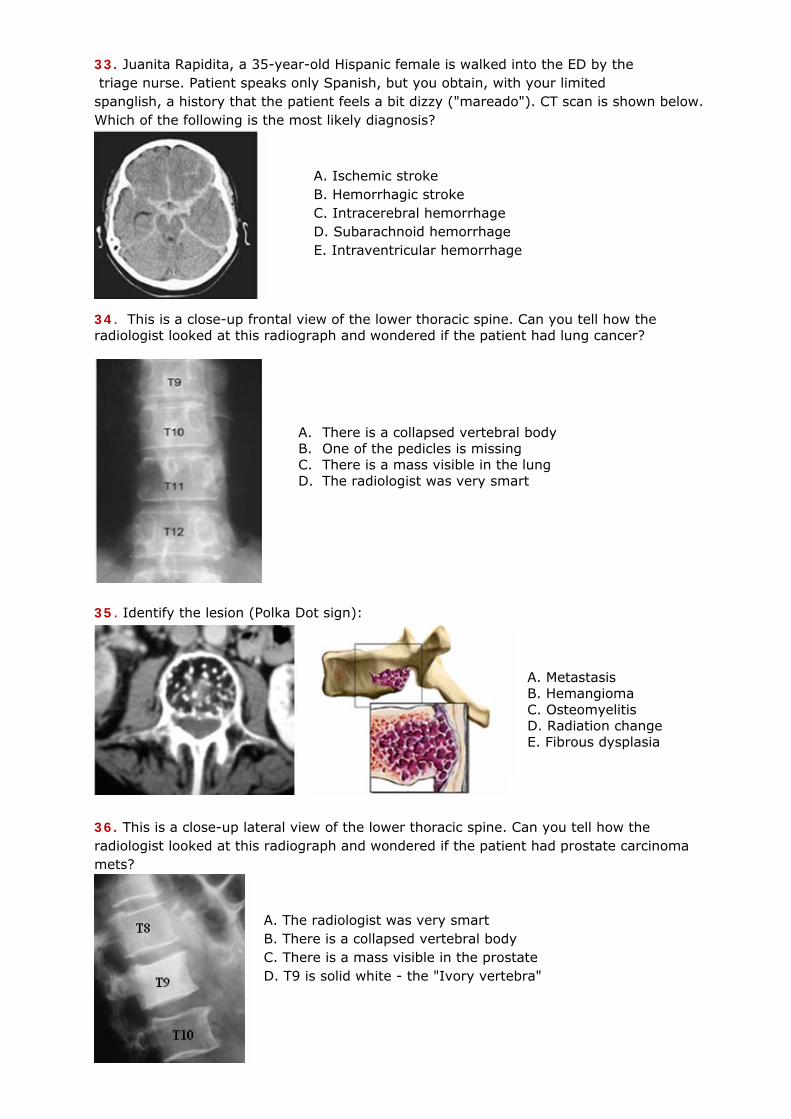
33. Juanita Rapidita, a 35-year-old Hispanic female is walked into the ED by the triage nurse. Patient speaks only Spanish, but you obtain, with your limited spanglish, a history that the patient feels a bit dizzy ("mareado"). CT scan is shown below. Which of the following is the most likely diagnosis?
A. Ischemic stroke B. Hemorrhagic stroke C. Intracerebral hemorrhage D. Subarachnoid hemorrhage E. Intraventricular hemorrhage
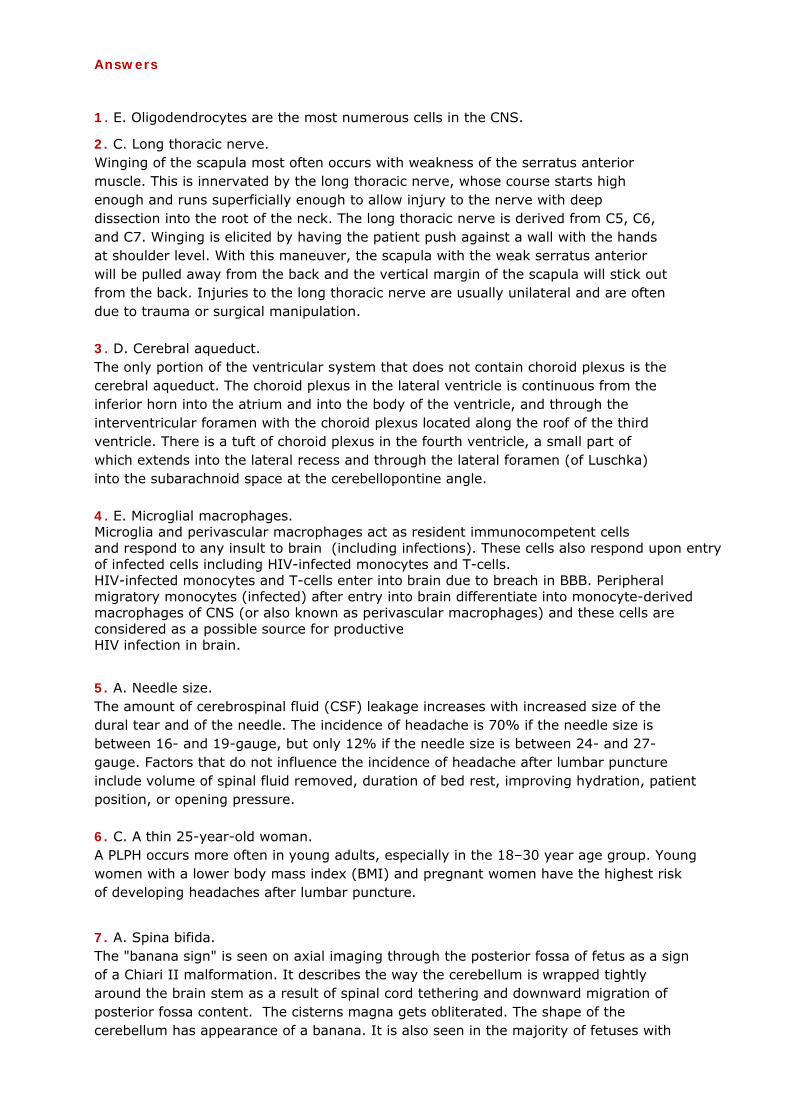
34. This is a close-up frontal view of the lower thoracic spine. Can you tell how the radiologist looked at this radiograph and wondered if the patient had lung cancer?
A. There is a collapsed vertebral body B. One of the pedicles is missing C. There is a mass visible in the lung D. The radiologist was very smart
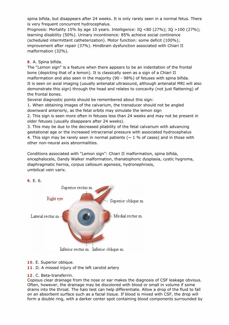
35. Identify the lesion (Polka Dot sign):
A. Metastasis B. Hemangioma C. Osteomyelitis D. Radiation change E. Fibrous dysplasia
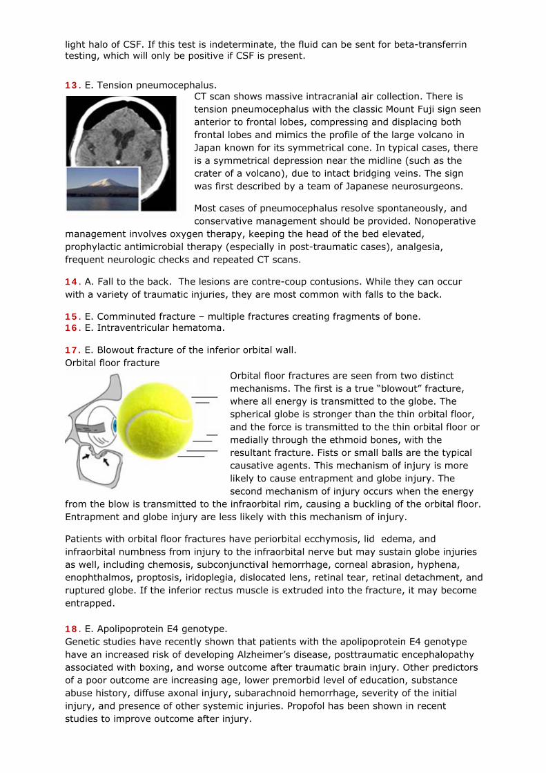
36. This is a close-up lateral view of the lower thoracic spine. Can you tell how the radiologist looked at this radiograph and wondered if the patient had prostate carcinoma mets?
A. The radiologist was very smart B. There is a collapsed vertebral body C. There is a mass visible in the prostate D. T9 is solid white - the "Ivory vertebra"

37. Which of the following is associated with the typical Polka Dot appearance?
A. Metastasis B. Hemangioma C. Osteomyelitis D. Radiation change E. Fibrous dysplasia
The festive red polka dots on feather white background of the
teacup.
Princess Diana in Polka Dot Jacket
(1988).
Answers
1. E. Oligodendrocytes are the most numerous cells in the CNS.
2. C. Long thoracic nerve. Winging of the scapula most often occurs with weakness of the serratus anterior muscle. This is innervated by the long thoracic nerve, whose course starts high enough and runs superficially enough to allow injury to the nerve with deep dissection into the root of the neck. The long thoracic nerve is derived from C5, C6, and C7. Winging is elicited by having the patient push against a wall with the hands at shoulder level. With this maneuver, the scapula with the weak serratus anterior will be pulled away from the back and the vertical margin of the scapula will stick out from the back. Injuries to the long thoracic nerve are usually unilateral and are often due to trauma or surgical manipulation. 3. D. Cerebral aqueduct. The only portion of the ventricular system that does not contain choroid plexus is the cerebral aqueduct. The choroid plexus in the lateral ventricle is continuous from the inferior horn into the atrium and into the body of the ventricle, and through the interventricular foramen with the choroid plexus located along the roof of the third ventricle. There is a tuft of choroid plexus in the fourth ventricle, a small part of which extends into the lateral recess and through the lateral foramen (of Luschka) into the subarachnoid space at the cerebellopontine angle. 4. E. Microglial macrophages. Microglia and perivascular macrophages act as resident immunocompetent cells and respond to any insult to brain (including infections). These cells also respond upon entry of infected cells including HIV-infected monocytes and T-cells. HIV-infected monocytes and T-cells enter into brain due to breach in BBB. Peripheral migratory monocytes (infected) after entry into brain differentiate into monocyte-derived macrophages of CNS (or also known as perivascular macrophages) and these cells are considered as a possible source for productive HIV infection in brain.
5. A. Needle size. The amount of cerebrospinal fluid (CSF) leakage increases with increased size of the dural tear and of the needle. The incidence of headache is 70% if the needle size is between 16- and 19-gauge, but only 12% if the needle size is between 24- and 27-gauge. Factors that do not influence the incidence of headache after lumbar puncture include volume of spinal fluid removed, duration of bed rest, improving hydration, patient position, or opening pressure. 6. C. A thin 25-year-old woman. A PLPH occurs more often in young adults, especially in the 18–30 year age group. Young women with a lower body mass index (BMI) and pregnant women have the highest risk of developing headaches after lumbar puncture.
7. A. Spina bifida. The "banana sign" is seen on axial imaging through the posterior fossa of fetus as a sign of a Chiari II malformation. It describes the way the cerebellum is wrapped tightly around the brain stem as a result of spinal cord tethering and downward migration of posterior fossa content. The cisterns magna gets obliterated. The shape of the cerebellum has appearance of a banana. It is also seen in the majority of fetuses with
spina bifida, but disappears after 24 weeks. It is only rarely seen in a normal fetus. There is very frequent concurrent hydrocephalus. Prognosis: Mortality 15% by age 10 years. Intelligence: IQ <80 (27%); IQ >100 (27%); learning disability (50%). Urinary incontinence: 85% achieve social continence (scheduled intermittent catheterization). Motor function: some deficit (100%); improvement after repair (37%). Hindbrain dysfunction associated with Chiari II malformation (32%). 8. A. Spina bifida. The “Lemon sign” is a feature when there appears to be an indentation of the frontal bone (depicting that of a lemon). It is classically seen as a sign of a Chiari II malformation and also seen in the majority (90 - 98%) of fetuses with spina bifida. It is seen on axial imaging (usually antenatal ultrasound, although antenatal MRI will also demonstrate this sign) through the head and relates to concavity (not just flattening) of the frontal bones. Several diagnostic points should be remembered about this sign: 1. When obtaining images of the calvarium, the transducer should not be angled downward anteriorly, as the fetal orbits may simulate the lemon sign 2. This sign is seen more often in fetuses less than 24 weeks and may not be present in older fetuses (usually disappears after 24 weeks). 3. This may be due to the decreased pliability of the fetal calvarium with advancing gestational age or the increased intracranial pressure with associated hydrocephalus 4. This sign may be rarely seen in normal patients (~ 1 % of cases) and in those with other non-neural axis abnormalities. Conditions associated with “Lemon sign”: Chiari II malformation, spina bifida, encephalocele, Dandy Walker malformation, thanatophoric dysplasia, cystic hygroma, diaphragmatic hernia, corpus callosum agenesis, hydronephrosis, umbilical vein varix. 9. E. 6.
10. E. Superior oblique. 11. D. A missed injury of the left carotid artery
12. C. Beta-transferrin. Copious clear drainage from the nose or ear makes the diagnosis of CSF leakage obvious. Often, however, the drainage may be discolored with blood or small in volume if some drains into the throat. The halo test can help differentiate. Allow a drop of the fluid to fall on an absorbent surface such as a facial tissue. If blood is mixed with CSF, the drop will form a double ring, with a darker center spot containing blood components surrounded by
light halo of CSF. If this test is indeterminate, the fluid can be sent for beta-transferrin testing, which will only be positive if CSF is present.
13. E. Tension pneumocephalus. CT scan shows massive intracranial air collection. There is tension pneumocephalus with the classic Mount Fuji sign seen anterior to frontal lobes, compressing and displacing both frontal lobes and mimics the profile of the large volcano in Japan known for its symmetrical cone. In typical cases, there is a symmetrical depression near the midline (such as the crater of a volcano), due to intact bridging veins. The sign was first described by a team of Japanese neurosurgeons.
Most cases of pneumocephalus resolve spontaneously, and conservative management should be provided. Nonoperative
management involves oxygen therapy, keeping the head of the bed elevated, prophylactic antimicrobial therapy (especially in post-traumatic cases), analgesia, frequent neurologic checks and repeated CT scans.
14. A. Fall to the back. The lesions are contre-coup contusions. While they can occur with a variety of traumatic injuries, they are most common with falls to the back.
15. E. Comminuted fracture – multiple fractures creating fragments of bone. 16. E. Intraventricular hematoma. 17. E. Blowout fracture of the inferior orbital wall. Orbital floor fracture
Orbital floor fractures are seen from two distinct mechanisms. The first is a true “blowout” fracture, where all energy is transmitted to the globe. The spherical globe is stronger than the thin orbital floor, and the force is transmitted to the thin orbital floor or medially through the ethmoid bones, with the resultant fracture. Fists or small balls are the typical causative agents. This mechanism of injury is more likely to cause entrapment and globe injury. The second mechanism of injury occurs when the energy
from the blow is transmitted to the infraorbital rim, causing a buckling of the orbital floor. Entrapment and globe injury are less likely with this mechanism of injury.
Patients with orbital floor fractures have periorbital ecchymosis, lid edema, and infraorbital numbness from injury to the infraorbital nerve but may sustain globe injuries as well, including chemosis, subconjunctival hemorrhage, corneal abrasion, hyphena, enophthalmos, proptosis, iridoplegia, dislocated lens, retinal tear, retinal detachment, and ruptured globe. If the inferior rectus muscle is extruded into the fracture, it may become entrapped. 18. E. Apolipoprotein E4 genotype. Genetic studies have recently shown that patients with the apolipoprotein E4 genotype have an increased risk of developing Alzheimer’s disease, posttraumatic encephalopathy associated with boxing, and worse outcome after traumatic brain injury. Other predictors of a poor outcome are increasing age, lower premorbid level of education, substance abuse history, diffuse axonal injury, subarachnoid hemorrhage, severity of the initial injury, and presence of other systemic injuries. Propofol has been shown in recent studies to improve outcome after injury.
Other genes for which evidence exists for an association with poorer outcome are P53, COMT, DND2, and CACNA1A. Progesterone may have a neuroprotective effect, and prognosis in females may be better if TBI is sustained at a time of the menstrual cycle when progesterone levels are high.
19. B. Fat embolism. 20. D. Right parietal lobe. This is the classical history of someone with a nondominant (right) parietal lobe lesion causing “neglect”; the patient may eagerly hunt for the examiner’s face on the right when the voice is called from the left, and the patient may deny that the left side of the body exists (even a dense hemiparesis may be cheerfully “overlooked”), or the patient may fail to acknowledge as one’s own the left arm held up in plane view by the examiner.
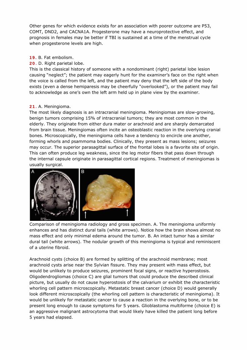
21. A. Meningioma. The most likely diagnosis is an intracranial meningioma. Meningiomas are slow-growing, benign tumors comprising 15% of intracranial tumors; they are most common in the elderly. They originate from either dura mater or arachnoid and are sharply demarcated from brain tissue. Meningiomas often incite an osteoblastic reaction in the overlying cranial bones. Microscopically, the meningioma cells have a tendency to encircle one another, forming whorls and psammoma bodies. Clinically, they present as mass lesions; seizures may occur. The superior parasagittal surface of the frontal lobes is a favorite site of origin. This can often produce leg weakness, since the leg motor fibers that pass down through the internal capsule originate in parasagittal cortical regions. Treatment of meningiomas is usually surgical.
Comparison of meningioma radiology and gross specimen. A. The meningioma uniformly enhances and has distinct dural tails (white arrows). Notice how the brain shows almost no mass effect and only minimal edema around the tumor. B. An intact tumor has a similar dural tail (white arrows). The nodular growth of this meningioma is typical and reminiscent of a uterine fibroid. Arachnoid cysts (choice B) are formed by splitting of the arachnoid membrane; most arachnoid cysts arise near the Sylvian fissure. They may present with mass effect, but would be unlikely to produce seizures, prominent focal signs, or reactive hyperostosis. Oligodendrogliomas (choice C) are glial tumors that could produce the described clinical picture, but usually do not cause hyperostosis of the calvarium or exhibit the characteristic whorling cell pattern microscopically. Metastatic breast cancer (choice D) would generally look different microscopically (the whorling cell pattern is characteristic of meningioma). It would be unlikely for metastatic cancer to cause a reaction in the overlying bone, or to be present long enough to cause symptoms for 5 years. Glioblastoma multiforme (choice E) is an aggressive malignant astrocytoma that would likely have killed the patient long before 5 years had elapsed.
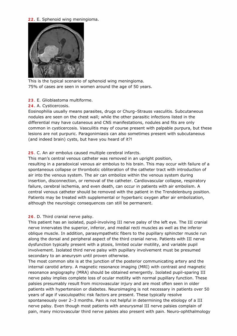
22. E. Sphenoid wing meningioma.
This is the typical scenario of sphenoid wing meningioma. 75% of cases are seen in women around the age of 50 years.
23. E. Glioblastoma multiforme. 24. A. Cysticercosis. Eosinophilia usually means parasites, drugs or Churg–Strauss vasculitis. Subcutaneous nodules are seen on the chest wall; while the other parasitic infections listed in the differential may have cutaneous and CNS manifestations, nodules and fits are only common in cysticercosis. Vasculitis may of course present with palpable purpura, but these lesions are not purpuric. Paragonimiasis can also sometimes present with subcutaneous (and indeed brain) cysts, but have you heard of it?!
25. C. An air embolus caused multiple cerebral infarcts. This man’s central venous catheter was removed in an upright position, resulting in a paradoxical venous air embolus to his brain. This may occur with failure of a spontaneous collapse or thrombotic obliteration of the catheter tract with introduction of air into the venous system. The air can embolize within the venous system during insertion, disconnection, or removal of the catheter. Cardiovascular collapse, respiratory failure, cerebral ischemia, and even death, can occur in patients with air embolism. A central venous catheter should be removed with the patient in the Trendelenburg position. Patients may be treated with supplemental or hyperbaric oxygen after air embolization, although the neurologic consequences can still be permanent.
26. D. Third cranial nerve palsy. This patient has an isolated, pupil-involving III nerve palsy of the left eye. The III cranial nerve innervates the superior, inferior, and medial recti muscles as well as the inferior oblique muscle. In addition, parasympathetic fibers to the pupillary sphincter muscle run along the dorsal and peripheral aspect of the third cranial nerve. Patients with III nerve dysfunction typically present with a ptosis, limited ocular motility, and variable pupil involvement. Isolated third nerve palsy with pupillary involvement must be presumed secondary to an aneurysm until proven otherwise. The most common site is at the junction of the posterior communicating artery and the internal carotid artery. A magnetic resonance imaging (MRI) with contrast and magnetic resonance angiography (MRA) should be obtained emergently. Isolated pupil-sparing III nerve palsy implies complete loss of ocular motility with normal pupillary function. These palsies presumably result from microvascular injury and are most often seen in older patients with hypertension or diabetes. Neuroimaging is not necessary in patients over 50 years of age if vasculopathic risk factors are present. These typically resolve spontaneously over 2–3 months. Pain is not helpful in determining the etiology of a III nerve palsy. Even though most patients with aneurysmal III nerve palsies complain of pain, many microvascular third nerve palsies also present with pain. Neuro-ophthalmology
or neurology consult is warranted for patients presenting to the emergency department with a III nerve palsy. (Visual Diagnosis in Emergency and Critical Care Medicine Christopher P. Holstege © 2006 by Blackwell Publishing Ltd UK) 27. E. Posterior communicating artery aneurysm. Posterior communicating aneurysms (one of the most common locations for aneurysms of the circle of Willis) can present with headache and third nerve palsy. In a diabetic third nerve palsy, there is usually sparing of pupillary reaction. An aneurysm on the posterior communicating artery is especially likely to compress the oculomotor (third) nerve. Because the pupilloconstrictor fibers lie superficially on this nerve, problems with pupillary activity are routinely early phenomena. An ischemic injury to the third cranial nerve, such as that seen with diabetes mellitus, will usually spare these superficial fibers, presumably because they have a vascular supply that is fairly distinct from that of the rest of the third nerve. The pupillary response to both direct and consensual stimulation will be impaired with compression of these parasympathetic nerve fibers. This means that the pupil in the right eye will not constrict in response to light shining into either the right or the left eye. The normal pupil on the left will constrict with light shining into either the left or the right eye because the sensory input from the right eye is unimpaired. As the aneurysm enlarges, it impinges upon the third-nerve fibers that supply the medial rectus muscle, weakness of which was responsible for this woman’s double vision. Lesions of the superior cerebellar artery and posterior cerebral artery can also compress the third nerve, which exits between them. It is therefore important that a complete angiogram, evaluating all four vessels, is performed in the evaluation for subarachnoid hemorrhage and third-nerve palsy.
28. E. Arteriovenous malformation. A vascular malformation can produce a localized mass within the cerebral hemisphere and produce small hemorrhages that induce gliosis in surrounding brain parenchyma.
29. B. Pituitary tumor apoplexy. Coronal gadolinium-enhanced T1-weighted images revealed a large soft-tissue mass in the pituitary fossa, with areas of intermediate and high intensity signal suggestive of hemorrhage. His neurologic finding (right-sided ptosis with a fixed and dilated pupil pointing downward and outward) was consistent with a right sided 3rd nerve palsy caused by extension of hemorrhage into the right cavernous sinus. Pituitary tumor apoplexy is defined as hemorrhage or infarction of a pituitary gland associated with the presence of a preexisting pituitary adenoma. It manifests as a sudden, severe headache, and it is sometimes associated with neurologic and hormonal dysfunction. The word "apoplexy" stems from a Greek term meaning to "have a stroke". Neurologic symptoms and signs are secondary to displacement of the optic nerve and impingement of the 3rd, 4th, and 6th cranial nerves. Hormonal dysfunction results from destruction of the anterior pituitary gland. 30. A. Meningioma.
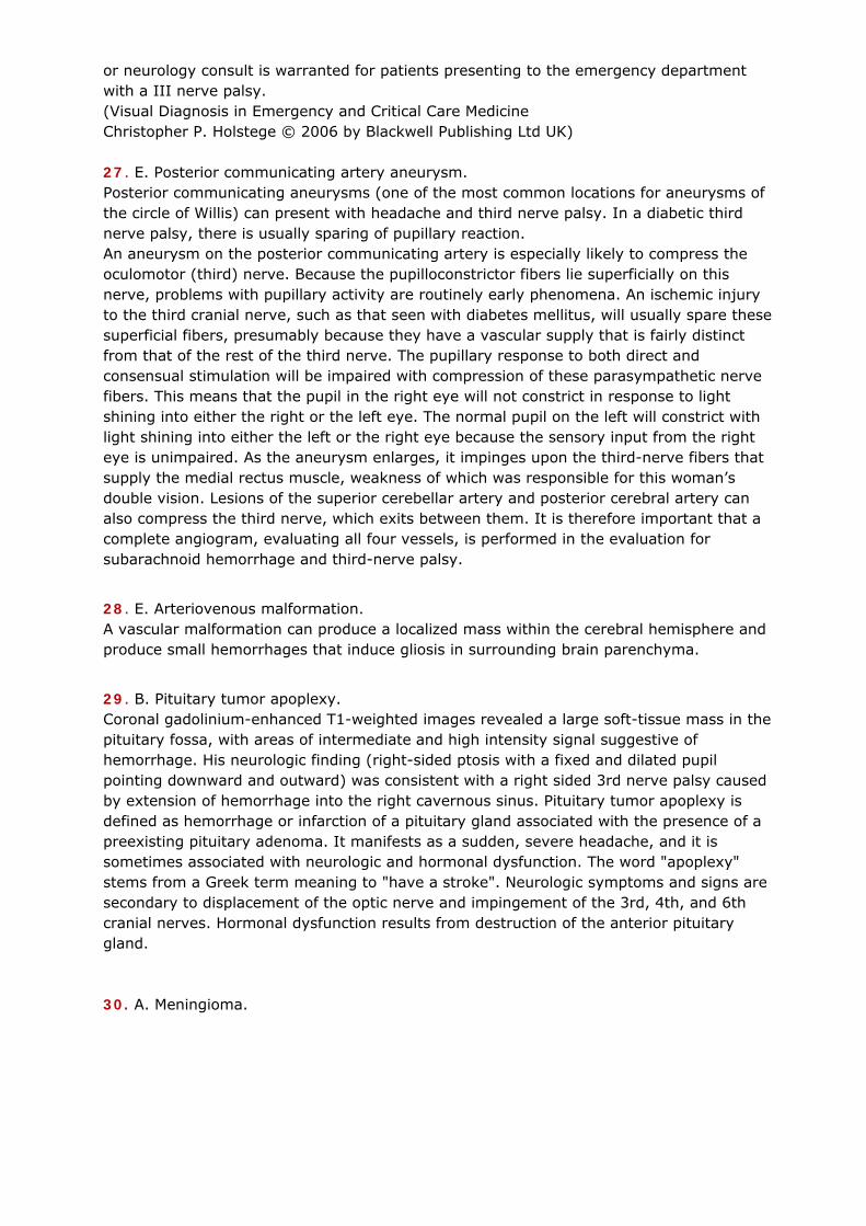
Mother in law sign - used to describe lesions that enhance early during the arterial phase and remain opacified well after the venous phase. The sign is jokingly named after the uncanny ability of mothers in law to arrive early and stay late. Such behaviour matches the classic angiographic enhancement pattern of meningiomas. The above lateral image is from a common carotid artery injection during the venous phase and demonstrates a large olfactory groove meningioma.
31. The answer is 1J, 2D, 3C, 4B, 5G. This was obtained using the Cere-TOM CT scanner. The CT angiogram has the advantage of imaging arteries and veins simultaneously. In this situation, the superior temporal artery enters the intracranial space via the surgical skull defect and anastomoses with the MCA, representing an EC-IC bypass. The other vessels are standard normal anatomy.
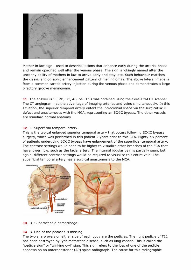
32. E. Superficial temporal artery. This is the typical enlarged superior temporal artery that occurs following EC-IC bypass surgery, which was performed in this patient 2 years prior to this CTA. Eighty-six percent of patients undergoing EC-IC bypass have enlargement of the superficial temporal artery. The contrast settings would need to be higher to visualize other branches of the ECA that have lower flow, such as the facial artery. The internal jugular vein is partially seen, but again, different contrast settings would be required to visualize this entire vein. The superficial temporal artery has a surgical anastomosis to the MCA.
33. D. Subarachnoid hemorrhage. 34. B. One of the pedicles is missing. The two sharp ovals on either side of each body are the pedicles. The right pedicle of T11 has been destroyed by lytic metastatic disease, such as lung cancer. This is called the “pedicle sign” or “winking owl” sign. This sign refers to the loss of one of the pedicle shadows on an anteroposterior (AP) spine radiograph. The cause for this radiographic
finding is most frequently a metastatic vertebral lesion that has extended into the pedicle region and caused destruction of the pedicle. 35. B. Hemangioma. A hemangioma is the most common benign slow-growing bone tumor of the spine that involves the body of the vertebra, made of newly formed blood vessels. Most often located in lower thoracic, upper lumbar spine. Skull is second most common location (spoke-wheel appearance). Mostly asymptomatic. More frequent in females. Peak incidence in 40’s.Multiple in up to 1/3 of cases. Most often occur in the medullary cavity of bone. Microscopically, there is hamartomatous proliferation of vascular tissue. Classified as to cavernous, capillary, arteriovenous and venous. Spine hemangiomas are usually capillary type; skull are cavernous. No known malignant potential. Over 40, patients may present with pain from compression fracture. CT: Corduroy (aka accordion, honeycomb, polka-dot) spine from coarse trabeculae seen in cross section. Thickened vertebral trabeculae produce a polka-dot appearance. Bone destruction and soft tissue extension may be present but are rare. Treatment: Treatment is instituted only if they are symptomatic and may include: vascular embolization prior to surgery, surgical excision, vertebroplasty, ethanol injection. 36. D. T9 is solid white - the "Ivory vertebra". 37. B. Hemangioma.