Embed Size (px)
Citation preview
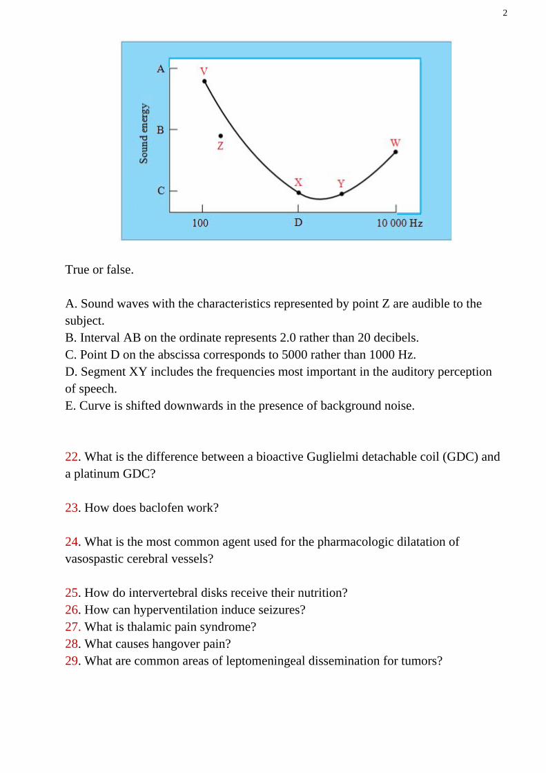
EANS/UEMS European examination in neurosurgery Part I and II Variants of questions with answers (compilation - Vyacheslav S. Botev, Department of Neurosurgery, M.Gorky Donetsk National Medical University) NEUROPHYSIOLOGY QUESTIONS I. Basic Concepts 1. What is the normal rate of cerebral blood flow (CBF)? 2. Decreases in CBF below what level result in deficits of neural function? 3. What factors can increase CBF? 4. How are body fluid levels regulated by the CNS? 5. How does hypocalcemia lead to tetany? 6. What causes night blindness? 7. What causes decerebrate rigidity? 8. What causes vasogenic edema? Cytotoxic edema? 9. What are the visual field findings in patients with ischemic optic neuropathy? 10. What is the effect of fentanyl on cerebral blood flow? Ketamine? 11. What is the drug of choice in the treatment of malignant hyperthermia? 12. Which is the best antiepileptic drug in the treatment of absence seizures? 13. What is the APO E gene? 14. What cellular elements compose the BBB? 15. What happens to platelet function after a subarachnoid hemorrhage? 16. What happens to cerebral blood flow immediately after a subarachnoid hemorrhage? 17. What is the proposed mechanism of action of steroid treatment in blunt spinal cord injury? 18. What is S100B and how is it related to traumatic brain injury? 19. What is the (intracranial) Windkessel phenomenon? 20. What is the ischemic penumbra? 21. In figure below, the line VXYW represents the threshold of hearing at various frequencies for a normal subject.
2
True or false. A. Sound waves with the characteristics represented by point Z are audible to the subject. B. Interval AB on the ordinate represents 2.0 rather than 20 decibels. C. Point D on the abscissa corresponds to 5000 rather than 1000 Hz. D. Segment XY includes the frequencies most important in the auditory perception of speech. E. Curve is shifted downwards in the presence of background noise. 22. What is the difference between a bioactive Guglielmi detachable coil (GDC) and a platinum GDC? 23. How does baclofen work? 24. What is the most common agent used for the pharmacologic dilatation of vasospastic cerebral vessels? 25. How do intervertebral disks receive their nutrition? 26. How can hyperventilation induce seizures? 27. What is thalamic pain syndrome? 28. What causes hangover pain? 29. What are common areas of leptomeningeal dissemination for tumors?
3
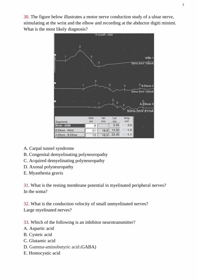
30. The figure below illustrates a motor nerve conduction study of a ulnar nerve, stimulating at the wrist and the elbow and recording at the abductor digiti minimi. What is the most likely diagnosis?
A. Carpal tunnel syndrome B. Congenital demyelinating polyneuropathy C. Acquired demyelinating polyneuropathy D. Axonal polyneuropathy E. Myasthenia gravis 31. What is the resting membrane potential in myelinated peripheral nerves? In the soma? 32. What is the conduction velocity of small unmyelinated nerves? Large myelinated nerves? 33. Which of the following is an inhibitor neurotransmitter? A. Aspartic acid B. Cysteic acid C. Glutamic acid D. Gamma-aminobutyric acid (GABA) E. Homocystic acid
4
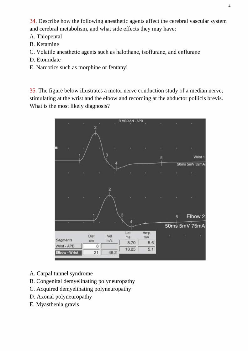
34. Describe how the following anesthetic agents affect the cerebral vascular system and cerebral metabolism, and what side effects they may have: A. Thiopental B. Ketamine C. Volatile anesthetic agents such as halothane, isoflurane, and enflurane D. Etomidate E. Narcotics such as morphine or fentanyl 35. The figure below illustrates a motor nerve conduction study of a median nerve, stimulating at the wrist and the elbow and recording at the abductor pollicis brevis. What is the most likely diagnosis?
A. Carpal tunnel syndrome B. Congenital demyelinating polyneuropathy C. Acquired demyelinating polyneuropathy D. Axonal polyneuropathy E. Myasthenia gravis
5
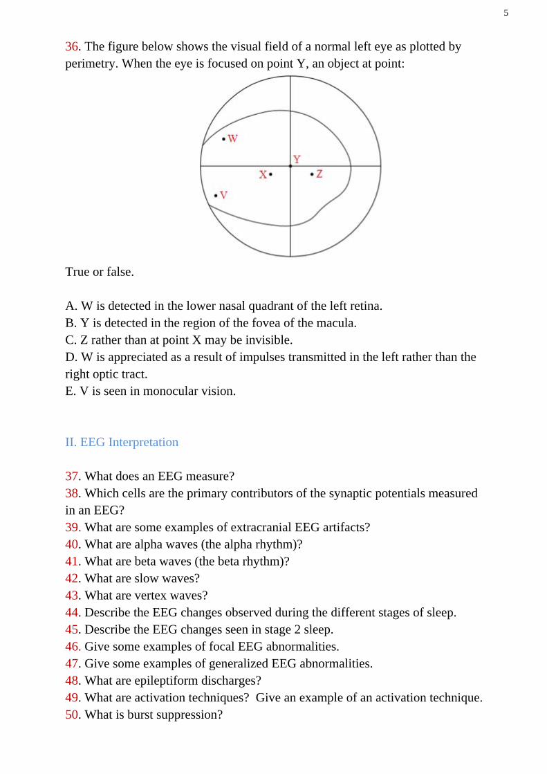
36. The figure below shows the visual field of a normal left eye as plotted by perimetry. When the eye is focused on point Y, an object at point:
True or false. A. W is detected in the lower nasal quadrant of the left retina. B. Y is detected in the region of the fovea of the macula. C. Z rather than at point X may be invisible. D. W is appreciated as a result of impulses transmitted in the left rather than the right optic tract. E. V is seen in monocular vision. II. EEG Interpretation 37. What does an EEG measure? 38. Which cells are the primary contributors of the synaptic potentials measured in an EEG? 39. What are some examples of extracranial EEG artifacts? 40. What are alpha waves (the alpha rhythm)? 41. What are beta waves (the beta rhythm)? 42. What are slow waves? 43. What are vertex waves? 44. Describe the EEG changes observed during the different stages of sleep. 45. Describe the EEG changes seen in stage 2 sleep. 46. Give some examples of focal EEG abnormalities. 47. Give some examples of generalized EEG abnormalities. 48. What are epileptiform discharges? 49. What are activation techniques? Give an example of an activation technique. 50. What is burst suppression?
6
III. EMG Interpretation 51. What is an electromyogram (EMG)? 52. What criteria are used to evaluate the motor unit action potential? 53. What are the constituents of the motor unit? 54. Between which diagnoses can EMG help differentiate? 55. What are some common patient complaints for which EMG may be ordered? 56. What is nerve conduction velocity? 57. What is insertional activity? 58. In what pathological situations would insertional activity be increased? 59. Describe the electrical activity seen in normal resting muscle (spontaneous activity). 60. Describe the electrical activity seen in myopathic resting muscle (spontaneous activity). 61. What are fibrillation potentials? 62. What are positive sharp waves? 63. What are complex repetitive discharges (CRDs)? 64. What are myotonic discharges? 65. What are fasciculation potentials? 66. Describe the electrical activity seen during normal minimal voluntary contraction. 67. Describe the electrical activity seen during minimal voluntary contraction in the (relatively rare) setting of acute complete denervation. 68. Describe the electrical activity seen during minimal voluntary contraction in the (more common) setting of chronic partial denervation. 69. Describe the electrical activity seen during minimal voluntary contraction in the setting of myopathic disease. 70. Describe the electrical activity seen during normal maximal voluntary contraction. 71. Describe the electrical activity seen during maximal voluntary contraction in the setting of myopathic disease. 72. Describe the electrical activity seen during maximal voluntary contraction in the setting of neuropathic disease. 73. How does repetitive nerve stimulation alter the observed amplitude in Eaton-Lambert syndrome? Myasthenia gravis (MG)?
7
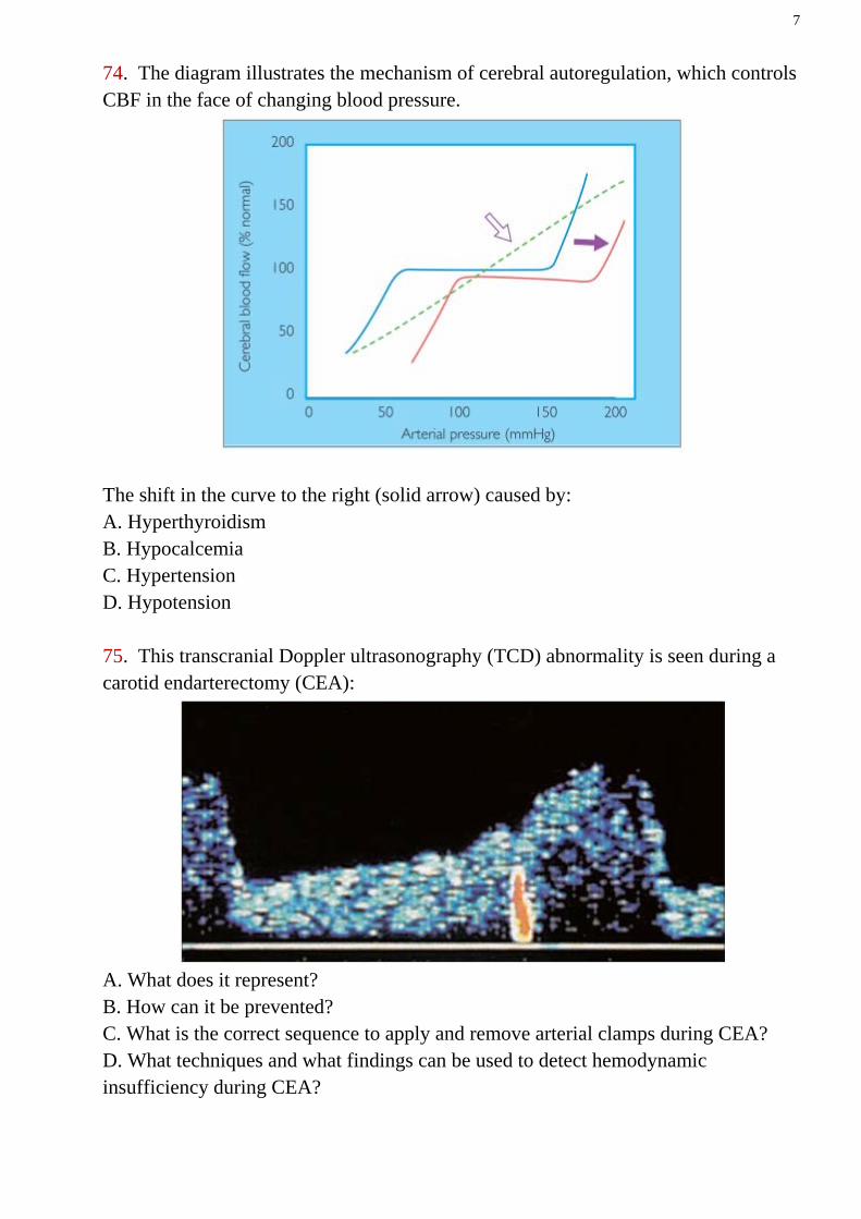
74. The diagram illustrates the mechanism of cerebral autoregulation, which controls CBF in the face of changing blood pressure.
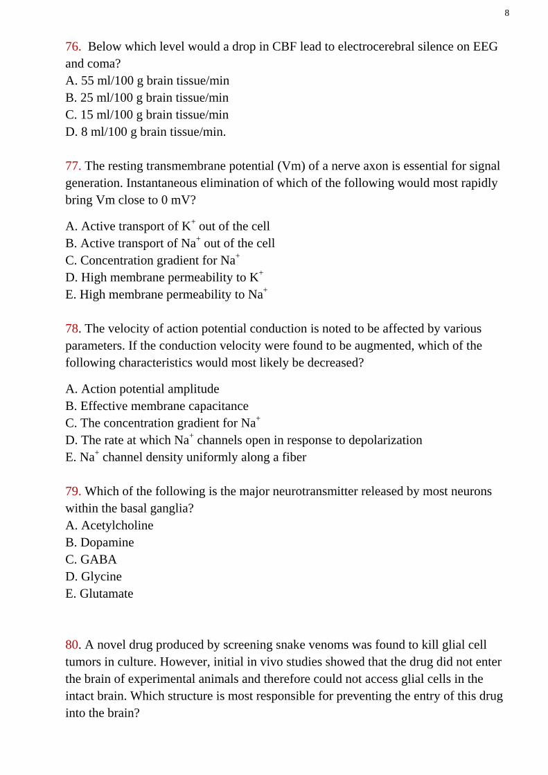
The shift in the curve to the right (solid arrow) caused by: A. Hyperthyroidism B. Hypocalcemia C. Hypertension D. Hypotension 75. This transcranial Doppler ultrasonography (TCD) abnormality is seen during a carotid endarterectomy (CEA):
A. What does it represent? B. How can it be prevented? C. What is the correct sequence to apply and remove arterial clamps during CEA? D. What techniques and what findings can be used to detect hemodynamic insufficiency during CEA?
8
76. Below which level would a drop in CBF lead to electrocerebral silence on EEG and coma? A. 55 ml/100 g brain tissue/min B. 25 ml/100 g brain tissue/min C. 15 ml/100 g brain tissue/min D. 8 ml/100 g brain tissue/min. 77. The resting transmembrane potential (Vm) of a nerve axon is essential for signal generation. Instantaneous elimination of which of the following would most rapidly bring Vm close to 0 mV?
A. Active transport of K+ out of the cell B. Active transport of Na+ out of the cell C. Concentration gradient for Na+ D. High membrane permeability to K+ E. High membrane permeability to Na+ 78. The velocity of action potential conduction is noted to be affected by various parameters. If the conduction velocity were found to be augmented, which of the following characteristics would most likely be decreased?
A. Action potential amplitude B. Effective membrane capacitance C. The concentration gradient for Na+ D. The rate at which Na+ channels open in response to depolarization E. Na+ channel density uniformly along a fiber 79. Which of the following is the major neurotransmitter released by most neurons within the basal ganglia? A. Acetylcholine B. Dopamine C. GABA D. Glycine E. Glutamate 80. A novel drug produced by screening snake venoms was found to kill glial cell tumors in culture. However, initial in vivo studies showed that the drug did not enter the brain of experimental animals and therefore could not access glial cells in the intact brain. Which structure is most responsible for preventing the entry of this drug into the brain?
9
A. Arachnoid mater B. Brain capillary endothelium C. Choroid plexus epithelium D. Dura mater E. Pia mater 81. In a laboratory experiment, a cortical neuron was electrically stimulated to produce action potentials. The stimulated neuron made synaptic contact with another neuron in which a recording electrode was located. The recording electrode detected a small depolarization following the electrical stimulation of the first neuron. Which neurotransmitter was most likely to be released at the synapse between these neurons? A. GABA B. Glutamate C. Glycine D. Met-enkephalin E. Somatostatin 82.
Direction: Each set of matching questions in this section consists of a list lettered options followed by several numbered items. For each numbered item, select the ONE lettered option that is most closely associated with it. To avoid spending too much time on matching sets with large numbers of options, it is generally advisable to begin each set by reading the list of options. Then, for each item in the set, try to generate the correct answer and locate it in the option list, rather than evaluating each option individually. Each lettered option may be selected once, more than once, or not at all.
Questions 1-6
A. Adrenergic α receptors B. Adrenergic β1 receptors C. Adrenergic β2 receptors D. Cholinergic muscarinic receptors E. Cholinergic nicotinic receptors Match each phenomenon with the autonomic receptors that mediate it. 1. Increased heart rate. 2. Secretion of epinephrine by the adrenal medulla 3. Ion channels for Na+ and K+ at the neuromuscular junction.
10
4. Blocked by hexamethonium at the ganglia, but not at the neuromuscular junction. 5. Atropine decreases gastrointestinal (GI) motility by blocking this receptor. 6. Low concentrations of epinephrine released from the adrenal medulla cause vasodilatation. Questions 7-9
A. Optic nerve B. Optic chiasm C. Optic tract D. Geniculocalcarine tract Match each deficit with the appropriate optic pathway. 7. Cutting on the left side causes total blindness in the left eye. 8. Cutting on the right side causes total blindness in the temporal field of the left eye and the nasal field of the right eye. 9. Cutting causes blindness in the temporal fields of the left and right eyes. Questions 10-11
A. Primary motor cortex B. Premotor cortex and supplementary motor cortex C. Prefrontal cortex D. Basal ganglia E. Cerebellum
Match each description with the correct structure. 10. Active during programming and “mental rehearsal” of complex motor sequences in the absence of movement. 11. Primary function is to coordinate rate, range, force, and direction of movement.
Questions 12-13
A. Stretch reflex (myotatic) B. Golgi tendon reflex (inverse myotatic) C. Flexor withdrawal reflex D. Subliminal occlusion reflex Match each description with the correct reflex. 12. Polysynaptic excitation of contralateral extensors. 13. Monosynaptic excitation of ipsilateral homonymous muscle.

11
Questions 14-16
A. α-Motoneurons B. γ- Motoneurons C. Group Ia fibers D. Group Ib fibers Match each description with the correct nerve fiber. 14. Detect increases in muscle tension 15. Stimulation leads to contraction of the bulk of skeletal muscle. 16. Muscle stretch leads to a direct increase in its firing rate.
12
NEUROPHYSIOLOGY ANSWERS I. Basic Concepts 1. 50 to 55 mL/100 g/min. 2. 23 mL/100 g/min. 3. Increases in paCO2 (acidosis) or serum H+, decreases in pH or paO2. 4. 1. ADH secreted from the supraoptic nucleus of the anterior hypothalamus decreases renal excretion of water. 2. The lateral hypothalamus increases water intake by the sensation of thirst. Both ways are activated or inhibited in response to decreases or increases (respectively) in serum osmolarity sensed by two circumventricular organs: the subforniceal organ and the vascular organ of the lamina terminalis. 5. When there is less Ca2+ in the interstitial fluid, the Na+ opens sooner (at about −80 mV), so the membrane is more excitable. In other words, hypocalcemia causes a lower threshold of membrane depolarization and action potential initiation. 6. This is due to a severe deficiency in vitamin A. There is a decreased amount of photosensitive pigment present to detect decreased light. This is remedied by rapid intravenous (IV) infusion of vitamin A. 7. This is caused by a lesion between the pons and the midbrain. This results in blockage of normal stimulation input to the medullary reticular formation from the red nucleus, basal ganglia, and cortex. As a result, there is unopposed antigravity muscle tone that is stimulated by the lateral vestibular nucleus and pontine reticular nucleus. 8. Vasogenic edema is caused by increased BBB permeability to proteins and macromolecules. This type of edema is extracellular and is caused by vessel damage and inflammation. Cytotoxic edema is caused by an impaired Na+/K+ pump as occurs in ischemia. Water and electrolytes accumulate inside the cells. It is an intracellular type of edema. 9. Ischemic optic neuropathy is the most common cause of painless monocular blindness in the elderly. This is caused by occlusion of the central retinal artery. This causes an altitudinal field deficit. One third of cases are bilateral. 10. Decreases and increases, respectively. 11. Dantrolen.
13
12. Ethosuximide. 13. Apoprotein E is produced mainly in astrocytes and is responsible for transportation of lipids within the brain. The protein mediates neuronal protection, interactions with estrogens, and modulation of synaptic proteins. Possession of the APO E4 allele has been shown to result in greater propensity to develop age-related cognitive impairment, a decrease in the synapse/neuron ratio, and increased susсeptibility to exogenous neurotoxins. 14. Endothelial cells, astrocyte endfeet, and pericytes. The capillary endothelial cells are connected together by tight junctions. 15. It is enhanced leading to an increase in platelet aggregates in the cerebral microcirculation. 16. Decreases. 17. Steroid treatment in blunt spinal cord injury is controversial. Steroids (when given within 8 hours of injury) are thought to have effects on local blood flow, inhibition of immunologic injury, and free radical-mediated lipid peroxidation and neuronal damage. 18. S100B is a protein belonging to a multigenic family of low-molecular-weight calcium-binding S100 proteins abundant in astrocytes. After traumatic brain injury, S100B protein is released by astrocytes; this protein may be neuroprotective and/or neurotrophic. 19. The Windkessel phenomenon is the ability of the cerebral vasculature to expand and the ability of the cerebrospinal fluid (CSF) and venous blood to translocate to accommodate arterial pulsations and provide a smooth capillary flow in the brain. 20. The term ischemic penumbra has been used to define a region in which cerebral blood flow reduction has passed the threshold that leads to the failure of electrical but not membrane function. The neuron is functionally disturbed, but remains structurally intact. 21. A. False. Anything below the line is inaudible, having less energy than the threshold value for detection at a particular frequency (Hz). B. False. AB and BC both represent 20 decibels; thus sounds at the extremes of the hearing range need relatively high energy to be heard. C. False. It corresponds to 1000 Hz; the frequency (or pitch) scale is logarithmic.

14
D. True. The ear is most sensitive to sounds in the range 1000–3000 Hz (XY), which includes the frequencies most important in distinguishing the different words in speech. E. False. It is shifted upwards since extraneous (masking) noise raises auditory threshold, that is the lowest energy level at which a sound of a particular frequency can just be detected. 22. The bioactive coil accelerates clot maturation and promotes the development of mature connective tissue and neointimal formation. The polymer used in bioactive coils is polyglycolic/poly-L-lactic acid (PGA/PLLA). 23. Baclofen is an agonist of gamma-aminobutyric acid (GABA); it reduces the release of presynaptic neurotransmitters in excitatory spinal pathways. 24. Papaverine hydrochloride is a potent, nonspecific, endothelium-independent smooth muscle relaxant that produces dilatation of arteries and arterioles, as well as veins. Intraarterial papaverine is usually administered superselectively via a microcatheter positioned proximal to the spastic vessel. 25. Intervertebral disks receive nutrition through passive diffusion from a network of capillary beds in the subchondral endplate region of the vertebral body. 26. Hyperventilation causes a respiratory alkalosis, which increases the pH. Increasing pH increases the membrane excitability and can induce seizures. 27. Also known as Dejerine-Roussy syndrome, this is usually due to a posteroventral thalamic stroke and its abnormal subsequent facilitation of the medial thalamic nucleus. These patients usually have a contralateral hemianesthesia at first, with increased pain in that area in the following weeks to months. 28. Chemical irritation to the meninges. 29. The most common areas of leptomeningeal dissemination of CNS tumors are the basilar cisterns, sylvian fissures, and cauda equina, most likely because of both gravity and slower rate of CSF flow in these areas. 30. C. Acquired demyelinating polyneuropathy. The figure illustrates a motor nerve conduction study of a ulnar nerve, stimulating at the wrist and the elbow and recording at the abductor digiti minimi. There is a dramatic reduction of the nerve conduction velocity in the demyelinating range with
15
conduction block and dispersion of the compound muscle action potential. These findings are suggestive of acquired demyelinating polyneuropathy. Compound Muscle Action Potential (CMAP)
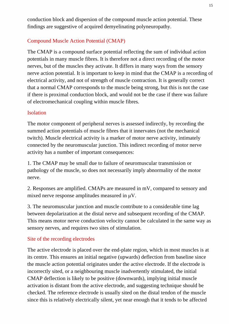
The CMAP is a compound surface potential reflecting the sum of individual action potentials in many muscle fibres. It is therefore not a direct recording of the motor nerves, but of the muscles they activate. It differs in many ways from the sensory nerve action potential. It is important to keep in mind that the CMAP is a recording of electrical activity, and not of strength of muscle contraction. It is generally correct that a normal CMAP corresponds to the muscle being strong, but this is not the case if there is proximal conduction block, and would not be the case if there was failure of electromechanical coupling within muscle fibres.
Isolation
The motor component of peripheral nerves is assessed indirectly, by recording the summed action potentials of muscle fibres that it innervates (not the mechanical twitch). Muscle electrical activity is a marker of motor nerve activity, intimately connected by the neuromuscular junction. This indirect recording of motor nerve activity has a number of important consequences:
1. The CMAP may be small due to failure of neuromuscular transmission or pathology of the muscle, so does not necessarily imply abnormality of the motor nerve.
2. Responses are amplified. CMAPs are measured in mV, compared to sensory and mixed nerve response amplitudes measured in μV.
3. The neuromuscular junction and muscle contribute to a considerable time lag between depolarization at the distal nerve and subsequent recording of the CMAP. This means motor nerve conduction velocity cannot be calculated in the same way as sensory nerves, and requires two sites of stimulation.
Site of the recording electrodes
The active electrode is placed over the end-plate region, which in most muscles is at its centre. This ensures an initial negative (upwards) deflection from baseline since the muscle action potential originates under the active electrode. If the electrode is incorrectly sited, or a neighbouring muscle inadvertently stimulated, the initial CMAP deflection is likely to be positive (downwards), implying initial muscle activation is distant from the active electrode, and suggesting technique should be checked. The reference electrode is usually sited on the distal tendon of the muscle since this is relatively electrically silent, yet near enough that it tends to be affected
16
by similar noise to the active electrode. Noise will therefore be minimized in the final output since this is the potential difference between active and reference electrodes.
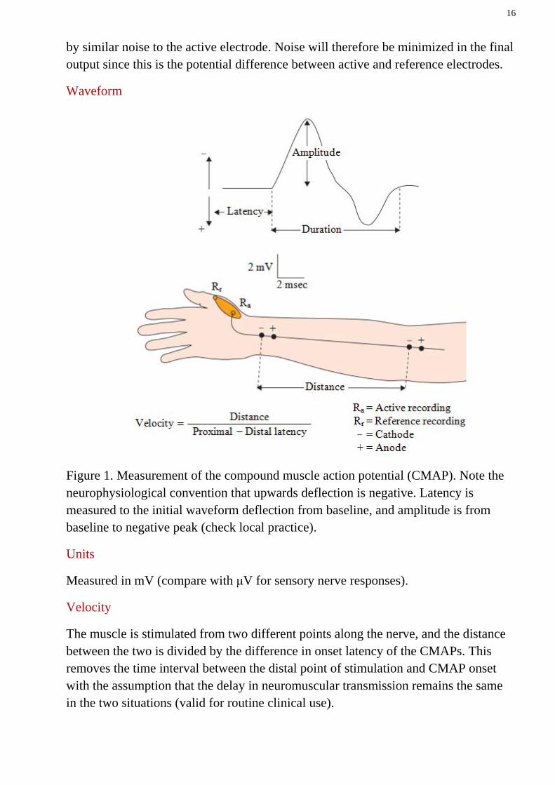
Waveform
Figure 1. Measurement of the compound muscle action potential (CMAP). Note the neurophysiological convention that upwards deflection is negative. Latency is measured to the initial waveform deflection from baseline, and amplitude is from baseline to negative peak (check local practice).
Units
Measured in mV (compare with μV for sensory nerve responses).
Velocity
The muscle is stimulated from two different points along the nerve, and the distance between the two is divided by the difference in onset latency of the CMAPs. This removes the time interval between the distal point of stimulation and CMAP onset with the assumption that the delay in neuromuscular transmission remains the same in the two situations (valid for routine clinical use).
17
31. −90 mV and −65 mV, respectively. This membrane potential is largely determined by the potential of K+, which is ~100 times more permeable than Na+. 32. About 0.5 m/s and 120 m/s, respectively. 33. D. Gamma-aminobutyric acid (GABA). Amino acids have been separated into two general classes: excitatory and inhibitory. The former group depolarizes neurons in mammalian cells and is formed by aspartic acid, cysteic acid, glutamic acid, and homocystic acid. The latter group hyperpolarizes neurons in mammals and is formed by GABA, glycine, taurine, and beta-alanine. 34. A. Thiopental is a cerebral vasoconstrictor that decreases CBF and CMRO2 but produces cardiovascular depression. Thiopental can protect the brain against the metabolic effects of cerebral ischemia and can reduce ICP. B. Ketamine is a dissociative anesthetic that is a pronounced cerebral vasodilator. It increases CBF and CMRO2 during normocapnia. The vasodilatation results in an increase in ICP. Ketamine can also induce seizure discharges. C. Halothane is a cerebral vasodilator and increases CBF. When given to patients with even mild intracranial hypertension it causes a further increase in ICP. It decreases CMRO2, however. Isoflurane and enflurane both cause a depression in cerebral metabolism that is associated with cerebral vasodilatation. Among volatile anesthetic agents, isoflurane causes the least increase in CBF. Enflurane can produce seizures. Hypotension and loss of cerebral autoregulation results when the inspired concentration of halothane, isoflurane or enflurane is increased. D. Etomidate decreases CBF and CMRO2 but suppresses the adrenocortical response to stress. E. Morphine in incremental doses causes a progressive and parallel decrease in CMRO2 and CBF. Morphine is a cerebral vasoconstrictor; this effect is abolished by hypercapnic vasodilatation. During normocapnia a combination of morphine and nitrous oxide does not significantly affect CBF or cerebral autoregulation in humans. Fentanyl in normal humans does not have a significant influence on either CBF or CMRO2 under normocapnia.
18
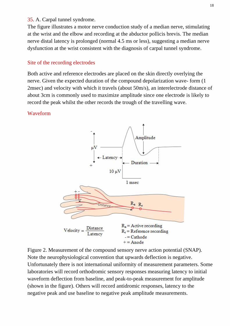
35. A. Carpal tunnel syndrome. The figure illustrates a motor nerve conduction study of a median nerve, stimulating at the wrist and the elbow and recording at the abductor pollicis brevis. The median nerve distal latency is prolonged (normal 4.5 ms or less), suggesting a median nerve dysfunction at the wrist consistent with the diagnosis of carpal tunnel syndrome. Site of the recording electrodes
Both active and reference electrodes are placed on the skin directly overlying the nerve. Given the expected duration of the compound depolarization wave- form (1 2msec) and velocity with which it travels (about 50m/s), an interelectrode distance of about 3cm is commonly used to maximize amplitude since one electrode is likely to record the peak whilst the other records the trough of the travelling wave.
Waveform
Figure 2. Measurement of the compound sensory nerve action potential (SNAP). Note the neurophysiological convention that upwards deflection is negative. Unfortunately there is not international uniformity of measurement parameters. Some laboratories will record orthodromic sensory responses measuring latency to initial waveform deflection from baseline, and peak-to-peak measurement for amplitude (shown in the figure). Others will record antidromic responses, latency to the negative peak and use baseline to negative peak amplitude measurements.
19
Units
Measured in μV (compare with mV for the CMAP).
Velocity
The distance between the centre of the stimulating cathode and the active recording electrode is divided by the time taken to travel between the two.
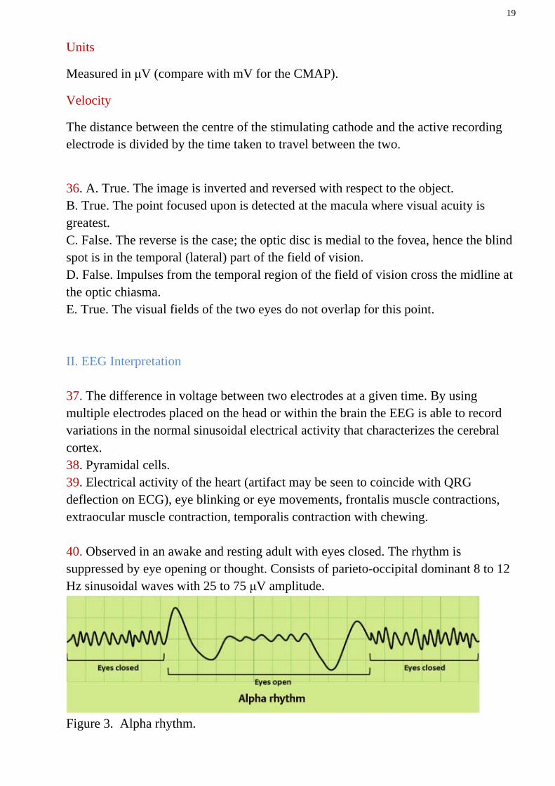
36. A. True. The image is inverted and reversed with respect to the object. B. True. The point focused upon is detected at the macula where visual acuity is greatest. C. False. The reverse is the case; the optic disc is medial to the fovea, hence the blind spot is in the temporal (lateral) part of the field of vision. D. False. Impulses from the temporal region of the field of vision cross the midline at the optic chiasma. E. True. The visual fields of the two eyes do not overlap for this point. II. EEG Interpretation 37. The difference in voltage between two electrodes at a given time. By using multiple electrodes placed on the head or within the brain the EEG is able to record variations in the normal sinusoidal electrical activity that characterizes the cerebral cortex. 38. Pyramidal cells. 39. Electrical activity of the heart (artifact may be seen to coincide with QRG deflection on ECG), eye blinking or eye movements, frontalis muscle contractions, extraocular muscle contraction, temporalis contraction with chewing. 40. Observed in an awake and resting adult with eyes closed. The rhythm is suppressed by eye opening or thought. Consists of parieto-occipital dominant 8 to 12 Hz sinusoidal waves with 25 to 75 μV amplitude.
Figure 3. Alpha rhythm.
20
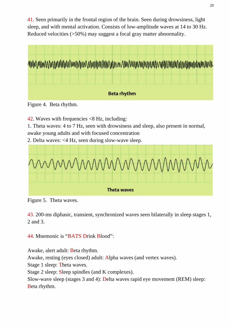
41. Seen primarily in the frontal region of the brain. Seen during drowsiness, light sleep, and with mental activation. Consists of low-amplitude waves at 14 to 30 Hz. Reduced velocities (>50%) may suggest a focal gray matter abnormality.
Figure 4. Beta rhythm. 42. Waves with frequencies <8 Hz, including: 1. Theta waves: 4 to 7 Hz, seen with drowsiness and sleep, also present in normal, awake young adults and with focused concentration 2. Delta waves: <4 Hz, seen during slow-wave sleep.
Figure 5. Theta waves. 43. 200-ms diphasic, transient, synchronized waves seen bilaterally in sleep stages 1, 2 and 3. 44. Mnemonic is “BATS Drink Blood”: Awake, alert adult: Beta rhythm. Awake, resting (eyes closed) adult: Alpha waves (and vertex waves). Stage 1 sleep: Theta waves. Stage 2 sleep: Sleep spindles (and K complexes). Slow-wave sleep (stages 3 and 4): Delta waves rapid eye movement (REM) sleep: Beta rhythm.
21
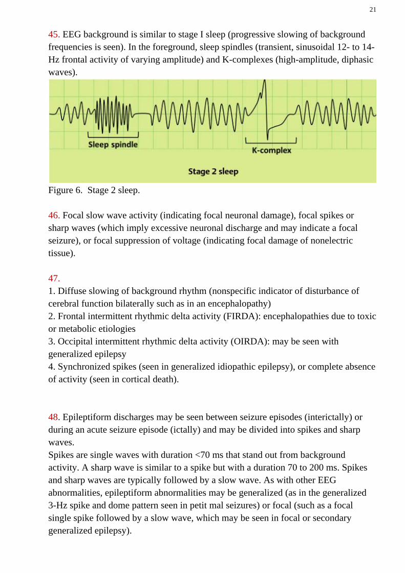
45. EEG background is similar to stage I sleep (progressive slowing of background frequencies is seen). In the foreground, sleep spindles (transient, sinusoidal 12- to 14-Hz frontal activity of varying amplitude) and K-complexes (high-amplitude, diphasic waves).
Figure 6. Stage 2 sleep. 46. Focal slow wave activity (indicating focal neuronal damage), focal spikes or sharp waves (which imply excessive neuronal discharge and may indicate a focal seizure), or focal suppression of voltage (indicating focal damage of nonelectric tissue). 47. 1. Diffuse slowing of background rhythm (nonspecific indicator of disturbance of cerebral function bilaterally such as in an encephalopathy) 2. Frontal intermittent rhythmic delta activity (FIRDA): encephalopathies due to toxic or metabolic etiologies 3. Occipital intermittent rhythmic delta activity (OIRDA): may be seen with generalized epilepsy 4. Synchronized spikes (seen in generalized idiopathic epilepsy), or complete absence of activity (seen in cortical death). 48. Epileptiform discharges may be seen between seizure episodes (interictally) or during an acute seizure episode (ictally) and may be divided into spikes and sharp waves. Spikes are single waves with duration <70 ms that stand out from background activity. A sharp wave is similar to a spike but with a duration 70 to 200 ms. Spikes and sharp waves are typically followed by a slow wave. As with other EEG abnormalities, epileptiform abnormalities may be generalized (as in the generalized 3-Hz spike and dome pattern seen in petit mal seizures) or focal (such as a focal single spike followed by a slow wave, which may be seen in focal or secondary generalized epilepsy).

22
49. Stimuli applied to the EEG subject in an effort to augment EEG abnormalities. Examples include photic stimulation, hyperventilation (causes cerebral vasoconstriction) and sleep deprivation. Other stimuli that may increase seizure frequency include emotion, fever, loud noises, and trauma. 50. A pattern suggesting a severe bilateral cerebral dysfunction. May also be induced iatrogenically for neuroprotection. Consists of stereotyped bursts occurring every 2 to 10 seconds separated by intervals of suppression (where no electrical activity is seen).
Figure 7. Burst suppression. III. EMG Interpretation 51. A clinical study of the electrical activity of muscle fibers using surface or needle electrodes. Electrical activity is measured during needle insertion, during periods of muscle rest (spontaneous activity), and during periods of minimal or maximal voluntary muscle contraction. 52. Maximum peak-to-peak amplitude, rise time of the initial positive spike/peak, duration of the action potential and number of phases 53. The alpha motor neuron whose cell body lies in the ventral horn of the spinal cord, its axon, and all motor fiber innervated by the cell. 54. 1. Radiculopathy 2. Entrapment neuropathy 3. Neuritis 4. Nonneurological condition 55. 1. Pain 2. Weakness 3. Numbness/paresthesias
23
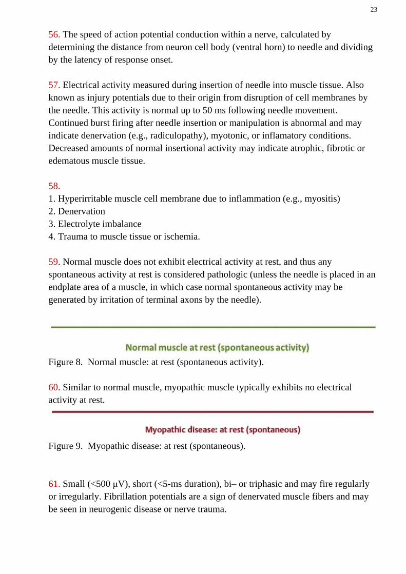
56. The speed of action potential conduction within a nerve, calculated by determining the distance from neuron cell body (ventral horn) to needle and dividing by the latency of response onset. 57. Electrical activity measured during insertion of needle into muscle tissue. Also known as injury potentials due to their origin from disruption of cell membranes by the needle. This activity is normal up to 50 ms following needle movement. Continued burst firing after needle insertion or manipulation is abnormal and may indicate denervation (e.g., radiculopathy), myotonic, or inflamatory conditions. Decreased amounts of normal insertional activity may indicate atrophic, fibrotic or edematous muscle tissue. 58. 1. Hyperirritable muscle cell membrane due to inflammation (e.g., myositis) 2. Denervation 3. Electrolyte imbalance 4. Trauma to muscle tissue or ischemia. 59. Normal muscle does not exhibit electrical activity at rest, and thus any spontaneous activity at rest is considered pathologic (unless the needle is placed in an endplate area of a muscle, in which case normal spontaneous activity may be generated by irritation of terminal axons by the needle).
Figure 8. Normal muscle: at rest (spontaneous activity). 60. Similar to normal muscle, myopathic muscle typically exhibits no electrical activity at rest.
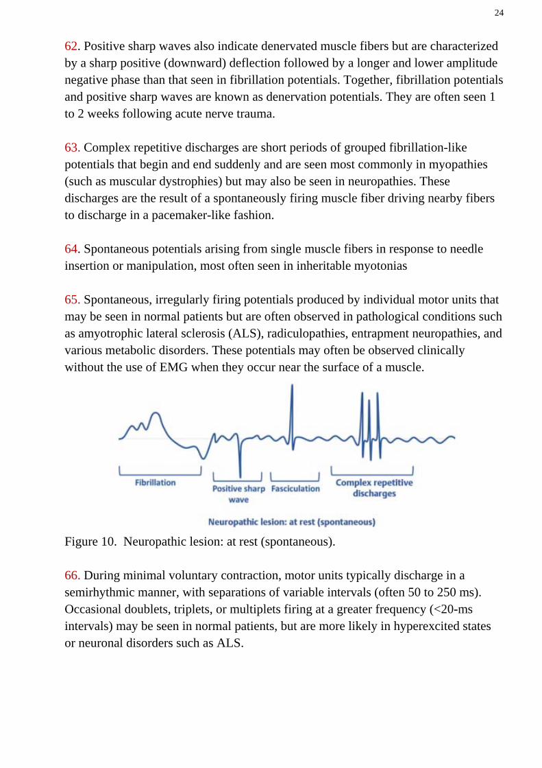
Figure 9. Myopathic disease: at rest (spontaneous). 61. Small (<500 μV), short (<5-ms duration), bi– or triphasic and may fire regularly or irregularly. Fibrillation potentials are a sign of denervated muscle fibers and may be seen in neurogenic disease or nerve trauma.
24
62. Positive sharp waves also indicate denervated muscle fibers but are characterized by a sharp positive (downward) deflection followed by a longer and lower amplitude negative phase than that seen in fibrillation potentials. Together, fibrillation potentials and positive sharp waves are known as denervation potentials. They are often seen 1 to 2 weeks following acute nerve trauma. 63. Complex repetitive discharges are short periods of grouped fibrillation-like potentials that begin and end suddenly and are seen most commonly in myopathies (such as muscular dystrophies) but may also be seen in neuropathies. These discharges are the result of a spontaneously firing muscle fiber driving nearby fibers to discharge in a pacemaker-like fashion. 64. Spontaneous potentials arising from single muscle fibers in response to needle insertion or manipulation, most often seen in inheritable myotonias 65. Spontaneous, irregularly firing potentials produced by individual motor units that may be seen in normal patients but are often observed in pathological conditions such as amyotrophic lateral sclerosis (ALS), radiculopathies, entrapment neuropathies, and various metabolic disorders. These potentials may often be observed clinically without the use of EMG when they occur near the surface of a muscle.
Figure 10. Neuropathic lesion: at rest (spontaneous). 66. During minimal voluntary contraction, motor units typically discharge in a semirhythmic manner, with separations of variable intervals (often 50 to 250 ms). Occasional doublets, triplets, or multiplets firing at a greater frequency (<20-ms intervals) may be seen in normal patients, but are more likely in hyperexcited states or neuronal disorders such as ALS.
25
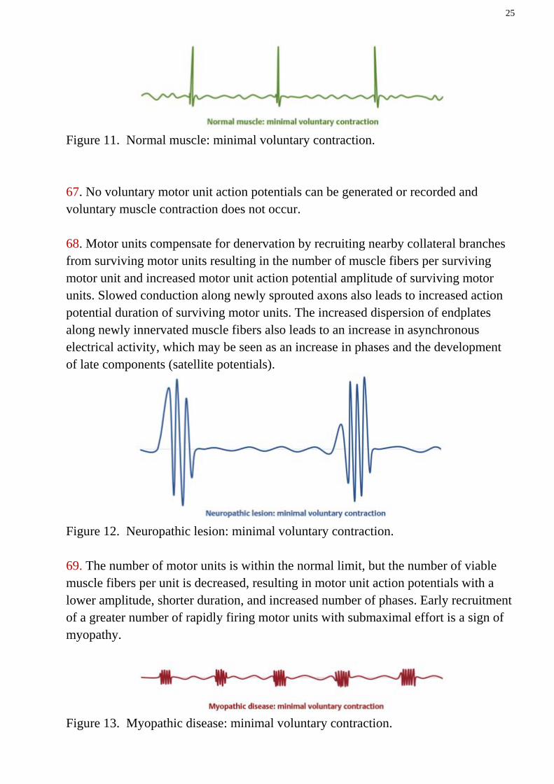
Figure 11. Normal muscle: minimal voluntary contraction. 67. No voluntary motor unit action potentials can be generated or recorded and voluntary muscle contraction does not occur. 68. Motor units compensate for denervation by recruiting nearby collateral branches from surviving motor units resulting in the number of muscle fibers per surviving motor unit and increased motor unit action potential amplitude of surviving motor units. Slowed conduction along newly sprouted axons also leads to increased action potential duration of surviving motor units. The increased dispersion of endplates along newly innervated muscle fibers also leads to an increase in asynchronous electrical activity, which may be seen as an increase in phases and the development of late components (satellite potentials).
Figure 12. Neuropathic lesion: minimal voluntary contraction. 69. The number of motor units is within the normal limit, but the number of viable muscle fibers per unit is decreased, resulting in motor unit action potentials with a lower amplitude, shorter duration, and increased number of phases. Early recruitment of a greater number of rapidly firing motor units with submaximal effort is a sign of myopathy.
Figure 13. Myopathic disease: minimal voluntary contraction.
26
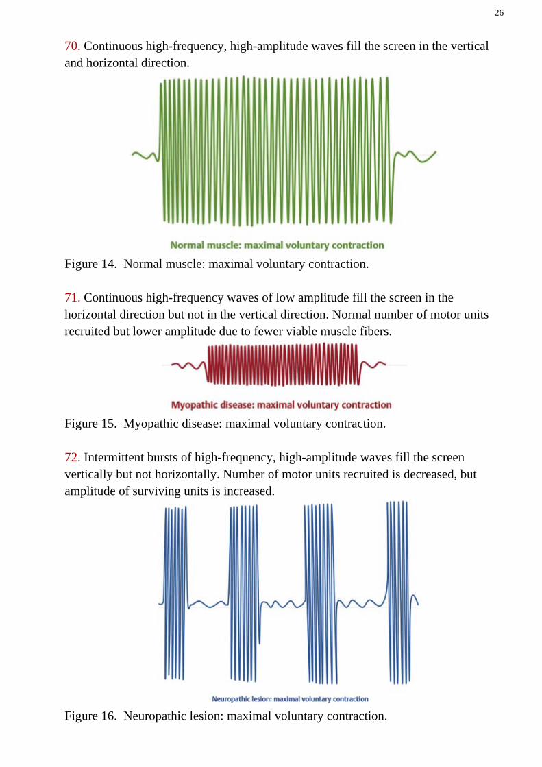
70. Continuous high-frequency, high-amplitude waves fill the screen in the vertical and horizontal direction.
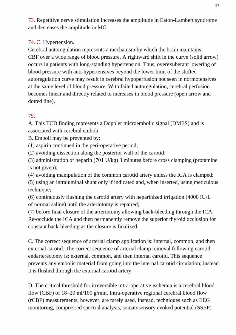
Figure 14. Normal muscle: maximal voluntary contraction. 71. Continuous high-frequency waves of low amplitude fill the screen in the horizontal direction but not in the vertical direction. Normal number of motor units recruited but lower amplitude due to fewer viable muscle fibers.
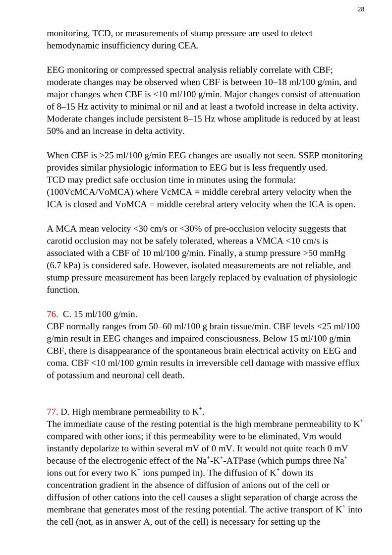
Figure 15. Myopathic disease: maximal voluntary contraction. 72. Intermittent bursts of high-frequency, high-amplitude waves fill the screen vertically but not horizontally. Number of motor units recruited is decreased, but amplitude of surviving units is increased.
Figure 16. Neuropathic lesion: maximal voluntary contraction.
27
73. Repetitive nerve stimulation increases the amplitude in Eaton-Lambert syndrome and decreases the amplitude in MG. 74. C. Hypertension. Cerebral autoregulation represents a mechanism by which the brain maintains CBF over a wide range of blood pressure. A rightward shift in the curve (solid arrow) occurs in patients with long-standing hypertension. Thus, overexuberant lowering of blood pressure with anti-hypertensives beyond the lower limit of the shifted autoregulation curve may result in cerebral hypoperfusion not seen in normotensives at the same level of blood pressure. With failed autoregulation, cerebral perfusion becomes linear and directly related to increases in blood pressure (open arrow and dotted line). 75. A. This TCD finding represents a Doppler microembolic signal (DMES) and is associated with cerebral emboli. B. Emboli may be prevented by: (1) aspirin continued in the peri-operative period; (2) avoiding dissection along the posterior wall of the carotid; (3) administration of heparin (701 U/kg) 3 minutes before cross clamping (protamine is not given); (4) avoiding manipulation of the common carotid artery unless the ICA is clamped; (5) using an intraluminal shunt only if indicated and, when inserted, using meticulous technique; (6) continuously flushing the carotid artery with heparinized irrigation (4000 IU/L of normal saline) until the arteriotomy is repaired; (7) before final closure of the arteriotomy allowing back-bleeding through the ICA. Re-occlude the ICA and then permanently remove the superior thyroid occlusion for constant back-bleeding as the closure is finalized. C. The correct sequence of arterial clamp application is: internal, common, and then external carotid. The correct sequence of arterial clamp removal following carotid endarterectomy is: external, common, and then internal carotid. This sequence prevents any embolic material from going into the internal carotid circulation; instead it is flushed through the external carotid artery. D. The critical threshold for irreversible intra-operative ischemia is a cerebral blood flow (CBF) of 18–20 ml/100 g/min. Intra-operative regional cerebral blood flow (rCBF) measurements, however, are rarely used. Instead, techniques such as EEG monitoring, compressed spectral analysis, somatosensory evoked potential (SSEP)
28
monitoring, TCD, or measurements of stump pressure are used to detect hemodynamic insufficiency during CEA. EEG monitoring or compressed spectral analysis reliably correlate with CBF; moderate changes may be observed when CBF is between 10–18 ml/100 g/min, and major changes when CBF is <10 ml/100 g/min. Major changes consist of attenuation of 8–15 Hz activity to minimal or nil and at least a twofold increase in delta activity. Moderate changes include persistent 8–15 Hz whose amplitude is reduced by at least 50% and an increase in delta activity. When CBF is >25 ml/100 g/min EEG changes are usually not seen. SSEP monitoring provides similar physiologic information to EEG but is less frequently used. TCD may predict safe occlusion time in minutes using the formula: (100VcMCA/VoMCA) where VcMCA = middle cerebral artery velocity when the ICA is closed and VoMCA = middle cerebral artery velocity when the ICA is open. A MCA mean velocity <30 cm/s or <30% of pre-occlusion velocity suggests that carotid occlusion may not be safely tolerated, whereas a VMCA <10 cm/s is associated with a CBF of 10 ml/100 g/min. Finally, a stump pressure >50 mmHg (6.7 kPa) is considered safe. However, isolated measurements are not reliable, and stump pressure measurement has been largely replaced by evaluation of physiologic function. 76. C. 15 ml/100 g/min. CBF normally ranges from 50–60 ml/100 g brain tissue/min. CBF levels <25 ml/100 g/min result in EEG changes and impaired consciousness. Below 15 ml/100 g/min CBF, there is disappearance of the spontaneous brain electrical activity on EEG and coma. CBF <10 ml/100 g/min results in irreversible cell damage with massive efflux of potassium and neuronal cell death. 77. D. High membrane permeability to K+. The immediate cause of the resting potential is the high membrane permeability to K+ compared with other ions; if this permeability were to be eliminated, Vm would instantly depolarize to within several mV of 0 mV. It would not quite reach 0 mV because of the electrogenic effect of the Na+-K+-ATPase (which pumps three Na+ ions out for every two K+ ions pumped in). The diffusion of K+ down its concentration gradient in the absence of diffusion of anions out of the cell or diffusion of other cations into the cell causes a slight separation of charge across the membrane that generates most of the resting potential. The active transport of K+ into the cell (not, as in answer A, out of the cell) is necessary for setting up the
29
concentration gradient that results in the diffusion of K+ out of the cell. This gradient (and therefore Vm) would take a long time to dissipate if active transport were stopped. Because the membrane is effectively impermeable to Na+ at rest, the transport and concentration gradient for Na+ has very little effect on the resting potential (answers B, C, and E). 78. B. Effective membrane capacitance. Effective membrane capacitance is decreased in many mammalian axons by myelination—the tight wrapping of many glial membranes around the axon, which is functionally equivalent to increasing the thickness of the membrane. Because conduction velocity is inversely related to membrane capacitance, which is related inversely to effective membrane thickness, a decrease in membrane capacitance increases conduction velocity. Decreasing action potential amplitude (answer A) will decrease rather than increase action potential velocity, as will decreasing the concentration gradient for Na+ (because this will reduce action potential amplitude). In addition, decreases in the opening rate or density of Na+ channels will decrease conduction velocity. NEUROPHYSIOLOGY PEARLS � The resting potential is generated by the high permeability of the membrane to K+ compared with other ions, which allows a very small amount of K+ to diffuse out of the cell in the absence of net diffusion of other ions, causing a charge separation across the membrane. � The resting potential and action potential in simple excitable systems, such as axons, that are permeable only to K+ and Na+, can be described by the Goldman-Hodgkin-Katz equation, which states that Vm is determined by opposing currents carried by K+ and Na+, which are determined entirely by (1) the ratio of permeabilities to K+ and Na+ and (2) their concentration gradients across the cell membrane. � An action potential is generated when membrane depolarization reaches a level at which voltage-gated Na+ channels open, increasing PNa (or, in electrical terms, gNa), which results in an inward current of Na+, which causes further depolarization, opening additional Na+ channels in a positive feedback cycle. � Inactivation of Na+ channels during an action potential prevents subsequent action potential initiation during the brief absolute refractory period, whereas the relative refractory period continues shortly thereafter because the delayed rectifier K+ channels remain open for a somewhat longer period. � The spread of depolarization in front of an active region of membrane during an action potential occurs by electrotonic propagation, which is characterized by an exponential decay of the depolarization with distance along the fiber.
30
� The velocity of action potential conduction is increased by myelinating axons, which decreases their effective membrane capacitance, and by increasing the fiber diameter, which decreases the intracellular resistance. 79. C. GABA. The majority of synapses from neurons in the basal ganglia are inhibitory and release GABA. The other major inhibitory neurotransmitter, glycine, is not important in the basal ganglia and is released primarily from inhibitory interneurons in the spinal cord. Dopamine is an important neurotransmitter that is released by neurons in the substantia nigra, but these neurons account for only a small fraction of the total number of neurons in the basal ganglia. In addition, other neurotransmitters (eg, acetylcholine, NO, various neuropeptides) are released from some neurons in the basal ganglia, but the largest number of synapses are GABAergic. NEUROPHYSIOLOGY PEARLS � The four principal nuclei of the basal ganglia are the striatum, globus pallidus, substantia nigra, and subthalamic nucleus. � The motor loop comprises two parallel pathways that travel from the cortex through the basal ganglia and then to the thalamus and back to the cortex. In the direct pathway, excitatory input to the basal ganglia excites thalamic neurons by inhibiting the inhibitory output neurons in the internal segment of the globus pallidus. In the indirect pathway, excitatory input to the basal ganglia further inhibits thalamic neurons by disinhibiting the inhibitory output neurons in the internal segment of the globus pallidus. � GABA is the most prominent inhibitory neurotransmitter in most of the brain, although glycine is an important inhibitory neurotransmitter in the spinal cord and brainstem. � Dopamine release from pars compacta neurons in the substantia nigra excites the direct pathway (by D1 receptors), which inhibits the inhibitor output neurons, thereby increasing thalamic activity and ultimately facilitating movements initiated in the cortex, whereas dopamine simultaneously inhibits the indirect pathway (by D2 receptors), further increasing the inhibition of the inhibitory output neurons. � Loss of dopaminergic neurons in Parkinson disease reduces the activation of the direct pathway as well as the inhibition of the indirect pathway, allowing greater inhibition of thalamic neurons and greater suppression of movements initiated in the cortex, resulting in the hypokinetic signs of this disease. 80.B. Brain capillary endothelium. The blood-brain barrier prevents the free access of most solutes from plasma to the brain extracellular fluid. The bloodbrain barrier is formed by the capillary endothelial
31
cells and supporting glial cells. The choroid plexus also contributes, to a lesser extent, by actively regulating cerebrospinal fluid composition. 81. B. Glutamate. The neurotransmitter produces an excitatory postsynaptic potential in this case. Glutamate is the most common excitatory neurotransmitter in the central nervous system; the others are all inhibitory transmitters. Enkephalin and somatostatin are large molecule transmitters present in lower abundance than the other examples given. 82. 1. B. Adrenergic β1 receptors. Heart rate is increased by the stimulatory effect of norepinephrine on β1 receptors in the sinoatrial (SA) node. There are also sympathetic β1 receptors in heart that regulate contractility. 2. E. Cholinergic nicotinic receptors. Preganglionic parasympathetic fibers synapse on the chromaffin cells of the adrenal medulla at a nicotinic receptor, releasing epinephrine (and, to a lesser extent, norepinephrine) into the circulation. 3. E. Cholinergic nicotinic receptors. Nicotinic receptors at the neuromuscular junction are not only the postsynaptic receptors for acetylcholine (Ach), but also the ion channels for Na+ and K+ when they are opened by Ach, they depolarize the muscle end plate, producing the end plate potential. 4. E. Cholinergic nicotinic receptors. Hexamethonium is a nicotinic blocker, but it acts only at ganglionic (not neuromuscular junction) nicotinic receptors. This pharmacologic distinction emphasizes that nicotinic receptors at these two locations, although similar, are not identical. 5. D. Cholinergic muscarinic receptors. Atropine is a specific inhibitor of muscarinic receptors for acetylcholine (ACh). Atropine would be expected to block all effects of ACh that are mediated by muscarinic receptors. Therefore, atropine will decrease gastrointestinal (GI) motility, decrease bladder tone, dilate bronchiolar smooth muscle, and increase heart rate. 6. C. Adrenergic β2 receptors.
32
β2 Receptors on vascular smooth muscle produce vasodilatation. α Receptors on vascular smooth muscle produce vasoconstriction. Because β2 receptors are more sensitive to epinephrine than are α receptors, low doses of epinephrine produce vasodilatation, and high doses produce vasoconstriction. 7. A. Optic nerve. Cutting the optic nerve from the left eye causes blindness in the left eye because the fibers have not yet crossed at the optic chiasm. 8. C. Optic tract. Fibers from the left temporal field and the right nasal field ascend together in the right optic tract. 9. B. Optic chiasm. Optic nerve fibers from both temporal receptor fields cross at the optic chiasm. 10. B. Premotor cortex and supplementary motor cortex. The premotor cortex (area 6) is responsible for generating a plan for movement before movement occurs. The supplementary motor cortex is responsible for the “mental rehearsal” of the movement. 11. E. Cerebellum.
Output of Purkinje cells from the cerebellum cortex to deep cerebellar nuclei is inhibitory. This output modulates movement and is responsible for the coordination that allows one to “catch a fly”. 12. C. Flexor withdrawal reflex. Flexor withdrawal is a polysynaptic reflex that is used when a person touches a hot stove or steps on a tack. On the ipsilateral side of the painful stimulus, there is flexion (withdrawal); on the contralateral side, there is extension to maintain balance. 13. A. Stretch reflex (myotatic). The stretch reflex is a monosynaptic response to stretching of a muscle. The reflex produces contraction and then shortening of the muscle that was originally stretched (homonymous muscle). 14. D. Group Ib fibers. Group Ib fibers are the afferent fibers from Golgi tendon organs that detect increases in muscle tension. The Golgi tendon reflex (inverse myotatic) compensates for the increased tension by causing relaxation of the homonymous muscle.
33
15. A. α-Motoneurons. Extrafusal fibers constitute the bulk of muscle and are innervated by α-motoneurons. 16. C. Group Ia fibers. Group Ia afferent fibers innervate intrafusal fibers of the muscle spindle. When the intrafusal fibers are stretched, the group Ia fibers fire and activate the stretch reflex, which causes the muscle to return to its resting length. References 1. Arthur C. Guyton, John E. Hall. Textbook of Medical Physiology. Elsevier Saunders, 11th Ed., Philadelphia, 2006.
2. Andrew W. Michell. Understanding EMG. Oxford University Press, UK, 2013.
3. Jasper R. Daube, Devon I. Rubin. Clinical Neurophysiology. 3rd Ed., Oxford University Press, UK, 2009.
4. Greenberg MS. Handbook of Neurosurgery, 7th Ed., Thieme Medical Publishers, Inc., NY, 2010.
5. Kranzler L.I. The Greenberg Rapid Review. A Companion to the Seventh Edition. Thieme Medical Publishers, Inc., NY, 2011.
6. Cargill H. Alleyne. Neurosurgery Board Review. Thieme Medical Publishers, Inc., NY, 1997.
7. Mark Shaya, Remi Nader, Anil Nanda. Neurosurgery Practice Questions and Answers. Thieme Medical Publishers, Inc., NY, 2010.
8. Thomas G. Psarros, Shawn P. Moore. Intensive Neurosurgery Board Review. Lippincott Williams & Wilkins, Philadelphia, 2006. 9. Mark R. Shaya. Neurosurgery Rounds: Questions and Answers. Thieme Medical Publishers, Inc., NY, 2011.