Embed Size (px)
Citation preview

Dysfunction of GRAP, encoding the GRB2-relatedadaptor protein, is linked to sensorineural hearing lossChong Lia,1,2, Guney Bademcib,1, Asli Subasiogluc, Oscar Diaz-Hortab, Yi Zhua, Jiaqi Liud, Timothy Gavin Mitchella,Clemer Abadb, Serhat Seyhanb, Duygu Dumane, Filiz Basak Cengizb, Suna Tokgoz-Yilmazf, Susan H. Blantonb,g,h,Amjad Farooqi, Katherina Walzb,g, R. Grace Zhaia,d,3, and Mustafa Tekinb,g,h,3
aDepartment of Molecular and Cellular Pharmacology, University of Miami Miller School of Medicine, Miami, FL 33136; bJohn P. Hussman Institute forHuman Genomics, University of Miami Miller School of Medicine, Miami, FL 33136; cDepartment of Medical Genetics, Izmir Ataturk Education and ResearchHospital, 35360 Izmir, Turkey; dSchool of Pharmacy, Key Laboratory of Molecular Pharmacology and Drug Evaluation (Yantai University), Ministry ofEducation, Collaborative Innovation Center of Advanced Drug Delivery System and Biotech Drugs in Universities of Shandong, Yantai University, 264005Yantai, Shandong, China; eDivision of Pediatric Genetics, Ankara University School of Medicine, 06260 Ankara, Turkey; fDepartment of Audiology, AnkaraUniversity School of Medicine, 06260 Ankara, Turkey; gDr. John T. Macdonald Foundation Department of Human Genetics, University of Miami Miller Schoolof Medicine, Miami, FL 33136; hDepartment of Otolaryngology, Miller School of Medicine, University of Miami, Miami, FL 33136; and iDepartment ofBiochemistry and Molecular Biology, Miller School of Medicine, University of Miami, Miami, FL 33136
Edited by Mary-Claire King, University of Washington, Seattle, WA, and approved December 6, 2018 (received for review June 29, 2018)
We have identified a GRAP variant (c.311A>T; p.Gln104Leu) cose-gregating with autosomal recessive nonsyndromic deafness in twounrelated families. GRAP encodes a member of the highly conservedgrowth factor receptor-bound protein 2 (GRB2)/Sem-5/drk family ofproteins, which are involved in Ras signaling; however, the functionof the growth factor receptor-bound protein 2 (GRB2)-relatedadaptor protein (GRAP) in the auditory system is not known. Here,we show that, in mouse, Grap is expressed in the inner ear and theprotein localizes to the neuronal fibers innervating cochlear and utric-ular auditory hair cells. Downstream of receptor kinase (drk), theDrosophila homolog of human GRAP, is expressed in Johnston’s or-gan (JO), the fly hearing organ, and the loss of drk in JO causesscolopidium abnormalities. drk mutant flies present deficits in nega-tive geotaxis behavior, which can be suppressed by human wild-typebut not mutant GRAP. Furthermore, drk specifically colocalizes withsynapsin at synapses, suggesting a potential role of such adap-tor proteins in regulating actin cytoskeleton dynamics in thenervous system. Our findings establish a causative link betweenGRAP mutation and nonsyndromic deafness and suggest a functionof GRAP/drk in hearing.
adaptor proteins | drk | Drosophila | GRAP | nonsyndromic hearing loss
Hearing loss (HL) is the most common sensory disorder withan incidence of 1–3 in 1,000 births (1). Genetic factors show
high heterogeneity and are responsible for more than half of thecases with congenital HL (2). It is estimated that 70% of geneticHL is nonsyndromic sensorineural HL and mostly shows anautosomal recessive inheritance pattern (1, 3). The identificationof DNA variants causing HL is rapidly advancing due to tech-nological developments in genome sequencing. However, delin-eating causality for a novel rare variant in a gene not previouslyassociated with HL is difficult and often requires functionalstudies in model organisms.The mouse has been shown to be an excellent model organism
to study human deafness due to the anatomic and physiologicsimilarities of their auditory systems with those of humans (4).Recent studies of Drosophila’s hearing organ, Johnston’s organ(JO), highlight the molecular and genetic conservations of themechanosensory transduction in flies and vertebrates (5, 6).Several key genes first identified in Drosophila, such as atonal,crinkled, and nanchung, that are required for the specification orfunction of auditory cell types in the JO are also important forthe normal development or function of the vertebrate ear (5). Inaddition, 20% of Drosophila auditory-associated genes have hu-man homologs and are implicated in hearing disorders (7).In this paper, via exome and genome sequencing of over 60
consanguineous multiplex families with autosomal recessive non-syndromic HL, which are negative for variants in known deafnessgenes (details of families are given in SI Appendix, Table S1), we
have identified a GRAP variant in two unrelated Turkish families.We show thatGrap is expressed in the inner ear of mice during thedevelopment and in adults. Moreover, Grap localizes in the neu-ronal fibers that innervate the auditory hair cells. To provide ev-idence for the causality of the variant, we established a loss-of-Drosophila ortholog (downstream of receptor kinase, drk) model,which recapitulates aspects of the human phenotype. In addition,our functional studies of growth factor receptor-bound protein 2(Grb2)-related adaptor protein (GRAP) in vivo suggest a causallink between mutant GRAP and deafness.
ResultsAffected Individuals Diagnosed with Nonsyndromic Profound Deafness.Four affected individuals from two consanguineous families ofTurkish origin (Fig. 1A) showed congenital profound sensori-neural hearing loss (Fig. 1B). Audiograms of one unaffectedmember from each family were normal (Fig. 1B). The remainderof the physical examinations were normal. In the affected indi-viduals, there was no history of pre- or postnatal exposure to
Significance
Specialized cells in the inner ear translate sound into electricalsignals for hearing, which are transferred to the brain throughthe cochlear nerve. Many of the molecular components of theinner ear are currently unknown. This paper uses a geneticapproach to identify GRAP as a gene mutated in human deaf-ness. In mice, Grap is present at the neuronal fibers innervatingthe auditory hair cells. The Drosophila homolog of the humanGrb2-related adaptor protein (GRAP), drk, localizes to thesynaptic terminals of neurons, and its disruption leads tohearing organ abnormalities associated with defects in loco-motor behavior. We conclude that GRAP/drk plays an in-dispensable role in hearing.
Author contributions: K.W., R.G.Z., and M.T. designed research; C.L., G.B., A.S., O.D.-H.,Y.Z., J.L., T.G.M., C.A., S.S., D.D., F.B.C., S.T.-Y., A.F., R.G.Z., and M.T. performed research;C.L., G.B., A.S., S.H.B., K.W., R.G.Z., and M.T. analyzed data; and C.L., G.B., K.W., R.G.Z.,and M.T. wrote the paper.
The authors declare no conflict of interest.
This article is a PNAS Direct Submission.
Published under the PNAS license.1C.L. and G.B. contributed equally to this work.2Present address: Institute of Molecular Biotechnology of the Austrian Academy of Sci-ence (IMBA), 1030 Vienna, Austria.
3To whom correspondence may be addressed. Email: [email protected] or [email protected].
This article contains supporting information online at www.pnas.org/lookup/suppl/doi:10.1073/pnas.1810951116/-/DCSupplemental.
Published online January 4, 2019.
www.pnas.org/cgi/doi/10.1073/pnas.1810951116 PNAS | January 22, 2019 | vol. 116 | no. 4 | 1347–1352
GEN
ETICS

ototoxic medications, head or sound trauma, neonatal jaundice,premature birth, intrauterine infections, or perinatal hypoxia.They all had normal gross motor development without balanceproblems, vertigo, dizziness, or nystagmus. Tandem walking wasnormal, and the Romberg test was negative.
Exome and Genome Sequencing Identifies a GRAP Variant. Thecoverages of targeted regions in the exome data for the 10-foldread depth were 87% and 86%, and the average read depths were50× and 52× in the probands of family 1 and family 2, respectively.Genome sequencing showed average sequencing read depths of66× and 44×; coverages of, at least, 4× were 96.1% and 99.7% ofthe genome in the probands of family 1 and family 2, respectively.The analysis of data did not reveal a single nucleotide polymorphism,indel, or copy number variant in any of the previously recognizedgenes for nonsyndromic HL (https://hereditaryhearingloss.org)or syndromic HL (OMIM; www.omim.org). After applying thefiltering criteria specified under the Materials and Methods sec-tion, we identified a variant mapping to a run of homozygosity(SI Appendix, Tables S2 and S3), chr17:18,927,685 (Hg19) andNM_006613.3: c.311A>T (p.Gln104Leu) in GRAP in both pro-bands. Sanger sequencing showed that the variant cosegregateswith deafness as an autosomal recessive trait in both families (Fig.1 A and C). The variant has been seen in only 1 of 242,154 allelesin the gnomAD database (allele freq: 0.000004129) and is absentfrom over 500 Turkish exomes in our in-house database. Exonsand intron–exon boundaries of homozygous runs in both samplesare well covered with exome and genome sequencing (SI Appen-dix, Table S3). The variant affects a conserved residue (GERP:4.45), and multiple in silico analysis tools predict it as damaging(SI Appendix, Table S4).Screening of the variant in 690 unrelated Turkish probands
with nonsyndromic HL did not show a positive sample. A twopoint logarithm of the odds score of 3.7364 was calculated for thetwo families between the identified variant and the phenotypeassuming autozygosity. Shared ancestry of the variant encom-passing ∼1.9 Mb was demonstrated via flanking single nucleotidevariant genotypes (SI Appendix, Table S5).Sanger sequencing of complementary DNA (cDNA) covering
exon-exon boundaries obtained from a saliva sample of a pro-band did not show abnormal splicing caused by the GRAP
c.311A>T variant (SI Appendix, Fig. S1; the primer sequencesused are available in SI Appendix, Table S6).MYO15A is a known nonsyndromic HL gene located ∼900 kb
away from the GRAP c.311A>T variant. Whole genome se-quencing did not identify a potentially causative variant withinMYO15A (SI Appendix, Table S7).
The p.Gln104Leu Variant Most Likely Alters Interactions of GRAP withIts Cellular Partners. GRAP encodes a protein consisting of acentral Src homology 2 (SH2) domain flanked by two Src ho-mology 3 (SH3) domains (8). The SH2 domain is required forinteracting with phosphotyrosine-containing sites of the activatedreceptors and cytoplasmic proteins (8). The p.Gln104Leu variantis located within the SH2 domain of GRAP. We modeled theatomic structure of its SH2 domain in a complex with a tyrosine-phosphorylated (pY) peptide harboring the pYVNV sequence(SI Appendix, Fig. S2). The Gln104 and the preceding Asp103residues are both located close to the peptide-binding pocket,although they do not interact directly with pY peptide residues.This suggests that p.Gln104Leu is unlikely to completely disruptinteractions of the SH2 domain of GRAP with its cellular part-ners and that it may be involved in attuning these interactions.The conservation at GRAP residue 104 extends only through
mammals (Fig. 1D). In many birds, such as finches, the AspGlndyad located at positions 103/104 in human GRAP is altered toresidues, such as LysAsp (Bengalese finch; Lonchura striatadomestica) and LysLeu (medium ground finch; Geospiza fortis). Asanatomy and physiology (such as the audible sound frequencyrange) of the cochlea between mammals and finches are consid-erably different, it is quite conceivable that the p.Gln104Leu variantmay lead to more serious consequences in the context of GRAPsignaling in humans than its naturally occurring variants in finches.
Grap Is Expressed in the Mouse Inner Ear. Human (NP_006604.1)and mouse (NP_082093.1) GRAP/Grap are composed of 217amino acids sharing 92% identity. Total RNA was isolated fromtissues of WT animals at E17.5 and P15. Reverse transcriptionpolymerase chain reaction (RT-PCR) using specific primer pairs(SI Appendix, Table S6) produced a unique band of 166 bpcorresponding to the WT Grap messenger RNA in all testedtissues. Gapdh expression was utilized as the control (Fig. 2A).
Fig. 1. Nonsyndromic profound deafness is diagnosed in affected individuals who are homozygous for a GRAP variant. (A) Pedigrees and segregation of theGRAP c.311A>T variant in families 1 and 2. (B) Hearing thresholds obtained from pure tone audiograms of the affected individuals showing severe toprofound HL. Unaffected individuals display normal hearing. Ages indicated are at the time of the audiograms. (C) Electropherograms showing the identifiedvariant. The wild-type (WT) traces are from an unrelated individual. Hom, homozygous mutant, Het, heterozygous mutant. (D) Amino acid sequences of thepartial SH2 domain of GRAP in different species. Amino acid Gln104 (Q104) is highly conserved in mammals.
1348 | www.pnas.org/cgi/doi/10.1073/pnas.1810951116 Li et al.

Both the Shield (https://shield.hms.harvard.edu/) and gEARdatabases (https://umgear.org/) show low levels of Grap ex-pression in the inner and outer auditory hair cells. The gEARdatabase shows that Grap might be expressed also in the utricle.To evaluate the localization of Grap in the inner ear, we firstvalidated an anti-GRAP antibody (SI Appendix, Fig. S3) andthen utilized it for immunofluorescence performed in wholemount cochleae from P0 WT mice as well as a cross section of
the inner ear (Fig. 2B and SI Appendix, Figs. S4 and S5).Immunostaining shows that Grap is localized in the SGN fibersinnervating auditory hair cells, both inner and outer, as well asthe utricular hair cells (Fig. 2B and SI Appendix, Figs. S4 andS5). An antibody recognizing the neurofilament heavy chainwas utilized to counterstain hair cell innervation (Fig. 2 Band C).
drk, the Drosophila Homolog of GRAP, Is Expressed in the DrosophilaHearing Organ, JO. GRAP and GRB2, the mammalian homologsof Drosophila drk and Caenorhabditis elegans Sem-5, belong tothe same protein family and share identical protein architectures(SH3-SH2-SH3) (9, 10). In humans, GRAP and GRB2 havedistinct expression patterns and possibly functions (8, 11). Bio-informatics analysis of amino acid sequences suggests that hu-man GRAP and Drosophila drk share 54% identity and 68%similarity (Fig. 3 A and B and SI Appendix, Fig. S6).JO is the component of the Drosophila auditory system re-
quired for sensing gravity, wind flow, and near-field sound (12,13). To examine the function of the Drosophila homolog ofGRAP in hearing, we first examined the distribution of drk inJO. There are around 200 scolopidia suspended within JO; theseare the functional units of hearing and share evolutionarilyconserved mechanosensory transduction mechanisms with ver-tebrate hair cells (7) (Fig. 3 C and D). Immunostaining of JOcryosections reveals the expression pattern of drk in scolopidia,including mechanosensory neurons, scolopale cells, and cap cells(Fig. 3 E and F).drk is distributed at the cell bodies and neurites of JO neurons
(Fig. 3F); however, the localization of drk at the neuronal ter-minals has not been examined. drk was first identified through agenetic screen for modifiers of signaling mediated by the protein
Fig. 2. Grap is expressed in the mouse inner ear. (A) RT-PCR of Grap expressionin different tissues at embryonic 17.5 and postnatal day 15, in different mousetissues. Gapdh expression was used as the control. Co, cochlea; Cx, cortex; Hi,hippocampus; Ki, kidney; Li, liver; Lu, lung. (B) Representative z-stack projection ofP0 whole mount cochlea stained for Grap (green). The red signals correspond tothe neurofilament (NF) heavy chain that stains the spiral ganglion neuron (SGN)innervation of hair cells. Cell nuclei are counterstained with 4′,6-diamidino-2-phenylindole (DAPI) (blue). Note the localization of Grap at the length of thenerves. (Scale bar: 10 μm.) (C) Black and white image indicates colocalization ofGrap and NF as a result of the colocalization highlighter plugin (ImageJ).
Fig. 3. drk is the Drosophila homolog of human GRAP and is expressed in the scolopidia. (A) Homology of human, mouse, and Drosophila adaptor proteinsGRAP/GRB2/drk scored by full-length amino acid identity and similarity. (B) Diagram illustrating the protein structure of GRAP and drk. Sequence alignment ofa partial SH2 domain shows evolutionary conservation (the black background indicating identical amino acid and the gray background indicating a similaramino acid). The red arrow head indicates the mutation identified in humans with nonsyndromic HL. The yellow arrow head indicates a Drosophila mutantallele. (C) Schematic shows Drosophila antenna. Mechanosensory neurons (labeled with cyan) are suspended within the JO, which is the A2 segment of the flyantenna. (D) Schematic shows the organization of one scolopidium (boxed area in C). (E) Confocal micrographs show the expression pattern of drk in the JO.DAPI labels nuclei, horseradish peroxidase (HRP) labels neuronal membranes, and phalloidin labels cap rods, scolopale rods, and actin bundles in the cilium. (F)Confocal micrographs of the high magnification of scolopidia (boxed area in E). (Scale bars: E and F, 10 μm.)
Li et al. PNAS | January 22, 2019 | vol. 116 | no. 4 | 1349
GEN
ETICS
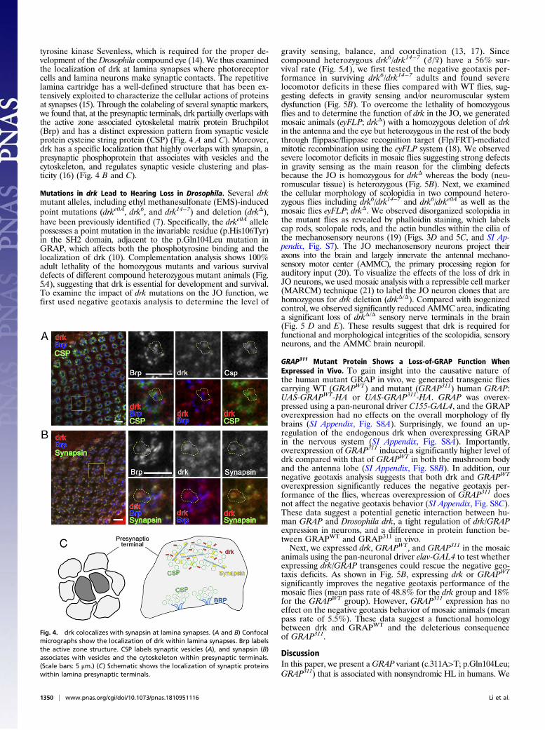
tyrosine kinase Sevenless, which is required for the proper de-velopment of theDrosophila compound eye (14). We thus examinedthe localization of drk at lamina synapses where photoreceptorcells and lamina neurons make synaptic contacts. The repetitivelamina cartridge has a well-defined structure that has been ex-tensively exploited to characterize the cellular actions of proteinsat synapses (15). Through the colabeling of several synaptic markers,we found that, at the presynaptic terminals, drk partially overlaps withthe active zone associated cytoskeletal matrix protein Bruchpilot(Brp) and has a distinct expression pattern from synaptic vesicleprotein cysteine string protein (CSP) (Fig. 4 A and C). Moreover,drk has a specific localization that highly overlaps with synapsin, apresynaptic phosphoprotein that associates with vesicles and thecytoskeleton, and regulates synaptic vesicle clustering and plas-ticity (16) (Fig. 4 B and C).
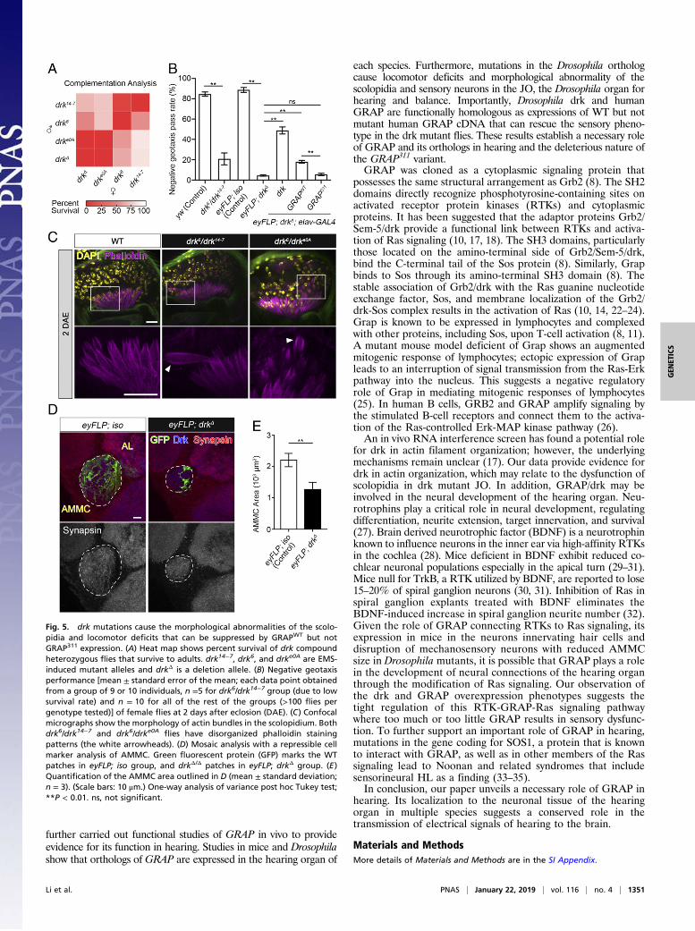
Mutations in drk Lead to Hearing Loss in Drosophila. Several drkmutant alleles, including ethyl methanesulfonate (EMS)-inducedpoint mutations (drke0A, drk6, and drk14−7) and deletion (drkΔ),have been previously identified (7). Specifically, the drke0A allelepossesses a point mutation in the invariable residue (p.His106Tyr)in the SH2 domain, adjacent to the p.Gln104Leu mutation inGRAP, which affects both the phosphotyrosine binding and thelocalization of drk (10). Complementation analysis shows 100%adult lethality of the homozygous mutants and various survivaldefects of different compound heterozygous mutant animals (Fig.5A), suggesting that drk is essential for development and survival.To examine the impact of drk mutations on the JO function, wefirst used negative geotaxis analysis to determine the level of
gravity sensing, balance, and coordination (13, 17). Sincecompound heterozygous drk6/drk14−7 (_/\) have a 56% sur-vival rate (Fig. 5A), we first tested the negative geotaxis per-formance in surviving drk6/drk14−7 adults and found severelocomotor deficits in these flies compared with WT flies, sug-gesting defects in gravity sensing and/or neuromuscular systemdysfunction (Fig. 5B). To overcome the lethality of homozygousflies and to determine the function of drk in the JO, we generatedmosaic animals (eyFLP; drkΔ) with a homozygous deletion of drkin the antenna and the eye but heterozygous in the rest of the bodythrough flippase/flippase recognition target (Flp/FRT)-mediatedmitotic recombination using the eyFLP system (18). We observedsevere locomotor deficits in mosaic flies suggesting strong defectsin gravity sensing as the main reason for the climbing defectsbecause the JO is homozygous for drkΔ whereas the body (neu-romuscular tissue) is heterozygous (Fig. 5B). Next, we examinedthe cellular morphology of scolopidia in two compound hetero-zygous flies including drk6/drk14−7 and drk6/drke0A as well as themosaic flies eyFLP; drkΔ. We observed disorganized scolopidia inthe mutant flies as revealed by phalloidin staining, which labelscap rods, scolopale rods, and the actin bundles within the cilia ofthe mechanosensory neurons (19) (Figs. 3D and 5C, and SI Ap-pendix, Fig. S7). The JO mechanosensory neurons project theiraxons into the brain and largely innervate the antennal mechano-sensory motor center (AMMC), the primary processing region forauditory input (20). To visualize the effects of the loss of drk inJO neurons, we used mosaic analysis with a repressible cell marker(MARCM) technique (21) to label the JO neuron clones that arehomozygous for drk deletion (drkΔ/Δ). Compared with isogenizedcontrol, we observed significantly reduced AMMC area, indicatinga significant loss of drkΔ/Δ sensory nerve terminals in the brain(Fig. 5 D and E). These results suggest that drk is required forfunctional and morphological integrities of the scolopidia, sensoryneurons, and the AMMC brain neuropil.
GRAP311 Mutant Protein Shows a Loss-of-GRAP Function WhenExpressed in Vivo. To gain insight into the causative nature ofthe human mutant GRAP in vivo, we generated transgenic fliescarrying WT (GRAPWT) and mutant (GRAP311) human GRAP:UAS-GRAPWT-HA or UAS-GRAP311-HA. GRAP was overex-pressed using a pan-neuronal driver C155-GAL4, and the GRAPoverexpression had no effects on the overall morphology of flybrains (SI Appendix, Fig. S8A). Surprisingly, we found an up-regulation of the endogenous drk when overexpressing GRAPin the nervous system (SI Appendix, Fig. S8A). Importantly,overexpression ofGRAP311 induced a significantly higher level ofdrk compared with that of GRAPWT in both the mushroom bodyand the antenna lobe (SI Appendix, Fig. S8B). In addition, ournegative geotaxis analysis suggests that both drk and GRAPWT
overexpression significantly reduces the negative geotaxis per-formance of the flies, whereas overexpression of GRAP311 doesnot affect the negative geotaxis behavior (SI Appendix, Fig. S8C).These data suggest a potential genetic interaction between hu-man GRAP and Drosophila drk, a tight regulation of drk/GRAPexpression in neurons, and a difference in protein function be-tween GRAPWT and GRAP311 in vivo.Next, we expressed drk, GRAPWT, and GRAP311 in the mosaic
animals using the pan-neuronal driver elav-GAL4 to test whetherexpressing drk/GRAP transgenes could rescue the negative geo-taxis deficits. As shown in Fig. 5B, expressing drk or GRAPWT
significantly improves the negative geotaxis performance of themosaic flies (mean pass rate of 48.8% for the drk group and 18%for the GRAPWT group). However, GRAP311 expression has noeffect on the negative geotaxis behavior of mosaic animals (meanpass rate of 5.5%). These data suggest a functional homologybetween drk and GRAPWT and the deleterious consequenceof GRAP311.
DiscussionIn this paper, we present aGRAP variant (c.311A>T; p.Gln104Leu;GRAP311) that is associated with nonsyndromic HL in humans. We
Fig. 4. drk colocalizes with synapsin at lamina synapses. (A and B) Confocalmicrographs show the localization of drk within lamina synapses. Brp labelsthe active zone structure. CSP labels synaptic vesicles (A), and synapsin (B)associates with vesicles and the cytoskeleton within presynaptic terminals.(Scale bars: 5 μm.) (C) Schematic shows the localization of synaptic proteinswithin lamina presynaptic terminals.
1350 | www.pnas.org/cgi/doi/10.1073/pnas.1810951116 Li et al.
further carried out functional studies of GRAP in vivo to provideevidence for its function in hearing. Studies in mice and Drosophilashow that orthologs of GRAP are expressed in the hearing organ of
each species. Furthermore, mutations in the Drosophila orthologcause locomotor deficits and morphological abnormality of thescolopidia and sensory neurons in the JO, the Drosophila organ forhearing and balance. Importantly, Drosophila drk and humanGRAP are functionally homologous as expressions of WT but notmutant human GRAP cDNA that can rescue the sensory pheno-type in the drk mutant flies. These results establish a necessary roleof GRAP and its orthologs in hearing and the deleterious nature ofthe GRAP311 variant.GRAP was cloned as a cytoplasmic signaling protein that
possesses the same structural arrangement as Grb2 (8). The SH2domains directly recognize phosphotyrosine-containing sites onactivated receptor protein kinases (RTKs) and cytoplasmicproteins. It has been suggested that the adaptor proteins Grb2/Sem-5/drk provide a functional link between RTKs and activa-tion of Ras signaling (10, 17, 18). The SH3 domains, particularlythose located on the amino-terminal side of Grb2/Sem-5/drk,bind the C-terminal tail of the Sos protein (8). Similarly, Grapbinds to Sos through its amino-terminal SH3 domain (8). Thestable association of Grb2/drk with the Ras guanine nucleotideexchange factor, Sos, and membrane localization of the Grb2/drk-Sos complex results in the activation of Ras (10, 14, 22–24).Grap is known to be expressed in lymphocytes and complexedwith other proteins, including Sos, upon T-cell activation (8, 11).A mutant mouse model deficient of Grap shows an augmentedmitogenic response of lymphocytes; ectopic expression of Grapleads to an interruption of signal transmission from the Ras-Erkpathway into the nucleus. This suggests a negative regulatoryrole of Grap in mediating mitogenic responses of lymphocytes(25). In human B cells, GRB2 and GRAP amplify signaling bythe stimulated B-cell receptors and connect them to the activa-tion of the Ras-controlled Erk-MAP kinase pathway (26).An in vivo RNA interference screen has found a potential role
for drk in actin filament organization; however, the underlyingmechanisms remain unclear (17). Our data provide evidence fordrk in actin organization, which may relate to the dysfunction ofscolopidia in drk mutant JO. In addition, GRAP/drk may beinvolved in the neural development of the hearing organ. Neu-rotrophins play a critical role in neural development, regulatingdifferentiation, neurite extension, target innervation, and survival(27). Brain derived neurotrophic factor (BDNF) is a neurotrophinknown to influence neurons in the inner ear via high-affinity RTKsin the cochlea (28). Mice deficient in BDNF exhibit reduced co-chlear neuronal populations especially in the apical turn (29–31).Mice null for TrkB, a RTK utilized by BDNF, are reported to lose15–20% of spiral ganglion neurons (30, 31). Inhibition of Ras inspiral ganglion explants treated with BDNF eliminates theBDNF-induced increase in spiral ganglion neurite number (32).Given the role of GRAP connecting RTKs to Ras signaling, itsexpression in mice in the neurons innervating hair cells anddisruption of mechanosensory neurons with reduced AMMCsize in Drosophila mutants, it is possible that GRAP plays a rolein the development of neural connections of the hearing organthrough the modification of Ras signaling. Our observation ofthe drk and GRAP overexpression phenotypes suggests thetight regulation of this RTK-GRAP-Ras signaling pathwaywhere too much or too little GRAP results in sensory dysfunc-tion. To further support an important role of GRAP in hearing,mutations in the gene coding for SOS1, a protein that is knownto interact with GRAP, as well as in other members of the Rassignaling lead to Noonan and related syndromes that includesensorineural HL as a finding (33–35).In conclusion, our paper unveils a necessary role of GRAP in
hearing. Its localization to the neuronal tissue of the hearingorgan in multiple species suggests a conserved role in thetransmission of electrical signals of hearing to the brain.
Materials and MethodsMore details of Materials and Methods are in the SI Appendix.
Fig. 5. drk mutations cause the morphological abnormalities of the scolo-pidia and locomotor deficits that can be suppressed by GRAPWT but notGRAP311 expression. (A) Heat map shows percent survival of drk compoundheterozygous flies that survive to adults. drk14−7, drk6, and drke0A are EMS-induced mutant alleles and drkΔ is a deletion allele. (B) Negative geotaxisperformance [mean ± standard error of the mean; each data point obtainedfrom a group of 9 or 10 individuals, n =5 for drk6/drk14−7 group (due to lowsurvival rate) and n = 10 for all of the rest of the groups (>100 flies pergenotype tested)] of female flies at 2 days after eclosion (DAE). (C) Confocalmicrographs show the morphology of actin bundles in the scolopidium. Bothdrk6/drk14−7 and drk6/drke0A flies have disorganized phalloidin stainingpatterns (the white arrowheads). (D) Mosaic analysis with a repressible cellmarker analysis of AMMC. Green fluorescent protein (GFP) marks the WTpatches in eyFLP; iso group, and drkΔ/Δ patches in eyFLP; drkΔ group. (E)Quantification of the AMMC area outlined in D (mean ± standard deviation;n = 3). (Scale bars: 10 μm.) One-way analysis of variance post hoc Tukey test;**P < 0.01. ns, not significant.
Li et al. PNAS | January 22, 2019 | vol. 116 | no. 4 | 1351
GEN
ETICS
Human Subjects. This paperwas approvedby theUniversity ofMiami InstitutionalReview Board and the Ankara University Medical School Ethics Committee(Turkey). A signed informed-consent form was obtained from each participantor, in the case of a minor, from the parents. Detailed past medical historiesand thorough physical examinations including otoscopy and ophthalmoscopyrevealed no abnormalities, and electrocardiograms were unremarkable.
DNA Sequencing and Bioinformatics. Exome and genome sequencing wereperformed on the probands of each family by using a previously publishedprotocol (36, 37). Variants were filtered with minor allele frequency thresholdsof 0.005 for recessive and 0.0005 for dominant variants as recommended (38).We used GERP > 2 for conservation (39). We also filtered variants by using thecriteria of the combined annotation-dependent depletion (CADD) score > 20(40), the deep annotation-dependent neural network (DANN) score > 0.95(41), and they were damaging for Provean (provean.jcvi.org/index.php) andFATHMM (fathmm.biocompute.org.uk/) for missense variants. XHMM andConifer were used for the copy number variant (CNV) detection with exomedata (42); CNVs were called using a CNVnator with genome data (43). Sangersequencing was used for confirmation and cosegregation of the variant. Ho-mozygous runs were detected from exome data and were used during thefiltering of variants (SI Appendix, Tables S2 and S3).
Mouse Studies. WT C57BL/6 mice were bred and maintained at the Universityof Miami. All procedures were approved by the University of Miami In-stitutional Animal Care and followed the National Institutes of Health (NIH)Guidelines, “Using Animals in Intramural Research” (44). Cochlear expression
of Grap was checked in embryos of 17.5 dpc and P15 via RT-PCR. Immuno-fluoresence was performed on P0 using a goat anti-GRAP polyclonal anti-body (ab9703; Abcam) as the primary antibody.
Drosophila Studies. The following fly strains were used in the studies: drk14−7
(27622), drk6 (27623), drke0A (5691), actin-GAL4, FRT42Diso, eyFLP; FRT42D,w+, and cl−, obtained from Bloomington Drosophila Stock Center; drkΔP24
(named as drkΔ in this paper) from E. Skoulakis’s laboratory. All drk alleleswere normalized to the w− background and balanced with the CyO, KrGFPchromosome. For mosaic analysis, drkΔ was recombined with the FRT42Disochromosome. Climbing behavior was measured as previously described (45).For immunofluorescence studies in Drosophila, the following primary andsecondary antibodies were used in this paper: anti-drk (from E. Skoulakis’slaboratory), anti-Brp (DSHB, AB_2314866), anti-Csp (DSHB AB_528183), anti-synapsin (DSHB, AB_2313867), Cy5 conjugated anti-HRP (123175021; JacksonImmunoLab), Alexa 546 conjugated phalloidin (A22283; ThermoFisher Sci-entific), and Cy5 conjugated anti-HRP and secondary antibodies conjugatedto Alexa 488/568/647 (ThermoFisher Scientific).
ACKNOWLEDGMENTS. This paper was supported by NIH Grants R01DC009645and R01DC012836 (to M.T.), the Dr. John T. Macdonald Foundation (to C.L.),the Lois Pope LIFE Fellows Program (to C.L., Y.Z., and T.G.M.), NIH GrantR21GM119018 (to R.G.Z.), by Taishan Scholar Project (Shandong Province, People’sRepublic of China) (to R.G.Z.), and by funds from the Sylvester ComprehensiveCancer Center (to A.F.).
1. Morton CC, NanceWE (2006) Newborn hearing screening–A silent revolution. N Engl JMed 354:2151–2164.
2. Dror AA, Avraham KB (2010) Hearing impairment: A panoply of genes and functions.Neuron 68:293–308.
3. Korver AM, et al. (2017) Congenital hearing loss. Nat Rev Dis Primers 3:16094.4. Brown SD, Hardisty-Hughes RE, Mburu P (2008) Quiet as a mouse: Dissecting the
molecular and genetic basis of hearing. Nat Rev Genet 9:277–290.5. Boekhoff-Falk G (2005) Hearing in Drosophila: Development of Johnston’s organ and
emerging parallels to vertebrate ear development. Dev Dyn 232:550–558.6. Lewis MA, Steel KP (2012) A cornucopia of candidates for deafness. Cell 150:879–881.7. Senthilan PR, et al. (2012) Drosophila auditory organ genes and genetic hearing de-
fects. Cell 150:1042–1054.8. Feng GS, et al. (1996) Grap is a novel SH3-SH2-SH3 adaptor protein that couples ty-
rosine kinases to the Ras pathway. J Biol Chem 271:12129–12132.9. Clark SG, Stern MJ, Horvitz HR (1992) C. elegans cell-signalling gene sem-5 encodes a
protein with SH2 and SH3 domains. Nature 356:340–344.10. Olivier JP, et al. (1993) A Drosophila SH2-SH3 adaptor protein implicated in coupling
the sevenless tyrosine kinase to an activator of Ras guanine nucleotide exchange, Sos.Cell 73:179–191.
11. Trüb T, Frantz JD, Miyazaki M, Band H, Shoelson SE (1997) The role of a lymphoid-restricted, Grb2-like SH3-SH2-SH3 protein in T cell receptor signaling. J Biol Chem 272:894–902.
12. Yorozu S, et al. (2009) Distinct sensory representations of wind and near-field soundin the Drosophila brain. Nature 458:201–205.
13. Kamikouchi A, et al. (2009) The neural basis of Drosophila gravity-sensing andhearing. Nature 458:165–171.
14. Simon MA, Bowtell DD, Dodson GS, Laverty TR, Rubin GM (1991) Ras1 and a putativeguanine nucleotide exchange factor perform crucial steps in signaling by the seven-less protein tyrosine kinase. Cell 67:701–716.
15. Hamanaka Y, Meinertzhagen IA (2010) Immunocytochemical localization of synapticproteins to photoreceptor synapses of Drosophila melanogaster. J Comp Neurol 518:1133–1155.
16. Vasin A, et al. (2014) Synapsin regulates activity-dependent outgrowth of synapticboutons at the Drosophila neuromuscular junction. J Neurosci 34:10554–10563.
17. Sun Y, et al. (2009) TRPA channels distinguish gravity sensing from hearing in John-ston’s organ. Proc Natl Acad Sci USA 106:13606–13611.
18. Li T, et al. (2016) The E3 ligase Ubr3 regulates Usher syndrome and MYH9 disorderproteins in the auditory organs of Drosophila and mammals. eLife 5:e15258.
19. Boekhoff-Falk G, Eberl DF (2014) The Drosophila auditory system. Wiley InterdiscipRev Dev Biol 3:179–191.
20. Murthy M (2010) Unraveling the auditory system of Drosophila. Curr Opin Neurobiol20:281–287.
21. Lee T, Luo L (2001) Mosaic analysis with a repressible cell marker (MARCM) for Dro-sophila neural development. Trends Neurosci 24:251–254.
22. Skolnik EY, et al. (1993) The function of GRB2 in linking the insulin receptor to Rassignaling pathways. Science 260:1953–1955.
23. Buday L, Downward J (1993) Epidermal growth factor regulates p21ras through theformation of a complex of receptor, Grb2 adapter protein, and Sos nucleotide ex-change factor. Cell 73:611–620.
24. Simon MA, Dodson GS, Rubin GM (1993) An SH3-SH2-SH3 protein is required forp21Ras1 activation and binds to sevenless and Sos proteins in vitro. Cell 73:169–177.
25. Shen R, et al. (2002) Grap negatively regulates T-cell receptor-elicited lymphocyteproliferation and interleukin-2 induction. Mol Cell Biol 22:3230–3236.
26. Vanshylla K, et al. (2018) Grb2 and GRAP connect the B cell antigen receptor to Erk
MAP kinase activation in human B cells. Sci Rep 8:4244.27. Bibel M, Barde YA (2000) Neurotrophins: Key regulators of cell fate and cell shape in
the vertebrate nervous system. Genes Dev 14:2919–2937.28. Pirvola U, et al. (1994) Coordinated expression and function of neurotrophins and
their receptors in the rat inner ear during target innervation. Hear Res 75:131–144.29. Bianchi LM, et al. (1996) Degeneration of vestibular neurons in late embryogenesis of
both heterozygous and homozygous BDNF null mutant mice. Development 122:
1965–1973.30. Fritzsch B, Silos-Santiago I, Bianchi LM, Fariñas I (1997) The role of neurotrophic
factors in regulating the development of inner ear innervation. Trends Neurosci 20:
159–164.31. Fritzsch B, Silos-Santiago I, Bianchi LM, Farinas I (1997) Effects of neurotrophin and
neurotrophin receptor disruption on the afferent inner ear innervation. Semin Cell
Dev Biol 8:277–284.32. Mullen LM, et al. (2012) Ras/p38 and PI3K/Akt but not Mek/Erk signaling mediate
BDNF-induced neurite formation on neonatal cochlear spiral ganglion explants. BrainRes 1430:25–34.
33. Bademci G, et al. (2016) Variations in multiple syndromic deafness genes mimic non-
syndromic hearing loss. Sci Rep 6:31622.34. Sarkozy A, Digilio MC, Dallapiccola B (2008) Leopard syndrome.Orphanet J Rare Dis 3:
13.35. van Trier DC, et al. (2015) External ear anomalies and hearing impairment in Noonan
Syndrome. Int J Pediatr Otorhinolaryngol 79:874–878.36. Bademci G, et al. (2016) Comprehensive analysis via exome sequencing uncovers
genetic etiology in autosomal recessive nonsyndromic deafness in a large multiethnic
cohort. Genet Med 18:364–371.37. Bowling KM, et al. (2017) Genomic diagnosis for children with intellectual disability
and/or developmental delay. Genome Med 9:43.38. Shearer AE, et al. (2014) Utilizing ethnic-specific differences in minor allele frequency
to recategorize reported pathogenic deafness variants. Am J Hum Genet 95:445–453.39. Cooper GM, et al.; NISC Comparative Sequencing Program (2005) Distribution and
intensity of constraint in mammalian genomic sequence. Genome Res 15:901–913.40. Kircher M, et al. (2014) A general framework for estimating the relative pathogenicity
of human genetic variants. Nat Genet 46:310–315.41. Quang D, Chen Y, Xie X (2015) DANN: A deep learning approach for annotating the
pathogenicity of genetic variants. Bioinformatics 31:761–763.42. Bademci G, et al. (2014) Identification of copy number variants through whole-exome
sequencing in autosomal recessive nonsyndromic hearing loss. Genet Test Mol
Biomarkers 18:658–661.43. Abyzov A, Urban AE, Snyder M, Gerstein M (2011) CNVnator: An approach to dis-
cover, genotype, and characterize typical and atypical CNVs from family and pop-
ulation genome sequencing. Genome Res 21:974–984.44. National Research Council (2011) Guide for the Care and Use of Laboratory Animals
(National Academies Press, Washington, DC), 8th Ed.45. Li C, et al. (2017) Spermine synthase deficiency causes lysosomal dysfunction and
oxidative stress in models of Snyder-Robinson syndrome. Nat Commun 8:1257.
1352 | www.pnas.org/cgi/doi/10.1073/pnas.1810951116 Li et al.


![A Sample Design in Programming with [1] zaferguney@aydin ... · software development for programmers, designers and educators. ... for a solution as teaching strategies (Guney, 2019)](https://img.pdfslide.us/doc/110x75/5faa2dc6e2e9566da6166a3d/a-sample-design-in-programming-with-1-zaferguneyaydin-software-development.jpg)