Embed Size (px)
Citation preview

pubs.acs.org/Biochemistry Published on Web 06/18/2010 r 2010 American Chemical Society
5978 Biochemistry 2010, 49, 5978–5988
DOI: 10.1021/bi1004359
Binding of the ERR Nuclear Receptor to DNA Is Coupled to Proton Uptake†
Brian J. Deegan, Kenneth L. Seldeen, Caleb B. McDonald, Vikas Bhat, and Amjad Farooq*
Department of Biochemistry and Molecular Biology and USylvester Braman Family Breast Cancer Institute,Leonard Miller School of Medicine, University of Miami, Miami, Florida 33136
Received March 23, 2010; Revised Manuscript Received May 24, 2010
ABSTRACT: Nuclear receptors act as ligand-modulated transcription factors and orchestrate a plethora ofcellular functions central to health and disease. Although studied formore than half a century, manymysteriessurrounding the mechanism of action of nuclear receptors remain unresolved. Herein, using isothermaltitration calorimetry (ITC) in conjunction with macromolecular modeling (MM), we provide evidence thatthe binding of the ERR nuclear receptor to its DNA response element is coupled to proton uptake by twoionizable residues, H196 and E203, located at the protein-DNA interface. Alanine substitution of theseionizable residues decouples protonation and hampers the binding of ERR to DNA by nearly 1 order ofmagnitude. Remarkably, H196 and E203 are predominantly conserved across ∼50 members of the nuclearreceptor family, implying that proton-coupled equilibrium may serve as a key regulatory switch formodulating protein-DNA interactions central to nuclear receptor function and regulation. Taken together,our findings unearth an unexpected but critical step in the molecular action of nuclear receptors and suggestthat they may act as sensors of intracellular pH.
Estrogen receptor R (ERR)1 is a member of a family of ligand-modulated transcription factors that have come to be known asnuclear receptors (NRs) (1-4). All members of NR family areevolutionarily related and share a core modular architecturecomprised of a central DNA-binding (DB) domain flankedbetween an N-terminal transactivation (TA) domain and aC-terminal ligand-binding (LB) domain (5-7). A typical scenariofor the activation of nuclear receptors involves the secretion oflipophilic messengers such as hormones and vitamins by appro-priate tissues. Upon their diffusion through the cell membrane,these ligands bind to the LB domain of nuclear receptors andallow their translocation into the nucleus to modulate geneexpression (8-10). While the DB domain recognizes specificpromoter elements, the LB domain additionally serves as a plat-form for the recruitment of a multitude of cellular proteins, suchas transcription factors, coactivators, and corepressors, to the siteof DNA transcription, thereby allowing nuclear receptors toexert their action at the genomic level in a concerted fash-ion (11, 12). The TA domain is believed to be responsive togrowth factors acting through the MAPK signaling and mayfurther synergize the action of various coactivators and core-pressors recruited by the LB domain at the site of DNA
transcription (13, 14). In this manner, nuclear receptors orches-trate a diverse array of cellular functions from embryonicdevelopment to metabolic homeostasis, and their malfunctionhas been widely implicated in disease (5, 15-19).
Discovered more than half a century ago, ERR mediates theaction of estrogens such as estradiol, and its hyperactivation leadsto the genesis of large fractions of breast cancer (20-26). The DBdomain of ERR binds as a homodimer to the AGGTCAnnnT-GACCT consensus motif, termed the estrogen response element(ERE), located within the promoters of target genes (27). DNAbinding is accomplished through a pair of tandem C4-type zincfingers, with each finger containing a Zn2þ ion coordinated ina tetrahedral arrangement by four highly conserved cysteineresidues (28, 29). The first zinc finger (ZF-I) within each mono-mer of DB domain recognizes the hexanucleotide sequence 50-AGGTCA-30 within the major groove at each end of the EREduplex, while the second zinc finger (ZF-II) is responsible for thehomodimerization of the DB domain upon DNA binding. Closescrutiny of the three-dimensional structure of the DB domain ofERR in complex with the ERE duplex reveals that a triplet ofionizable residues (D190, H196, and E203) either protrudes deepinto the comfort of the major groove at the protein-DNAinterface or appears to reside within touching distance of theDNA backbone (28) (Figure 1). Given that the placement ofthese residues in the proximity of the negatively charged phos-phate backbone of DNA would be energetically unfavorablebecause of electrostatic repulsions, we hypothesized that the sidechainmoieties of D190, H196, andE203may become protonatedupon the binding of ERR to DNA to neutralize the intermole-cular repulsions and further enhance the favorable role ofelectrostatic forces central to driving protein-DNA interactions.This notion is further corroborated by the fact that the imidazoleside chain of H196 stacks against the highly basic side chain ofK206, a scenario that could reduce the side chain pKa of H196and thereby render it more amenable to protonation upon the
†This workwas supported by theNational Institutes ofHealth (GrantR01-GM083897) and the USylvester Braman Family Breast CancerInstitute (A.F.). C.B.M. is a recipient of a postdoctoral fellowship fromthe National Institutes of Health (T32-CA119929). B.J.D. and A.F. aremembers of the Sheila and David Fuente Graduate Program in CancerBiology at the Sylvester ComprehensiveCancer Center of theUniversityof Miami.*To whom correspondence should be addressed. E-mail: amjad@
farooqlab.net. Phone: (305) 243-2429. Fax: (305) 243-3955.1Abbreviations: DB, DNA-binding; ERR, estrogen receptor R; ERE,
estrogen response element; ITC, isothermal titration calorimetry; LB,ligand-binding; MALDI-TOF, matrix-assisted laser desorpton ioniza-tion time-of-flight; MAPK, mitogen-activated protein kinase; NMR,nuclear magnetic resonance; NR, nuclear receptor; SEC, size-exclusionchromatography; TA, transactivation; Trx, thioredoxin; ZF, zinc finger.

Article Biochemistry, Vol. 49, No. 29, 2010 5979
binding of the DB domain to DNA. It is also of note that theacidic side chains of D190 and E203 are positioned in the proxi-mity of each other within the DB domain. It is thus conceivablethat the more acidic side chain of D190 may be able to increasethe pKa value of the side chain of E203, allowing it to becomeprotonated more easily upon the binding of the DB domain toDNA at its own expense. Although both D190 and E203 arelocated at the protein-DNA interface, protonation of E203would be more desirable as it directly inserts into the majorgroove of DNA.
In an effort to test our hypothesis, we have employed hereisothermal titration calorimetry (ITC) in conjunction withmacromolecular modeling (MM) to analyze the binding of theDB domain of ERR to a 21-mer dsDNA oligo containing theERE motif, hereafter termed the ERE duplex. Our data revealthat H196 and E203, but not D190, indeed become protonatedupon the binding of ERR to DNA. Furthermore, alanine sub-stitution of these ionizable residues decouples protonation andhampers the binding of ERR toDNAby nearly 1 order of magni-tude. Our study suggests that the proton-coupled equilibriumobserved here may be a general feature of the nuclear receptorfamily.
MATERIALS AND METHODS
Protein Preparation. TheDB domain (residues 176-250) ofhuman ERR (Expasy entry P03372) was cloned into the pET102bacterial expression vector, with anN-terminal thioredoxin (Trx)tag and a C-terminal polyhistidine (His) tag, using InvitrogenTOPO technology. The Trx tagwas included tomaximize proteinexpression in the soluble fraction, while the His tag was added toaid in protein purification through Ni-NTA affinity chromato-graphy. Additionally, thrombin protease sites were introduced at
both the N- and C-termini of the DB domain to aid in theremoval of tags after purification. The protein was subsequentlyexpressed in the Escherichia coli BL21*(DE3) bacterial strain(Invitrogen) and purified on a Ni-NTA affinity column usingstandard procedures. Briefly, bacterial cells were grown at 20 �Cin LB medium supplemented with 50 μM ZnCl2 to an opticaldensity of 0.5 at 600 nmprior to inductionwith 0.5mM isopropylβ-D-1-thiogalactopyranoside (IPTG). The bacterial culture wasfurther grown overnight at 20 �C, and the cells were subsequentlyharvested and disrupted using a BeadBeater (Biospec). Afterseparation of cell debris via high-speed centrifugation, the celllysatewas loaded onto aNi-NTA columnandwashed extensivelywith 20 mM imidazole to eliminate nonspecific binding of bac-terial proteins to the column. The recombinant protein was sub-sequently eluted with 200 mM imidazole and dialyzed against anappropriate buffer to remove excess imidazole. Further treatmenton a Hiload Superdex 200 size-exclusion chromatography (SEC)column coupled in-line with a GE Akta FPLC system led topurification of the recombinant DB domain to apparent homo-geneity as judged by SDS-PAGE analysis. The identity of therecombinant protein was confirmed by MALDI-TOF massspectrometry analysis. The final yield was typically between 5and 10mgof protein of apparent homogeneity per liter of bacterialculture. Treatment with thrombin protease significantly destabi-lized the recombinantDB domain, and it appeared to be proteoly-tically unstable. For this reason, except for control experiments toensure that the tags had no effect on the binding of theDBdomainto DNA, all experiments reported herein were conducted with therecombinant fusion DB domain containing a Trx tag at theN-terminus and a His tag at the C-terminus. The protein con-centration was determined by the fluorescence-based Quant-Itassay (Invitrogen) and spectrophotometrically using an extinction
FIGURE 1: Three-dimensional atomic model of the DB domain of human ERR in complex with the ERE duplex containing the AGGTCA-cagTGACCT consensus sequence. Note that the DB domain binds to DNA as a homodimer. One monomer of the DB domain is colored greenand the other blue. TheZn2þ divalent ions are depicted as gray spheres, and the side chainmoieties ofD190,H196, E203, andK206within theDBmonomers are colored red.TheDNAbackbone is colored yellow, and thebases are colored gray for the sakeof clarity.Thenumerals at the terminiof DB monomers indicate the boundaries of the DB domain within the amino acid sequence of human ERR.

5980 Biochemistry, Vol. 49, No. 29, 2010 Deegan et al.
coefficient of 29045 M-1 cm-1 calculated for the recombinantfusionDBdomain using the online software ProtParamat ExPasyServer (30). Results from both methods were in an excellentagreement.Site-Directed Mutagenesis. The pET102 bacterial expres-
sion vector expressing the wild-type DB domain of ERR wassubjected to the QuikChange Lightening kit (Stratagene) togenerate single mutants D190A (DB_D190A), S193A (DB_S193A),H196A (DB_H196A),Y197A (DB_Y197A), S201A (DB_S201A), E203A (DB_E203A), K206A (DB_K206A), K210A(DB_K210A), and R211A (DB_R211A) and double mutantH196A/E203A (DB_AA).AllmutantDBdomainswere expressed,purified, and characterized as described above. When analyzed bysize-exclusion chromatography (SEC) using a Hiload Superdex200 column, all mutant DB domains exhibited elution volumesvirtually indistinguishable from those observed for the wild-typeDBdomain, implying that the point substitution of specific residuesdid not lead to protein unfolding and that the mutant DB domainsretained the compact globular fold characteristic of the wild-typeDB domain. These observations were further confirmed by circulardichroism (CD) analysis.DNA Synthesis. 21-mer DNA oligos containing the ERE
consensus site (AGGTCAnnnTGACCT) were commerciallyobtained fromSigmaGenosys. The complete nucleotide sequencesof the sense and antisense oligos constituting the EREduplex wereas follows:
Oligo concentrations were determined spectrophotome-trically on the basis of their extinction coefficients derivedfrom their nucleotide sequences using the online softwareOligoAnalyzer 3.0 (IntegratedDNATechnologies) basedon the nearest-neighbor model (31). To obtain double-stranded DNA (dsDNA) annealed oligos to generate theERE duplex, equimolar amounts of sense and antisenseoligos weremixed together and heated at 95 �C for 10minand then allowed to cool to room temperature. Theefficiency of oligo annealing to generate dsDNA wasclose to 100% as judged by Native-PAGE and circulardichroism (CD) analysis.ITC Measurements. Isothermal titration calorimetry (ITC)
experiments were performed on a Microcal VP-ITC instrument,and data were acquired and processed using fully automizedfeatures in Microcal ORIGIN. Measurements were repeatedthree or four times in phosphate, Hepes, Tricine, or Tris buffer.All buffers were taken to a final concentration of 50 mM con-taining 5 mM β-mercaptoethanol (pH 7.0). Additionally, 0-105mMNaClwas added to adjust the ionic strength of all buffersto 110 mM. This ionic strength was sufficiently high to preventnonspecific binding of the DB domain of ERR to DNA yetsufficiently low to allow ITC analysis to be conducted with a highsignal-to-noise ratio. Various constructs of the DB domain andthe ERE duplex were prepared in an appropriate buffer anddegassed using the ThermoVac accessory for 5 min. The experi-ments were initiated via injection of 25 � 10 μL aliquots of50-100 μM ERE duplex from the syringe into the calorimetriccell containing 1.8 mL of a 5-10 μM DB domain solution at25 �C. The change in thermal power as a function of eachinjection was automatically recorded using Microcal ORIGIN,
and the raw data were further processed to yield binding iso-therms of heat release per injection as a function of the molarratio of ERE duplex to dimer-equivalent DB domain. The heatsof mixing and dilution were subtracted from the heat of bindingper injection by conducting a control experiment in which thesame buffer in the calorimetric cell was titrated against theERE duplex in an identical manner. Control experiments withscrambled dsDNA oligos generated a thermal power similar tothat obtained for the buffer alone, implying that there was nononspecific binding of the DB domain to noncognate DNA.Experiments that aimed to examine the binding of the thrombin-cleaved DB domain to DNA gave results similar to those ofexperiments conducted with the recombinant fusion protein,implying that the tags had no effect on DNA binding. However,because of the poor stability and low yield of the thrombin-cleaved DB domain and particularly in the case of mutant DBdomains, all experiments reported here were conducted with therecombinant fusion DB domain containing a Trx tag at theN-terminus and a His tag at the C-terminus. Additionally,titration of a protein construct containing thioredoxin with aC-terminalHis tag (Trx-His) in the calorimetric cell with the EREduplex in the syringe produced no observable signal, implyingthat the tags do not interact with the ERE duplex. In a similarmanner, titration of wild-type or mutant DB domains in thecalorimetric cell with the Trx-His construct in the syringeproduced no observable signal, implying that the tags do notinteract with any of the wild-type or mutant DB domains. Toextract the observed affinity (Kobs) and observed enthalpy(ΔHobs), the binding isothermswere iteratively fit to the followingbuilt-in function by nonlinear least-squares regression analysisusing integrated Microcal ORIGIN:
qðiÞ ¼ ðnVPΔHobs=2Þf1þL=nPþKobs=nP
- ½ð1þL=nPþKobs=nPÞ2 - 4L=nP�1=2g ð1Þwhere q(i) is the heat release (kilocalories per mole) for the ithinjection, n is the binding stoichiometry,V is the effective volumeof protein solution in the calorimetric cell (1.46mL),P is the totaldimer-equivalent concentration of the DB domain in the calori-metric cell (micromolar), and L is the concentration of EREduplex added (micromolar). Equation 1 is derived from thebinding of a ligand to a macromolecule using the law of massaction assuming a one-site model (32). The observed free energyof binding (ΔGobs) was calculated from the relationship
ΔGobs ¼ RT ln Kobs ð2ÞwhereR is the universal molar gas constant (1.99 cal mol-1 K-1)andT is the absolute temperature (298K). The observed entropiccontribution (TΔSobs) to binding was calculated from therelationship
TΔSobs ¼ ΔHobs -ΔGobs ð3ÞThe net change in the number of protons (Δm) absorbed orreleased per DB monomer upon binding to DNA and theintrinsic binding enthalpy (ΔHint) due to direct protein-DNAinteractions and protonation of ionizable moieties were calcu-lated from the slope and y-intercept of ΔHobs - ΔHion plots bylinear fits of data to the equation
ΔHobs ¼ 2ΔmΔHion þΔHint ð4Þwhere ΔHobs is the observed binding enthalpy and ΔHion is theionization enthalpy of each buffer. The ΔHion values of various

Article Biochemistry, Vol. 49, No. 29, 2010 5981
buffers were 1.22 kcal/mol for phosphate buffer, 5.02 kcal/molforHepes buffer, 7.64 kcal/mol for Tricine buffer, and 11.35 kcal/mol for Tris buffer (33-35).Macromolecular Modeling. Macromolecular modeling
(MM) was employed to generate a three-dimensional atomicmodel of the DB domain of ERR in complex with the EREduplex using MODELER based on homology modeling (36).The X-ray structure of the DB domain of ERR in complex with adsDNA oligo containing the ERE motif but with varyingflanking sequences was used as a template (Protein Data Bankentry 1HCQ). A total of 100 atomic models were calculated, andthe structure with the lowest energy, as judged by the MODE-LER Objective Function, was selected for further analysis. Theatomic model was rendered using RIBBONS (37), and theelectrostatic surface potentials were generated using MOL-MOL (38).
RESULTS AND DISCUSSION
Binding of the DBDomain of ERR to DNA Is Coupled toProton Uptake. To test our hypothesis that the binding of ERRtoDNA is coupled to proton uptake, wemeasured the binding ofthe DB domain of ERR to the ERE duplex in buffers of varyingionization enthalpies using ITC. Figure 2 shows representativeITC isotherms obtained from such measurements, while detailedthermodynamic parameters are listed in Table 1. It should benoted here that a classical test for ligand binding coupled toproton exchange is the dependence of the observed enthalpy(ΔHobs) on ionization enthalpy (ΔHion) of the reaction buffer.Because different buffers are characterized by distinct ionizationenthalpies, the observed enthalpy of ligand binding displays asharp dependence on the buffer employed due to varyingcontributions from coupled protonation and deprotonation.Our data indeed suggest that the ΔHobs for the binding of theDB domain of ERR toDNA is highly dependent on the nature ofbuffer conditions employed (Figure 2). Thus, the ΔHobs of
binding goes from being highly exothermic (-30.52 kcal/mol)in phosphate buffer to being endothermic (9.82 kcal/mol) in Trisbuffer and thereby mirrors the ΔHion of the respective buffersranging from 1.22 to 11.35 kcal/mol (33-35). This salientobservation demonstrates that the binding of the DB domainof ERR to DNA is directly coupled to proton uptake. Althoughsuch a coupled equilibrium could result from the protonation ofDNA bases, the side chain moieties of D190, H196, and E203within the DB domain must be considered as the major suspectsfor proton uptake because of their proximity to the negativelycharged phosphate backbone of DNA, a situation that wouldbe inconceivable on thermodynamic grounds barring their pro-tonation.
It is also worth noting that while the enthalpy is favorable forthe binding of ERR to DNA in phosphate buffer, it contributes asubstantial energetic penalty inTris buffer (Table 1). In fact, closescrutinization of thermodynamic parameters observed for thebinding of ERR to DNA in various buffers suggests that whileenthalpy solely drives this protein-DNA interaction in phos-phate and Hepes buffers of low ionization enthalpies, entropyplays amajor role in Tris and Tricine buffers with high ionizationenthalpies. In particular, in the case of Tris buffer, it is theentropy that drives binding against the backdrop of enthalpicpenalty. These observations imply that physiological settingswith low ionization enthalpies are likely to favor the binding ofERR to DNA, while the opposing conditions may be somewhatinhibitory. This is indeed corroborated by the fact that thebinding affinity for formation of the ERR-DNA complexdecreases by nearly 1 order of magnitude from 43 nM inphosphate buffer to 336 nM in Tris buffer (Table 1). It shouldbe borne in mind that the favorable enthalpic contributions tobinding largely result from the release of heat upon the formationof tight electrostatic interactions, hydrogen bonding, and hydro-phobic contacts between protein andDNA. Thus, in buffers withlow ionization enthalpies, the favorable enthalpic contribution is
FIGURE 2: Representative ITC isotherms for the binding of the ERE duplex to the wild-type DB domain of ERR in phosphate (a), Hepes (b),Tricine (c), andTris (d) buffers at pH7.0 and25 �C.The toppanels show the raw ITCdata expressed as the change in thermal powerwith respect totime over the period of titration. In the bottompanels, the change inmolar heat is expressed as a function of the molar ratio of the ERE duplex tothe dimer-equivalent DB domain (b). The solid lines in the bottom panels show the fit of data to a one-site model, as embodied in eq 1, usingMicrocal Origin. Note also that the DB domain shows no nonspecific binding to scrambled dsDNA oligos (O).

5982 Biochemistry, Vol. 49, No. 29, 2010 Deegan et al.
only slightly offset to compensate for the proton-coupled equi-librium, rendering enthalpy as the sole driving force accompaniedby an entropic penalty. In contrast, in buffers with high ioniza-tion enthalpies, the favorable enthalpic contribution is largelyoffset and even overridden by the enthalpic penalty due to theproton-coupled equilibrium with entropy either contributingfavorably or serving as the sole driving force at the expense ofenthalpy.
This reciprocal relationship between enthalpy and entropy liesin the enthalpy-entropy compensation phenomenon (39-43);macromolecular interactions are compensated by equal butopposing entropic changes such that there is little or no net gainin the overall free energy. Thus, buffers with high ionizationenthalpies gain a substantial increase in entropy upon the releaseof a proton, presumably due to an increase in the degrees offreedom that become available to water molecules after they arefreed from their hydration shell surrounding the exchangeableproton prior to its release. However, in the case of buffers withlow ionization enthalpies, the exchangeable proton would beexpected to be more “economically” hydrated such that therelease of water molecules from the rather small hydration shellcontributes relatively little to the overall entropy gain but at thesame time draws less heat to be removed. Such enthalpy-entropycompensations for the binding of the DB domain to the EREduplex in various buffers are illustrated in Figure 3a. Consistentwith the foregoing arguments, it should also be noted that whilethe increase in the binding affinity of the DB domain for DNAcorrelates with the overall favorable enthalpy change in variousbuffers, the increase in the favorable entropy change seems tooppose such protein-DNA interactions (Figure 3b,c).ResiduesH196 andE203 Serve as Sole ProtonAcceptors
upon the Binding of ERR to DNA. For processes in whichligand binding is coupled to proton exchange, the observedenthalpy (ΔHobs) is related to the ionization enthalpy (ΔHion)by the relationshipΔHobs=2ΔmΔHion þ ΔHint, where Δm is thenet change in the number of protons absorbed or released per DBmonomer upon binding to DNA and ΔHint is the intrinsicbinding enthalpy due to direct protein-DNA interactions andprotonation of ionizable moieties. A plot of ΔHobs versus ΔHion
should thus yield a linear curve with the slope 2Δm and y-inter-cept equal to ΔHint. As shown in Figure 4, such analysis revealsthat the binding of the wild-type DB domain (DB_WT) of ERRtoDNA results in the uptake of two protons perDBmonomer. Itshould be noted here that a positive slope equates to protonuptake and a negative slope to proton release in this analysis. Thefact that the binding of each monomer of the DB domain toDNA is coupled to a net uptake of two protons implies that atleast two of the three possible residues (D190, H196, and E203)may serve as proton acceptors. Could it be possible that only two
of these residues are involved in proton uptake, or do all threeresidues fractionally contribute to a net uptake of two protons?
To address this question, we introduced single alanine sub-stitutions at positions D190, H196, and E203 within the DBdomain and then bound these mutant domains to the EREduplex using ITC. TheΔHobs- ΔHion plot for the binding of theD190Amutant of the DB domain (DB_D190A) to DNA revealsthat there is no net change in the number of protons exchangedrelative to the DB_WT domain (Figure 4), implying that D190 ismost likely not responsible for the proton-coupled equilibriumobserved here. In striking contrast, the ΔHobs - ΔHion plots forthe binding of H196A (DB_H196A) and E203A (DB_E203A)mutants of theDBdomain toDNA reveal that only one proton isexchanged in each case (Figure 4), arguing strongly that residuesH196 and E203 are the sole sites of protonation.
Table 1: Observed Thermodynamic Parameters for the Binding of the ERE
Duplex to the Wild-Type DB Domain of ERR in Various Buffers at pH
7.0 and 25 �Ca
Kobs (nM)
ΔHobs
(kcal/mol)
TΔSobs
(kcal/mol)
ΔGobs
(kcal/mol)
phosphate 43( 13 -30.52( 0.27 -20.44 ( 0.38 -10.08( 0.20
Hepes 59( 6 -17.22 ( 0.50 -7.34( 0.53 -9.88 ( 0.07
Tricine 238( 84 -6.06( 1.54 3.02( 1.68 -9.08( 0.23
Tris 336( 6 9.82( 0.64 19.10( 0.52 -8.94( 0.12
aThe binding stoichiometries to the fits agreed within (10%. Errorswere calculated from three or four independent measurements. All errorsare given to one standard deviation.
FIGURE 3: Interdependence of the observed enthalpic change (ΔHobs),entropic change (TΔSobs), and free energy change (ΔGobs) for thebinding of the ERE duplex to the wild-type DB domain of ERR invarious buffers: (a)ΔHobs-TΔSobs plot, (b)ΔHobs-ΔGobs plot, and(c)TΔSobs-ΔGobs plot.Note that the solid lines represent linear fits tothe data in all plots. All error bars were calculated from three or fourindependent measurements and are given to one standard deviation.

Article Biochemistry, Vol. 49, No. 29, 2010 5983
Table 2 provides complete thermodynamic parameters for thebinding of wild-type and various mutants of the DB domain tothe ERE duplex in phosphate buffer. It is clearly evident fromthese data that while the D190A mutation has little effect on thebinding affinity of the DB domain for DNA, H196A and E203Amutations both reduce the binding affinity by severalfold.Remarkably, the binding of the H196A/E203A double mutantof the DB domain (DB_AA) to DNA is ∼1 order of magnitudeweaker than that of the wild-typeDBdomain (DB_WT), arguing
further that both H196 and E203 are likely involved in protonuptake upon the binding of ERR toDNA. It should, however, benoted that the poor stability of the DB_AA construct mademeasurements feasible only in phosphate buffer, and no reliableanalysis could be conducted in other buffers for direct compar-ison with the DB_WT construct. It is also of interest that whilethe H196A and E203A mutations result in the reduction ofenthalpy for the binding of the DB domain to DNA by ∼5-7 kcal/mol due to removal of the enthalpic contribution ofprotonation at H196 or E203 and corresponding protein-DNAinteractions at these positions, the enthalpy change for theD190A mutation is nearly 2-fold more favorable relative tothe wild-type DB domain (Table 2). It is thus conceivable thatthe D190A mutation results in local secondary and tertiarystructural changes within the DB domain and that the partialfolding of theDB_D190Amutant domain upon binding toDNAalso favorably contributes to the binding enthalpy. However,such an enhancement in favorable enthalpy does not translateinto a higher binding affinity of the D190A mutant domain forDNA due to an equally compensating entropic contribution asdiscussed in the previous section.pH Tightly Regulates the Binding of the DB Domain of
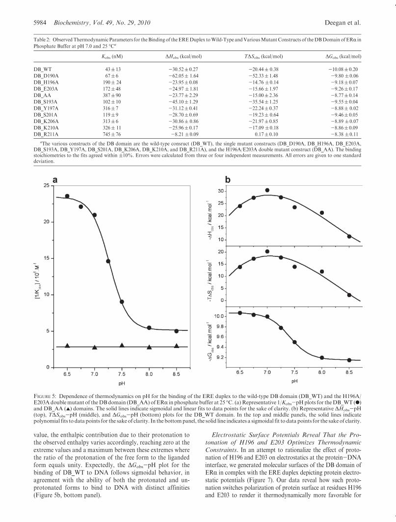
ERR to DNA. In an effort to further support the notion that theresidues H196 and E203 serve as the sole sites of protonationupon the binding of ERR to DNA, we analyzed the binding ofwild-type (DB_WT) and double mutant H196A/E203A (DB_AA) constructs of the DB domain to the ERE duplex as afunction of solution pH (Figure 5). Our data reveal that while thebinding affinity of the DB_WT construct for DNA is sharplydependent on solution pH in a sigmoidal fashion, the bindingaffinity of the DB_AA construct for DNA is independent ofsolution pH (Figure 5a). Taken collectively, our data suggeststrongly that the binding of the DB domain of ERR is coupled toproton uptake and that the side chainmoieties of H196 and E203serve as sole proton acceptors in this capacity.
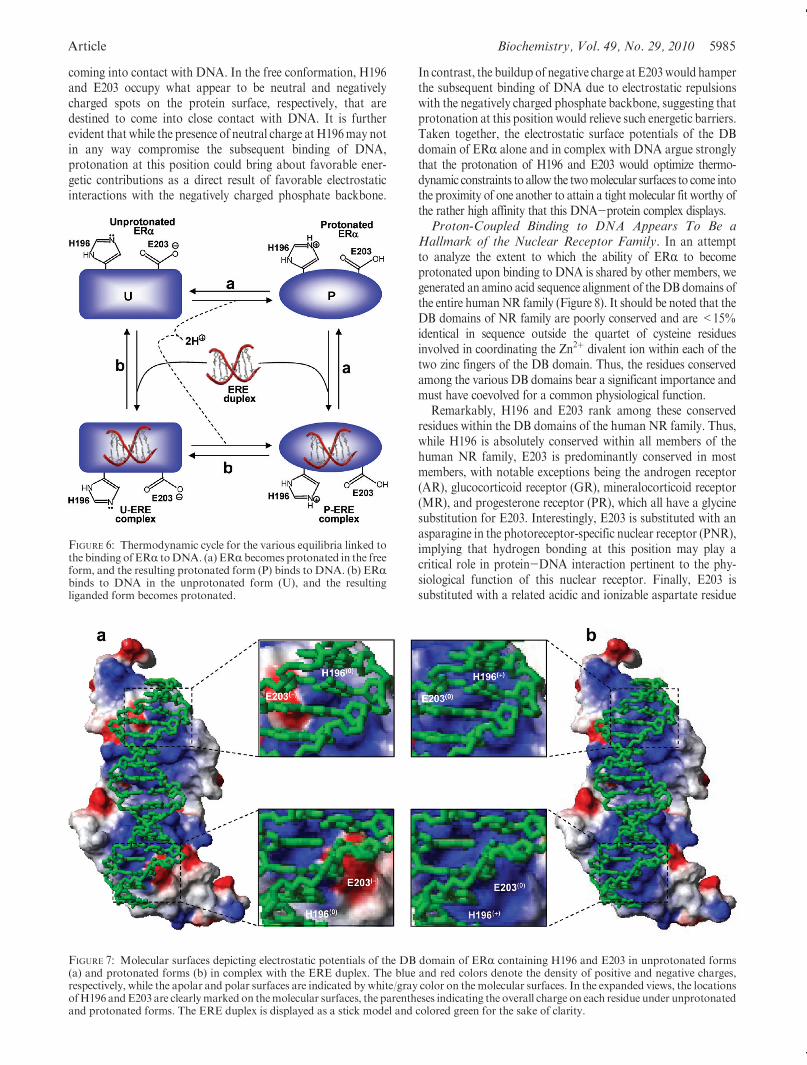
It is, however, noteworthy that the protonation of H196 andE203within ERR could precede or follow the subsequent bindingof DNA. In this manner, ERR could bind to DNA in both theprotonated and unprotonated forms. Figure 6 provides a ther-modynamic cycle for the various equilibria linked to the bindingof ERR to DNA. It is clearly evident from such a cycle that thebinding of ERR to DNA may or may not be coupled to protonuptake depending on solution pH. Thus, at low pH values, ERRmay become fully protonated prior to binding DNA. On theother hand, at high pH values, ERR may become fully unpro-tonated and the proton uptake may be decoupled for its bindingto DNA. The fact that ERR can bind to DNA both in theprotonated form and in the unprotonated form is furthersupported by the sigmoidal response of the binding affinity(Kobs) of DB_WT for DNA as a function of pH (Figure 5a).Thus, the plateau values of the 1/Kobs-pH plot at low and highpH values correspond to the intrinsic binding affinities of theprotonated and unprotonated forms of DB_WT domain forDNA, respectively.
It should also be noted that the ΔHobs-pH and TΔSobs-pHplots for the binding of theDB_WTdomain toDNAdisplay bell-shaped curves characteristic of a proton-coupled ligand bind-ing event (Figure 5b, top and middle panels). Such behaviorarises due to the fact that the enthalpy of protonation of thefree form is different from that of the liganded form. Since theratio of free and liganded forms of the protein varies as afunction of pH in going from a low pH value to a high pH
FIGURE 4: Dependence of the observed enthalpy (ΔHobs) as a func-tion of ionization enthalpy (ΔHion) of various buffers upon thebinding of the ERE duplex to the wild-type DB domain (DB_WT),the D190A single mutant of the DB domain (DB_D190A), theH196A single mutant of the DB domain (DB_H196A), and theE203A single mutant of the DB domain (DB_E203A) of ERR atpH 7.0 and 25 �C. TheΔHion values of various buffers usedwere 1.22kcal/mol for phosphate buffer, 5.02 kcal/mol for Hepes buffer, 7.64kcal/mol for Tricine buffer, and 11.35 kcal/mol for Tris buffer(33-35). The solid lines within each panel represent fits of datapoints to eq 4. Note that the net change in the number of protons(Δm) absorbed or released per DB monomer upon binding to DNAand the intrinsic binding enthalpy (ΔHint) due to direct protein-DNA interactions and protonation of ionizablemoieties for eachDBconstruct are provided within the corresponding panels. Error barswere calculated from three or four independent measurements. Allerrors are given to one standard deviation.
5984 Biochemistry, Vol. 49, No. 29, 2010 Deegan et al.
value, the enthalpic contribution due to their protonation tothe observed enthalpy varies accordingly, reaching zero at theextreme values and a maximum between these extremes wherethe ratio of the protonation of the free form to the ligandedform equals unity. Expectedly, the ΔGobs-pH plot for thebinding of DB_WT to DNA follows sigmoidal behavior, inagreement with the ability of both the protonated and un-protonated forms to bind to DNA with distinct affinities(Figure 5b, bottom panel).
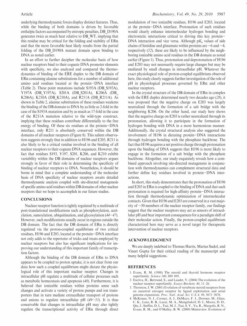
Electrostatic Surface Potentials Reveal That the Pro-tonation of H196 and E203 Optimizes ThermodynamicConstraints. In an attempt to rationalize the effect of proto-nation of H196 and E203 on electrostatics at the protein-DNAinterface, we generated molecular surfaces of the DB domain ofERR in complex with the ERE duplex depicting protein electro-static potentials (Figure 7). Our data reveal how such proto-nation switches polarization of protein surface at residues H196and E203 to render it thermodynamically more favorable for
Table 2: Observed Thermodynamic Parameters for the Binding of the EREDuplex toWild-Type andVariousMutantConstructs of theDBDomain of ERR in
Phosphate Buffer at pH 7.0 and 25 �Ca
Kobs (nM) ΔHobs (kcal/mol) TΔSobs (kcal/mol) ΔGobs (kcal/mol)
DB_WT 43( 13 -30.52( 0.27 -20.44( 0.38 -10.08( 0.20
DB_D190A 67( 6 -62.05( 1.64 -52.33( 1.48 -9.80 ( 0.06
DB_H196A 190( 24 -23.95( 0.08 -14.76 ( 0.14 -9.18( 0.07
DB_E203A 172( 48 -24.97 ( 1.81 -15.66( 1.97 -9.26( 0.17
DB_AA 387( 90 -23.77( 2.29 -15.00( 2.36 -8.77( 0.14
DB_S193A 102( 10 -45.10( 1.29 -35.54( 1.25 -9.55( 0.04
DB_Y197A 316( 7 -31.12( 0.41 -22.24( 0.37 -8.88( 0.02
DB_S201A 119( 9 -28.70( 0.69 -19.23( 0.64 -9.46( 0.05
DB_K206A 313( 6 -30.86( 0.86 -21.97( 0.85 -8.89 ( 0.07
DB_K210A 326( 11 -25.96( 0.17 -17.09 ( 0.18 -8.86( 0.09
DB_R211A 745( 76 -8.21 ( 0.09 0.17( 0.10 -8.38 ( 0.11
aThe various constructs of the DB domain are the wild-type consruct (DB_WT), the single mutant constructs (DB_D190A, DB_H196A, DB_E203A,DB_S193A, DB_Y197A, DB_S201A, DB_K206A, DB_K210A, and DB_R211A), and the H196A/E203A double mutant construct (DB_AA). The bindingstoichiometries to the fits agreed within (10%. Errors were calculated from three or four independent measurements. All errors are given to one standarddeviation.
FIGURE 5: Dependence of thermodynamics on pH for the binding of the ERE duplex to the wild-type DB domain (DB_WT) and the H196A/E203Adoublemutant of theDBdomain (DB_AA) ofERR in phosphate buffer at 25 �C. (a)Representative 1/Kobs-pHplots for theDB_WT (b)and DB_AA (2) domains. The solid lines indicate sigmoidal and linear fits to data points for the sake of clarity. (b) Representative ΔHobs-pH(top), TΔSobs-pH (middle), and ΔGobs-pH (bottom) plots for the DB_WT domain. In the top and middle panels, the solid lines indicatepolynomial fits to datapoints for the sakeof clarity. In the bottompanel, the solid line indicates a sigmoidal fit to datapoints for the sakeof clarity.
Article Biochemistry, Vol. 49, No. 29, 2010 5985
coming into contact with DNA. In the free conformation, H196and E203 occupy what appear to be neutral and negativelycharged spots on the protein surface, respectively, that aredestined to come into close contact with DNA. It is furtherevident that while the presence of neutral charge at H196may notin any way compromise the subsequent binding of DNA,protonation at this position could bring about favorable ener-getic contributions as a direct result of favorable electrostaticinteractions with the negatively charged phosphate backbone.
In contrast, the buildupof negative charge at E203would hamperthe subsequent binding of DNA due to electrostatic repulsionswith the negatively charged phosphate backbone, suggesting thatprotonation at this position would relieve such energetic barriers.Taken together, the electrostatic surface potentials of the DBdomain of ERR alone and in complex with DNA argue stronglythat the protonation of H196 and E203 would optimize thermo-dynamic constraints to allow the twomolecular surfaces to come intothe proximity of one another to attain a tight molecular fit worthy ofthe rather high affinity that this DNA-protein complex displays.Proton-Coupled Binding to DNA Appears To Be a
Hallmark of the Nuclear Receptor Family. In an attemptto analyze the extent to which the ability of ERR to becomeprotonated upon binding to DNA is shared by other members, wegenerated an amino acid sequence alignment of theDB domains ofthe entire humanNR family (Figure 8). It should be noted that theDB domains of NR family are poorly conserved and are <15%identical in sequence outside the quartet of cysteine residuesinvolved in coordinating the Zn2þ divalent ion within each of thetwo zinc fingers of the DB domain. Thus, the residues conservedamong the various DB domains bear a significant importance andmust have coevolved for a common physiological function.
Remarkably, H196 and E203 rank among these conservedresidues within the DB domains of the human NR family. Thus,while H196 is absolutely conserved within all members of thehuman NR family, E203 is predominantly conserved in mostmembers, with notable exceptions being the androgen receptor(AR), glucocorticoid receptor (GR), mineralocorticoid receptor(MR), and progesterone receptor (PR), which all have a glycinesubstitution for E203. Interestingly, E203 is substituted with anasparagine in the photoreceptor-specific nuclear receptor (PNR),implying that hydrogen bonding at this position may play acritical role in protein-DNA interaction pertinent to the phy-siological function of this nuclear receptor. Finally, E203 issubstituted with a related acidic and ionizable aspartate residue
FIGURE 6: Thermodynamic cycle for the various equilibria linked tothe binding of ERR toDNA. (a) ERR becomes protonated in the freeform, and the resulting protonated form (P) binds to DNA. (b) ERRbinds to DNA in the unprotonated form (U), and the resultingliganded form becomes protonated.
FIGURE 7: Molecular surfaces depicting electrostatic potentials of the DB domain of ERR containing H196 and E203 in unprotonated forms(a) and protonated forms (b) in complex with the ERE duplex. The blue and red colors denote the density of positive and negative charges,respectively, while the apolar and polar surfaces are indicated by white/gray color on themolecular surfaces. In the expanded views, the locationsofH196 andE203 are clearlymarked on themolecular surfaces, the parentheses indicating the overall charge on each residue under unprotonatedand protonated forms. The ERE duplex is displayed as a stick model and colored green for the sake of clarity.

5986 Biochemistry, Vol. 49, No. 29, 2010 Deegan et al.
in hepatocyte nuclear factor 4a (HNF4a), hepatocyte nuclearfactor 4g (HNF4g), and the tail-less orphan receptor (TLX),indicating that protonation at this position upon binding toDNA may also be critical for these nuclear receptors. Thus, thehighly conserved nature of H196 and E203 among the function-ally diverse members of the human NR family argues stronglythat the proton-coupled binding to DNA may have evolved as ageneral mechanism for nuclear receptor function and regulation.
It is also worth noting that residue D190 is absolutelyconserved among allmembers of the humanNR family, implying
that although it does not serve as a proton acceptor, it must alsoplay a pivotal role in protein-DNA interactions pertinent tonuclear receptors. Additionally, our thermodynamic data indi-cate that the D190A substitution has no bearable effect on thebinding affinity of theDB domain of ERR forDNA (Table 2), anobservation that is in conflict with evolutionary constraints beingplaced upon this residue in the human NR family. Furtherscrutiny of the binding of the wild-type DB domain (DB_WT)versus the D190A mutant (DB_D190A) to DNA suggests thatalthough they bind with virtually indistinguishable affinities, the
FIGURE 8: Amino acid sequence alignment of DB domains of all known members of the human NR family. Absolutely conserved residues arecolored red, while all other residues are colored black. Eachmember is denoted by its acronym in the left column, with the corresponding Expasycode provided in the right column for access to the complete proteomic details of each member. The numerals hyphenated to the amino acidsequence at each end denote the boundaries of DB domains for each member. The cysteine residues within each of the two zinc fingers of DBdomains, denoted ZF-I and ZF-II, that coordinate the Zn2þ ion in a tetrahedral arrangement are marked by asterisks. Residues D190, H196,E203, and K206, located within the DB domain of ERR, are indicated by vertical arrows.
Article Biochemistry, Vol. 49, No. 29, 2010 5987
underlying thermodynamic forces display distinct features. Thus,while the binding of both domains is driven by favorableenthalpic factors accompanied by entropic penalties, DB_D190Agenerates twice as much heat relative to DB_WT, implying thatthis residue may be critical for the folding and stability of ERRand that the more favorable heat likely results from the partialfolding of the DB_D190A mutant domain upon binding toDNA as noted earlier.
In an effort to further decipher the molecular basis of hownuclear receptors bind to their cognate DNA promoter elementswith specificity, we also analyzed and compared the thermo-dynamics of binding of the ERE duplex to the DB domain ofERR containing alanine substitutions for a number of additionalamino acid residues located at the protein-DNA interface(Table 2). These point mutations include S193A (DB_S193A),Y197A (DB_Y197A), S201A (DB_S201A), K206A (DB_K206A), K210A (DB_K210A), and R211A (DB_R211A). Asshown in Table 2, alanine substitution of these residues weakensthe binding of the DB domain toDNA by as little as 2-fold in thecase of the S193Amutation and by as much as 17-fold in the caseof the R211A mutation relative to the wild-type construct,implying that these residues contribute differentially to the freeenergy of binding. Of these six residues at the protein-DNAinterface, only R211 is absolutely conserved within the DBdomains of all nuclear receptors (Figure 8). This salient observa-tion suggests strongly that in addition toH196 and E203, R211 isalso likely to be a critical residue involved in the binding of allnuclear receptors to their cognate DNA sequences. However, thefact that residues S193, Y197, S201, K206, and K210 exhibitvariability within the DB domains of nuclear receptors arguesstrongly in favor of their role in determining the specificity ofbinding of nuclear receptors to DNA. Nonetheless, it should beborne in mind that a complete understanding of the molecularbasis of DNA specificity of nuclear receptors awaits detailedthermodynamic analysis coupled with site-directed mutagenesisof specific amino acid residueswithinDBdomains of other nuclearreceptors that we hope to accomplish in our future studies.
CONCLUSIONS
Nuclear receptor function is tightly regulated by amultitude ofpost-translational modifications such as phosphorylation, acet-ylation, sumoylation, ubiquitination, and glycosylation (44-47).However, suchmodifications usually occur in regions outside theDB domain. The fact that the DB domain of ERR is directlyregulated via the proton-coupled equilibrium of two criticalresidues, H196 and E203, located at the protein-DNA interfacenot only adds to the repertoire of tricks and treats employed bynuclear receptors but also has significant implications for im-proving our understanding of this important family of transcrip-tion factors.
Although the binding of the DB domain of ERR to DNAappears to be coupled to proton uptake, it is not clear from ourdata how such a coupled equilibrium might dictate the physio-logical role of this important nuclear receptor. Changes inintracellular pH regulate a multitude of cellular processes suchas metabolic homeostasis and apoptosis (48). Furthermore, it isbelieved that ionizable residues within proteins sense suchchanges and activate a variety of proton pumps and ion trans-porters that in turn mediate extracellular transport of protonsand anions to regulate intracellular pH (49-51). It is thusconceivable that changes in intracellular pH may also tightlyregulate the transcriptional activity of ERR through direct
modulation of two ionizable residues, H196 and E203, locatedat the protein-DNA interface. Protonation of such residueswould clearly enhance intermolecular hydrogen bonding andelectrostatic interactions critical to driving this key protein-DNA interaction and vice versa. Although pKa values of sidechains of histidine and glutamate within proteins are∼6 and∼4,respectively (52), these are likely to be influenced by the neigh-boring ionizable amino acid residues in the DB domain as notedearlier (Figure 1). Thus, protonation and deprotonation of H196and E203 may not necessarily require large changes but may bemediated by small changes in intracellular pH. Whatever theexact physiological role of proton-coupled equilibrium observedhere, this study clearly suggests further investigation of the role ofpH in physiological processes governed by ERR and othernuclear receptors.
In the crystal structure of the DB domain of ERR in complexwith the ERE duplex determined nearly two decades ago (28), itwas proposed that the negative charge on E203 was largelyneutralized through the formation of a salt bridge with theneighboring K206. On the other hand, our study here showsthat the negative charge on E203 is rather neutralized through itsprotonation, allowing it to participate in the formation ofhydrogen bonding with DNA in a more harmonious manner.Additionally, the crystal structural analysis also suggested theinvolvement of H196 in dictating protein-DNA interactionsthrough hydrogen bonding with the phosphate backbone. Thefact thatH196 acquires a net positive charge through protonationupon the binding of DNA suggests that H196 is more likely toengage in the formation of a salt bridge with the phosphatebackbone. Altogether, our study exquisitely reveals how a com-bined approach involving site-directed mutagenesis in conjunc-tion with thermodynamics can complement structural data andfurther define key residues involved in protein-DNA inter-actions.
In short, this study demonstrates that the protonation of H196and E203 in ERR is coupled to the binding ofDNAand that suchprotonation is required for high-affinity protein-DNA interac-tion through thermodynamic optimization of intermolecularcontacts.Given thatH196 andE203 are conserved in a vastmajo-rity of ∼50 members of the nuclear receptor family, our findingssuggest that the nuclear receptors may act as sensors of intracel-lular pHandbear important consequences for a paradigm shift oftheir molecular action. Finally, the proton-coupled equilibriumcharacterized here may serve as a novel target for therapeuticintervention of nuclear receptors.
ACKNOWLEDGMENT
We are deeply indebted to Thomas Harris, Marius Sudol, andVineet Gupta for their critical reading of the manuscript andmany helpful suggestions.
REFERENCES
1. Evans, R. M. (1988) The steroid and thyroid hormone receptorsuperfamily. Science 240, 889–895.
2. Escriva, H., Bertrand, S., and Laudet, V. (2004) The evolution of thenuclear receptor superfamily. Essays Biochem. 40, 11–26.
3. Thornton, J.W. (2001) Evolution of vertebrate steroid receptors froman ancestral estrogen receptor by ligand exploitation and serialgenome expansions. Proc. Natl. Acad. Sci. U.S.A. 98, 5671–5676.
4. McKenna, N. J., Cooney, A. J., DeMayo, F. J., Downes, M., Glass,C. K., Lanz, R. B., Lazar, M. A., Mangelsdorf, D. J., Moore, D. D.,Qin, J., Steffen, D. L., Tsai,M. J., Tsai, S. Y., Yu, R.,Margolis, R.N.,Evans, R. M., and O’Malley, B. W. (2009) Minireview: Evolution of

5988 Biochemistry, Vol. 49, No. 29, 2010 Deegan et al.
NURSA, the Nuclear Receptor Signaling Atlas.Mol. Endocrinol. 23,740–746.
5. McEwan, I. J. (2009)Nuclear receptors:One big family.MethodsMol.Biol. 505, 3–18.
6. Barnett, P., Tabak, H. F., and Hettema, E. H. (2000) Nuclearreceptors arose from pre-existing protein modules during evolution.Trends Biochem. Sci. 25, 227–228.
7. Kumar, R., and Thompson, E. B. (1999) The structure of the nuclearhormone receptors. Steroids 64, 310–319.
8. Egea, P. F., Klaholz, B. P., and Moras, D. (2000) Ligand-proteininteractions in nuclear receptors of hormones. FEBS Lett. 476, 62–67.
9. Claessens, F., and Gewirth, D. T. (2004) DNA recognition by nuclearreceptors. Essays Biochem. 40, 59–72.
10. Green, S., and Chambon, P. (1988) Nuclear receptors enhance ourunderstanding of transcription regulation. Trends Genet. 4, 309–314.
11. Darimont, B. D., Wagner, R. L., Apriletti, J. W., Stallcup, M. R.,Kushner, P. J., Baxter, J. D., Fletterick, R. J., and Yamamoto, K. R.(1998) Structure and specificity of nuclear receptor-coactivator inter-actions. Genes Dev. 12, 3343–3356.
12. Ham, J., and Parker, M. G. (1989) Regulation of gene expression bynuclear hormone receptors. Curr. Opin. Cell Biol. 1, 503–511.
13. Kato, S., Endoh, H., Masuhiro, Y., Kitamoto, T., Uchiyama, S.,Sasaki, H., Masushige, S., Gotoh, Y., Nishida, E., Kawashima, H.,Metzger, D., and Chambon, P. (1995) Activation of the estrogenreceptor through phosphorylation by mitogen-activated protein ki-nase. Science 270, 1491–1494.
14. Warnmark, A., Treuter, E., Wright, A. P., and Gustafsson, J. A.(2003) Activation functions 1 and 2 of nuclear receptors: Molecularstrategies for transcriptional activation. Mol. Endocrinol. 17, 1901–1909.
15. Noy, N. (2007) Ligand specificity of nuclear hormone receptors:Sifting through promiscuity. Biochemistry 46, 13461–13467.
16. Gottlieb, B., Beitel, L. K.,Wu, J., Elhaji, Y. A., and Trifiro,M. (2004)Nuclear receptors and disease: Androgen receptor. Essays Biochem.40, 121–136.
17. Gurnell, M., and Chatterjee, V. K. (2004) Nuclear receptors indisease: Thyroid receptor β, peroxisome-proliferator-activated recep-tor γ and orphan receptors. Essays Biochem. 40, 169–189.
18. Brzozowski, A.M., Pike, A. C., Dauter, Z., Hubbard, R. E., Bonn, T.,Engstrom, O., Ohman, L., Greene, G. L., Gustafsson, J. A., andCarlquist, M. (1997) Molecular basis of agonism and antagonism inthe oestrogen receptor. Nature 389, 753–758.
19. Sonoda, J., Pei, L., and Evans, R. M. (2008) Nuclear receptors:Decoding metabolic disease. FEBS Lett. 582, 2–9.
20. Jensen, E. V., and Jacobson, H. (1962) Basic guides to the mechanismof estrogen action. Recent Prog. Horm. Res. 18, 318–414.
21. Jensen, E. V. (1962) On the mechanism of estrogen action. Perspect.Biol. Med. 6, 47–59.
22. Toft, D., and Gorski, J. (1966) A receptor molecule for estrogens:Isolation from the rat uterus and preliminary characterization. Proc.Natl. Acad. Sci. U.S.A. 55, 1574–1581.
23. Toft, D., Shyamala, G., and Gorski, J. (1967) A receptor molecule forestrogens: Studies using a cell-free system. Proc. Natl. Acad. Sci. U.S.A. 57, 1740–1743.
24. Jensen, E. V., and Jordan, V. C. (2003) The estrogen receptor: Amodel for molecular medicine. Clin. Cancer Res. 9, 1980–1989.
25. Heldring, N., Pike, A., Andersson, S., Matthews, J., Cheng, G.,Hartman, J., Tujague, M., Strom, A., Treuter, E., Warner, M., andGustafsson, J. A. (2007) Estrogen receptors: How do they signal andwhat are their targets. Physiol. Rev. 87, 905–931.
26. Kumar, V., Green, S., Stack, G., Berry, M., Jin, J. R., and Chambon,P. (1987) Functional domains of the human estrogen receptor.Cell 51,941–951.
27. Klinge, C. M. (2001) Estrogen receptor interaction with estrogenresponse elements. Nucleic Acids Res. 29, 2905–2919.
28. Schwabe, J.W., Chapman,L., Finch, J. T., andRhodes,D. (1993) Thecrystal structure of the estrogen receptor DNA-binding domainbound to DNA: How receptors discriminate between their responseelements. Cell 75, 567–578.
29. Schwabe, J. W., Neuhaus, D., and Rhodes, D. (1990) Solutionstructure of the DNA-binding domain of the oestrogen receptor.Nature 348, 458–461.
30. Gasteiger, E., Hoogland, C., Gattiker, A., Duvaud, S., Wilkins,M. R., Appel, R. D., and Bairoch, A. (2005) in The Proteomics
Protocols Handbook (Walker, J. M., Ed.) pp 571-607, HumanaPress, Totowa, NJ.
31. Cantor, C. R.,Warshaw,M.M., and Shapiro, H. (1970) Oligonucleo-tide interactions. 3. Circular dichroism studies of the conformation ofdeoxyoligonucleotides. Biopolymers 9, 1059–1077.
32. Wiseman, T.,Williston, S., Brandts, J. F., and Lin, L.N. (1989) Rapidmeasurement of binding constants and heats of binding using a newtitration calorimeter. Anal. Biochem. 179, 131–137.
33. Fukada, H., and Takahashi, K. (1998) Enthalpy and heat capacitychanges for the proton dissociation of various buffer components in0.1 M potassium chloride. Proteins 33, 159–166.
34. Kozlov, A. G., and Lohman, T. M. (2000) Large contributions ofcoupled protonation equilibria to the observed enthalpy and heatcapacity changes for ssDNA binding to Escherichia coli SSB protein.Proteins 4 (Suppl.), 8–22.
35. Ortiz-Salmeron, E., Yassin, Z., Clemente-Jimenez, M. J., Las Heras-Vazquez, F. J., Rodriguez-Vico, F., Baron, C., and Garcia-Fuentes,L. (2001) Thermodynamic analysis of the binding of glutathione toglutathione S-transferase over a range of temperatures. Eur. J.Biochem. 268, 4307–4314.
36. Marti-Renom, M. A., Stuart, A. C., Fiser, A., Sanchez, R., Melo, F.,and Sali, A. (2000) Comparative Protein StructureModeling ofGenesand Genomes. Annu. Rev. Biophys. Biomol. Struct. 29, 291–325.
37. Carson, M. (1991) Ribbons 2.0. J. Appl. Crystallogr. 24, 958–961.38. Koradi, R., Billeter, M., and Wuthrich, K. (1996) MOLMOL: A
program for display and analysis of macromolecular structures.J. Mol. Graphics 14, 51–55.
39. Lumry, R., and Rajender, S. (1970) Enthalpy-entropy compensationphenomena in water solutions of proteins and small molecules: Aubiquitous property of water. Biopolymers 9, 1125–1227.
40. Eftink, M. R., Anusiem, A. C., and Biltonen, R. L. (1983) Enthalpy-entropy compensation and heat capacity changes for protein-ligandinteractions: General thermodynamicmodels and data for the bindingof nucleotides to ribonuclease A. Biochemistry 22, 3884–3896.
41. Cooper, A., Johnson, C. M., Lakey, J. H., and Nollmann, M. (2001)Heat does not come in different colours: Entropy-enthalpy compen-sation, free energy windows, quantum confinement, pressure pertur-bation calorimetry, solvation and the multiple causes of heat capacityeffects in biomolecular interactions. Biophys. Chem. 93, 215–230.
42. Sharp, K. (2001) Entropy-enthalpy compensation: Fact or artifact?Protein Sci. 10, 661–667.
43. Starikov, E.B., andNorden,B. (2007)Enthalpy-entropy compensation:A phantom or something useful? J. Phys. Chem. B 111, 14431–14435.
44. Chen, Y. X., Du, J. T., Zhou, L. X., Liu, X. H., Zhao, Y. F.,Nakanishi, H., and Li, Y. M. (2006) Alternative O-GlcNAcylation/O-phosphorylation of Ser16 induce different conformational distur-bances to the N terminus of murine estrogen receptor β. Chem. Biol.13, 937–944.
45. Faus, H., and Haendler, B. (2006) Post-translational modifications ofsteroid receptors. Biomed. Pharmacother. 60, 520–528.
46. Popov, V. M., Wang, C., Shirley, L. A., Rosenberg, A., Li, S.,Nevalainen, M., Fu, M., and Pestell, R. G. (2007) The functionalsignificance of nuclear receptor acetylation. Steroids 72, 221–230.
47. Weigel, N. L., and Moore, N. L. (2007) Steroid receptor phosphor-ylation: A key modulator of multiple receptor functions. Mol. En-docrinol. 21, 2311–2319.
48. Boron, W. F. (2004) Regulation of intracellular pH. Adv. Physiol.Educ. 28, 160–179.
49. Khaled, A. R., Moor, A. N., Li, A., Kim, K., Ferris, D. K., Muegge,K., Fisher, R. J., Fliegel, L., and Durum, S. K. (2001) Trophic factorwithdrawal: p38 mitogen-activated protein kinase activates NHE1,which induces intracellular alkalinization. Mol. Cell. Biol. 21, 7545–7557.
50. Khaled, A. R., Reynolds, D. A., Young, H. A., Thompson, C. B.,Muegge, K., and Durum, S. K. (2001) Interleukin-3 withdrawalinduces an early increase in mitochondrial membrane potential un-related to the Bcl-2 family. Roles of intracellular pH, ADP transport,and F0F1-ATPase. J. Biol. Chem. 276, 6453–6462.
51. Puceat, M., Roche, S., and Vassort, G. (1998) Src family tyrosinekinase regulates intracellular pH in cardiomyocytes. J. Cell Biol. 141,1637–1646.
52. Harris, T. K., and Turner, G. J. (2002) Structural basis of perturbedpKa values of catalytic groups in enzyme active sites. IUBMBLife 53,85–98.