Embed Size (px)
Citation preview
1864 Biophysical Journal Volume 106 May 2014 1864–1870
Dynamic Mechanical Responses of Arabidopsis Thylakoid Membranesduring PSII-Specific Illumination
Casper H. Clausen,† Matthew D. Brooks,‡k Tai-De Li,† Patricia Grob,k Gigi Kemalyan,k Eva Nogales,§{k
Krishna K. Niyogi,‡§k and Daniel A. Fletcher†§*†Bioengineering Department and ‡Department of Plant and Microbial Biology, University of California, Berkeley, California; §PhysicalBiosciences Division, Lawrence Berkeley National Laboratory, Berkeley, California; {Molecular and Cell Biology Department, University ofCalifornia, Berkeley, California; and kHoward Hughes Medical Institute, University of California, Berkeley, California
ABSTRACT Remodeling of thylakoidmembranes in response to illumination is an important process for the regulation of photo-synthesis. We investigated the thylakoid network from Arabidopsis thaliana using atomic force microscopy to capture dynamicchanges in height, elasticity, and viscosity of isolated thylakoidmembranes caused by changes in illumination.We also correlatedthemechanical response of the thylakoid network with membrane ultrastructure using electronmicroscopy.We find that the elas-ticity of the thylakoid membranes increases immediately upon PSII-specific illumination, followed by a delayed height change.Direct visualization by electron microscopy confirms that there is a significant change in the packing repeat distance of the mem-brane stacks in response to illumination. Although experiments with Gramicidin show that the change in elasticity dependsprimarily on the transmembrane pH gradient, the height change requires both the pH gradient andSTN7-kinase-dependent phos-phorylation of LHCII. Our studies indicate that lumen expansion in response to illumination is not simply a result of the influx ofwater, and we propose a dynamic model in which protein interactions within the lumen drive these changes.
INTRODUCTION
The mechanisms controlling how a plant acclimates to lightand maintains photosynthetic efficiency are of great interestfor understanding photosynthesis. The photosyntheticmachinery used to convert light to chemical energy islocated in the chloroplast, with reactions carried out bypigment-protein complexes in the thylakoid membranenetwork. Imbalances between the amount of light that isabsorbed by the photosystems and the amount that can beused for photosynthesis necessitate multiple mechanismsto dissipate excess energy (1).
Photosynthetic organisms respond to changes in lightintensity and wavelength by several processes, includingenergy-dependent quenching of chlorophyll (1–3) and statetransitions of thylakoid membranes (4). Although certainaspects of these processes have been extensively studied,many others are not yet fully understood (5). A particulararea of recent interest is how the light-harvesting complexof photosystem II (LHCII) and the thylakoid membraneare reorganized in response to light to protect the plantfrom damage, optimize electron transport, and facilitaterepair (6–8). One of the proposed mechanisms behind thesechanges of the thylakoid membrane involves phos-phorylation and movement of LHCII, which is thought tobalance light harvesting of Photosystem I (PSI) andPhotosystem II (PSII) by both molecular and ultrastructuralreorganization (6,7,9).
The effects on thylakoid membrane remodeling as a resultof illumination with PSI- and PSII-specific wavelengths
Submitted September 3, 2013, and accepted for publicationMarch 10, 2014.
*Correspondence: [email protected]
Editor: Jochen Guck.
� 2014 by the Biophysical Society
0006-3495/14/05/1864/7 $2.00
have been intensely investigated (6,7,10,11). Research hasfocused on identifying structural changes occurring inthylakoid membranes before and after illumination usingtechniques such as fluorescence microscopy, electron micro-scopy (EM), or atomic force microscopy (AFM) (6,7,12).AFM has been used in previous studies to image the compo-sition of the thylakoid membrane (10,13), the thylakoidsuperstructure (6,14,15), and changes in the protein arrange-ment (16). However, there are no reports describingisolated thylakoid mechanical properties or the dynamicsof structural remodeling occurring in response to light inreal time.
In this study, we have characterized the changes in thyla-koid membrane mechanical properties occurring during astate transition in response to PSII-specific illumination.The height and elasticity changes of isolated thylakoidnetworks were investigated in liquid using an AFM custom-ized for rheology measurements, in which the cantilever washeld in contact with the sample at a constant force to mea-sure height changes and a small sinusoidal movement ofthe cantilever was applied to capture viscoelastic propertiesof the thylakoid membrane. An inverted optical microscopewas used to control illumination wavelength and intensityand to obtain chlorophyll fluorescence images of the thyla-koid membrane, and EM provided a description of the mem-brane ultrastructure before and after illumination.
MATERIALS AND METHODS
Plant material
Arabidopsis thaliana wild-type (WT) (Col-0) and stn7 plants (17) were
grown in 7.5-cm soil pots in a chamber with a 10-h day, 14-h night cycle
http://dx.doi.org/10.1016/j.bpj.2014.03.016
A
Dynamic Mechanical Responses of Thylakoid Membranes 1865
at 21.5�C and a light intensity of 150 mmol photon m�2 s�1. Plants were
used for experiments before bolting at an age of 7–10 weeks.
B
C D
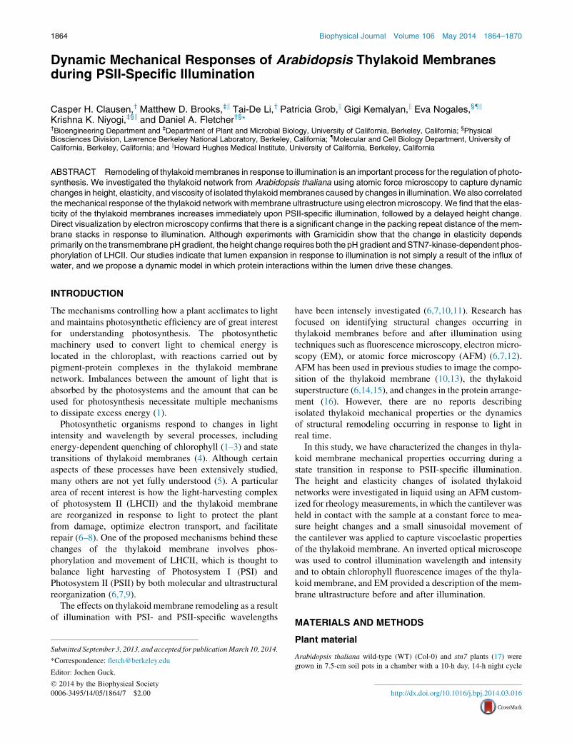
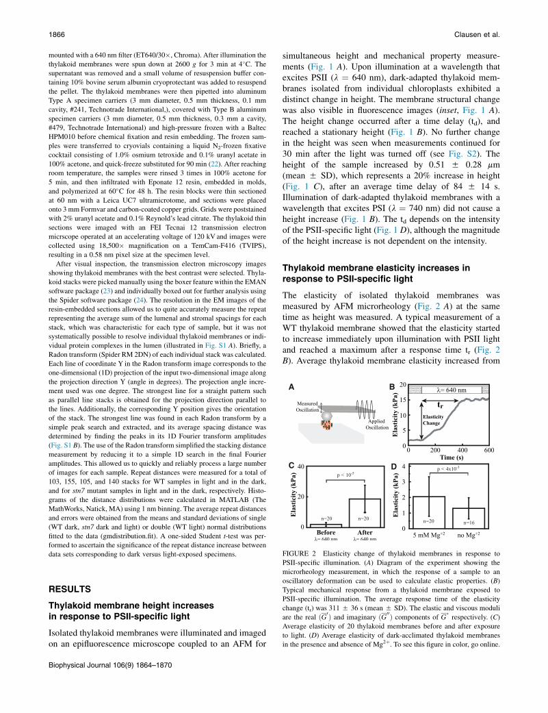
FIGURE 1 Height change of isolated thylakoid membranes in response
to PSII-specific illumination. (A) Diagram of the experiment showing
Thylakoid isolation
Thylakoid membranes were isolated according to Casazza et al. (18). The
whole preparation was carried out at 4�C and in the dark. Leaves from
Arabidopsis plants (10–20 g) as well as all the equipment used were cooled
to 4�C 1 h before the isolation process. The leaves were shredded in a
blender by eight 0.5-s pulses in 100 ml buffer, containing 0.4 M sorbitol,
5 mM EGTA, 5 mM EDTA, 5 mM MgCl2, 10 mM NaHCO3, and 20 mM
Tricine at pH 8.4. The mixture was filtered through six layers of cheese-
cloth, and at the end the cloth was gently squeezed to obtain a higher yield.
The solution was centrifuged for 3 min at 2600 � g. The supernatant was
discarded, and the pellet was resuspended in 10 mL of resuspension buffer
containing 0.3 M sorbitol, 2.5 mM EDTA, 5 mMMgCl2, 10 mM NaHCO3,
20 mM HEPES at pH 7.6. This step was repeated three times. After the last
centrifugation, the supernatant was discarded, and the pellet was resus-
pended for 5 min in 10 mL of hypotonic buffer containing 2.5 mM
EDTA, 5 mM MgCl2, 10 mM NaHCO3, 20 mM HEPES at pH 7.6. The
solution was then centrifuged for 3 min at 200 � g, and the supernatant
was collected and centrifuged for 3 min at 2600 � g. Finally, the superna-
tant was discarded, and the pellet was resuspended in measuring buffer
containing 0.3 M sorbitol, 5 mM MgCl2, 10 mM NaHCO3, 20 mM HEPES
at pH 7.6. To keep the thylakoid membranes active, the experiments were
carried out within 2 h of isolation.
isolated thylakoid membranes illuminated and imaged by an inverted
microscopy, whereas the height (h) is measured by AFM. The fluorescence
image shows reduced punctate structures after PSII-specific illumination
(640 nm), indicating that unstacking has occurred. Scale bar ¼ 5 mm. (B)
A typical height response from a thylakoid membrane sample exposed to
PSII-specific illumination. There is a time delay (td) between the light
and the height change of the thylakoid membrane. (C) Average height of
20 thylakoid membranes before and after the PSII-specific illumination.
(D) Average of the time delay at different light intensities. (1 mmol
photons ¼ 186.6 mJ for 640 nm light) To see this figure in color, go online.
AFM measurements and fluorescence imaging
Before the experiments, glass coverslips were coated with poly-L-lysine by
adding 0.1% (w/v) poly-L-lysine solution on the coverslips for 30 min and
then washed with measuring buffer four times. The suspension containing
the thylakoid membranes was added and left to settle for 10 min followed
by four washing steps with the buffer used during the AFM measurements.
An incubation period of 10 min was used for the supplementation of elec-
tron acceptors (10 mM ferredoxin and 0.6 mM NADPþ) and uncoupler
(gramicidin D) to the buffer used during the AFM measurements. Polysty-
rene beads (5 mm diameter) were attached (glued, Norland 61) to a tipless
uncoated cantilever (Veeco, customized MLCT cantilever with a nominal
spring constant of 0.01 N/m), and their spring constants were individually
determined by the thermal vibration method before each measurement (19).
The experiments were carried out at room temperature in the dark. Samples
were mounted on a Zeiss Observer Z1 microscope. The excitation wave-
lengths were 484 nm (S484/15�, Chroma), 640 nm (ET640/30�, Chroma),
and 740 nm (HQ740/40�, Chroma) for chlorophyll fluorescence imaging,
PSII excitation, and PSI excitation. The fluorescence was passed through a
long-pass filter (HQ665LP, Chroma). The intensity of the illumination,
which was 150 mmol photons m�2 s�1 for the 640 nm light and
100 mmol photons m�2 s�1 for the 740 nm light, was measured with a power
meter (Melles Griot 13PEM001). Optical images were acquired with an EM
CCD camera (Andor Ixonþ).
Height and rheology measurements were simultaneously monitored with
an AFM (Bruker Bioscope Catalyst) customized for measuring mechanical
properties. The wavelength of the AFM laser is 850 nm, which is not
absorbed by thylakoid membranes (20). The AFM was mounted on the
inverted microscope so that the sample could be imaged by epifluorescence
without obstruction by the cantilever (Fig. 1A). Tomonitor the height change
of the thylakoid membranes, the polystyrene bead was in contact with the
thylakoid membrane at a constant average force of 0.2 nN (corresponding
to ~0.14mmindentation or ~6%strain) thatwasmaintained by feedback con-
trol. To measure viscoelastic properties, we used a rheological technique in
which a sinusoidal oscillation with amplitude of 20 nm was applied to the
cantilever vertically at 2 Hz. The amplitude and phase shift of the oscillatory
cantilever deflection caused by viscoelasticity of the thylakoids was detected
with a lock-in amplifier (Signal Recovery 7270) and recorded every 10 ms.
Nearly constant force was maintained during the periodic oscillations by
setting a low gain in the force feedback control software. The time constant
of the lock-in amplifierwas 1 swith the sensitivity at 500mV.TheAFMmea-
surements were controlled and recorded with a LabView program together
with a National Instrument DAQcard (PCI-6229) and an electronic signal
filter (Krohn-Hite 3364). For the analysis of the storage and loss moduli,
calculations were based on the extended common Hertz model (21). The
elastic and viscous moduli were determined by,
G� ¼ ð1� nÞ$k$A4$d$
ffiffiffiffiffiffiffi
Rd0p $expði$fÞ; (1)
where G* is the dynamic modulus, n is the Poisson’s ratio assumed to be
0.5 (21), k is the spring constant of the cantilever, A is the measured ampli-
tude, R is the radius of the bead, d0 is the average indentation, d is given as
applied amplitude minus the measured amplitude, and f is the phase of the
measured signal. The elastic and viscous moduli are the real (G0) and
imaginary (G00) components of G*, respectively. Details of the derivation
are provided in the Supporting Material.
Electron microscopy
Thylakoid membranes were prepared as described previously and kept in
the dark before the experiments. They were illuminated for 5 min with
640 nm light at 150 mmol photons m�2 s�1 using a Schott KL 1500 LCD
Biophysical Journal 106(9) 1864–1870
A B
C D
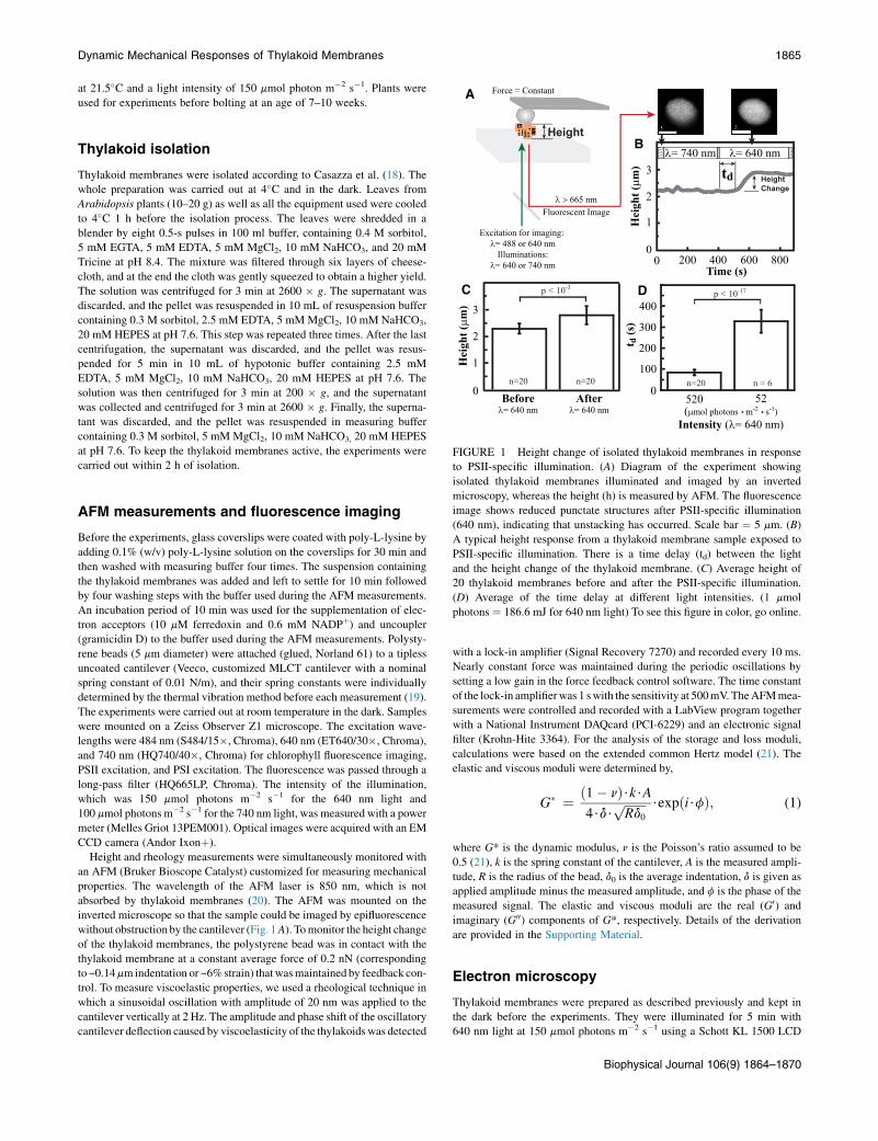
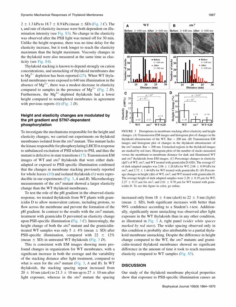
FIGURE 2 Elasticity change of thylakoid membranes in response to
1866 Clausen et al.
mounted with a 640 nm filter (ET640/30�, Chroma). After illumination the
thylakoid membranes were spun down at 2600 g for 3 min at 4�C. Thesupernatant was removed and a small volume of resuspension buffer con-
taining 10% bovine serum albumin cryoprotectant was added to resuspend
the pellet. The thylakoid membranes were then pipetted into aluminum
Type A specimen carriers (3 mm diameter, 0.5 mm thickness, 0.1 mm
cavity, #241, Technotrade International,), covered with Type B aluminum
specimen carriers (3 mm diameter, 0.5 mm thickness, 0.3 mm a cavity,
#479, Technotrade International) and high-pressure frozen with a Baltec
HPM010 before chemical fixation and resin embedding. The frozen sam-
ples were transferred to cryovials containing a liquid N2-frozen fixative
cocktail consisting of 1.0% osmium tetroxide and 0.1% uranyl acetate in
100% acetone, and quick-freeze substituted for 90 min (22). After reaching
room temperature, the samples were rinsed 3 times in 100% acetone for
5 min, and then infiltrated with Eponate 12 resin, embedded in molds,
and polymerized at 60�C for 48 h. The resin blocks were thin sectioned
at 60 nm with a Leica UC7 ultramicrotome, and sections were placed
onto 3 mm Formvar and carbon-coated copper grids. Grids were poststained
with 2% uranyl acetate and 0.1% Reynold’s lead citrate. The thylakoid thin
sections were imaged with an FEI Tecnai 12 transmission electron
micrscope operated at an accelerating voltage of 120 kV and images were
collected using 18,500� magnification on a TemCam-F416 (TVIPS),
resulting in a 0.58 nm pixel size at the specimen level.
After visual inspection, the transmission electron microscopy images
showing thylakoid membranes with the best contrast were selected. Thyla-
koid stacks were picked manually using the boxer feature within the EMAN
software package (23) and individually boxed out for further analysis using
the Spider software package (24). The resolution in the EM images of the
resin-embedded sections allowed us to quite accurately measure the repeat
representing the average sum of the lumenal and stromal spacings for each
stack, which was characteristic for each type of sample, but it was not
systematically possible to resolve individual thylakoid membranes or indi-
vidual protein complexes in the lumen (illustrated in Fig. S1 A). Briefly, a
Radon transform (Spider RM 2DN) of each individual stack was calculated.
Each line of coordinate Y in the Radon transform image corresponds to the
one-dimensional (1D) projection of the input two-dimensional image along
the projection direction Y (angle in degrees). The projection angle incre-
ment used was one degree. The strongest line for a straight pattern such
as parallel line stacks is obtained for the projection direction parallel to
the lines. Additionally, the corresponding Y position gives the orientation
of the stack. The strongest line was found in each Radon transform by a
simple peak search and extracted, and its average spacing distance was
determined by finding the peaks in its 1D Fourier transform amplitudes
(Fig. S1 B). The use of the Radon transform simplified the stacking distance
measurement by reducing it to a simple 1D search in the final Fourier
amplitudes. This allowed us to quickly and reliably process a large number
of images for each sample. Repeat distances were measured for a total of
103, 155, 105, and 140 stacks for WT samples in light and in the dark,
and for stn7 mutant samples in light and in the dark, respectively. Histo-
grams of the distance distributions were calculated in MATLAB (The
MathWorks, Natick, MA) using 1 nm binning. The average repeat distances
and errors were obtained from the means and standard deviations of single
(WT dark, stn7 dark and light) or double (WT light) normal distributions
fitted to the data (gmdistribution.fit). A one-sided Student t-test was per-
formed to ascertain the significance of the repeat distance increase between
data sets corresponding to dark versus light-exposed specimens.
PSII-specific illumination. (A) Diagram of the experiment showing themicrorheology measurement, in which the response of a sample to an
oscillatory deformation can be used to calculate elastic properties. (B)
Typical mechanical response from a thylakoid membrane exposed to
PSII-specific illumination. The average response time of the elasticity
change (tr) was 311 5 36 s (mean 5 SD). The elastic and viscous moduli
are the real ðG0Þ and imaginary ðG00Þ components of G�respectively. (C)
Average elasticity of 20 thylakoid membranes before and after exposure
to light. (D) Average elasticity of dark-acclimated thylakoid membranes
in the presence and absence of Mg2þ. To see this figure in color, go online.
RESULTS
Thylakoid membrane height increasesin response to PSII-specific light
Isolated thylakoid membranes were illuminated and imagedon an epifluorescence microscope coupled to an AFM for
Biophysical Journal 106(9) 1864–1870
simultaneous height and mechanical property measure-ments (Fig. 1 A). Upon illumination at a wavelength thatexcites PSII (l ¼ 640 nm), dark-adapted thylakoid mem-branes isolated from individual chloroplasts exhibited adistinct change in height. The membrane structural changewas also visible in fluorescence images (inset, Fig. 1 A).The height change occurred after a time delay (td), andreached a stationary height (Fig. 1 B). No further changein the height was seen when measurements continued for30 min after the light was turned off (see Fig. S2). Theheight of the sample increased by 0.51 5 0.28 mm(mean 5 SD), which represents a 20% increase in height(Fig. 1 C), after an average time delay of 84 5 14 s.Illumination of dark-adapted thylakoid membranes with awavelength that excites PSI (l ¼ 740 nm) did not cause aheight increase (Fig. 1 B). The td depends on the intensityof the PSII-specific light (Fig. 1 D), although the magnitudeof the height increase is not dependent on the intensity.
Thylakoid membrane elasticity increases inresponse to PSII-specific light
The elasticity of isolated thylakoid membranes wasmeasured by AFM microrheology (Fig. 2 A) at the sametime as height was measured. A typical measurement of aWT thylakoid membrane showed that the elasticity startedto increase immediately upon illumination with PSII lightand reached a maximum after a response time tr (Fig. 2B). Average thylakoid membrane elasticity increased from
A B
C D
Dynamic Mechanical Responses of Thylakoid Membranes 1867
25 1.3 kPa to 18.75 8.9 kPa (mean5 SD) (Fig. 2 C). Thetr and rate of elasticity increase were both dependent on illu-mination intensity (see Fig. S3). No change in the elasticitywas observed after the PSII light was turned off for 30 min.Unlike the height response, there was no time delay for theelasticity increase, but it took longer to reach the elasticitymaximum than the height maximum. Viscosity changes inthe thylakoid were also measured at the same time as elas-ticity (see Fig. S4).
Thylakoid stacking is known to depend strongly on cationconcentrations, and unstacking of thylakoid membranes dueto Mg2þ depletion has been reported (25). When WT thyla-koid membranes were exposed to 640 nm illumination in theabsence of Mg2þ, there was a modest decrease in elasticitycompared to samples in the presence of Mg2þ (Fig. 2 D).Furthermore, the Mg2þ-depleted thylakoids had a lowerheight compared to nondepleted membranes in agreementwith previous reports (6) (Fig. 2 D).
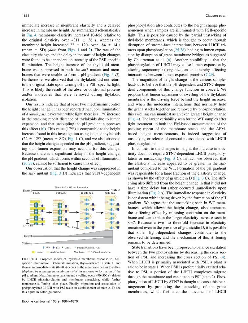
FIGURE 3 Disruptions in membrane stacking affect elasticity and height
changes. (A) Transmission EM images and histogram plot of changes in the
thylakoid ultrastructure of the WT. Bar ¼ 200 nm. (B) Transmission EM
images and histogram plot of changes in the thylakoid ultrastructure of
the stn7 mutant. Bar ¼ 200 nm. Unstacked regions in the thylakoid images
are marked by red stars. Histogram plots of the number of stacks measured
versus the membrane-to-membrane distance for dark and illuminated WT
and stn7 thylakoids from EM images. (C) Percentage changes in elasticity
(DG0) of WT, stn7, andWT treated with gramicidin D (GD). The average G0
of dark adapted samples was 2.065 1.26 kPa for WT, 2.065 0.99 kPa for
stn7, and 2.72 5 1.44 kPa for WT treated with gramicidin D. (D) Percent-
age changes in height (Dh) of WT, stn7, and WT treated with gramicidin D.
The average height of dark adapted samples were 2.285 0.19 mm for WT,
2.27 5 0.13 mm for stn7, and 2.61 5 0.70 mm for WT treated with gram-
icidin D. To see this figure in color, go online.
Height and elasticity changes are modulated bythe pH gradient and STN7-dependentphosphorylation
To investigate the mechanisms responsible for the height andelasticity changes, we carried out experiments on thylakoidmembranes isolated from the stn7mutant. This mutant lacksthe kinase responsible for phosphorylatingLHCII in responseto unbalanced excitation of PSII relative to PSI, and thus themutant is defective in state transitions (17). Transmission EMimages of WT and stn7 thylakoids that were either dark-adapted or exposed to PSII-specific illumination confirmedthat the changes in membrane stacking previously reportedfor whole leaves (10) and isolated thylakoids (6) were repro-ducible in our experiments (Fig. 3, A and B). Microrheologymeasurements of the stn7 mutant showed a larger elasticitychange than the WT thylakoid membranes.
To test the role of the pH gradient in the observed elasticresponse, we treated thylakoids from WT plants with gram-icidin D to allow monovalent cations, including protons, toflow across the membrane and prevent the formation of thepH gradient. In contrast to the results with the stn7 mutant,treatment with gramicidin D prevented an elasticity changeupon PSII-specific illumination (Fig. 3 C). Interestingly, theheight change of both the stn7 mutant and the gramicidin-treated WT samples was only 5 5 4% (mean 5 SD) afterPSII-specific illumination, compared with 22 5 12%(mean 5 SD) in untreated WT thylakoids (Fig. 3 D).
This is consistent with EM images showing more pro-found changes in organization for WT membranes, with asignificant increase in both the average and the variabilityof the stacking distance after light treatment, compared towhat is seen for the stn7 mutant (Fig. 3, A and B). In WTthylakoids, the stacking spacing repeat increased from205 10 nm (dark) to 21.35 10 nm up to 275 10 nm afterlight exposure, whereas in the stn7 mutant the spacing
increased only from 185 4 nm (dark) to 225 5 nm (light)(mean 5 SD), both significant increases with better than99% confidence according to a Student’s t-test. Addition-ally, significantly more unstacking was observed after lightexposure in the WT thylakoids than in any other condition,as illustrated in Fig. 3 A, right panel (wider white spacesmarked by red stars). The wider spacing observed only inthis condition is probably also attributable to a partial thyla-koid membrane unstacking. Despite the difference in heightchange compared to the WT, the stn7 mutants and grami-cidin-treated thylakoid membranes showed no significantdifference in the amount of time it took to reach maximumelasticity compared to WT samples (Fig. S5).
DISCUSSION
Our study of the thylakoid membrane physical propertiesshow that exposure to PSII-specific illumination causes an
Biophysical Journal 106(9) 1864–1870
1868 Clausen et al.
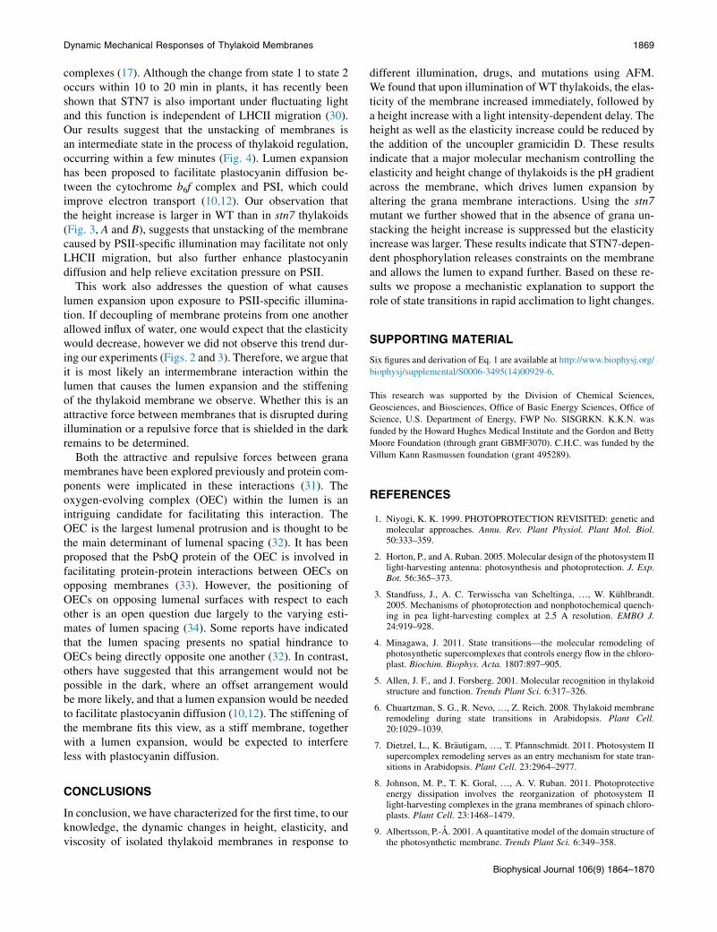
immediate increase in membrane elasticity and a delayedincrease in membrane height. As summarized schematicallyin Fig. 4, membrane elasticity increased 10-fold relative tothe original elasticity over ~311 5 36 s, whereas themembrane height increased 22 5 12% over ~84 5 14 s(mean 5 SD) (data from Figs. 1 and 2). The rate of theelasticity change and the delay in the start of height changeswere found to be dependent on intensity of the PSII-specificillumination. The height increase of the thylakoid mem-brane was suppressed in both the stn7 mutant and mem-branes that were unable to form a pH gradient (Fig. 3 D).Furthermore, we observed that the thylakoid did not returnto the original state upon turning off the PSII-specific light.This is likely the result of the absence of stromal proteinsand/or molecules that were removed during thylakoidisolation.
Our results indicate that at least two mechanisms controlthe height change. It has been reported that upon illuminationofArabidopsis leaveswithwhite light, there is a 17% increasein the stacking repeat distance of thylakoids due to lumenexpansion, and that uncoupling the pH gradient suppressesthis effect (10). This value (17%) is comparable to the heightincrease found in this investigation using isolated thylakoids(22 5 12% (mean 5 SD), Fig. 1 C), and we also observedthat the height change depended on the pH gradient, suggest-ing that lumen expansion may account for this change.Because there is a significant delay in the height change,the pH gradient, which forms within seconds of illumination(26,27), cannot be sufficient to cause this effect.
Our observation that the height change was suppressed inthe stn7 mutant (Fig. 3 D) indicates that STN7-dependent
FIGURE 4 Proposed model of thylakoid membrane response to PSII-
specific illumination. Before illumination, thylakoids are in state 1, and
then an intermediate state (0–90 s) occurs as the membrane begins to stiffen
(depicted by a change in membrane color) in response to formation of the
pH gradient. Next, lumen expansion and swelling occur (90–300 s), driven
by LHCII phosphorylation and membrane unstacking, while further
membrane stiffening takes place. Finally, migration and association of
phosphorylated LHCII with PSI result in establishment of state 2. To see
this figure in color, go online.
Biophysical Journal 106(9) 1864–1870
phosphorylation also contributes to the height change phe-nomenon when samples are illuminated with PSII-specificlight. This is possibly caused by the partial unstacking ofthylakoid membranes, which is thought to occur throughdisruption of stroma-face interactions between LHCII tri-mers upon phosphorylation (25,28) leading to lumen expan-sion by disruption of grana membrane bridges as suggestedby Chuartzman et al. (6). Another possibility is that thephosphorylation of LHCII may cause lumen expansion byaltering supercomplex organization and thereby affectinginteractions between lumen-exposed proteins (7,29).
The magnitude of height change in the various samplesleads us to believe that the pH-dependent and STN7-depen-dent components of this change function in concert. Wepropose that lumen expansion or swelling of the thylakoidmembrane is the driving force behind the height increase,and when the molecular interactions that normally holdthe grana stacks together are removed by phosphorylation,this swelling can manifest as an even greater height change(Fig. 4). The larger variability seen for the WT samples afterlight treatment, in both the EM-based measurements of thepacking repeat of the membrane stacks and the AFM-based height measurements, is indeed suggestive ofunstacking or release of constraints associated with LHCIIphosphorylation.
In contrast to the changes in height, the increase in elas-ticity does not require STN7-dependent LHCII phosphory-lation or unstacking (Fig. 3 C). In fact, we observed thatthe elasticity increase appeared to be greater in the stn7mutant compared to the WT. Formation of the pH gradientwas responsible for a large fraction of the elasticity change,as shown by the effect of gramicidin D (Fig. 3 C). The stiff-ening also differed from the height change in that it did nothave a time delay but rather occurred immediately uponillumination (Fig. 2 A). The immediate response in elasticityis consistent with it being driven by the formation of the pHgradient. We argue that the unstacking seen in WT mem-branes, which allows the height change, also attenuatesthe stiffening effect by releasing constraint on the mem-brane and can explain the larger elasticity increase seen instn7. Because a two- to threefold increase in elasticityremained even in the presence of gramicidin D, it is possiblethat other light-dependent changes contribute to theobserved stiffening, and the mechanism of this stiffeningremains to be determined.
State transitions have been proposed to balance excitationbetween the two photosystems by decreasing the cross sec-tion of PSII and increasing the cross section of PSI (4).When LHCII is primarily associated with PSII, a plant issaid to be in state 1. When PSII is preferentially excited rela-tive to PSI, a portion of the LHCII complexes migratethrough the membrane and can attach to PSI (state 2). Phos-phorylation of LHCII by STN7 is thought to cause this rear-rangement by promoting the unstacking of the granamembranes, which facilitates the movement of LHCII
Dynamic Mechanical Responses of Thylakoid Membranes 1869
complexes (17). Although the change from state 1 to state 2occurs within 10 to 20 min in plants, it has recently beenshown that STN7 is also important under fluctuating lightand this function is independent of LHCII migration (30).Our results suggest that the unstacking of membranes isan intermediate state in the process of thylakoid regulation,occurring within a few minutes (Fig. 4). Lumen expansionhas been proposed to facilitate plastocyanin diffusion be-tween the cytochrome b6f complex and PSI, which couldimprove electron transport (10,12). Our observation thatthe height increase is larger in WT than in stn7 thylakoids(Fig. 3, A and B), suggests that unstacking of the membranecaused by PSII-specific illumination may facilitate not onlyLHCII migration, but also further enhance plastocyanindiffusion and help relieve excitation pressure on PSII.
This work also addresses the question of what causeslumen expansion upon exposure to PSII-specific illumina-tion. If decoupling of membrane proteins from one anotherallowed influx of water, one would expect that the elasticitywould decrease, however we did not observe this trend dur-ing our experiments (Figs. 2 and 3). Therefore, we argue thatit is most likely an intermembrane interaction within thelumen that causes the lumen expansion and the stiffeningof the thylakoid membrane we observe. Whether this is anattractive force between membranes that is disrupted duringillumination or a repulsive force that is shielded in the darkremains to be determined.
Both the attractive and repulsive forces between granamembranes have been explored previously and protein com-ponents were implicated in these interactions (31). Theoxygen-evolving complex (OEC) within the lumen is anintriguing candidate for facilitating this interaction. TheOEC is the largest lumenal protrusion and is thought to bethe main determinant of lumenal spacing (32). It has beenproposed that the PsbQ protein of the OEC is involved infacilitating protein-protein interactions between OECs onopposing membranes (33). However, the positioning ofOECs on opposing lumenal surfaces with respect to eachother is an open question due largely to the varying esti-mates of lumen spacing (34). Some reports have indicatedthat the lumen spacing presents no spatial hindrance toOECs being directly opposite one another (32). In contrast,others have suggested that this arrangement would not bepossible in the dark, where an offset arrangement wouldbe more likely, and that a lumen expansion would be neededto facilitate plastocyanin diffusion (10,12). The stiffening ofthe membrane fits this view, as a stiff membrane, togetherwith a lumen expansion, would be expected to interfereless with plastocyanin diffusion.
CONCLUSIONS
In conclusion, we have characterized for the first time, to ourknowledge, the dynamic changes in height, elasticity, andviscosity of isolated thylakoid membranes in response to
different illumination, drugs, and mutations using AFM.We found that upon illumination of WT thylakoids, the elas-ticity of the membrane increased immediately, followed bya height increase with a light intensity-dependent delay. Theheight as well as the elasticity increase could be reduced bythe addition of the uncoupler gramicidin D. These resultsindicate that a major molecular mechanism controlling theelasticity and height change of thylakoids is the pH gradientacross the membrane, which drives lumen expansion byaltering the grana membrane interactions. Using the stn7mutant we further showed that in the absence of grana un-stacking the height increase is suppressed but the elasticityincrease was larger. These results indicate that STN7-depen-dent phosphorylation releases constraints on the membraneand allows the lumen to expand further. Based on these re-sults we propose a mechanistic explanation to support therole of state transitions in rapid acclimation to light changes.
SUPPORTING MATERIAL
Six figures and derivation of Eq. 1 are available at http://www.biophysj.org/
biophysj/supplemental/S0006-3495(14)00929-6.
This research was supported by the Division of Chemical Sciences,
Geosciences, and Biosciences, Office of Basic Energy Sciences, Office of
Science, U.S. Department of Energy, FWP No. SISGRKN. K.K.N. was
funded by the Howard Hughes Medical Institute and the Gordon and Betty
Moore Foundation (through grant GBMF3070). C.H.C. was funded by the
Villum Kann Rasmussen foundation (grant 495289).
REFERENCES
1. Niyogi, K. K. 1999. PHOTOPROTECTION REVISITED: genetic andmolecular approaches. Annu. Rev. Plant Physiol. Plant Mol. Biol.50:333–359.
2. Horton, P., and A. Ruban. 2005. Molecular design of the photosystem IIlight-harvesting antenna: photosynthesis and photoprotection. J. Exp.Bot. 56:365–373.
3. Standfuss, J., A. C. Terwisscha van Scheltinga, ., W. Kuhlbrandt.2005. Mechanisms of photoprotection and nonphotochemical quench-ing in pea light-harvesting complex at 2.5 A resolution. EMBO J.24:919–928.
4. Minagawa, J. 2011. State transitions—the molecular remodeling ofphotosynthetic supercomplexes that controls energy flow in the chloro-plast. Biochim. Biophys. Acta. 1807:897–905.
5. Allen, J. F., and J. Forsberg. 2001. Molecular recognition in thylakoidstructure and function. Trends Plant Sci. 6:317–326.
6. Chuartzman, S. G., R. Nevo, ., Z. Reich. 2008. Thylakoid membraneremodeling during state transitions in Arabidopsis. Plant Cell.20:1029–1039.
7. Dietzel, L., K. Brautigam, ., T. Pfannschmidt. 2011. Photosystem IIsupercomplex remodeling serves as an entry mechanism for state tran-sitions in Arabidopsis. Plant Cell. 23:2964–2977.
8. Johnson, M. P., T. K. Goral, ., A. V. Ruban. 2011. Photoprotectiveenergy dissipation involves the reorganization of photosystem IIlight-harvesting complexes in the grana membranes of spinach chloro-plasts. Plant Cell. 23:1468–1479.
9. Albertsson, P.-A. 2001. A quantitative model of the domain structure ofthe photosynthetic membrane. Trends Plant Sci. 6:349–358.
Biophysical Journal 106(9) 1864–1870
1870 Clausen et al.
10. Kirchhoff, H., C. Hall, ., Z. Reich. 2011. Dynamic control of proteindiffusion within the granal thylakoid lumen. Proc. Natl. Acad. Sci.USA. 108:20248–20253.
11. Rozak, P. R., R. M. Seiser, ., R. R. Wise. 2002. Rapid, reversiblealterations in spinach thylakoid appression upon changes in light inten-sity. Plant Cell Environ. 25:421–429.
12. Kirchhoff, H., S. Lenhert, ., J. Nield. 2008. Probing the organizationof photosystem II in photosynthetic membranes by atomic force micro-scopy. Biochemistry. 47:431–440.
13. Sznee, K., J. P. Dekker, ., R. N. Frese. 2011. Jumping mode atomicforce microscopy on grana membranes from spinach. J. Biol. Chem.286:39164–39171.
14. Yamada, T., H. Arakawa, ., A. Ikai. 2002. Use of AFM for imagingand measurement of the mechanical properties of light-convertibleorganelles in plants. Ultramicroscopy. 91:261–268.
15. Kaftan, D., V. Brumfeld,., Z. Reich. 2002. From chloroplasts to pho-tosystems: in situ scanning force microscopy on intact thylakoidmembranes. EMBO J. 21:6146–6153.
16. Scheuring, S., and J. N. Sturgis. 2005. Chromatic adaptation of photo-synthetic membranes. Science. 309:484–487.
17. Bellafiore, S., F. Barneche, ., J. D. Rochaix. 2005. State transitionsand light adaptation require chloroplast thylakoid protein kinaseSTN7. Nature. 433:892–895.
18. Casazza, A. P., D. Tarantino, and C. Soave. 2001. Preparation andfunctional characterization of thylakoids from Arabidopsis thaliana.Photosynth. Res. 68:175–180.
19. Stark, R. W., T. Drobek, and W. M. Heckl. 2001. Thermomechanicalnoise of a free v-shaped cantilever for atomic-force microscopy.Ultramicroscopy. 86:207–215.
20. Emerson, R., and M. L. Charlton. 1943. The dependence of the quan-tum yield of chlorella photosynthesis on wave length of light. Am. J.Bot. 30:165–178.
21. Mahaffy, R. E., S. Park, ., C. K. Shih. 2004. Quantitative analysis ofthe viscoelastic properties of thin regions of fibroblasts using atomicforce microscopy. Biophys. J. 86:1777–1793.
Biophysical Journal 106(9) 1864–1870
22. McDonald, K. L., and R. I. Webb. 2011. Freeze substitution in 3 hoursor less. J. Microsc. 243:227–233.
23. Ludtke, S. J., P. R. Baldwin, and W. Chiu. 1999. EMAN: semiauto-mated software for high-resolution single-particle reconstructions.J. Struct. Biol. 128:82–97.
24. Frank, J., M. Radermacher,., A. Leith. 1996. SPIDER andWEB: pro-cessing and visualization of images in 3D electron microscopy andrelated fields. J. Struct. Biol. 116:190–199.
25. McDonnel, A., and L. A. Staehelin. 1980. Adhesion between liposomesmediated by the chlorophyll a/b light-harvesting complex isolated fromchloroplast membranes. J. Cell Biol. 84:40–56.
26. Po, E. S. M., and J. W. Ho. 1997. Paraquat affects light-induced protontransport through chloroplast membranes in spinach. Comp. Biochem.Phys. C. 118:65–69.
27. Rumberg, B., and H. Muhle. 1976. Investigation of kinetics of protontranslocation across thylakoid membrane. Bioelectrochem. Bioenerg.3:393–403.
28. Barber, J. 1982. Influence of surface charges on thylakoid structure andfunction. Annu. Rev. Plant Physiol. 33:261–295.
29. Daum, B., D. Nicastro, ., W. Kuhlbrandt. 2010. Arrangement ofphotosystem II and ATP synthase in chloroplast membranes of spinachand pea. Plant Cell. 22:1299–1312.
30. Tikkanen, M., M. Grieco,., E. M. Aro. 2010. Thylakoid protein phos-phorylation in higher plant chloroplasts optimizes electron transferunder fluctuating light. Plant Physiol. 152:723–735.
31. Albertsson, P.-A. 1982. Interaction between the lumenal sides of thethylakoid membrane. FEBS Lett. 149:186–190.
32. Kou�ril, R., G. T. Oostergetel, and E. J. Boekema. 2011. Fine structureof granal thylakoid membrane organization using cryo electron tomog-raphy. Biochim. Biophys. Acta. 1807:368–374.
33. De Las Rivas, J., P. Heredia, and A. Roman. 2007. Oxygen-evolvingextrinsic proteins (PsbO,P,Q,R): bioinformatic and functional analysis.Biochim. Biophys. Acta. 1767:575–582.
34. Dekker, J. P., and E. J. Boekema. 2005. Supramolecular organization ofthylakoid membrane proteins in green plants. Biochim. Biophys. Acta.1706:12–39.
SUPPLEMENTARY MATERIALS
For
Dynamic mechanical responses of Arabidopsis thylakoid membranes during
PSII-specific illumination
by C. H. Clausen et al.
Figure S1
Figure S2
Figure S3
Figure S4
Figure S5
Figure S6
Derivation of Equation 1
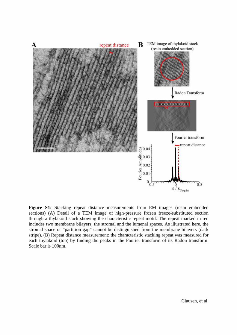
Figure S1: Stacking repeat distance measurements from EM images (resin embedded sections) (A) Detail of a TEM image of high-pressure frozen freeze-substituted section through a thylakoid stack showing the characteristic repeat motif. The repeat marked in red includes two membrane bilayers, the stromal and the lumenal spaces. As illustrated here, the stromal space or “partition gap” cannot be distinguished from the membrane bilayers (dark stripe). (B) Repeat distance measurement: the characteristic stacking repeat was measured for each thylakoid (top) by finding the peaks in the Fourier transform of its Radon transform. Scale bar is 100nm.
Clausen, et al.
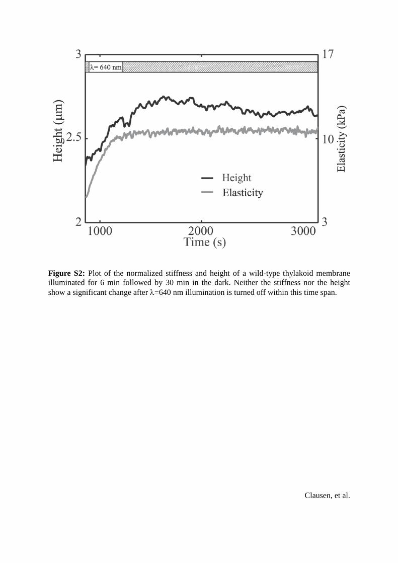
Figure S2: Plot of the normalized stiffness and height of a wild-type thylakoid membrane illuminated for 6 min followed by 30 min in the dark. Neither the stiffness nor the height show a significant change after λ=640 nm illumination is turned off within this time span.
Clausen, et al.
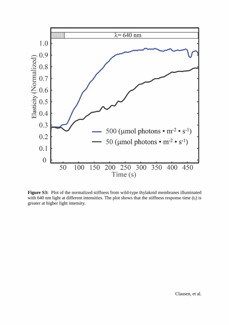
Figure S3: Plot of the normalized stiffness from wild-type thylakoid membranes illuminated with 640 nm light at different intensities. The plot shows that the stiffness response time (tr) is greater at higher light intensity.
Clausen, et al.
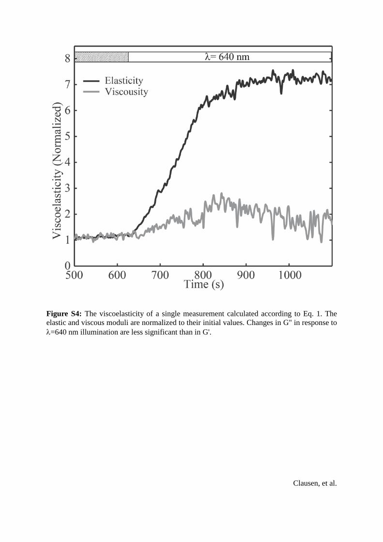
Figure S4: The viscoelasticity of a single measurement calculated according to Eq. 1. The elastic and viscous moduli are normalized to their initial values. Changes in G" in response to λ=640 nm illumination are less significant than in G'.
Clausen, et al.
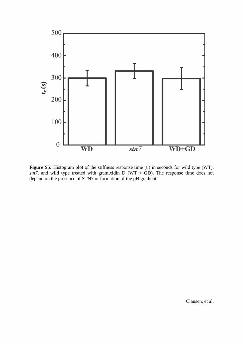
Figure S5: Histogram plot of the stiffness response time (tr) in seconds for wild type (WT), stn7, and wild type treated with gramicidin D (WT + GD). The response time does not depend on the presence of STN7 or formation of the pH gradient.
Clausen, et al.
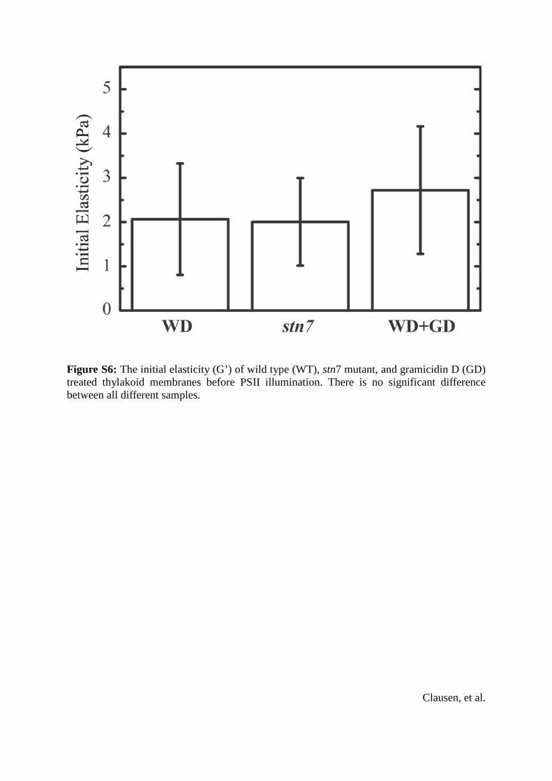
Figure S6: The initial elasticity (G’) of wild type (WT), stn7 mutant, and gramicidin D (GD) treated thylakoid membranes before PSII illumination. There is no significant difference between all different samples.
Clausen, et al.

Derivation of Equation 1:
Using Equation 5 from Mahaffy et al. 2004 𝐾𝐾1∗ = 𝑓𝑓𝑜𝑜𝑜𝑜𝑜𝑜∗
2𝛿𝛿∗(𝑅𝑅𝛿𝛿0)12
combined with the definitions 𝐾𝐾 = 𝐸𝐸
1−𝑣𝑣2 and 𝐺𝐺 = 𝐸𝐸
2(1+𝑣𝑣) , we obtain 𝐺𝐺∗ = (1−𝑣𝑣)𝑓𝑓𝑜𝑜𝑜𝑜𝑜𝑜∗
4𝛿𝛿∗(𝑅𝑅𝛿𝛿0)12 ,
where 𝑓𝑓𝑜𝑜𝑜𝑜𝑜𝑜∗ and 𝛿𝛿∗ are complex numbers describing force and displacement, respectively, that depend on angular frequency ω and phase ϕ. For negligible drag, which we confirmed for the frequencies used in our experiments, the applied force is given by 𝑓𝑓𝑜𝑜𝑜𝑜𝑜𝑜∗ = 𝑘𝑘 ∙ 𝐴𝐴 ∙ exp�𝑖𝑖(𝜔𝜔𝜔𝜔 + 𝜙𝜙)� , where k is the spring constant of the cantilever and A is the measured cantilever amplitude. The indentation is given by 𝛿𝛿∗ = δ ∙ exp(𝑖𝑖𝜔𝜔𝜔𝜔) , where δ is the applied amplitude minus the measured amplitude. Combining the equations for G*, fosc
*, and δ* , we obtain 𝐺𝐺∗ = (1−𝑣𝑣)∗𝑘𝑘∗𝐴𝐴∗exp�𝑖𝑖(𝑤𝑤𝑤𝑤+𝜙𝜙)�
4∗δ∗exp(𝑖𝑖𝑤𝑤𝑤𝑤)∗(𝑅𝑅𝛿𝛿0)12
= (1−𝑣𝑣)∙𝑘𝑘∙𝐴𝐴4∙δ∙�𝑅𝑅𝛿𝛿0
∙ exp(𝑖𝑖 ∙ 𝜙𝜙) ,
which is the form used in Equation 1 in the text.
Clausen, et al.