Embed Size (px)
Citation preview

DURAL AVF DURAL AVF ––CLASSIFICATION AND CLASSIFICATION AND
MANAGEMENTMANAGEMENT
Presented By : Presented By : RakeshRakesh K SinghK Singh

BRIEF ANATOMY OF DURA MATER
Dura mater, means "tough mother" (Latin - Toughest tissue in the body).
Embryologically develops from the mesoderm surrounding the neural tube.
Forms a tough and fibrous outer covering of CNS
Consists of two layers :- Outer or endosteal layer (Endocranium)- Inner or meningeal layer – continuous withspinal dura mater
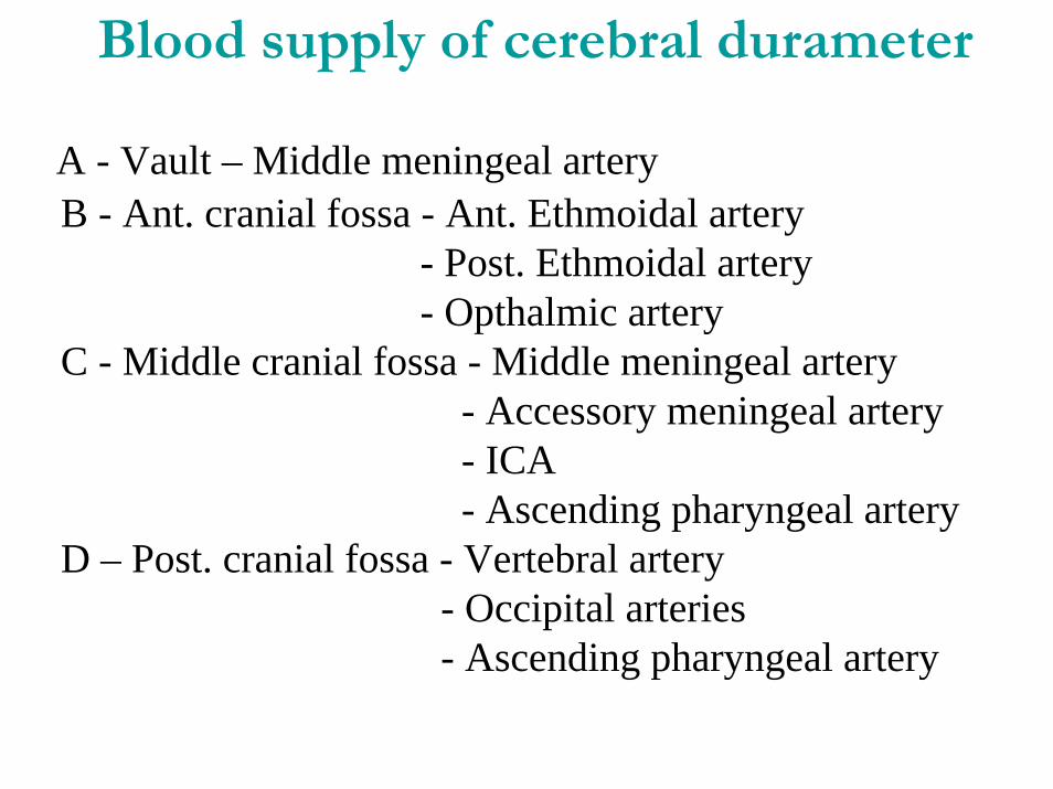
Blood supply of cerebral durameter
A - Vault – Middle meningeal arteryB - Ant. cranial fossa - Ant. Ethmoidal artery
- Post. Ethmoidal artery- Opthalmic artery
C - Middle cranial fossa - Middle meningeal artery- Accessory meningeal artery- ICA- Ascending pharyngeal artery
D – Post. cranial fossa - Vertebral artery- Occipital arteries- Ascending pharyngeal artery

DURAL AVFs
:INTRODUCTION:
Abnormal direct connection (fistula) between meningealartery and a meningeal vein or dural venous sinus within dural leaflets.
Nidus of AV shunting is contained solely within the duralleaflets, and is distinguishes DAVFs from pial AVM.
Arterial supply is usually derived from dural arteries and less frequently from osseous branches.

May drain through an adjacent dural sinus and/or other dural and leptomeningeal venous channels.
Retrograde leptomeningeal venous drainage is often tortuous and variceal, and may be frankly aneurysmal.
Broadly divided into –1 – Intracranial dural AVFs2 – Spinal dural AVFs

Figure shows a cranial DAVF as seen on a side-on or lateral view of a cerebral angiogram. The small fistulae are in the dura (red circles). The arterial supply is marked by the red arrows (branches of the external and internal carotid arteries). In this case, there is no distinct draining vein, but there is a thrombosed dural venous sinus in the vicinity of the fistulae. The blocked or thrombosed portion of the sinus is shown by dark-blue arrow heads, the normal open portion is shown by light-blue arrow heads.

Intracranial Dural AV F:EPIDEMIOLOGY: (Brown RJ et al, 1996).
Incidence= 0.17 per 100,000 persons
Comprise 10-15% of intracranial vascular malformation.
Found in 1.1% of consecutive angiograms and one fifth as common as AVMs.

Usually solitary (Multiple in < 7% cases, Malik et al,1988
May occur anywhere in the dura mater covering the brain (M.C. site - CS and T-S sinuses).
Usually located within or near the wall of a dural venous sinus, which is often narrowed or obstructed.
Most commonly involve occipital and meningeal arteries.

ETIOLOGY
May be due to : - Idiopathic – Most common- Sinus thrombosis - Head trauma : linear # across T.-sinus- Cranial surgery- Hormonal influence - Sinusitis : in CCF- Congenital (rare)
May associated with: - Meningioma- Ehlers-Danlos syndrome - Fibromuscular dysplasia- NF-1

NATURAL HISTORY
Highly variable
No or benign symptoms for many years.
May be incidental finding on angiogram.
May exhibit aggressive behaviours.
Benign DAVFs may spontaneously thrombose, occasionallyafter angiography or compression.
Acute or gradual shift of grade may occur.

Overall risk of hemorrhage – 1.6% / yr (Brown et al.)
Annual morbidity and mortality rates of with an aggressive presentation differ widely (1.8% to 20% per year).
Risk of re bleed – 35% within 2 weeks (Duffau et at.1999).

A largest prospective natural history study:(Van Dijk et al, 2002)
- Total pt.= 236- Two groups :
A – Pt. with CVR :N = 119
- Successful curative treatment =96- Lost follow up = 3
Rest 20 pt.: - Refused to treatment = 14- Partial treatment = 6
Mean follow up = 4.3 yr

RESULT :- Annual risk of ICH and NHND = 15 % (8.1+6.9)- Annual mortality rate = 10.4%
B – Pt. without CVR :N =117
- Lost follow up = 5- Rest 112 pts. median follow up = 27.9 months- Only observation = 68- Palliative treatment =44
(Endovascular = 43,Surgery=1)RESULT:
- Pts. with benign and well tolerated clinical course without any ICH or NHND =98%
- Long term angiographic follow up (N=50) shows 2-3%of risk of developing CVR

Pathophysiology
Mostly acquired, idiopathic lesion.
Varies in complexity from a single shunt in small region of the dura to an extensive malformation.
Two theories for origin :
1- Opening of preexisting microscopic vascular channels within duramater after venous hypertension secondary to sinus thrombosis.

2 - Formation of new vascular channels within duramater, a process stimulated and regulated by angiogenicfactors.(Pathology specimen contain- Angiogenic growth factor and basic fibroblast growth factor)
With aggressive symptoms, lesions seem to grow by recruitment of arterial feeders into nidus, favored by the AV shunting (Sump effect . Awad et al,1990).
Hypertrophy of dural arteries and reappearance of involutedembryonic arteries occur.

Formed as a consequence of thrombosis and subsequent recanalization of dural venous sinuses.
Sinus thrombosis seems to be related to either initiating or healing phases of the disease progression.
Postmenopausal and pregnant females more commonly experience CS and TS DAVF - thus possibility of a hormonal influence can not be ruled out.

Progressive arterialization of the pathologic dural leaflet results in hypertension in adjacent leptomeningeal channels.
Thus retrograde leptomeningeal venous drainage may occur.
Under arterialized pressure, these venous channels may become tortuous and eventually, varicose or aneurysmal.
Demyelination may occur around leptomeningeal veins due to venous hypertension.

LOCATION
Common locations of dural AVFs:
1 = Cavernous sinus (CS) (20%-40% of cases)2 = Transverse-sigmoid sinus (TS, SS) (20%-60%)3 = Tentorium (12%-14%), 4 = Superior sagittal sinus (SSS) (8%), and 5 = Anterior fossa (2%-3%).
CCF and T-S D-AVFs more common in female.
Ant. Cranial fossa and Tentorial D-AVFs more in males.
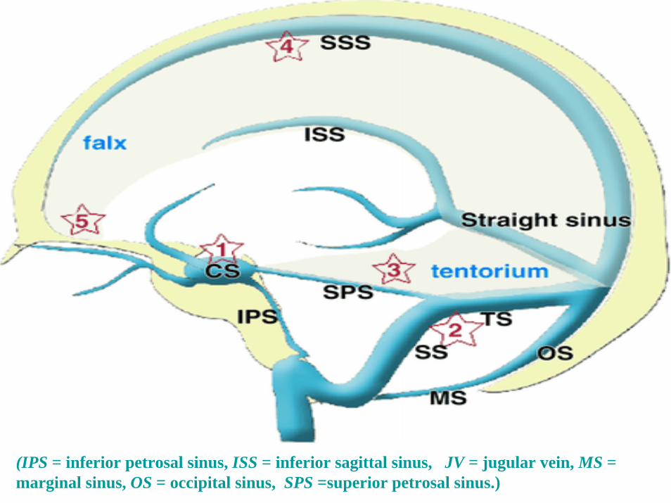
(IPS = inferior petrosal sinus, ISS = inferior sagittal sinus, JV = jugular vein, MS = marginal sinus, OS = occipital sinus, SPS =superior petrosal sinus.)

(IPS = inferior petrosal sinus, ISS = inferior sagittal sinus, JV = jugular vein, MS = marginal sinus, OS = occipital sinus, SPS =superior petrosal sinus.)

Classification
Most classification systems are based on :
1- Presence of venous stasis or occlusion2- Direction of flow3- Presence of leptomeningeal venous drainage,
aneurysm and single or multiple fistulas.


CLINICAL FEATURES
More common in adults (Age= 40-60 yr).
May be asymptomatic or benign symptoms to fatal hemorrhage.
Two groups of symptoms –A- Non aggressive : -Tinnitus (pulsatile)- M.C.
- HeadacheB- Aggressive : -Intracranial hemorrhage
-Non hemorrhagic neurological deficits

Symptoms of cranial dural AVF

Tinnitus :- Pulsatile- With high flow fistula in TS or SS location- Due to recruitment of arterial feeders and closeproximity to middle ear
- Bruit can be auscultated- Compression of carotid artery, jugular vein or occipitalartery may decrease the bruit intensity.
Headache :- U/L or generalized- Increase on physical activity or position change- Due to .. Engorgement of venous collaterals and duralsinus distention, edema, compression of 5th nerve or inflammation d/t venous thrombosis.

Hemorrhage :- May be - Intra ventricular
- Intraparenchymal- Subarachnoid- Subdural
- May be distant from the fistula site.

Most predictive predisposing factors for aggressive symptom are :
1 - Venous drainage pattern- Cortical venous drainage- Venous ecstasies- Galenic venous drainage
2- Location- Tentorial incisura (I.=8.4%) - Ant. Cranial fossa
At the transverse-sigmoid sinuses and cavernous sinuses - least likely associated with aggressive symptoms.

Any change in tinnitus or bruit warrants re-evaluation.

INVESTIGATION:
1- CT : - N- Hemorrhage- Hcp
2- MRI and MRA : - N- Dilated pial vessels- White matter edema- Stenosis or occlusion of dural sinuses

3- Intra arterial catheter angiography- Gold standard- Premature visualization of intracranial veins or
venous sinuses during arterial phase- Characteristic- Best views of nidus are often obtained during the
ultra early arterial phase and by injections of distant arterial feeders.
- Exact site of CVR must be determined

MANAGEMENT OPTIONS
1 - Observation2 – Transarterial Embolisation3 – Transvenous Embolisation4 – Surgery:5 - Stereo tactic Radio surgery

OBSERVATION:
- In lesions without CVR- Asymptomatic or tolerable symptoms- Follow up with serial MRI, MRA and DSA after 3 yr- 2-3% chance of developing CVR- Any change in symptoms needs evaluation.

TRANSARTERIAL EMBOLIZATION :- Rarely curative- Best agent : n-BCA- Commonly used as an adjuvant before surgery or TVE
- Palliative treatment for benign lesions with intolerable symptoms
Complications: - Stroke- conversion of benign fistula into aggressive one

TRANSVENOUS EMBOLIZATION :
- Through transfemoral route- Sacrifice the involve dural venous sinus- Only done when venous phase of DSA shows that the
involved sinus is not used by brain and alternatepathway for venous drainage is developed
Complications :- Venous hypertension and infarction- SAH

SURGERY:Indication –
- When endovascular therapy fails- When endovascular therapy not feasible
- 3 strategies :1- Obtain venous access for direct packing2- Complete excision of AVF3- Disconnection of CVR alone
Complications:1- Technically difficult2- Risk of exsanguination2- Risk of venous infarction


Management Strategy for D-AVF without CVR

Management Strategy for D-AVF with CVR

Management Strategy for D-AVF with CVR and sinus drainage


STEREOTACTIC RADIOSURGERY
Recent studies have demonstrated promising results in selected pts.
Pan and colleagues (2002) demonstrated complete angiographic resolution of the nidus in 47% of 19 T-S sinusDAVFs (Median follow up=19 month).
O,leary S et al (2002) in 17 pts of various sites lesion, demonstrated complete obliteration in 10 pts. - 2 pts showed considerable reduction in size.- 1 pts showed detoriation.- In 4 pts, no angiographic follow up available
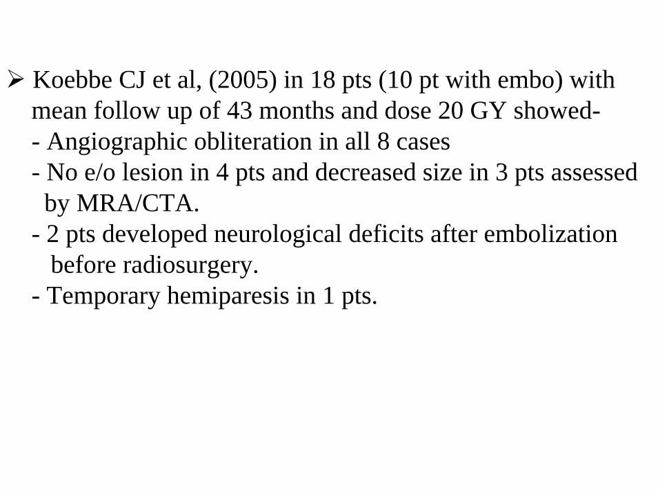
Koebbe CJ et al, (2005) in 18 pts (10 pt with embo) with mean follow up of 43 months and dose 20 GY showed-- Angiographic obliteration in all 8 cases- No e/o lesion in 4 pts and decreased size in 3 pts assessed by MRA/CTA.
- 2 pts developed neurological deficits after embolizationbefore radiosurgery.
- Temporary hemiparesis in 1 pts.
LIMITATIONS:
1- Long interval b/w treatment and therapeutic effect- Not acceptable in pts with CVR
2- Without CVR, most pts can be managed with observation alone or palliative transarterial embolization.

CAVERNOUS SINUS DURAL AVFs
Cavernous Sinus is the 2nd m. c. involved region.
First recorded treatment by Travers in 1809.(Ligation of CCA)
Dandy (1935) performed a craniotomy and Clipped the ICA just proximal to p-com artery.
Nunnelly (1865) and Rivington (1874) described the pathology and site of lesion.
Parkinson (1965) opened the CS and directly occlude the fistula.
Serbinenko (1974) developed and used a detachable balloon for vascular occlusion.
Guglielmi (1991) introduced soft detachable and retrieval platinum coils.

Relation of structures in cavernous sinus
A-A – Lateral view

Classification of CCFClassification of CCF
Direct CCF (High-flow)Most common (70-90%) 75% traumatic (assoc. basal skull fx)Defect in intra-cavernous ICASpontaneous: mid-age, HTN, post-menopausal female
Indirect CCF (Low-flow)Dural Shunt: Meningeal arteries (ICA, ECA or combo)Can be spontaneous or traumatic alsoMore insidious onset of symptoms

BarrowBarrow’’s Classification (1985)s Classification (1985)
Type A: Direct between ICA and CS
Type B: Dural ICA branches to CS (uncommon)
Type C: Dural ECA branches to CS
Type D: Dural ICA & ECA branches to CS

Angiographic Pattern Classification (Dae Chul Suh et al. : 2005)
- Categorized by the degrees and patterns of prominent arteriovenous shunt as well as venous flow.
Type – 1: Proliferative type (40%) Type – 2: Restrictive type (40%)Type – 3: Late restrictive type (20%)

Principle ECA feeders are :1- Int. maxillary artery – distal branches2- MMA – meningeal branch3- Accessory meningeal artery – meningeal branch4- Ascending pharyngeal artery- meningeal branch
ICA feeders – Dural branches from cavernous segment-C5 branches and the inferolateral trunk.

Carotid Cavernous Sinus FistulaCarotid Cavernous Sinus Fistula• Symptoms:
– Double vision– eyelid droop– facial pain/numbness
• Signs:– Proptosis– chemosis– ↑ IOP– ocular pulse pressure– orbital/temple bruit– ptosis– miosis– ophthalmoplegia (most commonly
CN VI)– facial hypoesthesia– optic disk swelling– retinal venous dilatation– intraretinal hemorrhage

Classic Sign:Limbal injection with arterialized conjunctival & episcleral vessels

Symptoms and signs are usually benign because CS has sufficient venous drainage routes.
Because the symptoms are very diverse and they fluctuate, analysis of the symptomatology related to the angiographic findings does not always correspond to the disease status.
Symptoms depend on the direction of venous drainage.

Severity depends on the rate of blood flow through shunt.
C/L eye symptoms in 11% of patients.
More common in -Postmenopausal elderly women- Pregnancy- Sphenoid sinusitis- Trauma
Over all mortality = 1-2%

PathophysiologyRetrograde venous drainage into orbit :–Venous HTN
Enlarged EOM’sRestriction DiplopiaProptosis Exposure keratopathy
Chemosis & Injection Red EyeIncreased episcleral & vortex venous pressure
Increased IOP Secondary Glaucoma
–Venous & Arterial StasisDecreased ocular/retinal perfusion
Decreased visual acuityAnterior Segment Ischemia
Decreased perfusion to intra CS cranial nervesOphthalmoplegias Diplopia

Pathophysiology
Venous HTN in other directions may also occur -
– Retrograde cortical venous drainage • 10-55% of CCF cases
Severe HA
Contralateral neurological deficits
30-40% risk of intracerebral hemorrhageMay be fatal

Diagnostic StudiesDiagnostic Studies
Orbital color Doppler U/S:- Reversed, arterialized flow
in S. Ophthalmic v.
CT/MRI:- Enlarged S. Ophthalmic v.- Enlarged EOM - Proptosis
Cerebral Angiography-“Gold Standard” diagnosis- View ICA, ECA, &
vertebral circulations

Schematic presentation of the types and progression of CSDAVF.
Schematic presentation of the types and progression of CSDAVF.

Treatment Options for Cavernous Sinus Dural AVFs(Listed in increasing order of potential risk and technical difficulty)

ManagementManagementIndications for Tx:
- Lack of spontaneous closure
- Risk to eye/vision,
- Intolerable symptoms
-“High-risk” for stroke,
-Venous thrombosis,
-Mental status changes

Interventional Radiology :(balloon occlusion/embolization)
- Primary treatment modality.- Trans-arterial route directly through tear or
embolization of feeding vessels.- Trans-venous through sup. ophthal. v. or inferior
petrosal sinus.
Surgical closure- Rare in last 30 years- Can be salvage option in : In failed embolization, contraindication of embo, in occluded ICA with patent fistula d/t previous intervention.

Endovascular ManagementEndovascular Management•Meyers, et al. Am J Ophthalmology, 2002
–Retrospective interventional case series•121 (90%) patients were cured clinically (mean f/u 56 mos)
•4% patients with moderate/severe disability
•6% with symptomatic complications
•133/135 consecutive cases had tx–Cerebral infarction, Decreased VA (2), Diabetes Insipidus, orbital eccymosis, retroperitoneal hematoma, DVT’s (2)
•No operative mortality
Conclusions:•High success rate
•Low complication/morbidity rate
•Patient’s ocular symptoms may be transiently worsened post procedure

First proposed by Hanneken, et al. in 1989.
–Direct access to cavernous sinus
Potential complications: puncture of S. ophthal v., orbital hemorrhage, infection, trochlea or other structure damage
Conclusions
•Especially effective with significant ICA contribution to CCF
•“technically straightforward, safe, and effective treatment”
Superior Ophthalmic Vein ApproachSuperior Ophthalmic Vein Approach

RADIORURGERY :- May be effective in treating some indirect CCF.
(Type –B,C or D)Dose : 20 GyLIMITATIONS are –- Takes a mean of 7.5 month to show effect on lesion.- Inappropriate for pts. with progressive visual loss,neurological deficit, or cortical venous drainage.
- 15% recurrence rate (Pollock et al.)- Risk of radiation induced malignancies.
Thus precise role remains to be determined.

Treatment of Choice in CCF
Type – A : TA detachable balloon (Silicon balloons filled with HEMA)
Type – B : TVE
Type – C : TAE
Type – D : TAE, if fail then – TVE or Surgery

PrognosisPrognosisDirect CCF:
-Poor visual prognosis (90% with severe vision loss) . -Ocular & optic nerve damage,
-Exposure keratopathy, secondary glaucoma, -Ant segment ischemia, -CRVO-Ischemic ON-Concern for intra-cerebral hemorrhage
Indirect CCF:-Prognosis less severe-Also concern for intra-cerebral hemorrhage-Exacerbation & remission is the hallmark-May close spontaneously (10-60%)

Transverse-Sigmoid Sinus Dural AVFsMost common location of IC dural AVFs.
Most common symptoms are benign (pulsatile tinnitus and headache).
More frequently associated with hemorrhagic and non hemorrhagic aggressive neurologic symptoms than CS duralAVFs.
Spontaneous regression is relatively rare (approx.5%) and usually occurs following hemorrhagic events.

Epicenter of fistula most often found at junction between the transverse and sigmoid sinus.
Arterial feeders :1- Occipital artery branches2- MMA – posterior and petrosal branches3- ICA- marginal tentorial branch4- Post. auricular artery5- Vertebral artery – post. meningeal branch6- A. pharyngeal artery – meningeal branch

Require treatment because of :
symptomatic events. - Relatively high rate of aggressive symptoms.
- Low rate of spontaneous regression without
Recent studies of stereo tactic radiation therapy for transverse-sigmoid sinus dural AVFs showed a relatively high occlusion rate of the AVF (approximately 60% of cases) several months after treatment without significant complications.

Although TVE showed higher occlusion rates (80%–100% of cases), this procedure requires sacrifice of sinus flow and may cause venous infarction if the sinus contributes to the drainage of normal cerebral tissue.
The rate of permanent complications in TVE is approximately 4%.

In the treatment of Grade 2 lesions, occlusion of the normal cortical venous drainage system should be avoided.
When there is a high risk of normal cortical venous drainage sacrifice at TVE, other treatments such as radiation therapy should be applied.
Surgical isolation of the sinus with preservation of normal cortical venous drainage may also be performed but is more invasive.

Grade 3 lesions can be treated with TVE, during which time the affected sinus and retrograde cortical drainage outlet should be tightly packed with coils.
Loose packing might cause recanalization, resulting in delayed hemorrhagic infarction after embolization.

Grade 4 lesions are the most difficult type of dural AVF to treat.
The standard techniques combine endovascular and neurosurgical elements (eg, TVE combined with a surgical approach).
In patients in poor general condition, other techniques (eg, TVE combined with other approaches, TAE with n-butyl-2-cyanoacrylate) may be used; however, they require more skill.

Recanalizationof a grade 3 transverse-sigmoid sinus dural AVF after TVE. (a) Early arterial phase left common carotid angiogram shows a Grade 3 transverse-sigmoid sinus dural AVF. outlet (arrows).

Recanalization of a grade 3 transverse-sigmoid sinus dural AVF after TVE.(b) Late arterial phase left common carotid angiogram shows that the left sigmoid sinus is occluded (arrow) and the duralAVF drains mainly into cortical veins and the posterior condylar vein (arrowheads).

Recanalizationof a grade 3 transverse-sigmoid sinus dural AVF after TVE.
(c) Superselectivevenogram shows a microcatheterthat has been advanced via the posterior condylarvein (arrowheads) into the affected sinus.

Recanalization of a grade 3 transverse-sigmoid sinus dural AVF after TVE.
d) Left common carotid angiogram obtained after TVE shows disappearance of the AVF.

Recanalizationof a grade 3 transverse-sigmoid sinus dural AVF after TVE.(e) CT scan obtained 2 months after TVE shows a massive hemorrhage in the left temporal lobe.

Recanalization of a grade 3 transverse-sigmoid sinus dural AVF after TVE.
f) Left common carotid angiogram shows recanalization of the dural AVF at the retrograde cortical drainage outlet (arrows).

Treatment options are listed in increasing order of potential risk and technical difficulty. (Numbers in parentheses indicate percentages of cases)
Treatment Options for Transverse-Sigmoid Sinus Dural AVFs

Recommended Treatment Strategies for Transverse-Sigmoid Sinus Dural AVFs


Tentorial Dural AVFsIncidence of intracranial hemorrhage _ 60% to 74%.
May cause fatal bleeding in the posterior fossa.
Tentorial dural AVFs drain through the retrograde leptomeningeal-cortical venous drainage system only (Cognard types III and IV, Borden type III), resulting in a high risk of hemorrhagic or nonhemorrhagic aggressive symptoms (19% and 10% of cases per year, respectively).
Complete cure requires aggressive treatment.
Interventional and surgical procedures are both used to disconnect the venous drainage system.
Because of the deep-seated location of such lesions, the difficult access route, and the need for n-butyl- 2-cyanoacrylate, these techniques require a high level of skill.

Treatment selection depends on : - Skill of the neurosurgeon and interventional radiologist
- On lesion accessibility
Stereotactic radiosurgery should be considered an option, especially in older patients or in those in poor general condition.

Treatment Options for Tentorial Dural AVFs

Type IV tentorial duralAVF with intracranial hemorrhage.(a)UnenhancedCT scan shows intracranial hemorrhage in the left occipital lobe and the lateral ventricle.

Type IV tentorial duralAVF with intracranial hemorrhage.
(b) Left external carotid angiogram shows a tentorial duralAVF (arrowheads) with leptomeningeal-cortical venous drainage and venous ectasia(arrow).

Type IV tentorialdural AVF with intracranial hemorrhage
(c) Digital subtraction angiogram obtained during the injection of diluted n-butyl-2 cyanoacrylatedemonstrates the tip of a microcatheter(arrow).

Type IV tentorialdural AVF with intracranial hemorrhage
(d) Left common carotid angiogram obtained after TAE shows complete obliteration of the AVF.

Arterial Supply of the Spinal Cord
By the vertebral arteries.
Also from the branches ultimately from the thoracic and abdominal aorta ( Radicular arteries).
Each vertebral artery (or PICA) gives rise to a posterior spinal artery, which proceeds along the line of attachment of the dorsal roots.
Each vertebral artery also gives rise to an anterior spinal artery.
SDAVFs
The two ant. spinal arteries fuse to form a single midline vessel along the anterior median fissure.
The posterior spinal arteries and the anterior spinal artery supply upper cervical levels with from the vertebral arteries.
Below this, all 3 spinal arteries form a more or less continuous series of anastomoses with radicular arteries.

Great radicular artery (of Adamkiewicz), present at the spinal cord level T12-L2 may provide the entire arterial supply for the caudal 2/3 of the spinal cord.
Very long anterior spinal artery is usually a continuous vessel for the length of the spinal cord.
It gives rise to hundreds of central and circumferential branches.
These supply the anterior 2/3 of the spinal cord.( posterior horn and a variable portion of the lateral corticospinal tract )

The posterior spinal arteries are really more of a plexiformnetwork of small arteries.
They supply the posterior columns, substantia gelatinosa, dorsal root entry zone, and a variable portion of the lateral corticospinal tract.
Venous Drainage of the Spinal Cord
By 6 irregular, plexiform channels.
There is one along: - The anterior and posterior midlines; - Along the line of attachment of the dorsal roots of
each side; - Along the line of attachment of the ventral roots of each side.
These are drained by the radicular veins.
Each, in turn empty into the epidural venous plexus.

Spinal Dural AVFsINTRODUCTION :
Rare and enigmatic disease entityAlso known as
- Long dorsal AVM- Angioma racemosum venosum (Wyburn-Mason)- Malformation retromedullaire (Djindjian)- Dorsal extra medullary AVMs
Clinical features and structural changes have been recognized since 1926 (Foix and Alajouanine), and the pathophysiology and the essentials of treatment since 1974.
Etiology unknown

Probably the first successful operation on an SDAVF was performed by Elsberg in 1916.
He ligated and excised an enlarged and thickened vein at the level of T8 in a patient with a sensory level at T9.
The patient made a full recovery
Kendall and Logue (1977) showed that the site of the fistula was not located in the spinal cord but on or in the dural root sleeve.
Caudal end of the spinal -first affected by congestive edema and ultimately infarction, regardless of the level of the fistula.
Thus initial clinical features often consist of sensory and motor symptoms ascending from the feet, suggesting a polyneuropathy or polyradiculopathy
EpidemiologySDAVFs are rare, but still make up the most common vascular anomaly of the spine, with a proportion of 60–80%.
5–10/million/year in the general population
Underdiagnosed disease.
Mostly in middle aged men.

Mean age at the time of diagnosis is 55–60 years.
Patients under the age of 30 are rarely reported
M:F = 5:1
M. C. location - Thoracolumbar region
Cervical and sacral SDAVF constitute just under 6% of patients with SDAVF.
Multiple SDAVFs are uncommon (0.5-4%).

Classification of spinal vascular lesions (Spetzler et al,2002 )
Neoplastic vascular lesions- Haemangioblastoma- Cavernous malformations
AneurysmsArteriovenous lesions
- Arteriovenous malformations- Arteriovenous fistulas
: Extradural: Intradural
Ventral - A: Small, B: Medium, C: LargeDorsal - A: Single arterial feeder
B: Multiple arterial feeder


Spinal Dural AVF, Extradural type
Type 1a: Single Feeders Type 1b: Multiple feeders

Causal factors and pathophysiology
An acquired condition – 0nset in middle age.
Venous hypertension - the main pathophysiological factor.
The shunt is most often formed within the dorsal surface of the dural root sleeve in the intervertebral foramen, where the radicular vein pierces the dura, together with one or more dural branches of the radicular artery.
Shunt is sometimes situated along the dura between two adjacent nerve roots.
Increased pressure causes the venous system to ‘arterialize’.
The radicular feeding artery is often a dural branch and in a minority, the medullary artery.
The shunt (low flow) results in venous hypertension in the spinal cord, because the intramedullary veins and the radicular vein share a common venous outflow.

Reduced arteriovenous pressure gradient:
- Decrease in tissue perfusion and venous infarction.
Apart from the increased pressure caused by the shunt, the venous outflow may be less efficient to start with than is the case in healthy individuals.
Lower thoracic region has relatively fewer venous outflow channels at a segmental level than the cervical or lumbosacral region.

These differences in segmental outflow probably contribute to the phenomenon that venous congestion is transmitted in a caudo-cranial direction throughout the spinal cord.
Thus, first symptoms of myelopathy tend to reflect dysfunction of the lowest part of the cord, (conusmedullaris), even though the shunt is at the higher level, or in some cases even near the skull base.
Venous outflow through the medullary vein and venous plexus is dorsal from the cord in 80–90%, and combined ventral and dorsal in 10–20%.

Table 3 Proportion of patients with symptoms present at the onset of thedisease and symptoms present at the time of diagnosis
Initial symptoms (%)
Symptoms at diagnosis (%)
Sensory disturbances 17–72 63–100
Gait difficulties and motor disturbances
50–81 78–100
Pain (either pain in the back or radicular pain)
13–64 17–86
Micturition difficulties 4–75 62–91
Defaecation problems 0–38 30–100
Sexual dysfunction 0–17 11–80
Clinical diagnosis
SDAVF is notoriously hard to diagnose, because of the misleading nature of the initial symptoms and the rarity of the disease.
The median time between onset and diagnosis ranges between 12 and 44 months.

Erroneous diagnoses often made initially are -- Sensory polyneuropathy, - Acute or chronic inflammatory demyelinating polyneuropathy
- Spinal muscular atrophy - Medullary tumour
Not infrequently patients are unsuccessfully operated for a lumbar disc prolapse.

At the time of diagnosis, two-thirds of patients show a combination of gait difficulties, sensory disturbances andinvolvement of sacral segments (micturition, defaecation orsexual dysfunction).
Bowel and micturition problems frequently occur, mostly later in the course of the disease .
Micturition disturbances consist of urinary retention.
Erectile dysfunction exists in 11–80% of men patients, and unwanted and involuntary ejaculations may occur after exercise.

In the majority of patients (40–63%) progression lasts for 1–3 years before the diagnosis is made, but a protracted course with a duration of >3 years occurs in 10–34%.
A gradually progressive course with stepwise deterioration is recorded in 11–32% of patients.
A spontaneous and complete disappearance of the fistula has occasionally been described, though the clinical deficits remained unchanged in these patients.

If symptoms develop within minutes to hours they can mimic an anterior spinal artery syndrome.
The sudden episodes mostly occur after exercise, prolongedstanding and even singing and may disappear after rest.
Acute worsening of symptoms may also be related to changes in posture such as bending over; even eating has been related with worsening of symptoms


INVESTIGATIONThe essential investigations to establish the diagnosis areMRI and catheter angiography.
MRI findings - Hypointens -T1WI - Hyperintens-T2-WI
- Increased signal intensity in the centre of the spinal cord and peripheral sparing on T2-WI is found in 67–100% of patients.
Hyper intensities extend over an average level of 5–7 vertebrae, with conus involvement over 80%, and are typically homogeneous.

MRI and angiography of SDAVF. (A) T2-weighted MRI image of a 66-year-old man with SDAVF. Multiple flow voids resembling an enlarged medullary draining vein can be seen. (B) Angiogram of a 57-year-old man showing an SDAVF at T7 on the right.

Increased signal intensity in the centre of the spinal cord with peripheral sparing on a T2-weighted MRI image of a 60-year-old male with a left-sided SDAVF at T8

Abnormal blood vessels may be seen on either the ventral or the dorsal side of the spinal cord (flow void phenomena in 35-91% patients).
Marked enhancement occurs some 40–45 min after contrast injection.
M R Angiography:
- reveals flow in serpentine perimedullary structures in up to 100% of patients.

Catheter angiography:
- Gold standard in the diagnosis of SDAVF
- Not only the intercostal and lumbar arteries should be visualized as potential feeding arteries of an abnormalshunt but also the median and lateral sacral artery, the deepcervical and ascending cervical arteries.

Figure 3 (above) shows a spinal DAVF during selective spinal angiography. The catheter through which the contrast is injected is marked by the light-blue arrow heads. The fistula itself (F; red circle) is in the spinal nerve root dural sleeve. The arterial supply is marked by the red arrow and the draining vein is marked by the dark-blue arrows.

Methods of treatmentThe choice of treatment is between endovascular embolization and surgical ligation of the fistula.
Embolization :- Liquid polymers [such as isobutyl 2-cyanoacrylate (IBCA), n-butyl 2-cyanoacrylate (NBCA)] is preferred over particles such as polyvinyl alcohol (PVA), because the use of particles leads to a recurrence rate as high as 30–93% (Nichols et al.,1992). In contrast, occlusion is successful with liquid polymers in 44–100%.

Embolization of SDAVF is not possible in -.
1- If the arterial feeder of the fistula is a segmental medullary artery.( high risk of spinal cord ischemia)
2- In technical difficulties such as arterial wall dissectionof the feeding vessels during the embolizationprocedure may prohibit introduction of the micro catheter close enough to the fistula.

If embolization is possible, then the success of treatment depends on endovascular occlusion of the draining vein, which means that recanalization will rarely occur when the draining vein is filled with glue (Jellema et al., 2005).
The reason is probably that the fistula is often made up of several small feeding arteries and a single draining vein, so that occlusion of only (one of) the arterial feeder(s) to the fistula will generally lead to development of new arterial feeders.

Intradurallocalization of the glue cast, indicating that the draining vein is filled with glue.

Surgical treatmentMulti-level laminectomy with stripping of the draining vein and decompression of the spinal cord (when pathophysiology of SDAVF was not clear )
Presently an intradural interruption of the vein draining the fistula is advocated (Huffmann et al., 1995).
As effective as total removal of the draining vein.

Figure shows the draining vein complex of a spinal DAVF immediately following open surgical disconnection of the fistula. The draining vein complex, a tangle of red-purple veins, can be seen. Normally, there should be no such tangle of veins on the spinal cord surface.

A meta-analysis of patients with SDAVF who were treated with either embolization or operation showed that almost 98% of surgical procedures were technically successful; this stands in contrast to the 46% of patientswho were successfully treated with embolization(Steinmetz et al., 2004).

Outcome after treatmentDepends on several factors:
- Duration of symptoms - Pre-treatment disability - Success of the procedure to close the fistula
Treatment is directed at halting the progression of symptoms or even reversing them.
Symptoms that generally respond well to treatment are :
- Gait difficulties- Muscle strength
(resulting in less disability and dependence)

Symptoms that often respond less well to treatment are :
- Micturition, - Pain- Muscle spasms

Aminoff–Logue disability scales for gait and micturition(Aminoff and Logue, 1974b)
Gait0 Normal1 Leg weakness, abnormal gait or stance, but no restriction of activity2 Restricted activity but not requiring support3 Requiring one stick for walking4 Requiring two sticks, crutches or walker5 Confined to wheelchair
Micturition0 Normal1 Hesitancy, urgency, frequency, altered sensation, but continent2 Occasional urinary incontinence or retention3 Total incontinence or persistent retention

REFRENCES1 – YOUMANS Neurological Surgery : Vol.-22 – Textbook of Neurological Surgery : Vol.-3
- H. Hunt and Christopher M.3 - Textbook of Neurosurgery : 2nd edition
- Prof. B. Ramamurtthi and Prof. P.N. Tandon4 – Neurosurgery : Vol.-2
- Setti S. Rengachary5 – Operative Neurosurgical Techniques : Vol.-2
- Schmidek and Sweet6 – Operative Neurosurgery : Vol.-2
- Kaye and Black7 - Internet

. Images of a brain dural arteriovenous fistula (DAVF).
Figure 2 (above collage) shows a cranial (brain) DAVF. Top left: Axial CT scan shows a ruptured DAVF (circled in red) located in the right paramedial cerebellum and located in the durasurrounding a very high-risk venous structure known as the torcula herophili. The patient presented with abrupt-onset headache and impaired consciousness. Bottom left: Preoperative cerebral angiogram (right external carotid artery injection) showing the actual fistula (circled in red) with its blood supply arising mainly from the occipital artery (OA) via many arterial channels (red arrow heads). The fistula is drained by an abnormal sac-like draining vein (DV). Top right: Intraoperative photograph showing the fistula being surgically disconnected by the placement of titanium microclips via a neurovascular clip applier (CA) across the draining vein.Bottom right: Postoperative cerebral angiogram (right external carotid artery injection) following surgical disconnection of the fistula. The angiogram shows complete obliteration of the fistula (nothing left to see in the red circled area!). The patient was discharged from hospital neurologically intact

External layer- derived from the internal layer of
periosteum and adherent to the inner surface of the skull.
Continuous with the ectocranial periosteumthrough the
foramina of the skull.
Internal layer - True meningeal duramater(cranial dura
mater) and adheres in most places to the external layer.
Separations of the two layers create the duralsinuses, the
dural reflections, and the trigeminal cave.
Internal layer continues through the foramen magnum to
encloses the spinal cord ending at S2 level.

Internal layer forms the five dural reflections :1 - Falx cerebri2 - Falx cerebelli3 - Tentorium cerebelli4 - Diaphragma sellae5 - Tentorium bulbus
olfactorium
Cranial duramater contains :- 8 paired venous sinuses- 7 unpaired venous sinuses

A - Valt – Ophthalmic branch of trigeminal nerve
B - Ant. cranial fossa :- Ant. Ethmoidal nerve- Maxillary nerve
C – Middle cranial fossa :Ant. half - Maxillary
nerve- Mandibular
nervePost. half- Trigeminal
ganglionD – Posterior cranial fossa :
- C1, C2, C3- 9th and 10th cranial
nerves

ANATOMY OF CSPlexus of veins encased in a double layer of
dura.
Located on either side of the body of sphenoid sinus.
Length = 2 cm, Width = 1 cm.
Extends from SOF to apex of petroustemporal bone
ICA given of 3 branches in the CS :1 – Meningohypophyseal trunk2 – A. of inf. CS3 – McConnell,s capsular arteries (30-
50%)

Venous drainage of CS include:- Superior ophthalmic vein (SOV) - Superior petrosal sinus (IPS) - Inferior petrosal sinus - Superficial middle cerebral vein (SMCV)- Coronary sinus to the opposite side of
CS.
CS and IPS drain veins of the brain after birth.
All the cerebral veins converge toward the posterior
sinuses, depending on the rapidity with which CS matures
and venous communication between CS and cerebral veins
takes place.

Djindjian Classification (1977)
Type -1: Immediate drainage into a sinus with antegrade
flow.
Type -2 : Initial drainage into a sinus with reflux into other
sinuses or cortical veins.
Type -3 : Initial drainage into cortical vein.
Type -4 : Initial drainage into cortical vein with a venous
pouch.
Borden Classification
- Three types: based upon their venous drainage:
Type I: Dural arterial supply drains anterograde into
venous sinus. Type II: Dural arterial supply drains into venous sinus.
High pressure in sinus results in both anterograde
drainage and retrograde drainage via
subarachnoid veins.
Type III: Dural arterial supply drains retrograde into
subarachnoid veins.

Type I :- Suppied by meningeal arteries
and drain into a meningeal vein or dural venous
sinus. The flow within the draining vein or
venous sinus is anterograde.
Type Ia -Simple dural arteriovenousfistula have a single
meningeal arterial supply.
Type Ib -More complex arteriovenousfistulas
are supplied my multiple meningeal
arteries.

Type IIThe high pressure causes blood to flow in
a retrograde fashion into subarachnoid veins which
normally drain into the sinus.
Typically this is because the sinus has outflow obstruction.
Such draining veins form venous varicesor aneuryms
which can bleed.
Type II fistulas need to be treated to prevent hemorrhage.

Type III
Drain directly into subarachnoid veins.
These veins can form aneurysms and bleed.
Type III dural fistulas need to be treated to prevent
hemorrhage.