Embed Size (px)
Citation preview
F E A T U R E D B A S I C S C I E N C E A R T I C L E
Dual-Target Binding Ligands with Modulated Pharmacokineticsfor Endoradiotherapy of Prostate Cancer
James M. Kelly1, Alejandro Amor-Coarasa1, Anastasia Nikolopoulou1, Till Wüstemann1, Peter Barelli2,3, Dohyun Kim4,Clarence Williams Jr.1, Xiwei Zheng5, Cong Bi5, Bao Hu6, J. David Warren2,3,7, David S. Hage5, Stephen G. DiMagno6,and John W. Babich1,3,7
1Division of Radiopharmaceutical Sciences and MI3, Department of Radiology, Weill Cornell Medicine, New York, New York;2Abby and Howard P. Milstein Synthetic Chemistry Core Facility, Weill Cornell Medicine, New York, New York; 3Department ofBiochemistry, Weill Cornell Medicine, New York, New York; 4Citigroup Biomedical Imaging Center, Weill Cornell Medicine,New York, New York; 5Department of Chemistry, University of Nebraska-Lincoln, Lincoln, Nebraska; 6Departments of MedicinalChemistry and Pharmacognosy & Chemistry, University of Illinois at Chicago, Chicago, Illinois; and 7Sandra and Edward MeyerCancer Center, Weill Cornell Medicine, New York, New York
Prostate-specific membrane antigen (PSMA)–targeted radiotherapy
of prostate cancer (PCa) has emerged recently as a promising ap-
proach to the treatment of disseminated disease. A small number ofligands have been evaluated in patients, and although early tumor
response is encouraging, relapse rate is high and these compounds
localize to the parotid, salivary, and lacrimal glands as well as to the
kidney, leading to dose-limiting toxicities and adverse events affect-ing quality of life. We envision that dual-target binding ligands dis-
playing high affinity for PSMA and appropriate affinity for human
serum albumin (HSA) may demonstrate a higher therapeutic index
and be suitable for treatment of PCa by targeted a-therapy.Methods:Six novel urea-based ligands with varying affinities for PSMA and
HSA were synthesized, labeled with 131I, and evaluated by in vitro
binding and uptake assays in LNCaP cells. Four compounds were
advanced for further evaluation in a preclinical model of PCa. Thecompounds were compared with MIP-1095, a PSMA ligand cur-
rently in clinical evaluation. Results: The compounds demonstrated
affinity for PSMA on the order of 4–40 nM and affinity for HSA in therange of 1–53 mM. Compounds with relatively high affinity for HSA
(#2 mM) showed high and sustained blood-pool activity and reduced
uptake in the kidneys. 131I-RPS-027, with a 50% inhibitory concentra-
tion (PSMA) of 15 nM and a dissociation constant (HSA) of 11.2 mM,cleared from the blood over the course of 48 h and showed good
tumor uptake (10 percentage injected dose per gram) and retention
and a greater than 5-fold decrease in kidney uptake relative to MIP-
1095. The tumor-to-kidney ratio of 131I-RPS-027 was greater than 3:1at 24 h after injection, increasing to 7:1 by 72 h. Conclusion: RPS-027shows dual targeting to PSMA and albumin, resulting in a high tumor
uptake, highly favorable tissue distribution, and promising therapeuticprofile in a preclinical model of prostate cancer. In comparison to
existing ligands proposed for targeted therapy of prostate cancer,
RPS-027 has tumor-to-tissue ratios that predict a significant reduction
in side effects during therapy. Using iodine/radioiodine as a surrogatefor the radiohalogen 211At, we therefore propose dual-target binding
ligands such as RPS-027 as next-generation radiopharmaceuticals for
targeted a-therapy using 211At.
Key Words: prostate cancer; PSMA; albumin; targeted radio-
therapy
J Nucl Med 2017; 58:1442–1449DOI: 10.2967/jnumed.116.188722
Targeted radiotherapy (TRT), the systemic application of radio-labeled drugs or biologicals to treat disseminated cancers, has reached
a tipping point with the pending approval of Lutathera (177Lu-
DOTATATE; Advanced Accelerator Applications Inc.) for the treat-
ment of neuroendocrine cancer (1) and with the publication of
promising clinical results for the treatment of metastatic castrate-
resistant prostate cancer with radiolabeled small-molecule inhibi-
tors of prostate-specific membrane antigen (PSMA) (2–8). These
low-molecular-weight peptidomimetic structures rapidly distribute
throughout the body and clear rapidly from the blood (9). In contrast
to larger targeting constructs such as immunoglobulins, the low mo-
lecular weight facilitates capillary transport and penetration into solid
tumors (10,11). Such rapid and systemic tissue access is desirable for
targeting of tumors and enabling access to widely disseminated tumor
lesions (12). However, rapid tissue access and distribution may also
enable off-target interactions in normal tissues that express physiologic
levels of the target or may lead to concentration of the therapeutic
radiopharmaceutical in excretory organs such as the kidney. These
interactions may lead to normal tissues receiving radiation doses that
cause potentially fatal irreversible tissue damage (13–15).To date, TRTof prostate cancer with low-molecular-weight PSMA
inhibitors has not required nephroprotection schemes despite de-
monstrable renal accumulation of the radiotherapeutics, though renal
radiation dose must be considered and tracked because modest
response rates and high rates of relapse mean that patients receive
several cycles of b-emitting compounds over their course of treat-
ment (2,3). PSMA therapy is associated, however, with concentration
of therapeutic radiopharmaceuticals in the lacrimal, salivary, and
parotid glands of patients; irradiation of these tissues can cause glan-
dular dysfunction leading to xerostomia or dry eye (2,3). Off-target
effects may become severe when patients receive multiple cycles of
b-emitter or in the case when the therapeutic radionuclide chosen is
an a-emitter (16).
Received Dec. 16, 2016; revision accepted Apr. 11, 2017.For correspondence or reprints contact: John W. Babich, Department of
Radiology, Weill Cornell Medicine, Belfer Research Building, Rm. 1600, 413East 69th St., New York, NY 10021.E-mail: [email protected] online Apr. 27, 2017.COPYRIGHT© 2017 by the Society of Nuclear Medicine and Molecular Imaging.
1442 THE JOURNAL OF NUCLEAR MEDICINE • Vol. 58 • No. 9 • September 2017
by SNMMI headquarters on September 7, 2017. For personal use only. jnm.snmjournals.org Downloaded from
Preliminary experiments in preclinical models have suggestedthat uptake in these structures can be reduced by treatment with2-(phosphonomethyl)-pentandioic acid, a high-affinity PSMAinhibitor (17,18). An alternative to such a pharmacologic dis-placement strategy is to modulate normal tissue access viacompartmentalization of the radiopharmaceutical in safe-haventissues. The blood pool becomes an attractive space to considercompartmentalizing a-emitters because it would allow access tothe tumor vascular bed but should hinder rapid diffusion intonormal tissues. At any one time, about 60%–70% of the entireblood volume in a human is contained in the veins (19). Hence, ana-emitter compartmentalized to the blood pool will reduce off-target effects because most veins have diameters considerablygreater (200–5,000 mm) than the a-particle length of radionu-clides such as 211At (25–100 mm) (20). Albumin targeting is idealfor such an application because the protein is abundant in bloodserum, has a long physiologic half-life, and is known to reversiblybind small negatively charged or hydrophobic molecules (21–23).Reversible binding to albumin has been shown to extend bloodclearance and change tissue distribution of biologics (24), peptides(25), drugs (26), contrast agents (27), and radiopharmaceuticals(23,28).We report herein the results of our efforts to design a new
class of dual-target binding small molecules that display high affinity(low nanomolar) chemical targeting to prostate cancer cells throughinteraction with PSMA, and display a range of moderate (micro-molar) affinities for albumin. We show that modulation ofalbumin affinity among potent PSMA ligands has a demonstrableeffect on tumor targeting and normal organ compartmentalizationand residence time. To study this phenomenon, we use iodine/radioiodine as a surrogate for the a-emitting radiohalogen 211At(29–31), and our results support investigation of such dual-target bind-ing ligands as next-generation radiopharmaceuticals for a-targetedtherapy of prostate cancer using 211At.
MATERIALS AND METHODS
Chemistry
A full description of the synthesis of compounds RPS-001, RPS-
005, RPS-020, RPS-022, RPS-023, RPS-025, RPS-026, and RPS-027 (Fig. 1) and the corresponding organostannane precursors
11–17 (the organostannanyl analog of RPS-026 was not prepared)can be found in the supplemental materials (available at http://jnm.
snmjournals.org).
Radiosynthesis
Radiolabeling was performed as previously described with modi-fication (2). One hundred microliters of a 250 mg/mL solution of
organnostannane precursor 11–17 in EtOH was added to a vial con-taining 74–740 MBq (2–20 mCi) of Na124I or Na131I in 30–60 mL of
aqueous NaOH. A 15% v/v H2O2/AcOH solution was prepared, and50 mL were transferred immediately to the reaction vial. The reaction
was mixed for 20 s and let stand for 5 min at room temperature. It wasthen diluted with 3 mL of H2O and passed through a preactivated
SOLA cartridge (Thermo Scientific). The cartridge was washed withH2O (3 mL) and dried with air. The radiolabeled intermediate was
eluted into a second vial with 1 mL of a 4 M HCl/dioxane solution.The reaction was mixed for 20 s and let stand for 40 min. It was then
diluted with H2O (9 mL) and passed through a preactivated BondElut Plexa cartridge (Agilent Technologies, Inc.). The cartridge
was washed with 5 mL of a 20% v/v EtOH/H2O solution and driedwith air. The radiolabeled product was eluted with dimethyl sulfoxide
(100–300 mL).
Human Serum Albumin (HSA) Affinity Determination
HSA was immobilized to high-performance liquid chromatography–grade silica by the Schiff base method as described previously and was
packed into 10 · 2.1 mm inner diameter microcolumns (32,33). Theprotein content of these columns was approximately 60 mg of HSA
per gram of silica (34). Control microcolumns were prepared in thesame manner but with no HSA being added during the immobilization
step. The retention factor for each compound was measured on both anHSA microcolumn and a control column by injecting 5-mL samples
that contained approximately 50 mM of the compound in 0.067 Mpotassium phosphate buffer (pH 7.4). All samples were injected in
triplicate at room temperature and at 1.0 mL/min, with the phosphatebuffer used as the mobile phase. Similar injections were made with
samples containing sodium nitrate, which was used as a void volumemarker. Elution of the injected compounds was monitored by absor-
bance detection. The dissociation constant (Kd) for each compoundwith HSA was determined using the measured retention factors, after
correction for any observed retention on the control column, alongwith the estimated content of active HSA in the column, as based on
injections made with warfarin and L-tryptophan (i.e., probes for Sudlowsites I and II of HSA) (34,35). The estimated precision of the Kd values
was 62%–14%.
Cell Culture
The PSMA-expressing human prostate cancer cell line LNCaP wasobtained from the American Type Culture Collection. Cell culture
supplies were from Invitrogen unless otherwise noted. LNCaP cellswere maintained in RPMI-1640 medium supplemented with 10% fetal
bovine serum (Hyclone), 4 mM L-glutamine, 1 mM sodium pyruvate,10 mM N-2-hydroxyethylpiperazine-N-2-ethanesulfonic acid, 2.5 mg/mL
D-glucose, and 50 mg/mL gentamicin in a humidified incubator at
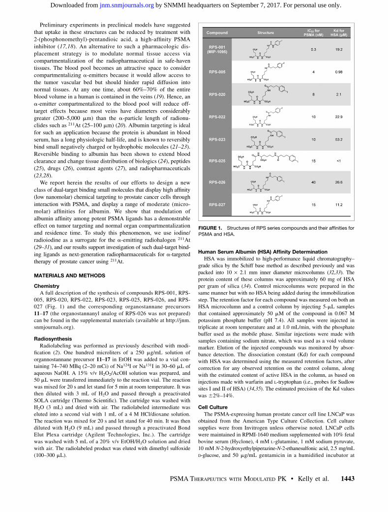
FIGURE 1. Structures of RPS series compounds and their affinities for
PSMA and HSA.
PSMA THERAPEUTICS WITH MODULATED PK • Kelly et al. 1443
by SNMMI headquarters on September 7, 2017. For personal use only. jnm.snmjournals.org Downloaded from
37�C/5% CO2. Cells were removed from flasks for passage or for
transfer to 12-well assay plates by incubating them with 0.25%trypsin/ethylenediaminetetraacetic acid.
In Vitro Determination of 50% Inhibitory Concentration (IC50)
IC50 values of the nonradioactive iodine–containing ligands were
determined by screening in a multiconcentration competitive bindingassay against 99mTc-((7S,12S,16S)-1-(1-(carboxymethyl)-1H-imidazol-
2-yl)-2-((1-(carboxymethyl)-1H-imidazol-2-yl)methyl)-9,14-dioxo-2,8,13,15-tetraazaoctadecane-7,12,16,18-tetracarboxylic acid tech-
netium tricarbonyl complex (99mTc-MIP-1427) for binding to PSMAon LNCaP cells, according to methods previously described (36). Briefly,
LNCaP cells were plated 48 h before the experiment to achieve a densityof approximately 5 · 105 cells/well (in triplicate) in RPMI-1640 medium
supplemented with 0.25% bovine serum albumin. The cells were incu-bated for 1 h with 1 nM 99mTc-MIP-1427 in serum-free RPMI-1640
medium in the presence of 0.1–10,000 nM test compounds. Radioactive
incubation medium was then removed by pipette, and the cells werewashed twice using 1 mL of ice-cold N-2-hydroxyethylpiperazine-N-2-
ethanesulfonic acid buffer. Cells were harvested from the plates and trans-ferred to tubes for radioactive counting using a Packard Cobra II g-Coun-
ter. IC50 values were determined by nonlinear regression using GraphPadPrism software (GraphPad Software).
Inoculation of Mice with Xenografts
All animal studies were approved by the Institutional Animal Careand Use Committee of Weill Cornell Medicine and were undertaken in
accordance with the guidelines set forth by the U.S. Public HealthService Policy on Humane Care and Use of Laboratory Animals.
Animals were housed under standard conditions in approved facilitieswith 12-h light–dark cycles. Food and water were provided ad libitum
throughout the course of the studies. Male inbred athymic nu/nu mice
were purchased from The Jackson Laboratory. For inoculation inmice, LNCaP cells were suspended at 4 · 107 cells/mL in a 1:1
mixture of phosphate-buffered saline:Matrigel (BD Biosciences).Each mouse was injected in the left flank with 0.25 mL of the cell
suspension. The mice were imaged when the tumors reached approx-imately 200–400 mm3, whereas biodistributions were conducted when
tumors were in the range 100–400 mm3.
Tissue Distribution Studies
A quantitative analysis of the tissue distribution of 131I-labeled
compounds was performed in separate groups of male NCr-nu/numice bearing LNCaP cell xenografts administered via the tail vein
as a bolus injection (;370 kBq [10 mCi]/mouse) in a volume of
0.05–0.1 mL of saline solution containing 2.5% v/v dimethyl sulfox-ide. The animals (n 5 3–5/time point) were euthanized by asphyxia-
tion with CO2 at the indicated time points after injection. Tissues,including blood, heart, lungs, liver, spleen, pancreas, kidneys, stom-
ach, large and small intestines (with contents), skeletal muscle, bone,and tumor, were dissected, excised, weighed wet (Sartorius analytic
balance), and counted in a Wizard automated g-counter (PerkinElmer). A 1 percentage injected dose per gram (%ID/g) standard
was counted with the tissue samples. Tissue time–radioactivitylevels expressed as %ID/g were determined. Blood pharmacoki-
netics were modeled as a dual-compartment system using biexpo-nential least-squares regression fit to the data. The regression
method was implemented in MATLAB R2015b (The MathWorks).An independent-samples t test was used to compare different time points
and between different compounds, and in all analyses the statistical sig-nificance (a-level) was set at a P value of less than 0.05.
Imaging Studies
LNCaP xenograft tumor–bearing mice (2 per compound) were in-
jected intravenously via the tail vein as a bolus injection of 7.03–7.77 MBq
(190–210 mCi) of 124I-RPS-027. The specific activity of 124I-RPS-027 was
in the range of 3–10 GBq/mmol. The mice were imaged by small-animalPET/CT (Inveon; Siemens Medical Solutions, Inc.) at 1, 3, 6, 24, and
48 h after injection. Total acquisition time was 30 min, and a CT scanwas obtained either immediately before or immediately after the acqui-
sition for both anatomic coregistration and attenuation correction. Thedata were reconstructed using the commercial Inveon software supplied
by the vendor. Image-derived tumor uptake was estimated by drawing aregion of interest.
RESULTS
Chemistry and Radiochemistry
Reference compounds and their trimethylstannyl precursorswere synthesized as described in Supplemental Figure 1. Synthesisyields ranged from 4% to 20% in 5–8 steps from L-Glu(OtBu)-OtBu•HCl. Trimethylstannylation was accomplished in 20%–82%yield from the protected 4-iodophenyl precursors.Radiolabeling was accomplished by an iododestannylation
reaction, followed by deprotection of the labeled intermediate inacidic conditions (Fig. 2). Total reaction time was 60 min, radio-chemical yield ranged from 43% to 72%, and radiochemical puritywas greater than 90% for all compounds tested. Specific activityvaried from 2 to 10 GBq/mmol according to the starting 124I or 131Iactivity. The deprotection step proved to be time sensitive: forreaction times below 40 min, incomplete deprotection was ob-served, whereas reaction times greater than 45 min led to theformation of an unidentified impurity that could not be removedduring purification by solid-phase extraction. The characteristicsof the radiosyntheses are described in Table 1. No significantdifference in labeling yield was observed when 124I was used inplace of 131I.
In Vitro Studies
The range of affinities for PSMAwas determined to be 4–40 nM,with most of the compounds clustered between 10 and 15 nM (Fig.1). In the same assay RPS-001, also known as MIP-1095, was foundto have an IC50 of 0.3 nM. Compounds bearing the p-(iodophenyl)butyric acid moiety were found to have a high affinity (1–2 mM) forHSA (Fig. 1). RPS-025 could not be eluted from the column, whichprevented the calculation of a precise Kd. The compound RPS-027had a modest affinity of 11 mM, whereas RPS-001, RPS-022, andRPS-026 were in the range 19–26 mM. RPS-023 (Kd 5 53.2 mM)was determined to have a weak affinity.
Biodistribution and Small-Animal PET/CT Imaging
The biodistribution studies of the 6 ligands demonstrated thatalbumin binding affinity contributed markedly to the differentpharmacokinetics observed in mice. RPS-001 (Kd 5 20 mM for
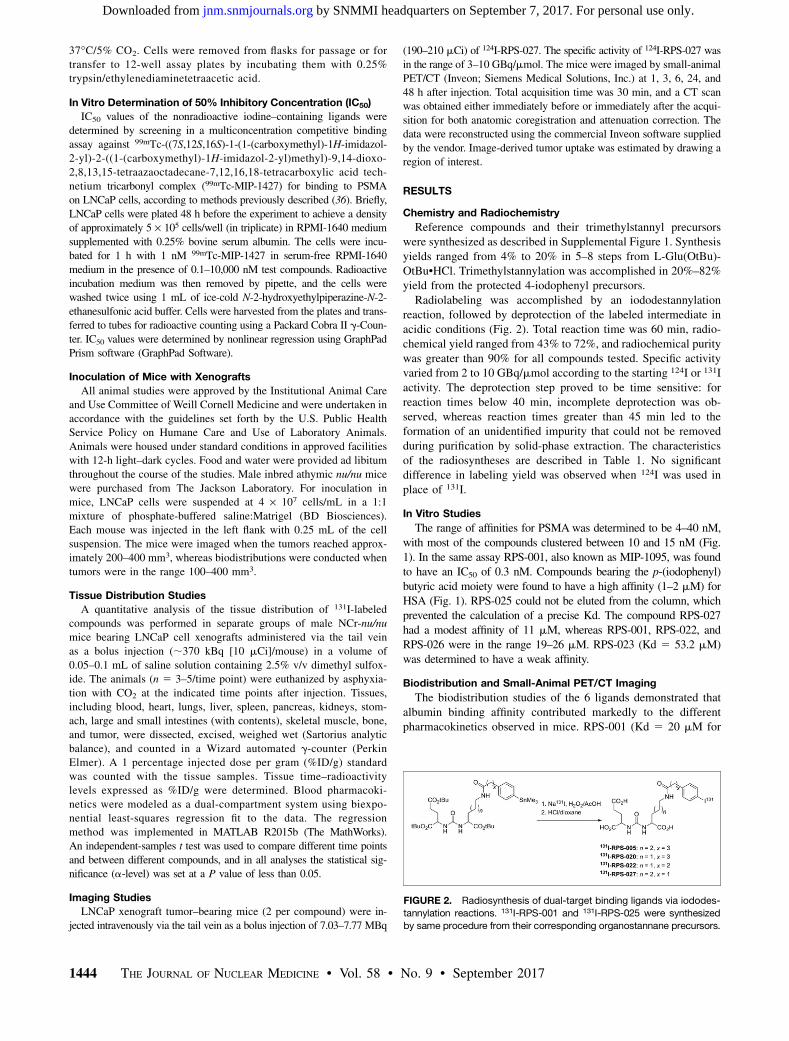
FIGURE 2. Radiosynthesis of dual-target binding ligands via iododes-
tannylation reactions. 131I-RPS-001 and 131I-RPS-025 were synthesized
by same procedure from their corresponding organostannane precursors.
1444 THE JOURNAL OF NUCLEAR MEDICINE • Vol. 58 • No. 9 • September 2017
by SNMMI headquarters on September 7, 2017. For personal use only. jnm.snmjournals.org Downloaded from
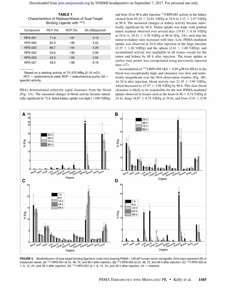
HSA) demonstrated relatively rapid clearance from the blood(Fig. 3A). The measured changes in blood activity became statisti-cally significant by 72 h. Initial kidney uptake was high (.100 %ID/g),
and from 24 to 96 h after injection 131I-RPS-001 activity in the kidneycleared from 65.24 6 22.61 %ID/g at 24 h to 2.12 6 2.47 %ID/gat 96 h. The measured changes in kidney activity became statis-tically significant by 48 h. Tumor uptake was high, with gradualtumor washout observed over several days (19.81 6 6.16 %ID/gat 24 h vs. 10.21 6 4.30 %ID/g at 96 h) (Fig. 3A), such that thetumor-to-kidney ratio increased with time. Low, PSMA-mediateduptake was observed at 24 h after injection in the large intestine(2.35 6 1.26 %ID/g) and the spleen (2.41 6 1.40 %ID/g), andaccumulated activity was negligible in all tissues except for thetumor and kidney by 48 h after injection. The tissue uptake atearlier time points was extrapolated using previously reporteddata (37).Accumulation of 131I-RPS-005 (Kd 5 0.89 mM for HSA) in the
blood was exceptionally high, and clearance was slow and statis-tically insignificant over the 96-h observation window (Fig. 3B).At 24 h after injection, blood activity was 21.35 6 3.99 %ID/g,which decreased to 15.57 6 3.98 %ID/g by 96 h. This slow bloodclearance is likely to be responsible for the non–PSMA-mediateduptake observed in tissues such as the heart (4.406 0.74 %ID/g at24 h), lungs (8.87 6 0.74 %ID/g at 24 h), and liver (3.41 6 0.56
TABLE 1Characteristics of Radiosynthesis of Dual-Target
Binding Ligands with 131I
Compound RCY (%) RCP (%) SA (GBq/μmol)*
RPS-001 71.8 .97 3.15
RPS-005 62.0 .96 3.52
RPS-020 66.7 .94 4.29
RPS-022 53.6 .90 3.06
RPS-025 43.5 .93 2.02
RPS-027 49.5 .98 9.18
*Based on a starting activity of 72–370 MBq (2–10 mCi).
RCY 5 radiochemical yield; RCP 5 radiochemical purity; SA 5specific activity.
FIGURE 3. Biodistribution of dual-target binding ligands in nude mice bearing PSMA1 LNCaP human tumor xenografts. Error bars represent SD of
measured values. (A) 131I-RPS-001 at 24, 48, 72, and 96 h after injection. (B) 131I-RPS-005 at 24, 48, 72, and 96 h after injection. (C) 131I-RPS-020 at
1, 6, 12, 24, and 48 h after injection. (D) 131I-RPS-022 at 1, 6, 12, 24, and 48 h after injection. Int 5 intestine.
PSMA THERAPEUTICS WITH MODULATED PK • Kelly et al. 1445
by SNMMI headquarters on September 7, 2017. For personal use only. jnm.snmjournals.org Downloaded from
%ID/g at 24 h). Kidney uptake (39.096 2.96 %ID/g) was lower at24 h after injection than observed for 131I-RPS-001, but clearancewas considerably slower. In combination with the comparativelylow tumor uptake of approximately 10% (9.37 6 1.56 %ID/g at24 h; 10.82 6 2.64 %ID/g at 96 h), these pharmacokinetics resultin poor tumor-to-background ratios at all time points.Because the tissue uptake of 131I-RPS-005 appeared to peak
within the first 24 h, 131I-RPS-020 (Kd 5 2.1 mM) was investi-gated at early time points as well. Prolonged blood retention wasalso observed, with an initial accumulation of 16.206 4.68 %ID/gat 1 h after injection only decreasing to 12.71 6 1.55 %ID/g at48 h (Fig. 3C), which was not statistically significant. This wasassociated with high off-target uptake, most notably in the lungs(6.12 6 0.95 %ID/g at 1 h after injection) and kidneys (16.21 60.97 %ID/g at 1 h). Activity continued to accumulate in the kidneys,peaking at 22.266 2.48 %ID/g at 24 h after injection. Tumor uptake(4.81 6 1.27 %ID/g) was lower than observed for RPS-005, as acomparison of PSMA affinities might predict, but with no statisti-cally significant change over the course of 48 h.In contrast, 131I-RPS-022 (Kd 5 22.9 mM) showed a rapid and
significant drop in blood activity over time, with negligible activ-ity detected as early as 12 h after injection (Fig. 3D). Considerableuptake was observed in the liver (12.46 6 1.63 %ID/g) and smallintestine (13.09 6 2.98 %ID/g) at 1 h after injection, thoughclearance from each organ was rapid. Kidney uptake, whichreached (52.18 6 5.35 %ID/g) at 1 h after injection, demonstrated
a significant clearance of activity, reaching 2.27 6 1.41 %ID/g by24 h, leading to favorable tumor-to-kidney and tumor-to-backgroundratios at later time points. However, tumor uptake peaked at 7.20 60.10 %ID/g at 1 h after injection and decreased significantly to3.35 6 1.70 %ID/g by 6 h.
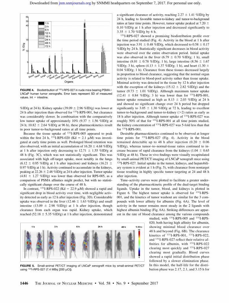
131I-RPS-027 showed a promising biodistribution profile overthe time period studied (Fig. 4). Activity in the blood at 1 h afterinjection was 3.91 6 0.48 %ID/g, which decreased to 0.58 6 0.17%ID/g by 24 h. Statistically significant decreases in blood activitywere observed over the entire observation period. Initial uptakewas also observed in the liver (6.79 6 0.70 %ID/g; 1 h), smallintestine (8.01 6 0.78 %ID/g; 1 h), large intestine (8.56 6 1.67%ID/g; 3 h), spleen (4.13 6 1.37 %ID/g; 1 h), and heart (1.30 60.04 %ID/g; 1 h). Clearance from these tissues decreased largelyin proportion to blood clearance, suggesting that the normal organactivity is related to blood-pool activity rather than tissue uptake.Minimal activity was detected in the tissue by 12 h after injectionwith the exception of the kidneys (15.12 6 2.82 %ID/g) and thetumor (9.73 6 1.01 %ID/g). Although maximum tumor uptake(12.41 6 0.84 %ID/g; 3 h) was lower than for 131I-RPS-001,tumor uptake remained as high as 8.13 6 2.03 %ID/g at 24 hand showed no significant change over 24 h period but droppedsignificantly to 3.05 6 1.30 %ID/g at 72 h, leading to excellenttumor-to-background and tumor-to-kidney (.2) ratios as early as18 h after injection. Although tumor uptake of 131I-RPS-027 wasroughly 50% of that for 131I-RPS-001 at all time points studied,the kidney concentration of 131I-RPS-027 was 5-fold less than thatfor 131I-RPS-001.Desirable pharmacokinetics continued to be observed at longer
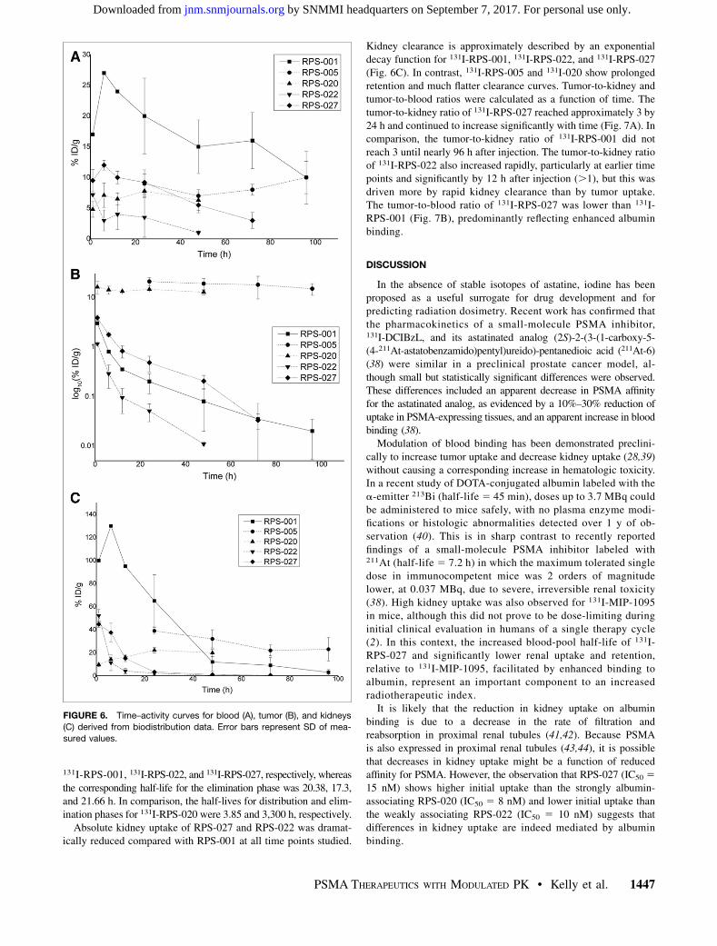
time points for 131I-RPS-027 (Fig. 4). Activity in the bloodremained detectable up to 48 h after injection (0.20 6 0.06%ID/g), whereas tumor–to–normal-tissue ratios continued to in-crease because of rapid clearance from the kidneys (1.04 6 0.65%ID/g at 48 h). These in vivo findings were visually recapitulatedby small-animal PET/CT imaging of LNCaP xenograft mice using124I-RPS-027. Initial uptake in the tumor, kidneys, and hepatobili-ary system is evident at 1 h (Fig. 5), with clearance from nontargettissue resulting in highly specific tumor targeting at 24 and 48 hafter injection.Time–activity curves were plotted to facilitate a greater under-
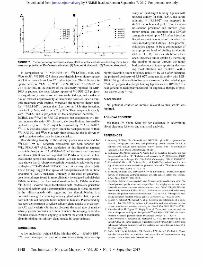
standing of the pharmacokinetic profile of the dual-target bindingligands. Uptake in the tumor, blood, and kidneys is plotted inFigure 6. The highest tumor uptake is observed for 131I-RPS-001, and the kinetics of tumor washout are similar for the 3 com-pounds with lower affinity for albumin (Fig. 6A). The level ofactivity in the tumor remains most steady in the 2 ligands withhighest albumin binding (Fig. 6A). Striking differences are appar-ent in the rate of blood clearance among the various compounds
studied, with 131I-RPS-005 and 131I-RPS-020, both having high affinity for albumin,showing minimal blood clearance over48 h and beyond (Fig. 6B). The clearancekinetics of 131I-RPS-001, 131I-RPS-022,and 131I-RPS-027 reflect their relative af-finities for albumin, with 131I-RPS-022clearing most quickly and 131I-RPS-027clearing most gradually. Blood curvesshowed a rapid initial distribution phasefollowed by a slower elimination phase.In this model, the half-life for the distri-bution phase was 2.17, 2.1, and 3.15 h for
FIGURE 4. Biodistribution of 131I-RPS-027 in nudemice bearing PSMA1LNCaP human tumor xenografts. Error bars represent SD of measured
values. Int 5 intestine.
FIGURE 5. Small-animal PET/CT imaging of LNCaP xenograft mice by small-animal PET/CT
using 124I-RPS-027 (7.4 MBq [200 μCi]).
1446 THE JOURNAL OF NUCLEAR MEDICINE • Vol. 58 • No. 9 • September 2017
by SNMMI headquarters on September 7, 2017. For personal use only. jnm.snmjournals.org Downloaded from
131I-RPS-001, 131I-RPS-022, and 131I-RPS-027, respectively, whereasthe corresponding half-life for the elimination phase was 20.38, 17.3,and 21.66 h. In comparison, the half-lives for distribution and elim-ination phases for 131I-RPS-020 were 3.85 and 3,300 h, respectively.Absolute kidney uptake of RPS-027 and RPS-022 was dramat-
ically reduced compared with RPS-001 at all time points studied.
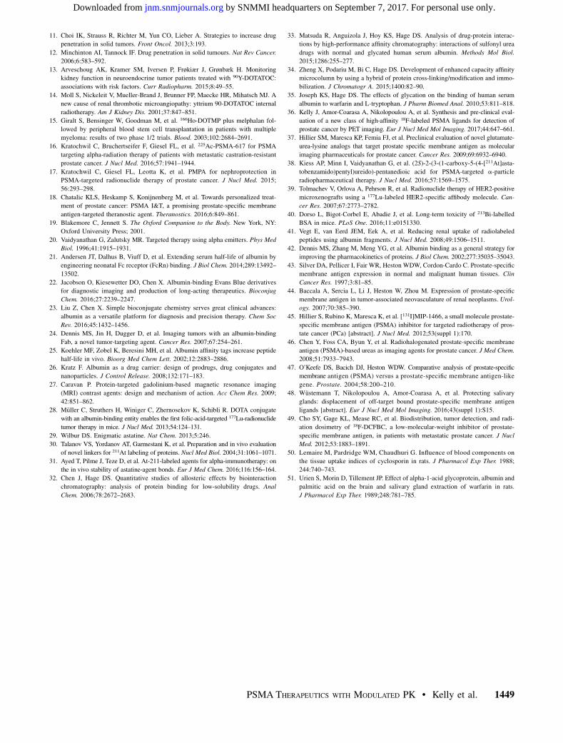
Kidney clearance is approximately described by an exponentialdecay function for 131I-RPS-001, 131I-RPS-022, and 131I-RPS-027(Fig. 6C). In contrast, 131I-RPS-005 and 131I-020 show prolongedretention and much flatter clearance curves. Tumor-to-kidney andtumor-to-blood ratios were calculated as a function of time. Thetumor-to-kidney ratio of 131I-RPS-027 reached approximately 3 by24 h and continued to increase significantly with time (Fig. 7A). Incomparison, the tumor-to-kidney ratio of 131I-RPS-001 did notreach 3 until nearly 96 h after injection. The tumor-to-kidney ratioof 131I-RPS-022 also increased rapidly, particularly at earlier timepoints and significantly by 12 h after injection (.1), but this wasdriven more by rapid kidney clearance than by tumor uptake.The tumor-to-blood ratio of 131I-RPS-027 was lower than 131I-RPS-001 (Fig. 7B), predominantly reflecting enhanced albuminbinding.
DISCUSSION
In the absence of stable isotopes of astatine, iodine has beenproposed as a useful surrogate for drug development and forpredicting radiation dosimetry. Recent work has confirmed thatthe pharmacokinetics of a small-molecule PSMA inhibitor,131I-DCIBzL, and its astatinated analog (2S)-2-(3-(1-carboxy-5-(4-211At-astatobenzamido)pentyl)ureido)-pentanedioic acid (211At-6)(38) were similar in a preclinical prostate cancer model, al-though small but statistically significant differences were observed.These differences included an apparent decrease in PSMA affinityfor the astatinated analog, as evidenced by a 10%–30% reduction ofuptake in PSMA-expressing tissues, and an apparent increase in bloodbinding (38).Modulation of blood binding has been demonstrated preclini-
cally to increase tumor uptake and decrease kidney uptake (28,39)without causing a corresponding increase in hematologic toxicity.In a recent study of DOTA-conjugated albumin labeled with thea-emitter 213Bi (half-life 5 45 min), doses up to 3.7 MBq couldbe administered to mice safely, with no plasma enzyme modi-fications or histologic abnormalities detected over 1 y of ob-servation (40). This is in sharp contrast to recently reportedfindings of a small-molecule PSMA inhibitor labeled with211At (half-life 5 7.2 h) in which the maximum tolerated singledose in immunocompetent mice was 2 orders of magnitudelower, at 0.037 MBq, due to severe, irreversible renal toxicity(38). High kidney uptake was also observed for 131I-MIP-1095in mice, although this did not prove to be dose-limiting duringinitial clinical evaluation in humans of a single therapy cycle(2). In this context, the increased blood-pool half-life of 131I-RPS-027 and significantly lower renal uptake and retention,relative to 131I-MIP-1095, facilitated by enhanced binding toalbumin, represent an important component to an increasedradiotherapeutic index.It is likely that the reduction in kidney uptake on albumin
binding is due to a decrease in the rate of filtration andreabsorption in proximal renal tubules (41,42). Because PSMAis also expressed in proximal renal tubules (43,44), it is possiblethat decreases in kidney uptake might be a function of reducedaffinity for PSMA. However, the observation that RPS-027 (IC50 515 nM) shows higher initial uptake than the strongly albumin-associating RPS-020 (IC50 5 8 nM) and lower initial uptake thanthe weakly associating RPS-022 (IC50 5 10 nM) suggests thatdifferences in kidney uptake are indeed mediated by albuminbinding.
FIGURE 6. Time–activity curves for blood (A), tumor (B), and kidneys
(C) derived from biodistribution data. Error bars represent SD of mea-
sured values.
PSMA THERAPEUTICS WITH MODULATED PK • Kelly et al. 1447
by SNMMI headquarters on September 7, 2017. For personal use only. jnm.snmjournals.org Downloaded from
In comparison to 131I-MIP-1095 (45), 131I-DCIBzL (46), and211At-6 (38), 131I-RPS-027 shows considerably lower kidney uptakeat all time points from 1 to 72 h after injection. The difference inuptake between 131I-MIP-1095 (131I-RPS-001) and 131I-RPS-027 at24 h is 20-fold. In the context of the dosimetry reported for MIP-1095 in patients, the lower kidney uptake of 131I-RPS-027 projectsto a significantly lower absorbed dose to the kidneys, and a reducedrisk of relevant nephrotoxicity at therapeutic doses or under a mul-tiple treatment cycle regime. Moreover, the tumor-to-kidney ratiofor 131I-RPS-027 is greater than 2 as soon as 18 h after injection,rises to 3 by 24 h, and exceeds 7 by 72 h. This compares favorablywith 211At-6, and a projection of the comparison between 131I-DCIBzL and 211At-6 to RPS-027 predicts that astatination will fur-ther increase the ratio (38). As such, the dose-limiting, irreversiblenephrotoxicity of 211At-6 might be resolved by 211At-RPS-027.131I-RPS-022 also shows higher tumor-to-background ratios than131I-RPS-001 and 211At-6 at early time points, but this is driven byrapid excretion rather than by tumor uptake.Dose-limiting toxicity to the salivary glands was observed for
131I-MIP-1095 (2). Moderate xerostomia has been reported for177Lu-PSMA-617 (3,6), but translation of this ligand to targeteda-particle therapy as 225Ac-PSMA-617 led to severe and sustainedxerostomia (18). It has been reported that PSMA is expressed at lowlevels in the parotid and lacrimal glands (47), and recent experimentshave shown that 2-(phosphonomethyl)-pentandioic acid can be usedto displace 68Ga-PSMA-HBED-CC from rat salivary glands (48).These findings suggest that uptake of radiopharmaceuticals in thesestructures is PSMA-mediated. Uniquely to the class of glutamate-urea heterodimers found in most clinically investigated radiolabeledPSMA inhibitors, the fluorinated small-molecule PSMA inhibitor18F-DCFBC showed tumor localization with moderately persistentblood-pool activity and a corresponding decrease in signal intensityin the salivary glands (49), suggesting that albumin-binding is apotential strategy for reducing salivary gland uptake that, a priori,does not rule out adequate tumor uptake in humans. Plasma bindinghas been demonstrated to reduce salivary gland uptake of cyclospor-ine (50) and warfarin (51) in rats. Because the small size of murinesalivary glands precludes detection of uptake by imaging or biodis-tribution studies, work is ongoing to confirm the effect of modulatedalbumin binding on salivary gland uptake in larger animals.
CONCLUSION
A low-molecular-weight PSMA inhibitor (IC50 5 15 nM), RPS-027, was developed as part of a structure–activity relationship
study on dual-target binding ligands withunequal affinity for both PSMA and serumalbumin. 131I-RPS-027 was prepared in49.5% radiochemical yield from its orga-nostannane precursor and showed goodtumor uptake and retention in a LNCaPxenograft model up to 72 h after injection.Rapid washout was observed in other tis-sues, including the kidneys. These pharma-cokinetics appear to be a consequence ofan appropriate level of binding to albumin(Kd 5 11 mM) that extends blood clear-ance, increases tumor uptake by increasingthe number of passes through the tumorbed, and reduces kidney uptake by decreas-ing renal filtration and reuptake. With a
highly favorable tumor-to-kidney ratio (.3 by 24 h after injection),the projected dosimetry of RPS-027 compares favorably with MIP-1095. Using iodine/radioiodine as a surrogate for the radiohalogen211At, we propose dual-target binding ligands such as RPS-027 as anext-generation radiopharmaceutical for targeted a-therapy of pros-tate cancer using 211At.
DISCLOSURE
No potential conflict of interest relevant to this article wasreported.
ACKNOWLEDGMENT
We thank Dr. Yeona Kang for her assistance in determiningblood clearance kinetics and statistical analysis.
REFERENCES
1. Strosberg JR, Wolin EM, Chasen B, et al. NETTER-1 phase III: progression-free
survival, radiographic response, and preliminary overall survival results in
patients with midgut neuroendocrine tumors treated with 177-Lu-dotatate
[abstract]. J Clin Oncol. 2016;34(suppl 4S):194.
2. Zechmann CM, Afshar-Oromieh A, Armor T, et al. Radiation dosimetry and first
therapy results with a 124I/131I-labeled small molecule (MIP-1095) targeting PSMA
for prostate cancer therapy. Eur J Nucl Med Mol Imaging. 2014;41:1280–1292.
3. Kratochwil C, Giesel FL, Stefanova M, et al. PSMA-Targeted radionuclide ther-
apy of metastatic castration-resistant prostate cancer with 177Lu-labeled PSMA-
617. J Nucl Med. 2016;57:1170–1176.
4. Baum RP, Kulkarni HR, Schuchardt C, et al. Lutetium-177 PSMA radioligand
therapy of metastatic castration-resistant prostate cancer: safety and efficacy.
J Nucl Med. 2016;57:1006–1013.
5. Heck MM, Retz M, D’Alessandria C, et al. Systemic radioligand therapy with 177Lu
labeled prostate specific membrane antigen ligand for imaging and therapy in pa-
tients with metastatic castration resistant prostate cancer. J Urol. 2016;196:382–391.
6. Fendler WP, Reinhardt S, Ilhan H, et al. Preliminary experience with dosimetry,
response and patient reported outcome after 177Lu-PSMA-617 therapy for met-
astatic castration-resistant prostate cancer. Oncotarget. 2017;8:3581–3590.
7. Rahbar K, Schmidt M, Heinzel A, et al. Response and tolerability of a single
dose of 177Lu-PSMA-617 in patients with metastatic castration-resistant prostate
cancer: a multicenter retrospective analysis. J Nucl Med. 2016;57:1334–1338.
8. Ahmadzadehfar H, Eppard E, Kürpig S, et al. Therapeutic response and side
effects of repeated radioligand therapy with 177Lu-PSMA-DKFZ-617 of castrate-
resistant metastatic prostate cancer. Oncotarget. 2016;7:12477–12488.
9. Afshar-Oromieh A, Hetzheim H, Kratochwil C, et al. The theranostic PSMA
ligand PSMA-617 in the diagnosis of prostate cancer by PET/CT: biodistribution
in humans, radiation dosimetry, and first evaluation of tumor lesions. J Nucl Med.
2015;56:1697–1705.
10. Dreher MR, Liu W, Michelich CR, Dewhirst MW, Yuan F, Chilkoti A. Tumor
vascular permeability, accumulation, and penetration of macromolecular drug
carriers. J Natl Cancer Inst. 2006;98:335–344.
FIGURE 7. Tumor-to-background ratios show effect of enhanced albumin binding. Error bars
were computed from SD of measured values. (A) Tumor-to-kidney ratio. (B) Tumor-to-blood ratio.
1448 THE JOURNAL OF NUCLEAR MEDICINE • Vol. 58 • No. 9 • September 2017
by SNMMI headquarters on September 7, 2017. For personal use only. jnm.snmjournals.org Downloaded from
11. Choi IK, Strauss R, Richter M, Yun CO, Lieber A. Strategies to increase drug
penetration in solid tumors. Front Oncol. 2013;3:193.
12. Minchinton AI, Tannock IF. Drug penetration in solid tumours. Nat Rev Cancer.
2006;6:583–592.
13. Arveschoug AK, Kramer SM, Iversen P, Frøkiær J, Grønbæk H. Monitoring
kidney function in neuroendocrine tumor patients treated with 90Y-DOTATOC:
associations with risk factors. Curr Radiopharm. 2015;8:49–55.
14. Moll S, Nickeleit V, Mueller-Brand J, Brunner FP, Maecke HR, Mihatsch MJ. A
new cause of renal thrombotic microangiopathy: yttrium 90-DOTATOC internal
radiotherapy. Am J Kidney Dis. 2001;37:847–851.
15. Giralt S, Bensinger W, Goodman M, et al. 166Ho-DOTMP plus melphalan fol-
lowed by peripheral blood stem cell transplantation in patients with multiple
myeloma: results of two phase 1/2 trials. Blood. 2003;102:2684–2691.
16. Kratochwil C, Bruchertseifer F, Giesel FL, et al. 225Ac-PSMA-617 for PSMA
targeting alpha-radiation therapy of patients with metastatic castration-resistant
prostate cancer. J Nucl Med. 2016;57:1941–1944.
17. Kratochwil C, Giesel FL, Leotta K, et al. PMPA for nephroprotection in
PSMA-targeted radionuclide therapy of prostate cancer. J Nucl Med. 2015;
56:293–298.
18. Chatalic KLS, Heskamp S, Konijnenberg M, et al. Towards personalized treat-
ment of prostate cancer: PSMA I&T, a promising prostate-specific membrane
antigen-targeted theranostic agent. Theranostics. 2016;6:849–861.
19. Blakemore C, Jennett S. The Oxford Companion to the Body. New York, NY:
Oxford University Press; 2001.
20. Vaidyanathan G, Zalutsky MR. Targeted therapy using alpha emitters. Phys Med
Biol. 1996;41:1915–1931.
21. Andersen JT, Dalhus B, Viuff D, et al. Extending serum half-life of albumin by
engineering neonatal Fc receptor (FcRn) binding. J Biol Chem. 2014;289:13492–
13502.
22. Jacobson O, Kiesewetter DO, Chen X. Albumin-binding Evans Blue derivatives
for diagnostic imaging and production of long-acting therapeutics. Bioconjug
Chem. 2016;27:2239–2247.
23. Liu Z, Chen X. Simple bioconjugate chemistry serves great clinical advances:
albumin as a versatile platform for diagnosis and precision therapy. Chem Soc
Rev. 2016;45:1432–1456.
24. Dennis MS, Jin H, Dugger D, et al. Imaging tumors with an albumin-binding
Fab, a novel tumor-targeting agent. Cancer Res. 2007;67:254–261.
25. Koehler MF, Zobel K, Beresini MH, et al. Albumin affinity tags increase peptide
half-life in vivo. Bioorg Med Chem Lett. 2002;12:2883–2886.
26. Kratz F. Albumin as a drug carrier: design of prodrugs, drug conjugates and
nanoparticles. J Control Release. 2008;132:171–183.
27. Caravan P. Protein-targeted gadolinium-based magnetic resonance imaging
(MRI) contrast agents: design and mechanism of action. Acc Chem Res. 2009;
42:851–862.
28. Müller C, Struthers H, Winiger C, Zhernosekov K, Schibli R. DOTA conjugate
with an albumin-binding entity enables the first folic-acid-targeted 177Lu-radionuclide
tumor therapy in mice. J Nucl Med. 2013;54:124–131.
29. Wilbur DS. Enigmatic astatine. Nat Chem. 2013;5:246.
30. Talanov VS, Yordanov AT, Garmestani K, et al. Preparation and in vivo evaluation
of novel linkers for 211At labeling of proteins. Nucl Med Biol. 2004;31:1061–1071.
31. Ayed T, Pilme J, Teze D, et al. At-211-labeled agents for alpha-immunotherapy: on
the in vivo stability of astatine-agent bonds. Eur J Med Chem. 2016;116:156–164.
32. Chen J, Hage DS. Quantitative studies of allosteric effects by biointeraction
chromatography: analysis of protein binding for low-solubility drugs. Anal
Chem. 2006;78:2672–2683.
33. Matsuda R, Anguizola J, Hoy KS, Hage DS. Analysis of drug-protein interac-
tions by high-performance affinity chromatography: interactions of sulfonyl urea
drugs with normal and glycated human serum albumin. Methods Mol Biol.
2015;1286:255–277.
34. Zheng X, Podariu M, Bi C, Hage DS. Development of enhanced capacity affinity
microcolumn by using a hybrid of protein cross-linking/modification and immo-
bilization. J Chromatogr A. 2015;1400:82–90.
35. Joseph KS, Hage DS. The effects of glycation on the binding of human serum
albumin to warfarin and L-tryptophan. J Pharm Biomed Anal. 2010;53:811–818.
36. Kelly J, Amor-Coarasa A, Nikolopoulou A, et al. Synthesis and pre-clinical eval-
uation of a new class of high-affinity 18F-labeled PSMA ligands for detection of
prostate cancer by PET imaging. Eur J Nucl Med Mol Imaging. 2017;44:647–661.
37. Hillier SM, Maresca KP, Femia FJ, et al. Preclinical evaluation of novel glutamate-
urea-lysine analogs that target prostate specific membrane antigen as molecular
imaging pharmaceuticals for prostate cancer. Cancer Res. 2009;69:6932–6940.
38. Kiess AP, Minn I, Vaidyanathan G, et al. (2S)-2-(3-(1-carboxy-5-(4-[211At]asta-
tobenzamido)pentyl)ureido)-pentanedioic acid for PSMA-targeted a-particle
radiopharmaceutical therapy. J Nucl Med. 2016;57:1569–1575.
39. Tolmachev V, Orlova A, Pehrson R, et al. Radionuclide therapy of HER2-positive
microxenografts using a 177Lu-labeled HER2-specific affibody molecule. Can-
cer Res. 2007;67:2773–2782.
40. Dorso L, Bigot-Corbel E, Abadie J, et al. Long-term toxicity of 213Bi-labelled
BSA in mice. PLoS One. 2016;11:e0151330.
41. Vegt E, van Eerd JEM, Eek A, et al. Reducing renal uptake of radiolabeled
peptides using albumin fragments. J Nucl Med. 2008;49:1506–1511.
42. Dennis MS, Zhang M, Meng YG, et al. Albumin binding as a general strategy for
improving the pharmacokinetics of proteins. J Biol Chem. 2002;277:35035–35043.
43. Silver DA, Pellicer I, Fair WR, Heston WDW, Cordon-Cardo C. Prostate-specific
membrane antigen expression in normal and malignant human tissues. Clin
Cancer Res. 1997;3:81–85.
44. Baccala A, Sercia L, Li J, Heston W, Zhou M. Expression of prostate-specific
membrane antigen in tumor-associated neovasculature of renal neoplasms. Urol-
ogy. 2007;70:385–390.
45. Hillier S, Rubino K, Maresca K, et al. [131I]MIP-1466, a small molecule prostate-
specific membrane antigen (PSMA) inhibitor for targeted radiotherapy of pros-
tate cancer (PCa) [abstract]. J Nucl Med. 2012;53(suppl 1):170.
46. Chen Y, Foss CA, Byun Y, et al. Radiohalogenated prostate-specific membrane
antigen (PSMA)-based ureas as imaging agents for prostate cancer. J Med Chem.
2008;51:7933–7943.
47. O’Keefe DS, Bacich DJ, Heston WDW. Comparative analysis of prostate-specific
membrane antigen (PSMA) versus a prostate-specific membrane antigen-like
gene. Prostate. 2004;58:200–210.
48. Wüstemann T, Nikolopoulou A, Amor-Coarasa A, et al. Protecting salivary
glands: displacement of off-target bound prostate-specific membrane antigen
ligands [abstract]. Eur J Nucl Med Mol Imaging. 2016;43(suppl 1):S15.
49. Cho SY, Gage KL, Mease RC, et al. Biodistribution, tumor detection, and radi-
ation dosimetry of 18F-DCFBC, a low-molecular-weight inhibitor of prostate-
specific membrane antigen, in patients with metastatic prostate cancer. J Nucl
Med. 2012;53:1883–1891.
50. Lemaire M, Pardridge WM, Chaudhuri G. Influence of blood components on
the tissue uptake indices of cyclosporin in rats. J Pharmacol Exp Ther. 1988;
244:740–743.
51. Urien S, Morin D, Tillement JP. Effect of alpha-1-acid glycoprotein, albumin and
palmitic acid on the brain and salivary gland extraction of warfarin in rats.
J Pharmacol Exp Ther. 1989;248:781–785.
PSMA THERAPEUTICS WITH MODULATED PK • Kelly et al. 1449
by SNMMI headquarters on September 7, 2017. For personal use only. jnm.snmjournals.org Downloaded from
Doi: 10.2967/jnumed.116.188722Published online: April 27, 2017.
2017;58:1442-1449.J Nucl Med. John W. BabichClarence Williams, Jr, Xiwei Zheng, Cong Bi, Bao Hu, J. David Warren, David S. Hage, Stephen G. DiMagno and James M. Kelly, Alejandro Amor-Coarasa, Anastasia Nikolopoulou, Till Wüstemann, Peter Barelli, Dohyun Kim, Endoradiotherapy of Prostate CancerDual-Target Binding Ligands with Modulated Pharmacokinetics for
http://jnm.snmjournals.org/content/58/9/1442This article and updated information are available at:
http://jnm.snmjournals.org/site/subscriptions/online.xhtml
Information about subscriptions to JNM can be found at:
http://jnm.snmjournals.org/site/misc/permission.xhtmlInformation about reproducing figures, tables, or other portions of this article can be found online at:
(Print ISSN: 0161-5505, Online ISSN: 2159-662X)1850 Samuel Morse Drive, Reston, VA 20190.SNMMI | Society of Nuclear Medicine and Molecular Imaging
is published monthly.The Journal of Nuclear Medicine
© Copyright 2017 SNMMI; all rights reserved.
by SNMMI headquarters on September 7, 2017. For personal use only. jnm.snmjournals.org Downloaded from