Embed Size (px)
Citation preview

ORIGINAL RESEARCHpublished: 24 September 2015doi: 10.3389/fphys.2015.00264
Frontiers in Physiology | www.frontiersin.org 1 September 2015 | Volume 6 | Article 264
Edited by:
Ovidiu Constantin Baltatu,
Camilo Castelo Branco University,
Brazil
Reviewed by:
Slade T. Matthews,
The University of Sydney, Australia
Sumit Sahni,
University of Illinois at Chicago, USA
*Correspondence:
Christoph Küper,
Department of Physiology, University
of Munich, Pettenkoferstrasse 12,
80336 Munich, Germany
Specialty section:
This article was submitted to
Integrative Physiology,
a section of the journal
Frontiers in Physiology
Received: 16 July 2015
Accepted: 07 September 2015
Published: 24 September 2015
Citation:
Küper C, Beck F-X and Neuhofer W
(2015) Dual effect of lithium on NFAT5
activity in kidney cells.
Front. Physiol. 6:264.
doi: 10.3389/fphys.2015.00264
Dual effect of lithium on NFAT5activity in kidney cellsChristoph Küper 1*, Franz-Xaver Beck 1 and Wolfgang Neuhofer 2
1Department of Physiology, University of Munich, Munich, Germany, 2Medical Clinic V, University Hospital Mannheim,
University of Heidelberg, Mannheim, Germany
Lithium salts are used widely for treatment of bipolar and other mental disorders. Lithium
therapy is accompanied frequently by renal side effects, such as nephrogenic diabetes
insipidus or chronic kidney disease (CKD), but the molecular mechanisms underlying
these effects are still poorly understood. In the present study we examined the effect
of lithium on the activity of the osmosensitive transcriptional activator nuclear factor
of activated T cells 5 (NFAT5, also known as TonEBP), which plays a key role in renal
cellular osmoprotection and urinary concentrating ability. Interestingly, we found different
effects of lithium on NFAT5 activity, depending on medium osmolality and incubation
time. When cells were exposed to lithium for a relative short period (24 h), NFAT5
activity was significantly increased, especially under isosmotic conditions, resulting in
an enhanced expression of the NFAT5 target gene heat shock protein 70 (HSP70).
Further analysis revealed that the increase of NFAT5 activity depended primarily on an
enhanced activity of the c-terminal transactivation domain (TAD), while NFAT5 protein
abundance was largely unaffected. Enhanced activity of the TAD is probably mediated
by lithium-induced inhibitory phosphorylation of glycogen synthase kinase 3β (GSK-3β),
which is in accordance with previous studies. When cells were exposed to lithium for
a longer period (96 h), cellular NFAT5 activity and subsequently expression of HSP70
significantly decreased under hyperosmotic conditions, due to diminished NFAT5 protein
abundance, also resulting from GSK-3β inhibition. Taken together, our results provide
evidence that lithium has opposing effects on NFAT5 activity, depending on environmental
osmolality and exposure duration. The potential impacts of these observations on the
diverse effects of lithium on kidney function are discussed.
Keywords: NFAT5, lithium, nephrogenic diabetes insipidus, GSK3β, urinary concentrating mechanism, chronic
kidney disease
Introduction
The beginning of the widespread use of lithium as a mood stabilizer in the treatment ofbipolar disorders in the early 1970s also saw the first reports of adverse renal side effectsaccompanying lithium treatment. The most common adverse effect is nephrogenic diabetesinsipidus (NDI), due to a decreased expression of the aquaporin-2 (AQP-2) water channel andincorrect trafficking of AQP-2 to the luminal membrane in collecting duct cells (Marples et al.,1995; Kwon et al., 2000). Lithium-induced NDI, affecting up to 40% of all patients, can bedetected within a few weeks after beginning lithium administration (Grünfeld and Rossier, 2009).

Küper et al. Dual effect of lithium on NFAT5 activity
Additionally, patients receiving lithium for more than 10 yearshave a significantly increased risk of developing chronic kidneydisease (CKD; Bendz et al., 2010).
Lithium affects multiple molecular targets and signalingpathways, making it difficult to characterize the exactmechanisms by which the positive effects on the mood ofpsychiatric patients or the renal side effects are induced. In thelast two decades, however, it has become increasingly evident thatinhibition of the glycogen synthase kinase 3β (GSK-3β; Klein andMelton, 1996; Stambolic et al., 1996) plays a crucial role for theeffects of lithium both in neuronal and kidney cells. GSK-3β isa ubiquitiously expressed serine/threonine kinase with multipledownstream targets, often transcriptional regulators (Doble andWoodgett, 2003). Under “normal” (non-stimulated) conditionsGSK-3β is in its active state. In most cases, phosphorylation byactive GSK-3β suppresses the activity of its downstream targets.Under stimulated conditions, phosphorylation of a serine residue(Ser9 in mouse, Ser21 in human) inactivates GSK-3β, therebyactivating downstream targets.
Recently, the osmosensitive transcription factor nuclear factorof activated T-cells 5 [NFAT5, also known as tonicity enhancerbinding protein (TonEBP) or osmolality response elementbinding protein (OREBP)] has been identified as GSK-3βdownstream target in renal cells (Zhou et al., 2013; Quadri andSiragy, 2014). NFAT5 is an osmosensitive transcription factorthat serves two important functions in the renal medulla. First,it regulates the expression of osmoprotective genes, such as heatshock protein 70 (HSP70;Woo et al., 2002), aldose reductase (AR;Ko et al., 2000), taurine transporter (TauT; Zhang et al., 2003),sodium-myo-inositol transporter (SMIT; Miyakawa et al., 1999),or betain-gaba transporter 1 (BGT-1; Miyakawa et al., 1998), allof which are essential for cell survival under the hyperosmoticconditions of the renal medulla. Second, NFAT5 (together withother factors) regulates the expression of components of theurinary concentration machinery, such as AQP-2 (Lam et al.,2004; Hasler et al., 2006), or the urea transporter-1 (UT-A; Nakayama et al., 2000). Accordingly, animal studies havedemonstrated that inhibition or downregulation of NFAT5 inthe kidney is associated with urinary concentration defects(Lam et al., 2004) and severe renal damages (López-Rodríguezet al., 2004). Generally, activation of NFAT5 in response tohyperosmotic stress is mediated by an increase of NFAT5protein abundance (Miyakawa et al., 1999) and by activationof the c-terminal transactivation domain (TAD; Ferraris et al.,2002), which in turn stimulates the transcriptional machinery.The regulation of NFAT5 activity is a complex process, andvarious kinases, in addition to GSK-3β, are reportedly involved,among them p38 (Ko et al., 2002), AKT (Roth et al., 2010),or phosphoinositide-3 kinase (PI-3K; Irarrazabal et al., 2006).Under isosmotic conditions GSK-3β is active and suppressesNFAT5 activation (the exact phosphorylation site within NFAT5has not yet been identified). High osmolality activates AKT, PKA,and PI-3K, which in turn inhibit GSK-3β, resulting in enhancedactivity of the c-terminal TAD of NFAT5 (Zhou et al., 2013).
The aim of the present study was to evaluate the effects oflithium on the activity of NFAT5. We present evidence for twoopposing effects: short exposure enhances NFAT5 activity by
stimulation of the c-terminal TAD, while long exposure decreasesNFAT5 activity due to diminished protein abundance.
Methods
MaterialsAntibodies were obtained as follows: anti-NFAT5 antibodywas from Santa Cruz Biotechnology (Santa Cruz, CA, USA);anti-actin antibody was from Sigma (Deisenhofen, Germany);anti-phospho-GSK-3β (Ser9), anti-GSK-3β, anti-phospho-p38(Thr180/Tyr182), and anti-p38 were purchased from CellSignaling (Beverly, MA, USA); anti-phospho-Akt (Ser473) andanti-AKT were from Genscript (Piscataway, NJ, USA). GSK-3β inhibitor VIII was obtained from Cayman Chemical (AnnArbor,MI, USA). Unless otherwise indicated, other reagents werepurchased from Biomol (Hamburg, Germany), Biozol (Eching,Germany), Carl Roth (Karlsruhe, Germany), or Sigma.
Cell CultureHEK293 or IMCD-3 cells were cultured in Dulbeccos modifiedEagles medium supplemented with 10% fetal bovine serum(Biochrom, Berlin, Germany), 100 units/ml penicillin, and100µg/ml streptomycin (Invitrogen, Karlsruhe, Germany) at37◦C in a humidified atmosphere (95% air/5% CO2). Cells weregrown in 24-well plates to confluency. For experiments, mediumosmolality was increased by addition of NaCl. Lithiumwas addedto the medium as Li2SO4, in concentrations of 1–10mM; controlcells were treated with corresponding concentrations of Na2SO4.GSK-3β inhibitor VIII, dissolved in DMSO, was used at a finalconcentration of 10µM.
qRT-PCR AnalysisFor determination of mRNA expression levels, total RNAfrom IMCD-3 cells was recovered using TriFast Reagent(Peqlab, Erlangen, Germany) according to the manufacturer’srecommendations. The primers (Metabion, Martinsried,Germany) used in these experiments were:
HSP70_fw: 5′-tga gtc cca cac tct cac ca-3′;HSP70_rev: 5′-ctg tgg gtg aag ctg tta agg -3′;NFAT5_fw: 5′-AAT CGC CCA AGT CCC TCT AC-3′;NFAT5_rev: 5′-GGT GGT AAA GGA GCT GCA AG -3′;actin_fw: 5′- CCA ACC GCG AGA AGA TGA-3′;actin_rev: 5′- CCA GAG GCG TAC AGG GAT AG -3′.
Experiments were performed on a CFX Connect Real TimePCR Detection System (BioRad, Hercules, CA, USA) using theSensiMix SYBR One-Step Kit (Bioline, Luckenwalde, Germany)according to the manufacturer’s recommendations. RelativemRNA expression of the respective genes was calculated by the2−11CT–method (Livak and Schmittgen, 2001), using β-actin ashousekeeping gene. Specificity of PCR product formation wasconfirmed by monitoring melting point analysis and by agarosegel electrophoresis.
Immunoblot AnalysisAliquots of cell extracts from IMCD-3 cells (5–30µg protein)were subjected to sodium dodecylsulphate polyacrylamide gel
Frontiers in Physiology | www.frontiersin.org 2 September 2015 | Volume 6 | Article 264

Küper et al. Dual effect of lithium on NFAT5 activity
electrophoresis (SDS-PAGE) and blotted onto nitrocellulosemembranes (GE Healthcare, Pittsburgh, PA, USA). Non-specificbinding sites were blocked with 5% non-fat dry milk inphosphate-buffered saline (PBS) containing 0.1% Tween-20(PBS-T) at room temperature for 1 h. Samples were incubatedwith primary antibodies in PBS-T containing 5% non-fat drymilk over night at 4◦C. Subsequently, the blots were washed threetimes with PBS-T for 5min each, and the membranes incubatedwith appropriate secondary antibody at room temperature for1 h in PBS-T containing 5% non-fat dry milk. After washingwith PBS-T three times for 5min each, immunocomplexes werevisualized by enhanced chemiluminescence.
Determination of NFAT5 Cellular ActivityNFAT5 transcriptional activity was assessed using the secretedalkaline phosphatase system (SEAP) with a reporter constructin which the SEAP open reading frame is under control oftwo TonE sites. Generation of stably transfected HEK293-pSeap-TonE cells is described elsewhere (Neuhofer et al., 2007). Aftergrowing to confluency, the cells were treated as indicated andSEAP activity in the medium determined as described in detailelsewhere (Neuhofer et al., 2007). In experiments in which cellswere exposed for 96 h to the respective stimulus, medium waschanged after 72 h to ensure that SEAP activity in the mediumreflects the actual NFAT5 cellular activity.
Determination of NFAT5 Transactivating ActivityNFAT5 transactivating activity in HEK293 cells was determinedusing the Gal4 binary assay as described elsewhere (Ferrariset al., 2002; Küper et al., 2012). Gal4-TonEBP-TAD contains theyeast Gal4 DNA binding domain fused in-frame to the TAD ofNFAT5 [amino acids 548-1531; kindly provided by Dr. J. Ferraris(NIH, Bethesda, MD, USA)]. pFR-SEAP (Agilent, Santa Clara,CA, USA) contains five tandem repeats of the Gal4 binding siteupstream of a minimal promoter and the SEAP ORF. Briefly,4 × 106 cells were transfected by electroporation with 20µgpGal4-TonEBP-TAD and 20µg pFR-SEAP (350V, 950µF; 4mmcuvette) using a Genepulser Xcell apparatus (Bio-Rad), andsubsequently plated into 4–6 wells of a 24-well plate. After 24–48 h, the cells were treated as indicated and SEAP activity in themedium was determined as described above.
Statistical AnalysesData are expressed as means ± S.E.M. The significance ofdifferences between the means was assessed by Student’s t-test.P < 0.05 was regarded as significant. All experiments wereperformed at least four times and representative results areshown.
Results
Short-term Exposure to LithiumFirst, the effect of a short-term exposure to lithium on NFAT5activity in renal cells was tested. For this purpose, IMCD-3 orHEK 293 cells were exposed to 10mM Li2SO4 or 10mMNa2SO4
(as control) for periods of 24 h, under isosmotic (300 mosm/kgH2O) or hyperosmotic (500 mosm/kg H2O) conditions.
Short-term Exposure to Lithium IncreasesCellular NFAT5 ActivityCellular NFAT5 activity was measured in HEK 293 cells stablytransfected with a TonE-driven reporter vector. Under isosmoticconditions a four-fold increase of reporter gene activity inresponse to lithium was observed, while under hypertonicconditions lithium caused a more moderate increase of reportergene activity about 25% (Figure 1). These data clearly indicatethat lithium, especially under isosmotic conditions, stimulatesbasal cellular NFAT5 activity, but also slightly increases NFAT5activity under hyperosmotic conditions.
Lithium Increases Expression of the NFAT5Target Gene HSP70To test the effect of increased cellular activity of NFAT5 onexpression of NFAT5 target genes, mRNA and protein levels ofHSP70 (as a representative NFAT5 target gene) were evaluatedin IMCD-3 cells (Figures 2A–C). Expression of HSP70 underisosmotic conditions was enhanced severalfold compared withcontrol cells. Under hyperosmotic conditions, lithium alsoincreased expression of HSP70 (although the increase was lesspronounced than under isosmotic conditions), consistent withthe results of lithium-induced enhanced cellular NFAT5 activity.
Cellular NFAT5 Activity Is Increased by EnhancedActivation of the TADTo determine the mechanism, by which lithium increases NFAT5cellular activity, NFAT5 expression and TAD activity wereassessed. NFAT5 expression was assessed in IMCD-3 cells at themRNA and protein levels. As shown in Figure 2, hyperosmolalityincreased NFAT5 expression, as expected, but lithium had nosignificant influence on NFAT5 mRNA or protein levels. Theactivity of the C-terminal TAD of NFAT5 was measured in HEK293 cells, transiently transfected with a binary reporter system.As shown in Figure 3, lithium increased basal TAD activityapproximately three-fold under isosmotic conditions, comparedwith control cells. Also under hyperosmotic conditions, lithiumalso significantly increased TAD activity. These results clearly
FIGURE 1 | Short-term exposure to lithium increases cellular NFAT5
activity. HEK 293 cells stably transfected with a reporter construct in which
the SEAP gene is under control of two TonE sites were incubated for 24 h in
isosmotic ( ; 300 mosm/kg H2O) or hyperosmotic (�; 500 mosm/kg H2O)
medium and exposed to 10mM Li2SO4 or 10mM Na2SO4 (as control). After
24 h, SEAP activity was measured as described in Methods. Data are means
± SEM for n = 6; *P < 0.05 vs. hyperosmotic control; #P < 0.05 vs.
isosmotic control.
Frontiers in Physiology | www.frontiersin.org 3 September 2015 | Volume 6 | Article 264

Küper et al. Dual effect of lithium on NFAT5 activity
FIGURE 2 | Short-term exposure to lithium increases expression of the NFAT5 target gene HSP70. Confluent IMCD-3 cells were incubated for 24 h in
isosmotic ( ; 300 mosm/kg H2O) or hyperosmotic (�; 500 mosm/kg H2O) medium and exposed to 10mM Li2SO4 or 10mM Na2SO4 (as control). (A) Cells were
processed for RNA extraction and the abundance of NFAT5 and HSP70 mRNA transcripts determined by qRT-PCR. Relative mRNA abundance was normalized to
that of β-actin to correct for differences in RNA input. Data are means ± SEM for n = 4; *P < 0.05 vs. hyperosmotic control; #P < 0.05 vs. isosmotic control. (B) Cells
were processed for immunoblotting. To demonstrate comparable protein loading, the blots were also probed for actin. A representative blot from four independent
experiments is shown. (C) Relative protein abundance of NFAT5 and HSP70 was quantified by densitometric analysis of immunoblots and normalized to that of actin
to correct for differences in protein loading. Data are means ± SEM for n = 4; *P < 0.05 vs. hyperosmotic control; #P < 0.05 vs. isosmotic control.
FIGURE 3 | Short-term exposure to lithium stimulates the
transactivation domain of NFAT5. HEK 293 cells were electroporated with
pGAL4-TonEBP-TAD and pFR-SEAP as described in Methods. Subsequently,
cells were incubated for 24 h in isosmotic ( ; 300 mosm/kg H2O) or
hyperosmotic (�; 500 mosm/kg H2O) medium and exposed to 10mM Li2SO4
or 10mM Na2SO4 (as control). Thereafter, SEAP activity was determined as
described in Methods. Data are means ± SEM for n = 6; *P < 0.05 vs.
hyperosmotic control; #P < 0.05 vs. isosmotic control.
indicate that an enhanced transactivating activity is by far themost important mechanism for lithium-induced increase ofcellular NFAT5 activity.
Lithium Mediates Inhibitory Phosphorylation ofGSK-3βNext, we examined the effect of lithium on GSK-3β in IMCD-3cells. Lithium induced an increase of inhibitory phosphorylationat Ser9, as shown in Figure 4A. We also tested phosphorylationstatus of the kinases AKT and p38, which have been previouslyshown to mediate inhibition of GSK-3β in renal cells under
hyperosmotic conditions. We could not detect any substantialimpact of lithium on AKT or p38 activity, under either iso-or hyper-osmotic conditions. Time-course analysis revealed arelatively slow lithium-induced inhibitory phosphorylation ofGSK-3β, which reached its maximum after approximately 24 h(Figure 4B).
Long-term Exposure to LithiumAdditionally, the effect of prolonged exposure to lithium onNFAT5 activity in renal cells was examined. For this purpose,IMCD-3 or HEK 293 cells were again exposed to 10mM Li2SO4
or 10mM Na2SO4 (as control) under isosmotic (300 mosm/kgH2O) or hyperosmotic (500 mosm/kg H2O) conditions, this timefor periods of 48–96 h.
Long-term Exposure to Lithium DecreasesCellular NFAT5 Activity under HyperosmoticConditionsHEK 293 cells stably transfected with a TonE-driven reportervector were incubated for 96 h. Whilst still slightly enhancedunder isosmotic conditions, reporter gene activity in response tolithium decreased dramatically under hyperosmotic conditions,compared with control cells (Figure 5A). These data clearlyindicate that prolonged exposure to lithium suppresses cellularNFAT5 activity under hyperosmotic conditions.
Long-term Exposure to Lithium DecreasesExpression of NFAT5 and HSP70Next, expression of NFAT5 during long-term lithium exposurewas assessed in IMCD-3 cells. While mRNA levels showed nosignificant differences compared to control cells (Figure 5B),NFAT5 protein abundance declined significantly after 48 h,especially under hyperosmotic conditions, but also under
Frontiers in Physiology | www.frontiersin.org 4 September 2015 | Volume 6 | Article 264

Küper et al. Dual effect of lithium on NFAT5 activity
FIGURE 4 | Lithium mediates inhibitory phosphorylation of GSK-3β. (A)
Confluent IMCD-3 cells were incubated for 24 h in isosmotic (300 mosm/kg
H2O) or hyperosmotic (500 mosm/kg H2O) medium and exposed to 10mM
Li2SO4 or 10mM Na2SO4 (as control). Thereafter, cells were processed for
immunoblotting and phosphorylation status of GSK-3β, AKT, and p38 was
determined as described in Methods. A representative blot from four
independent experiments is shown. (B) Time-course of GSK-3β inhibitory
phosphorylation. Confluent IMCD-3 cells were incubated for 0–24 h in
isosmotic medium (300 mosm/kg H2O) in the presence of 10mM Li2SO4.
Thereafter, cells were processed for immunoblotting and phosphorylation
status of GSK-3β was determined as described in Methods. A representative
blot from four independent experiments is shown.
isosmotic conditions; this decline was further enhanced after 96 h(Figures 5C,D). These results indicate that long-term exposureto lithium decreases NFAT5 expression and thereby cellularNFAT5 activity, probably mediated by posttranscriptionalmechanisms.
Expression of the NFAT5 target gene HSP70 under isosmoticconditions was slightly enhanced compared with control cells,even after 96 h. Under hyperosmotic conditions, HSP70 proteinlevels were significantly decreased after 96 h of exposure tolithium (Figures 5C,D). These results are consistent with theobserved attenuation of cellular NFAT5 activity.
GSK-3β inhibitory phosphorylation by lithium was still stableafter 96 h (data not shown).
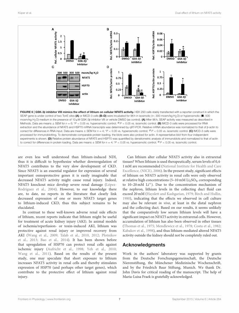
Pharmacological GSK-3β Inhibition Mimics theEffect of Lithium on NFAT5 ActivityTo determine whether lithium-induced downregulation ofNFAT5 is mediated by GSK-3β inhibition, we also examinedthe effect of the specific pharmacological GSK-3β inhibitor VIII.Similar to lithium treatment, under hyperosmotic conditionsboth cellular NFAT5 activity (Figure 6A) and expression ofNFAT5 and HSP70 (Figures 6B–D) were decreased significantlycompared with control cells. Under isosmotic conditions, cellularNFAT5 activity and HSP70 expression in cells treated with
GSK-3β inhibitor VIII were also less than in control cells, incontrast to lithium treatment. We assume that an even strongerdownregulation of NFAT5 by GSK-3β inhibitor VIII, comparedwith lithium, is responsible for this observation. Taken together,the results suggest that lithium-induced downregulation ofNFAT5 might be mediated by GSK-3β inhibition.
Discussion
The results presented in this study indicate that lithium has twoopposing effects on NFAT5 activity in renal cells: in the first,“rapid” response to lithium exposure, NFAT5 activity increases.This increase is probably mediated by inhibition of GSK-3βand subsequent activation of the c-terminal TAD of NFAT5,while NFAT5 protein levels are largely unaffected. The groupof Joan Ferraris showed recently that the TAD activity underisosmotic conditions is suppressed by GSK-3β, and that underhyperosmotic conditions, inhibitory phosphorylation of GSK-3β(mediated by AKT, PKA, and PI-3K) interrupts this suppression(Zhou et al., 2013). The lithium-induced increase of NFAT5activity was especially pronounced under isosmotic conditions.Normally, NFAT5 activity under isosmotic conditions is virtuallyzero, as illustrated by the minimal expression of the NFAT5 targetgene HSP70. Under these conditions, lithium increases NFAT5activity and expression of HSP70 severalfold (however, stillless than under hyperosmotic conditions). Under hyperosmoticconditions, the effect of lithium on NFAT5 activity is moremoderate, probably since GSK-3β is already inhibited underthese conditions, as mentioned above. An additional GSK-3β inhibition by lithium only slightly enhances TAD activity(25–50%).
When cells were exposed to lithium for prolonged periods(up to 96 h), a second, “slow” response was observed: underhyperosmotic conditions, cellular NFAT5 activity and expressionof NFAT5 target genes decreased, probably due to reducedNFAT5 protein abundance. The mechanisms, by which lithium-induced inhibition of GSK-3β mediates NFAT5 downregulation,are not clear. Normally, NFAT5 protein abundance increasesunder hyperosmotic conditions, which, in addition to activationof TAD, contributes to enhanced cellular NFAT5 activity underthese conditions. Enhanced transcription and increased mRNAstability have been identified as important mechanisms forhyperosmolality-induced upregulation of NFAT5 expression (Caiet al., 2005). Since NFAT5 mRNA levels were largely unaffectedby prolonged exposure to lithium, we assume that translationalor posttranslational mechanisms are probably responsible forthe observed downregulation of NFAT5. Regulation of NFAT5by such mechanisms has, to date, not been addressed in detail.In cardiac cells, doxorubicin induces ubiquitin-independentproteasomal degradation of NFAT5 protein (Ito et al., 2007),and recent investigations suggest that the long non-coding RNA“non-coding repressor of NFAT” (NRON) may be involved inproteasomal NFAT5 degradation (Umekita et al., 2013). Weattempted to establish whether the proteasomal inhibitor MG-132 influences lithium-induced degradation of NFAT5, but sinceexposure of IMCD-3 cells to the combination of hyperosmoticstress, lithium and MG-132 for periods longer than 24 h caused
Frontiers in Physiology | www.frontiersin.org 5 September 2015 | Volume 6 | Article 264

Küper et al. Dual effect of lithium on NFAT5 activity
FIGURE 5 | Long-term exposure to lithium decreases cellular NFAT5 activity. HEK 293 cells stably transfected with a reporter construct in which the SEAP
gene is under control of two TonE sites (A) or IMCD-3 cells (B–D) were incubated for 96 h in isosmotic ( ; 300 mosm/kg H2O) or hyperosmotic (�; 500 mosm/kg
H2O) medium and exposed to 10mM Li2SO4 or 10mM Na2SO4 (as control). (A) After 96 h, SEAP activity was measured as described in Methods. Data are means ±
SEM for n = 6; *P < 0.05 vs. hyperosmotic control; #P < 0.05 vs. isosmotic control. (B) IMCD-3 cells were processed for RNA extraction and the abundance of
NFAT5 and HSP70 mRNA transcripts was determined by qRT-PCR. Relative mRNA abundance was normalized to that of β-actin to correct for differences in RNA
input. Data are means ± SEM for n = 4; *P < 0.05 vs. hyperosmotic control; #P < 0.05 vs. isosmotic control. (C) IMCD-3 cells were processed for immunoblotting.
To demonstrate comparable protein loading, the blots were also probed for actin. A representative blot from four independent experiments is shown. (D) Relative
protein abundance of NFAT5 and HSP70 was quantified by densitometric analysis of immunoblots and normalized to that of actin to correct for differences in protein
loading. Data are means ± SEM for n = 4; *P < 0.05 vs. hyperosmotic control; #P < 0.05 vs. isosmotic control.
massive cell death, we were not able to obtain significant results(data not shown).
Of particular interest is the observation that the impactof prolonged lithium incubation (96 h) on NFAT5 activityalso depends on environmental osmolality. As discussed above,NFAT5 activity and expression of target gene HSP70 clearlydeclines under hyperosmotic conditions. Under isosmoticconditions, however, NFAT5 activity in lithium treated cellsis higher than in control cells, although NFAT5 proteinabundance slightly decreased.We assume that, in sum, the strongstimulatory effect of lithium on TAD activity of NFAT5 overridesthe decrease of NFAT5 protein, as also indicated by the increasedexpression of HSP70 under these conditions. Transferred to thesituation in the kidney, these results indicate that the effect oflithium on NFAT5 activity depends not only on the durationof exposure but also on the kidney region. Cortical collectingduct cells are not usually exposed to hyperosmotic stress, hence,NFAT5 activity in this region may be enhanced even during long-term lithium treatment. In contrast, in collecting duct cells ofthe outer and especially the inner medulla, which are regularlyexposed to hyperosmotic conditions, lithium probably decreasesNFAT5 activity and expression of its target genes. Accordingly, aproteomic analysis demonstrated that expression of the NFAT5target genes AR and HSP70 is significantly decreased in innermedullary collecting duct cells in lithium-treated rats (Nielsenet al., 2008).
The question arises as to whether altered NFAT5 activitycontributes to the well-known renal side effects of lithium
therapy. The molecular mechanisms underlying these side effectsare not completely understood (Behl et al., 2015). Among theseside effects, NDI is the most frequent, affecting up to 40% oflithium-treated patients. Lithium decreases AQP-2 expressionand trafficking to the luminal membrane of the principal cellsof the collecting duct. Moreover, the urea transporters UT-Aand UT-B, which are necessary for efficient urea circulation inthe kidney and hence for maximal urinary concentration ability,are downregulated by lithium in a rat model (Klein et al., 2002;Blount et al., 2010). NFAT5 has been identified as a positiveregulator of AQP-2 (Kasono et al., 2005; Hasler et al., 2006)and UT-A (Nakayama et al., 2000) expression; additionally,the NFAT5 target gene AR has been linked to the urinaryconcentration mechanism (Aida et al., 2000). Accordingly, in amouse model expressing a dominant-negative NFAT5 derivative,the urinary concentrating ability is decreased (Lam et al., 2004).In a rat model of sepsis-induced polyuria, inhibitory nitrosylationof NFAT5 is associated with decreased renal expression of AQP-2,UT-A1, ClC-K1 and its regulatory subunit barttin, also resultingin impaired urinary concentration ability (Küper et al., 2012).These data clearly indicate the importance of NFAT5 for urinaryconcentration, and thus it seems plausible that lithium-induceddownregulation of NFAT5, at least in medullary collecting ductcells, contributes to the development of NDI during lithiumtreatment.
Long-term treatment with lithium (>10 years) is alsoassociated with an enhanced risk of developing CKD (Markowitzet al., 2000; Presne et al., 2003). The underlying mechanisms
Frontiers in Physiology | www.frontiersin.org 6 September 2015 | Volume 6 | Article 264
Küper et al. Dual effect of lithium on NFAT5 activity
FIGURE 6 | GSK-3β inhibitor VIII mimics the effect of lithium on cellular NFAT5 activity. HEK 293 cells stably transfected with a reporter construct in which the
SEAP gene is under control of two TonE sites (A) or IMCD-3 cells (B–D) were incubated for 96 h in isosmotic ( ; 300 mosm/kg H2O) or hyperosmotic (�; 500
mosm/kg H2O) medium in the presence of 10µM GSK-3β inhibitor VIII or vehicle DMSO (as control). (A) After 96 h, SEAP activity was measured as described in
Methods. Data are means ± SEM for n = 6; *P < 0.05 vs. hyperosmotic control; #P < 0.05 vs. isosmotic control. (B) IMCD-3 cells were processed for RNA
extraction and the abundance of NFAT5 and HSP70 mRNA transcripts was determined by qRT-PCR. Relative mRNA abundance was normalized to that of β-actin to
correct for differences in RNA input. Data are means ± SEM for n = 4; *P < 0.05 vs. hyperosmotic control; #P < 0.05 vs. isosmotic control. (C) IMCD-3 cells were
processed for immunoblotting. To demonstrate comparable protein loading, the blots were also probed for actin. A representative blot from four independent
experiments is shown. (D) Relative protein abundance of NFAT5 and HSP70 was quantified by densitometric analysis of immunoblots and normalized to that of actin
to correct for differences in protein loading. Data are means ± SEM for n = 4; *P < 0.05 vs. hyperosmotic control; #P < 0.05 vs. isosmotic control.
are even less well understood than lithium-induced NDI,thus it is difficult to hypothesize whether downregulation ofNFAT5 contributes to the very slow development of CKD.Since NFAT5 is an essential regulator for expression of severalimportant osmoprotective genes it is easily imaginable thatdecreased NFAT5 activity might cause renal injury. Indeed,NFAT5 knockout mice develop severe renal damage (López-Rodríguez et al., 2004). However, to our knowledge thereare, to date, no reports in the literature that clearly linkdecreased expression of one or more NFAT5 target genesto lithium-induced CKD, thus this subject remains to beelucidated.
In contrast to these well-known adverse renal side effectsof lithium, recent reports indicate that lithium might be usefulfor treatment of acute kidney injury (AKI). In animal modelsof ischemia/reperfusion- or toxin-induced AKI, lithium wasprotective against renal injury or improved recovery fromAKI (Wang et al., 2009; Talab et al., 2010, 2012; Plotnikovet al., 2013; Bao et al., 2014). It has been shown beforethat upregulation of HSP70 can protect renal cells againstischemic injury (Aufricht et al., 1998; Yeh et al., 2010;Wang et al., 2011). Based on the results of the presentstudy, one may speculate that short exposure to lithiumincreases NFAT5 activity in renal cells and thereby stimulatesexpression of HSP70 (and perhaps other target genes), whichcontribute to the protective effect of lithium against renalinjury.
Can lithium alter cellular NFAT5 activity also in extrarenaltissues?When lithium is used therapeutically, serum levels of 0,4-1mM are recommended (National Institute for Health and CareExcellence, (NICE), 2006). In the present study, significant effectsof lithium on NFAT5 activity in renal cells were only observedat relative high concentrations (5–10mM Li2SO4, correspondingto 10–20mM Li+). Due to the concentration mechanism ofthe nephron, lithium levels in the collecting duct fluid canexceed 20mM (Hayslett and Kashgarian, 1979; Birch and Hullin,1980), indicating that the effects we observed in cell culturemay also be relevant in vivo, at least in the distal nephronand the collecting duct. Based on our results, it seems unlikelythat the comparatively low serum lithium levels will have asignificant impact onNFAT5 activity in extrarenal cells. However,accumulation of lithium has also been observed in other tissues(Thomas et al., 1975; Mendlewicz et al., 1978; Costa et al., 1982;Kabakov et al., 1998), and thus lithium-mediated altered NFAT5activity outside the kidney should not be completely ruled out.
Acknowledgments
Work in the authors’ laboratory was supported by grantsfrom the Deutsche Forschungsgemeinschaft, the DeutscheNierenstiftung, the Münchener Medizinische Wochenschrift,and by the Friedrich Baur Stiftung, Munich. We thank Dr.John Davis for critical reading of the manuscript. The help ofMaria-Luisa Fraek is gratefully acknowledged.
Frontiers in Physiology | www.frontiersin.org 7 September 2015 | Volume 6 | Article 264

Küper et al. Dual effect of lithium on NFAT5 activity
References
Aida, K., Ikegishi, Y., Chen, J., Tawata, M., Ito, S., Maeda, S., et al. (2000).
Disruption of aldose reductase gene (Akr1b1) causes defect in urinary
concentrating ability and divalent cation homeostasis. Biochem. Biophys. Res.
Commun. 277, 281–286. doi: 10.1006/bbrc.2000.3648
Aufricht, C., Lu, E., Thulin, G., Kashgarian, M., Siegel, N. J., and Van Why, S. K.
(1998). ATP releases HSP-72 from protein aggregates after renal ischemia. Am.
J. Physiol. 274, F268–F274.
Bao, H., Ge, Y., Wang, Z., Zhuang, S., Dworkin, L., Peng, A., et al. (2014). Delayed
administration of a single dose of lithium promotes recovery from AKI. J. Am.
Soc. Nephrol. 25, 488–500. doi: 10.1681/ASN.2013040350
Behl, T., Kotwani, A., Kaur, I., and Goel, H. (2015). Mechanisms of prolonged
lithium therapy-induced nephrogenic diabetes insipidus. Eur. J. Pharmacol.
755, 27–33. doi: 10.1016/j.ejphar.2015.02.040
Bendz, H., Schön, S., Attman, P. O., and Aurell, M. (2010). Renal failure occurs
in chronic lithium treatment but is uncommon. Kidney Int. 77, 219–224. doi:
10.1038/ki.2009.433
Birch, N. J., and Hullin, R. P. (1980). Lithium and the kidney. Br. Med. J. 280,
1148–1149. doi: 10.1136/bmj.280.6223.1148-b
Blount, M. A., Sim, J. H., Zhou, R., Martin, C. F., Lu, W., Sands, J. M., et al.
(2010). Expression of transporters involved in urine concentration recovers
differently after cessation of lithium treatment. Am. J. Physiol. Renal Physiol.
298, F601–F608. doi: 10.1152/ajprenal.00424.2009
Cai, Q., Ferraris, J. D., and Burg, M. B. (2005). High NaCl increases
TonEBP/OREBP mRNA and protein by stabilizing its mRNA. Am. J. Physiol.
Renal Physiol. 289, F803–F807. doi: 10.1152/ajprenal.00448.2004
Costa, J. L., Fay, D. D., Nurnberger, J. I., and Murphy, D. L. (1982). Preferential
accumulation of lithium in the dense bodies of human platelets. Biochem.
Pharmacol. 31, 3215–3218. doi: 10.1016/0006-2952(82)90552-4
Doble, B. W., and Woodgett, J. R. (2003). GSK-3: tricks of the trade for a
multi-tasking kinase. J. Cell Sci. 116, 1175–1186. doi: 10.1242/jcs.00384
Ferraris, J. D., Williams, C. K., Persaud, P., Zhang, Z., Chen, Y., and Burg, M. B.
(2002). Activity of the TonEBP/OREBP transactivation domain varies directly
with extracellular NaCl concentration. Proc. Natl. Acad. Sci. U.S.A. 99, 739–744.
doi: 10.1073/pnas.241637298
Grünfeld, J. P., and Rossier, B. C. (2009). Lithium nephrotoxicity revisited. Nat.
Rev. Nephrol. 5, 270–276. doi: 10.1038/nrneph.2009.43
Hasler, U., Jeon, U. S., Kim, J. A., Mordasini, D., Kwon, H. M., Féraille, E., et al.
(2006). Tonicity-responsive enhancer binding protein is an essential regulator
of aquaporin-2 expression in renal collecting duct principal cells. J. Am. Soc.
Nephrol. 17, 1521–1531. doi: 10.1681/ASN.2005121317
Hayslett, J. P., and Kashgarian, M. (1979). A micropuncture study of the renal
handling of lithium. Pflugers Arch. 380, 159–163. doi: 10.1007/BF00582152
Irarrazabal, C. E., Burg, M. B., Ward, S. G., and Ferraris, J. D. (2006).
Phosphatidylinositol 3-kinase mediates activation of ATM by high NaCl and
by ionizing radiation: role in osmoprotective transcriptional regulation. Proc.
Natl. Acad. Sci. U.S.A. 103, 8882–8887. doi: 10.1073/pnas.0602911103
Ito, T., Fujio, Y., Takahashi, K., and Azuma, J. (2007). Degradation of NFAT5,
a transcriptional regulator of osmotic stress-related genes, is a critical event
for doxorubicin-induced cytotoxicity in cardiac myocytes. J. Biol. Chem. 282,
1152–1160. doi: 10.1074/jbc.M609547200
Kabakov, A. Y., Karkanias, N. B., Lenox, R. H., and Papke, R. L. (1998). Synapse-
specific accumulation of lithium in intracellular microdomains: a model for
uncoupling coincidence detection in the brain. Synapse 28, 271–279.
Kasono, K., Saito, T., Saito, T., Tamemoto, H., Yanagidate, C., Uchida, S.,
et al. (2005). Hypertonicity regulates the aquaporin-2 promoter independently
of arginine vasopressin. Nephrol. Dial. Transplant 20, 509–515. doi:
10.1093/ndt/gfh677
Klein, J. D., Gunn, R. B., Roberts, B. R., and Sands, J. M. (2002). Down-regulation
of urea transporters in the renal inner medulla of lithium-fed rats. Kidney Int.
61, 995–1002. doi: 10.1046/j.1523-1755.2002.00210.x
Klein, P. S., and Melton, D. A. (1996). A molecular mechanism for the effect
of lithium on development. Proc. Natl. Acad. Sci. U.S.A. 93, 8455–8459. doi:
10.1073/pnas.93.16.8455
Ko, B. C., Lam, A. K., Kapus, A., Fan, L., Chung, S. K., and Chung, S. S. (2002).
Fyn and p38 signaling are both required for maximal hypertonic activation of
the osmotic response element-binding protein/tonicity-responsive enhancer-
binding protein (OREBP/TonEBP). J. Biol. Chem. 277, 46085–46092. doi:
10.1074/jbc.M208138200
Ko, B. C., Turck, C. W., Lee, K. W., Yang, Y., and Chung, S. S. (2000).
Purification, identification, and characterization of an osmotic response
element binding protein. Biochem. Biophys. Res. Commun. 270, 52–61. doi:
10.1006/bbrc.2000.2376
Küper, C., Fraek, M. L., Muller, H. H., Beck, F. X., and Neuhofer, W. (2012). Sepsis-
induced urinary concentration defect is related to nitric oxide-dependent
inactivation of TonEBP/NFAT5, which downregulates renal medullary solute
transport proteins and aquaporin-2. Crit. Care Med. 40, 1887–1895. doi:
10.1097/CCM.0b013e31824e1186
Kwon, T. H., Laursen, U. H., Marples, D., Maunsbach, A. B., Knepper, M.
A., Frokiaer, J., et al. (2000). Altered expression of renal AQPs and Na(+)
transporters in rats with lithium-induced NDI. Am. J. Physiol. Renal Physiol.
279, F552–F564.
Lam, A. K., Ko, B. C., Tam, S., Morris, R., Yang, J. Y., Chung, S. K., et al. (2004).
Osmotic response element-binding protein (OREBP) is an essential regulator
of the urine concentrating mechanism. J. Biol. Chem. 279, 48048–48054. doi:
10.1074/jbc.M407224200
Livak, K. J., and Schmittgen, T. D. (2001). Analysis of relative gene expression
data using real-time quantitative PCR and the 2(-Delta Delta C(T)) Method.
Methods 25, 402–408. doi: 10.1006/meth.2001.1262
López-Rodríguez, C., Antos, C. L., Shelton, J. M., Richardson, J. A., Lin, F.,
Novobrantseva, T. I., et al. (2004). Loss of NFAT5 results in renal atrophy and
lack of tonicity-responsive gene expression. Proc. Natl. Acad. Sci. U.S.A. 101,
2392–2397. doi: 10.1073/pnas.0308703100
Markowitz, G. S., Radhakrishnan, J., Kambham, N., Valeri, A. M., Hines,
W. H., and D’Agati, V. D. (2000). Lithium nephrotoxicity: a progressive
combined glomerular and tubulointerstitial nephropathy. J. Am. Soc. Nephrol.
11, 1439–1448.
Marples, D., Christensen, S., Christensen, E. I., Ottosen, P. D., and Nielsen,
S. (1995). Lithium-induced downregulation of aquaporin-2 water channel
expression in rat kidney medulla. J. Clin. Invest. 95, 1838–1845. doi:
10.1172/JCI117863
Mendlewicz, J., Verbanck, P., Linkowski, P., and Wilmotte, J. (1978).
Lithium accumulation in erythrocytes of manic-depressive patients:
an in vivo twin study. Br. J. Psychiatry 133, 436–444. doi: 10.1192/bjp.
133.5.436
Miyakawa, H., Woo, S. K., Chen, C. P., Dahl, S. C., Handler, J. S., and Kwon, H. M.
(1998). Cis- and trans-acting factors regulating transcription of the BGT1 gene
in response to hypertonicity. Am. J. Physiol. 274, F753–F761.
Miyakawa, H., Woo, S. K., Dahl, S. C., Handler, J. S., and Kwon, H. M. (1999).
Tonicity-responsive enhancer binding protein, a rel-like protein that stimulates
transcription in response to hypertonicity. Proc. Natl. Acad. Sci. U.S.A. 96,
2538–2542. doi: 10.1073/pnas.96.5.2538
Nakayama, Y., Peng, T., Sands, J. M., and Bagnasco, S. M. (2000). The
TonE/TonEBP pathway mediates tonicity-responsive regulation of UT-
A urea transporter expression. J. Biol. Chem. 275, 38275–38280. doi:
10.1074/jbc.M004678200
Neuhofer, W., Steinert, D., Fraek, M. L., and Beck, F. X. (2007). Prostaglandin
E2 stimulates expression of osmoprotective genes in MDCK cells and
promotes survival under hypertonic conditions. J. Physiol. 583, 287–297. doi:
10.1113/jphysiol.2007.135178
National Institute for Health and Care Excellence, (NICE) (2006). “Bipolar
disorder: the management of bipolar disorder in adults, children and
adolescents, in primary and secondary care,” in Clinical Guideline 38.
Nielsen, J., Hoffert, J. D., Knepper, M. A., Agre, P., Nielsen, S., and Fenton, R. A.
(2008). Proteomic analysis of lithium-induced nephrogenic diabetes insipidus:
mechanisms for aquaporin 2 down-regulation and cellular proliferation.
Proc. Natl. Acad. Sci. U.S.A. 105, 3634–3639. doi: 10.1073/pnas.08000
01105
Plotnikov, E. Y., Grebenchikov, O. A., Babenko, V. A., Pevzner, I. B., Zorova,
L. D., Likhvantsev, V. V., et al. (2013). Nephroprotective effect of GSK-
3beta inhibition by lithium ions and delta-opioid receptor agonist dalargin
on gentamicin-induced nephrotoxicity. Toxicol. Lett. 220, 303–308. doi:
10.1016/j.toxlet.2013.04.023
Frontiers in Physiology | www.frontiersin.org 8 September 2015 | Volume 6 | Article 264

Küper et al. Dual effect of lithium on NFAT5 activity
Presne, C., Fakhouri, F., Noël, L. H., Stengel, B., Even, C., Kreis, H., et al. (2003).
Lithium-induced nephropathy: rate of progression and prognostic factors.
Kidney Int. 64, 585–592. doi: 10.1046/j.1523-1755.2003.00096.x
Quadri, S., and Siragy, H. M. (2014). Regulation of (pro)renin receptor expression
in mIMCD via the GSK-3beta-NFAT5-SIRT-1 signaling pathway. Am. J.
Physiol. Renal Physiol. 307, F593–F600. doi: 10.1152/ajprenal.00245.2014
Roth, I., Leroy, V., Kwon, H. M., Martin, P. Y., Féraille, E., and Hasler, U. (2010).
Osmoprotective transcription factor NFAT5/TonEBP modulates nuclear
factor-kappaB activity. Mol. Biol. Cell 21, 3459–3474. doi: 10.1091/mbc.E10-
02-0133
Stambolic, V., Ruel, L., and Woodgett, J. R. (1996). Lithium inhibits glycogen
synthase kinase-3 activity and mimics wingless signalling in intact cells. Curr.
Biol. 6, 1664–1668. doi: 10.1016/S0960-9822(02)70790-2
Talab, S. S., Elmi, A., Emami, H., Nezami, B. G., Assa, S., Ghasemi, M., et al.
(2012). Protective effects of acute lithium preconditioning against renal
ischemia/reperfusion injury in rat: role of nitric oxide and cyclooxygenase
systems. Eur. J. Pharmacol. 681, 94–99. doi: 10.1016/j.ejphar.2012.
01.042
Talab, S. S., Emami, H., Elmi, A., Nezami, B. G., Assa, S., Deroee, A. F., et al. (2010).
Chronic lithium treatment protects the rat kidney against ischemia/reperfusion
injury: the role of nitric oxide and cyclooxygenase pathways. Eur. J. Pharmacol.
647, 171–177. doi: 10.1016/j.ejphar.2010.08.036
Thomas, R. C., Simon, W., and Oehme, M. (1975). Lithium accumulation by
snail neurones measured by a new Li+-sensitive microelectrode. Nature 258,
754–756. doi: 10.1038/258754a0
Umekita, K., Trenkmann, M., Kolling, C., Michel, B. A., Gay, R. E., Gay, S., et al.
(2013). Long noncoding RNANron regulates the cytoplasmic-nuclear shuttling
and activity of NFAT5 in rheumatoid arthritis synovial fibroblast [Abstract].
Arthritis Rheum. 65(Suppl. 10):2872. doi: 10.1002/art.2013.65.issue-s10
Wang, Y., Huang, W. C., Wang, C. Y., Tsai, C. C., Chen, C. L., Chang, Y. T.,
et al. (2009). Inhibiting glycogen synthase kinase-3 reduces endotoxaemic acute
renal failure by down-regulating inflammation and renal cell apoptosis. Br. J.
Pharmacol. 157, 1004–1013. doi: 10.1111/j.1476-5381.2009.00284.x
Wang, Z., Gall, J. M., Bonegio, R. G., Havasi, A., Hunt, C. R., Sherman, M. Y.,
et al. (2011). Induction of heat shock protein 70 inhibits ischemic renal injury.
Kidney Int. 79, 861–870. doi: 10.1038/ki.2010.527
Woo, S. K., Lee, S. D., Na, K. Y., Park, W. K., and Kwon, H. M.
(2002). TonEBP/NFAT5 stimulates transcription of HSP70 in response to
hypertonicity. Mol. Cell. Biol. 22, 5753–5760. doi: 10.1128/MCB.22.16.5753-
5760.2002
Yeh, C. H., Hsu, S. P., Yang, C. C., Chien, C. T., and Wang, N. P. (2010). Hypoxic
preconditioning reinforces HIF-alpha-dependent HSP70 signaling to reduce
ischemic renal failure-induced renal tubular apoptosis and autophagy. Life Sci.
86, 115–123. doi: 10.1016/j.lfs.2009.11.022
Zhang, Z., Ferraris, J. D., Brooks, H. L., Brisc, I., and Burg, M. B. (2003). Expression
of osmotic stress-related genes in tissues of normal and hyposmotic rats. Am. J.
Physiol. Renal Physiol. 285, F688–F693. doi: 10.1152/ajprenal.00028.2003
Zhou, X., Wang, H., Burg, M. B., and Ferraris, J. D. (2013). Inhibitory
phosphorylation of GSK-3beta by AKT, PKA, and PI3K contributes to high
NaCl-induced activation of the transcription factor NFAT5 (TonEBP/OREBP).
Am. J. Physiol. Renal Physiol. 304, F908–F917. doi: 10.1152/ajprenal.00591.2012
Conflict of Interest Statement: The authors declare that the research was
conducted in the absence of any commercial or financial relationships that could
be construed as a potential conflict of interest.
Copyright © 2015 Küper, Beck and Neuhofer. This is an open-access article
distributed under the terms of the Creative Commons Attribution License (CC BY).
The use, distribution or reproduction in other forums is permitted, provided the
original author(s) or licensor are credited and that the original publication in this
journal is cited, in accordance with accepted academic practice. No use, distribution
or reproduction is permitted which does not comply with these terms.
Frontiers in Physiology | www.frontiersin.org 9 September 2015 | Volume 6 | Article 264