Embed Size (px)
Citation preview
1
<R/Heads>PHARMACOLOGY
Drugs affecting the autonomic nervous system
Matthew Charlton
Jonathan P Thompson
Matthew Charlton MBChB FRCA is an Honorary Clinical Lecturer and Specialist Registrar in
Anaesthesia & Critical Care at the University Hospitals of Leicester NHS Trust, Leicester, UK.
Conflicts of interest: none declared.
Jonathan P Thompson BSc (Hons) MD FRCA FFICM is an Honorary Professor at the University of
Leicester and Consultant in Anaesthesia & Critical Care at the University Hospitals of Leicester
NHS Trust, Leicester Royal Infirmary, UK. Conflicts of interest: none declared.
Abstract
The Autonomic Nervous System (ANS) is a complex system of nervous and humoral
mechanisms that modulates the function of the autonomous or visceral organs. Autonomic
control of organs aims to maintain homeostasis in health. Many drugs used in clinical practice
can have either primary or secondary effects on the function of autonomic nervous system.
Keywords: autonomic nervous system, catecholamines, parasympathetic, sympathetic.
Royal College of Anaesthetists CPD Matrix: 1A02, 2C04.
Learning objectives
After reading this article you should be able to
Identify five sympathomimetic drugs, their indications and doses
Identify five sympatholytic drugs, their indications and doses
2
Explain the biosynthesis of endogenous catecholamines
The ANS is divided on anatomical, physiological and pharmacological grounds into Sympathetic
(SNS) and Parasympathetic (PNS) nervous systems (Tables 1 & 2). Both the SNS and PNS consist
of pre- and post-ganglionic neurones. Pre-ganglionic fibres of the SNS arise from the thoraco-
lumbar regions of the spinal cord, with pre-ganglionic fibres of the PNS arising from the cranio-
sacral regions. Pre-ganglionic transmission in both the SNS and PNS is mediated via
acetylcholine (ACh), acting at nicotinic acetylcholine receptors.
Post-ganglionic transmission in SNS neurones is primarily mediated by noradrenaline, acting via
specific adrenergic receptors, except in sweat glands and the adrenal gland. Sweat gland post-
ganglionic neurones release ACh. Pre-ganglionic fibres of the adrenal gland synapse directly
with the adrenal medulla, stimulating the release of adrenaline from enterochromaffin cells.
Adrenergic receptors are classified into three major types (α1, α2, and β), with further subtypes
in each class. Two subtypes of β-receptor (β1 and β2) are well defined on functional, anatomical
and pharmacological grounds and a third β-receptor subtype, β3, is found in adipocytes, skeletal
and ventricular muscle, and the vasculature. Dopaminergic (DA) receptors are now classified
separately from adrenoceptors but are included here due to overlap in their actions and
response to exogenous and endogenous catecholamines. There are 5 subtypes of DA receptors
(D1-D5) belonging to two subfamilies: D1-like and D2-like. D1-like receptors mediate
vasodilatation in vascular smooth muscle of the renal, splanchnic, coronary and cerebral
circulations; D2–like receptors are widespread in the central nervous system.
Post-ganglionic transmission in the PNS is mediated via acetylcholine, acting via muscarinic
acetylcholine receptors of which there are five subtypes (M1-5).
Postganglionic muscarinic and adrenergic receptors are coupled to membrane-bound G-
proteins and elicit a response through second and third messenger systems that vary with
receptor subtype (Table 1).
Termination of neurotransmitter activity is brought about by a number of mechanisms.
Acetylcholine is rapidly hydrolysed by the enzyme acetylcholinesterase to acetate and choline.
Noradrenaline is removed from the synaptic junction by the reuptake system, being returned to
the sympathetic nerve that released it. It is subsequently metabolised intra-neuronally by
3
monoamine oxidase (MAO) enzymes. Circulating catecholamines and metabolised by the
catechol-O-methyltransferase (COMT) enzyme system in the liver [1].
(TABLE 1 NEAR HERE PLEASE)
Drugs acting on the Sympathetic Nervous System
Drugs with effects that mimic stimulation of SNS or adrenal medullary discharge are termed
sympathomimetics; drugs that antagonise the sympathetic nervous system effects are called
sympatholytics. Other more recent methods of modulating the autonomic nervous system (such
as implantable carotid sinus stimulators or renal nerve ablation procedures) have been
introduced for the treatment of drug-resistant hypertension; these are outside the scope of this
article.
Sympathomimetics
Sympathomimetics mimic SNS stimulation by one of three mechanisms, acting directly on
adrenoceptors (e.g. catecholamines, phenylephrine and methoxamine), indirectly by stimulating
release of noradrenaline from nerve endings (e.g. amphetamine), or by combination of both
mechanisms (e.g. dopamine, ephedrine and metaraminol). Sympathomimetics can be classified
pharmacologically according to their structure (catecholamine/non-catecholamine); origin
(endogenous/synthetic) and site of action (adrenoceptor/non-adrenoceptor).
Catecholamines
Catecholamine drugs can be endogenous or synthetic. All catecholamines are based on a
benzene ring structure with hydroxyl groups at the C3 and C4 positions. Substitutions in the
amine side-chain lead to the different hormones. Catecholamines have very short half-lives in
vivo and are immediately inactivated in the gut by monoamine oxidase (MAO) enzymes. They are
therefore usually administered parenterally, with doses being titrated to clinical effect. Choice of
catecholamine depends on the clinical indication, desired therapeutic response and duration of
action.
Endogenous catecholamines
The endogenous catecholamines (dopamine, noradrenaline and adrenaline) are synthesised
from the essential amino acid phenylalanine (Figure 1).
(TABLE 2 AND Figure 1 NEAR HERE PLEASE)
Adrenaline (Epinephrine)
4
Adrenaline is the principle catecholamine (80-90 %) synthesised by the adrenal medulla and is a
potent, non-selective sympathomimetic. It can administered intravenously (IV), intramuscularly
(IM), topically or via nebulizer or tracheal tube. In non-emergency situations, adrenaline should
be administered IM to reduce the risk of cardiac arrhythmias and intense vasoconstriction. In
emergency situations (e.g. cardiac arrest, peri-arrest and anaphylaxis) the IV route is indicated.
The effects of adrenaline are dose-dependent. In low doses, β-effects predominate, leading to
bronchodilation, and an increase in heart rate, cardiac output and myocardial oxygen
consumption. Vasodilatation of skeletal muscle and splanchnic arterioles leads to a decrease in
peripheral vascular resistance seen clinically as reduced diastolic pressure. At higher infusion
rates or bolus doses, α1-effects predominate, with vasoconstriction leading to an increase in
systemic vascular resistance.
Adrenaline is included in a number of algorithms supported by the Resuscitation Council (UK),
including those for cardiac arrest and anaphylaxis [2]. In cardiac arrest scenarios, adrenaline is
administered at a dose of 1mg (10 ml of 1:10000 [0.1 mg/ml]) at alternating CPR cycles, with
commencement depending on presenting rhythm. In anaphylaxis adrenaline is usually
administered IM at a dose of 500 mcg (0.5 of 1:1000 [1 mg/ml] solution), although IV doses of
50-100 µg (0.5-1ml of 1:10000 [0.1 mg/ml]) can alternatively be used. Adrenaline can be given
by continuous infusion in shock states, administered at a dose range of 0.01-0.5 µg/kg/min.
Adrenaline is incorporated into local anaesthetic solutions, prolonging their duration of action by
decreasing systemic adsorption (due to localised vasoconstriction). Adrenaline is also used as a
topical vasoconstrictor to achieve local haemostasis and in the treatment of wide-angle
glaucoma.
Noradrenaline (Norepinephrine)
Noradrenaline is synthesised in the adrenal medulla and postganglionic sympathetic nerve
endings from the essential amino acid tyrosine as previously described. Noradrenaline acts
primarily on α-receptors to cause intense arteriolar and venous vasoconstriction, usually
accompanied by a reflex bradycardia. Noradrenaline is also an agonist at β-adrenoceptors and
therefore falls in to the class of ‘ino-constrictors’, alongside the other endogenous
catecholamines. Noradrenaline is primarily indicated in states of low systemic vascular
resistance, as seen in sepsis and post-cardiac bypass. Noradrenaline increases systolic and
diastolic pressures with an associated rise in pulmonary artery and central venous pressures.
Cardiac output may fall (due to increased afterload), with an increase in myocardial oxygenation
consumption. Noradrenaline is administered IV as an infusion through a central vein. The usual
concentration is a solution of 100 µg/ml administered at a dose of 0.05-1.5 µg/kg/min.
5
Dopamine
Dopamine is the natural precursor of adrenaline and noradrenaline (Figure 1). It directly
stimulates α, β and dopaminergic receptors (D1 and D2), whilst also stimulating the release of
noradrenaline from adrenergic nerve endings. Dopamine receptors are present throughout the
body but concentrated in the CNS (basal ganglia, chemoreceptor trigger zone and the pituitary
gland), with receptors also in the splanchnic and renal circulations.
The effects of dopamine are dose-dependent. At doses of 1-5 µg/kg/min, dopamine primarily
stimulates D1 receptors leading to renal and mesenteric vasodilatation, with an associated
increase in blood flow. This leads to increased glomerular filtration, diuresis and natriuresis,
though there is no evidence that dopamine in this range confers protection from renal
dysfunction [3]. Between 5-10 µg/kg/min, β1 effects predominate leading to increased inotropy,
and cardiac output with lesser effects on heart rate. At doses above 15µg/kg/min, α-mediated
effects predominate with an increase in systemic vascular resistance.
Dopamine has been used for the treatment of cardiogenic shock, refractory congestive cardiac
failure and following cardiac surgery, although its use is declining.
(TABLE 3 NEAR HERE PLEASE)
Synthetic catecholamines
Dobutamine
Dobutamine is a synthetic catecholamine similar in structure to dopamine. Dobutamine acts
primarily at β1-receptors, with some activity at β2-receptors. Dobutamine increases cardiac
contractility, heart rate and cardiac output (due to both an increase in contractility and mild
decrease in afterload). It also increases conduction through the AV node and should be used
with caution in patients with atrial fibrillation. Dobutamine is administered as an IV infusion, the
usual dose being 0.5-40 µg/kg/min.
Dopexamine
Dopexamine is a synthetic dopamine analogue that stimulates β2 and D1 receptors and may also
inhibit neuronal noradrenaline reuptake. It has minimal effect on D2 receptors. The combination
of β2 and D1 stimulation leads to increased chronotropy and inotropy of the heart, alongside
vasodilation and a decrease in systemic vascular resistance, seen particularly in the mesenteric,
skeletal and renal vascular beds. Dopexamine is used in the treatment of acute primary cardiac
failure and low cardiac output states. The dose range of dopexamine is 0.5-6 µg/kg/min.
6
Isoprenaline
Isoprenaline is a β-adrenoceptor agonist. It has been used for the temporary treatment of
bradyarrhythmias and atrioventricular block owing to its effects primarily on β1 receptors. This
leads to an increase in automaticity and heart rate (arrhythmias are common). β2-effects
produce bronchodilation, vasodilatation and a decrease in systemic vascular resistance.
Increased inotropy and decreased afterload lead to an increase in cardiac output. It is now only
available in the UK from special-order manufacturer or specialist importing companies.
Non catecholamine sympathomimetic drugs
Ephedrine is a naturally occurring sympathomimetic amine that acts both directly (stimulating
both α and β receptors) and indirectly (by causing release of noradrenaline from sympathetic
nerve terminals). Ephedrine increases heart rate, cardiac output, cardiac oxygen demand,
cerebral and coronary blood flow. It also induces bronchodilation and tachypnoea. These effects
last longer (onset within 1 minute and lasting up to one hour) than endogenous catecholamines
as ephedrine is not metabolised by COMT and MAO. Intravenous doses range from 3-30mg.
Owing to its indirect actions, tachyphylaxis can develop due to depletion of noradrenaline from
nerve terminals. Ephedrine easily crosses blood-brain barrier and can be used in the treatment
of narcolepsy and nocturnal enuresis. Ephedrine was used widely in obstetric anaesthesia as
uterine blood flow is relatively maintained. However it crosses the placenta to cause increased
foetal metabolic rate and metabolic acidosis so has been superseded by phenylephrine in
obstetric patients [4].
Phenylephrine is a direct-acting α1 agonist, causing vasoconstriction and a rapid rise in blood
pressures. Other effects include mydriasis. Phenylephrine can be administered via several routes
including oral, nasal (as a decongestant), topical eye drops and IV. MAO is present in the GI tract
and the bioavailability of phenylephrine is approximately 38%. Phenylephrine has largely
replaced ephedrine as the vasopressor of choice in obstetric anaesthesia. Phenylephrine can be
administered as a bolus of 50-100 µg IV or used as an infusion. Effects occur within 1 minute
with a duration of up to 45 minutes after bolus injection. Phenylephrine has no inotropic or
chronotropic effects directly but should be used with caution as marked reflex bradycardia can
occur.
Metaraminol is a direct and indirect-acting α and β-agonist. Acting mainly via α1 receptors, its
main actions are to increase blood pressure via an increase in systemic vascular resistance.
Pulmonary vascular resistance is also increased leading to pulmonary arterial hypertension. The
dose range is 0.5-2 mg IV titrated slowly as reflex bradycardia leading to cardiac arrest has been
reported. Effects are seen within 2 minutes and duration is 20-60 minutes.
7
α2-adrenoceptor agonists
Clonidine acts at central and peripheral α2 receptors as a partial agonist (some α1 activity α2: α1
>200:1) and as a full agonist at central imidazoline receptors. Transient hypertension and
bradycardia can occur after IV injection through stimulation of vascular α2 receptors followed by
centrally mediated hypotension. Bolus doses of 2-3 µg/kg attenuates sympathetic responses
during anaesthesia whilst 1-2 µg/kg neuraxially increases the potency and duration of analgesic
block. Clonidine is absorbed well orally with peak plasma levels at 60-90 minutes. Clonidine can
be used orally to treat hypertension and can be given as an intravenous infusion to provide
sedation on the critical care unit.
Dexmedetomidine is a full agonist at central and peripheral α2 receptors (α2: α1 >1,600:1), with a
selectivity 8-10 fold greater than clonidine for α2 receptors. Dexmedetomidine has similar effects
to clonidine on sympathetic responses under anaesthesia, has significant opioid sparing effects
and is used in the treatment of intractable neuropathic pain. Dexmedetomidine can be used as a
single-agent sedative in intensive care, administered as an infusion in the dose range of 0.2-1.4
µg/kg/hr.
Sympatholytic drugs
α-adreceptor antagonists
Non selective α antagonists
Phentolamine and phenoxybenzamine are used in the management of hypertensive crises and
phaeochromocytoma.
Phentolamine is a competitive non-selective α-blocker, predominantly acting at α1-
adrenoceptors (three times the affinity than for α2). It can be administered as an intravenous
bolus at a dose of 1-5 mg repeated as necessary to effector as an infusion at a rate of 0.1-0.2
mg/min. It has an onset of 1-2 minutes and duration of action between 5-20 minutes.
Phenoxybenzamine is a non-competitive irreversible α-blocker with a high affinity for α1. It is
administered orally, starting at a dose of 10 mg and increased as required; the usual maintenance
dose is 1-2 mg kg-1 daily in 2 divided doses. Onset time is several hours with a duration of action
lasting up to 3 days owing to its prolonged elimination, covalent binding and the need for new α-
adrenoceptors to be synthesised.
Both phentolamine and phenoxybenzamine can produce postural hypotension with reflex
tachycardia. Abdominal side-effects can also be present including cramps and diarrhoea.
8
Selective α1 antagonists
Drugs that selectively block α1-adrenoceptors are predominantly used for the symptomatic
treatment of benign prostatic hyperplasia as they cause relaxation of prostatic smooth muscle
and increase urinary flow. Doxazosin, Indoramin, and Prazosin are also licenced for the
treatment of hypertension, though the latter 2 drugs have largely been superseded for this
indication. All can cause postural hypotension and a condition associated with cataract surgery
known as “floppy iris syndrome”.
Doxazosin is a selective α1 antagonist used to treat BPH and hypertension. Its duration of action
is prolonged compared to earlier drugs such as prazosin and it has a better side effect profile in
terms of erectile function when used in BPH. The usual dose is 2-4 mg (maximum 16mg) daily
Tamsulosin predominantly blocks prostatic α1a receptors as opposed to vascular α1b receptors. It
is used to treat benign prostatic hyperplasia (BPH) as smooth muscle relaxation allows easier
urinary flow. Adverse effects include reactions to the sulphur moiety of the drug.
β-adrenoreceptor antagonists
β-blockers are classified according to their receptor selectivity (β1 or β2). All β-blockers are
competitive antagonists and bind avidly to their specific receptors. β -blockers decrease cardiac
contractility and automaticity, and increase refractory times at the SA and AV nodes. Heart rate,
cardiac work, propensity to arrhythmias, risk of myocardial ischaemia and arterial pressure are
all reduced. Non-cardiac effects include increased airway and peripheral vascular resistance;
these can lead to bronchospasm and worsen symptoms of peripheral vascular disease. Some β-
blockers act as partial agonists, membrane stabilisers and some antagonise other receptors.
Non selective β-antagonists
Propanolol is a non-selective β-blocker that also exhibits a membrane stabilising effect through
its action on Na+ channels (although not in clinically significant doses). It has no sympathomimetic
actions, is highly lipid-soluble and crosses the blood-brain barrier. Propanolol is used to treat the
sympathetic manifestations of anxiety, for thyrotoxicosis, portal hypertension, hypertrophic
obstructive cardiomyopathy, migraine but has been superseded in the treatment of hypertension
and angina. It is administered orally (30-320 mg/day) in 2-3 divided doses, or IV 1-10 mg titrated
to response.
Carvedilol and labetalol are non-selective β-blockers that also antagonise α1-receptors. Labetalol
is primarily used in the treatment of hypertensive crises particularly pregnancy-induced
9
hypertension and pre-eclampsia. The ratio of β to α activity varies with route of administration
(7:1 when administered IV). It is available as an oral formulation (dose up to 2.4 g/day) or IV
infusion of 2 mg/min. Bolus IV doses can be administered (e.g. 20 mg over 2 minutes). Adverse
effects include insomnia, drowsiness and rarely respiratory distress.
Selective β1 blockers
Atenolol is a relatively cardioselective (β1) beta blocker that can be administered orally (50-
100mg/day) or IV (2.5-10mg, max 1mg/min). Atenolol is predominantly excreted unchanged in
the urine and therefore the dose must be adjusted in patients with renal disease.
Metoprolol is used to treat hypertension and in the treatment/prevention of angina. It is available
in either oral (50% bioavailability), or IV preparations. There possesses no intrinsic
sympathomimetic activity. Oral doses vary with indication between 100-450 mg/day. Slow
release preparations are available.
Bisoprolol exhibits almost twice the selectivity for β1 receptors compared to atenolol and
propanolol. Bisoprolol also inhibits the secretion of renin. The oral dose is 2.5-20 mg/day (oral
bioavailability 90%) and metabolism is via hepatic and renal systems to inactive metabolites. The
half-life of bisoprolol is approximately 10-12 hours.
Esmolol is a short-acting β1-selective blocker used in the treatment of supraventricular
tachyarrhythmias (atrial flutter and fibrillation) and perioperative hypertension. Esmolol is
metabolised by erythrocyte esterases to produce methanol and a weaker active metabolite. It is
administered IV as bolus (0.5-2.0 mg/kg) or an infusion (25-500 µg/kg/min) with clinical effects
within 2 minutes and duration of action of approximately 10 minutes.
Drugs acting on the Parasympathetic nervous system
Agonists
Pilocarpine is a muscarinic agonist used topically in the treatment of glaucoma.
Antagonists
Atropine is a competitive muscarinic antagonist with widespread dose-dependent effects
including increased heart rate, decreased bladder tone, decreased salivary secretions, increased
intra-ocular pressure and mydriasis. Atropine is administered IV in a dose of 0.6-3.0 mg to
counter intense vagal stimulation (the dose in children is 20 µg/kg). At very low doses there is a
paradoxical bradycardia thought to be mediated by M2 receptor antagonism centrally in the CNS
10
(Bezold-Jarisch effect). The therapeutic dose effects are mediated by M3 receptors, whilst at high
doses, excitation/sedation, hallucinations, ventricular arrhythmias or hyperthermia can occur.
Hyoscine is available in two forms: hydrobromide and butylbromide. Hyoscine butylbromide is
used as an antispasmodic (genitourinary and gastrointestinal). The hydrobromide form increases
heart rate less but is a more effective antisialogogue than atropine and causes problems in the
elderly, as it crosses the blood brain barrier causing confusion, sedation and ataxia (central
anticholinergic syndrome). Hyoscine is used as an antiemetic, especially for motion sickness
(transdermal patch application).
Glycopyrrolate is a quarternary ammonium compound with similar effects to atropine but is
approximately 5 times more potent as an antisialogogue. Glycopyrrolate does not cross the
blood brain barrier so has no CNS effects. It is used to prevent bradycardia caused by neostigmine
as it has a similar onset and duration of action. The dose of glycopyrrolate in adults is 0.2-0.4 mg.
References:
1. McCorry LK. Physiology of the Autonomic Nervous System. American Journal of
Pharmaceutical Education. 2007;71(4):78.
2. Resuscitation Council (UK). 2015. Resuscitation Guidelines. [ONLINE] Available at:
https://www.resus.org.uk/resuscitation-guidelines. [Accessed 02 February 16].
3. ANZICS Clinical Trial Group. Low-dose dopamine in patients with early renal dysfunction: a
placebo-controlled randomised trial. The Lancet. Volume 356, Issue 9248 , 2139 – 2143.
4. Cooper DW, et al. Effect of intravenous vasopressor on spread of spinal anaesthesia and fetal acid-base equilibrium. Br. J. Anaesth. (2007) 98 (5): 649-656.
5. Caulfield MP, Birdsall NJ. "International Union of Pharmacology. XVII. Classification of
muscarinic acetylcholine receptors". Pharmacol. Rev.1998. 50 (2): 279–90
Further reading
Book chapter:
11
Thompson JP. Drugs acting on the cardiovascular system. In Aitkenhead AR, Moppett IK,
Thompson JP, eds Smith and Aitkenhead’s Textbook of Anaesthesia, 6th edn. Churchill
Livingstone, Elsevier 2013.
Journal article:
Bangash MN, Kong M-L, Pearse RM. Use of inotropes and vasopressor agents in critically ill
patients. British Journal of Pharmacology. 2012;165(7):2015-2033. doi:10.1111/j.1476-
5381.2011.01588.x.
Print and online:
British National Formulary. 2016. Cardiovascular System. [ONLINE] Available at:
https://www.evidence.nhs.uk/formulary/bnf/current/2-cardiovascular-system. [Accessed 02
February 16].
12
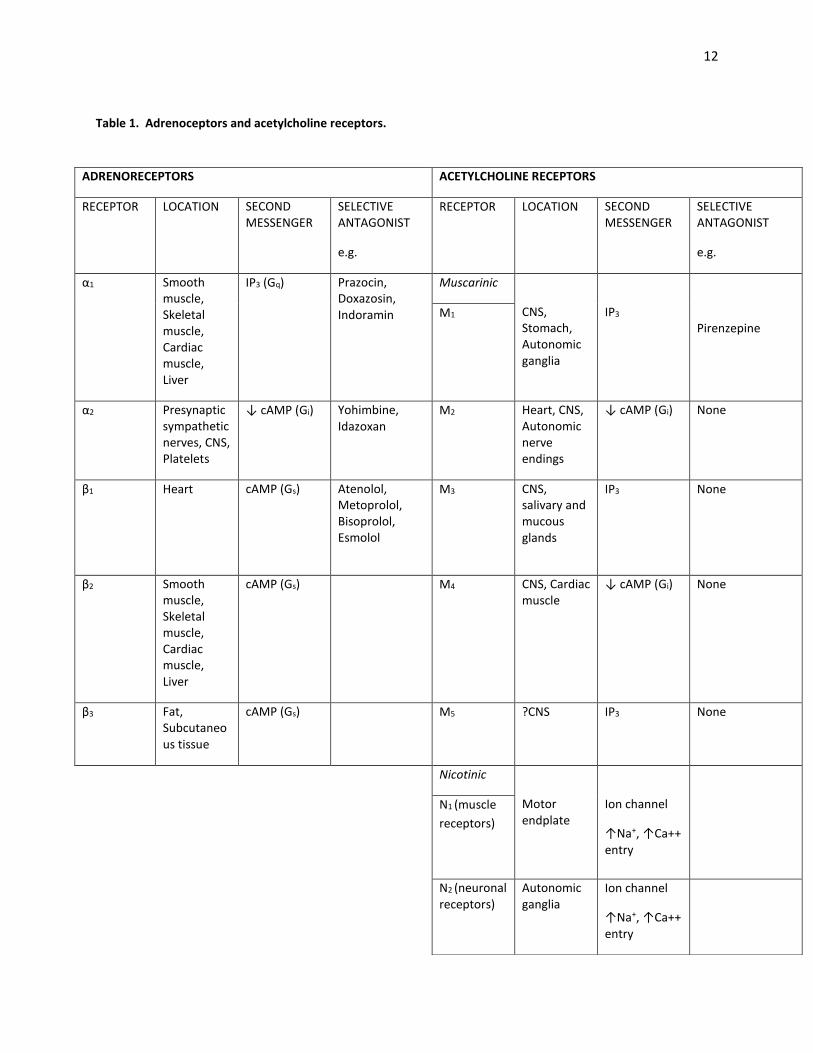
Table 1. Adrenoceptors and acetylcholine receptors.
ADRENORECEPTORS ACETYLCHOLINE RECEPTORS
RECEPTOR LOCATION SECOND MESSENGER
SELECTIVE ANTAGONIST
e.g.
RECEPTOR LOCATION SECOND MESSENGER
SELECTIVE ANTAGONIST
e.g.
α1 Smooth muscle, Skeletal muscle, Cardiac muscle, Liver
IP3 (Gq) Prazocin, Doxazosin, Indoramin
Muscarinic
CNS, Stomach, Autonomic ganglia
IP3
Pirenzepine M1
α2 Presynaptic sympathetic nerves, CNS, Platelets
↓ cAMP (Gi) Yohimbine, Idazoxan
M2 Heart, CNS, Autonomic nerve endings
↓ cAMP (Gi)
None
β1 Heart cAMP (Gs)
Atenolol, Metoprolol, Bisoprolol, Esmolol
M3 CNS, salivary and mucous glands
IP3 None
β2 Smooth muscle, Skeletal muscle, Cardiac muscle, Liver
cAMP (Gs)
M4 CNS, Cardiac muscle
↓ cAMP (Gi)
None
β3 Fat, Subcutaneous tissue
cAMP (Gs)
M5 ?CNS IP3 None
Nicotinic
Motor endplate
Ion channel
↑Na+, ↑Ca++ entry
N1 (muscle
receptors)
N2 (neuronal receptors)
Autonomic ganglia
Ion channel
↑Na+, ↑Ca++ entry
13
Footnote to Table 1 (table from the last edition):
Stimulation of β1-3 receptors results in the activation of GTP binding Gs proteins, which in turn activates adenylate cyclase enzymes, generating cAMP to mediate the associated altered cell function. Stimulated α2, M2 and M4 receptors interact with Gi proteins to inhibit adenylate cyclase and hence reduce cAMP. Stimulation of α1, M1 and M3 receptors causes an interaction with the Gq protein. This leads to activation of membrane bound phospholipase C, hydrolysing phosphatidylinositol biphosphate (PIP2) to Inositol triphosphate (IP3) and diacylglycerol (DAG). IP3 binds to its receptor, opening Ca2+ channels. Nicotinic receptors are associated with non-selective ion channels that open up on their activation to effect changes.
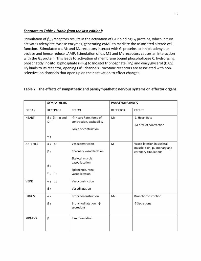
Table 2. The effects of sympathetic and parasympathetic nervous systems on effector organs.
SYMPATHETIC PARASYMPATHETIC
ORGAN RECEPTOR EFFECT RECEPTOR EFFECT
HEART β 1 , β 2 , α and D1
α 1
↑ Heart Rate, force of contraction, excitability
Force of contraction
M2 ↓ Heart Rate
↓Force of contraction
ARTERIES α 1 α 2
β 1
β 2
D1, β 2
Vasoconstriction
Coronary vasodilatation
Skeletal muscle vasodilatation
Splanchnic, renal vasodilatation
M Vasodilatation in skeletal muscle, skin, pulmonary and coronary circulations
VEINS α 1 α 2
β 2
Vasoconstriction
Vasodilatation
LUNGS α 1
β 2
Bronchoconstriction
Bronchodilatation , ↓ secretions
M3 Bronchoconstriction
↑Secretions
KIDNEYS β
Renin secretion
14
Footnote to Table 2 (table from the last edition): The 5 different muscarinic receptor subtypes (M1-5) have been classified using selective radioactively-labeled agonist and antagonist substances [5]. Note that in some cases the receptor subtype is unknown.
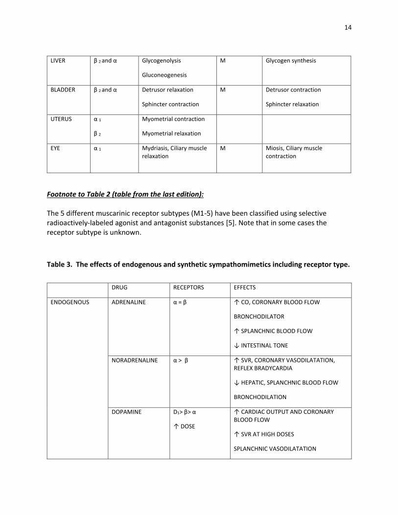
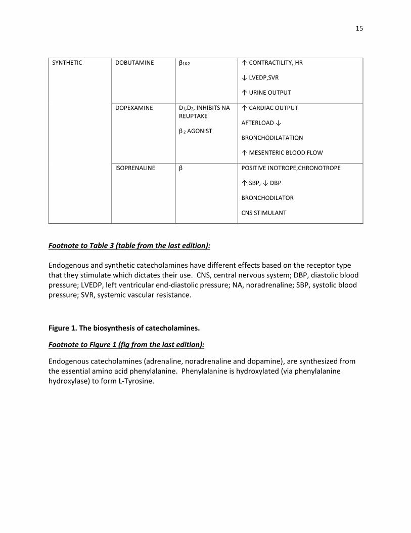
Table 3. The effects of endogenous and synthetic sympathomimetics including receptor type.
LIVER β 2 and α
Glycogenolysis
Gluconeogenesis
M Glycogen synthesis
BLADDER β 2 and α
Detrusor relaxation
Sphincter contraction
M Detrusor contraction
Sphincter relaxation
UTERUS α 1
β 2
Myometrial contraction
Myometrial relaxation
EYE α 1
Mydriasis, Ciliary muscle relaxation
M Miosis, Ciliary muscle contraction
DRUG RECEPTORS EFFECTS
ENDOGENOUS ADRENALINE α = β ↑ CO, CORONARY BLOOD FLOW
BRONCHODILATOR
↑ SPLANCHNIC BLOOD FLOW
↓ INTESTINAL TONE
NORADRENALINE α > β ↑ SVR, CORONARY VASODILATATION, REFLEX BRADYCARDIA
↓ HEPATIC, SPLANCHNIC BLOOD FLOW
BRONCHODILATION
DOPAMINE D1> β> α
↑ DOSE
↑ CARDIAC OUTPUT AND CORONARY BLOOD FLOW
↑ SVR AT HIGH DOSES
SPLANCHNIC VASODILATATION
15
Footnote to Table 3 (table from the last edition): Endogenous and synthetic catecholamines have different effects based on the receptor type that they stimulate which dictates their use. CNS, central nervous system; DBP, diastolic blood pressure; LVEDP, left ventricular end-diastolic pressure; NA, noradrenaline; SBP, systolic blood pressure; SVR, systemic vascular resistance.
Figure 1. The biosynthesis of catecholamines.
Footnote to Figure 1 (fig from the last edition):
Endogenous catecholamines (adrenaline, noradrenaline and dopamine), are synthesized from the essential amino acid phenylalanine. Phenylalanine is hydroxylated (via phenylalanine hydroxylase) to form L-Tyrosine.
SYNTHETIC DOBUTAMINE β1&2 ↑ CONTRACTILITY, HR
↓ LVEDP,SVR
↑ URINE OUTPUT
DOPEXAMINE D1,D2, INHIBITS NA REUPTAKE
β 2 AGONIST
↑ CARDIAC OUTPUT
AFTERLOAD ↓
BRONCHODILATATION
↑ MESENTERIC BLOOD FLOW
ISOPRENALINE β POSITIVE INOTROPE,CHRONOTROPE
↑ SBP, ↓ DBP
BRONCHODILATOR
CNS STIMULANT