Embed Size (px)
Citation preview
Clin. Invest. (Epub ahead of print) ISSN 2041-6792
Drug Profile
part of
10.4155/CLI.14.59 © 2014 Future Medicine Ltd
Clin. Invest.
Drug Profile4
8
2014
Mycosis fungoides (MF) is a rare, potentially life-threatening cutaneous T-cell lymphoma characterized by cutaneous homing of neoplastic T lymphocytes. MF can mimic other diseases; clinicopathologic evaluation and imaging studies are essential. Biopsy of suspicious skin sites is essential for diagnosis. Topical mechlorethamine has been clinically tested over decades for the treatment of MF. Safety concerns include contact dermatitis, pruritus and hyperpigmentation. Nonmelanoma skin cancers have been reported with topical mechlorethamine use, including in patients who received therapies known to cause nonmelanoma skin cancer. Noninferiority to mechlorethamine ointment in a Phase II controlled trial led to the US FDA 2013 approval of VALCHLOR™ (mechlorethamine gel) for treatment of stage IA/IB MF after prior skin-directed therapy.
Keywords: cutaneous T-cell lymphoma • mechlorethamine hydrochloride • mycosis fungoides • nitrogen mustard
Mycosis fungoidesCutaneous T-cell lymphoma (CTCL) com-prises approximately 4% of non-Hodgkin’s lymphomas in the USA [1]. Of the cutane-ous lymphomas reported in the USA from 2001–2005, CTCLs accounted for 71% of the cases [2], and have an overall annual age-adjusted incidence of 9.6 per million persons [1]. CTCLs, characterized by localization of neoplastic T lymphocytes to the skin, encom-pass a broad group of cutaneous lymphomas that include the slower progressing myco-sis fungoides (MF) and the more aggressive Sézary syndrome (SS).
MF is the most common type of CTCL (54% of CTCLs were MF in a USA study from 2001 to 2005) [2]. First described in the early 1800s, [3] MF is characterized by epidermal and dermal infiltration of clonal T cells. There are many variants of MF including folliculo-tropic, hypopigmented and granulomatous MF [4–6].
MF is a rare disease. Although prevalence is difficult to determine, the annual incidence of MF in the USA has been reported at between
3.6 [7] and 4.1 per million people a year [2], with no strong evidence of increasing inci-dence rates. MF incidence rates do increase with patient age, peaking at around 80 years [2,7], although MF cases in children have also been reported [1,7,8]. Incidence rates were simi-lar among males and females at ages younger than 30 years, but rose among males at older ages, with the male:female ratio doubled by age 60 years[2].
Mean age at diagnosis is 55 to 60 years of age [2,9,10], with reports in older and younger patients [11]. 71% of patients presented with early-stage disease [9,10]. Women presented with early onset of MF before the age of 40 years more often than men [12].
One of the challenges with treating MF is possible long-term misdiagnosis as other dis-ease states such as chronic contact dermatitis, folliculitis, eczema, vitiligo, pigmented pur-puric dermatoses, pityriasis lichenoides chron-ica, pityriasis lichenoides et varioliformis acuta or psoriasis [13–16]. Because of incorrect diagnosis, the proper treatment of MF may be delayed, resulting in poorly directed therapies.
Recent clinical evidence for topical mechlorethamine in mycosis fungoides
Joya Sahu1, Marjan Sepassi2, Mitchell Nagao2 & Youn H Kim*,3
1Dermatology, Pathology & Medical
Oncology, Jefferson Cutaneous
Lymphoma Clinic, Thomas Jefferson
University Hospitals, 833 Chestnut St,
Suite 704, Philadelphia, PA 19107, USA 2Medical Affairs, Actelion
Pharmaceuticals US Inc., 5000 Shoreline
Court, Suite 200, South San Francisco,
CA 94080, USA 3Multidisciplinary Cutaneous Lymphoma
Program, Stanford University School of
Medicine, 900 Blake Wilbur Dr MC 5356,
Office W1010, Stanford, CA 94305, USA
*Author for correspondence:
Tel.: +1 650 725 3292
Fax: +1 650 725 6937
For reprint orders, please contact [email protected]
10.4155/CLI.14.59 Clin. Invest. (Epub ahead of print) future science group
Drug Profile Sahu, Sepassi, Nagao & Kim
Adding to the difficulty of diagnosis, the pathophysi-ology of MF is not well understood. T cells found in MF seem to function as T cells under normal physiologic conditions that home to the skin, become activated and develop into a clonal state [13]. Chemokine receptors can play an important role in this process. The activation of T-cell integrins can lessen T-cell adhesion to skin endothelial cells and a gradient of chemokines (e.g., CC chemokine ligand 17 [CCL17] and 27 [CCL27]) attract chemokine receptors (e.g., CC chemokine recep-tor 4 [CCR4]) on the malignant T cells that help the T cell migrate to the epidermis. The CD4+ T cells often cluster around antigen-presenting dendritic cells, such as Langerhans cells, forming Pautrier’s microabscesses that result in T-cell activation and the release of inflam-matory cytokines. Kinases (e.g., PI3K and Akt) are upregulated, and downstream effectors are activated that can allow T cells to survive and proliferate [17,18].
Studying the band-like infiltrate of lymphocytes per-meating the papillary dermis from a skin biopsy may help with diagnosis of MF. Small, medium-sized and sometimes large mononuclear cells with atypia (pleo-morphic, hyperchromatic or cerebriform nuclei) may be seen [19,20]. Pautrier’s microabscesses are not frequently seen upon histologic examination; however, immuno-histochemical staining generally shows atypical CD4+ T cells [11]. Pautrier’s microabscesses have been seen in 4–37% of patch biopsies in patients diagnosed with early MF [21,22].
Advanced stages may lead to the formation of gener-alized erythroderma associated with lymphadenopathy and neoplastic T lymphocytes with cerebriform nuclei (Sézary cells) in skin, lymph nodes and peripheral blood, known as the leukemic variant of MF, SS. MF accounts for approximately 50–80% of CTCL, whereas SS accounts for approximately 1–3% of cases [1,2,23]. SS may be similar to MF except that cellular infiltrates are more likely a single type of cell and epidermotropism, or cellular movement towards the epidermis, may be absent [1,2,23].
Etiologic factors of MF are indeterminate but have included infectious agents, environmental exposures and genetic mutations [19]. It is believed that the uncon-trolled clonal accumulation of T lymphocytes is a result of chronic antigenic stimulation [13]. Ulceration of tumors, with secondary infection with Staphylococ-cus aureus, Enterobacteriaceae and Pseudomonas aeru-ginosa, is a common cause of morbidity [19]. Minimal evidence may also support viral etiology for MF sec-ondary to Epstein–Barr virus and cytomegalovirus [24]. Patients with later-stage MF and SS are at a significantly increased risk of developing a second lymphoma, in par-ticular Hodgkin lymphoma, as well as nonhematologic malignancies [1,25].
To help diagnose MF, staining skin-biopsy speci-mens with a panel of lymphocyte markers and poly-merase chain reaction analysis of T-cell receptor genes to determine clonality may be performed [13]. If a patient appears to have advanced disease or if their lymph nodes are enlarged on physical examination or imaging studies, lymph node biopsies may be carried out. If SS is suspected, peripheral blood is examined for the presence of circulating malignant cells [19].
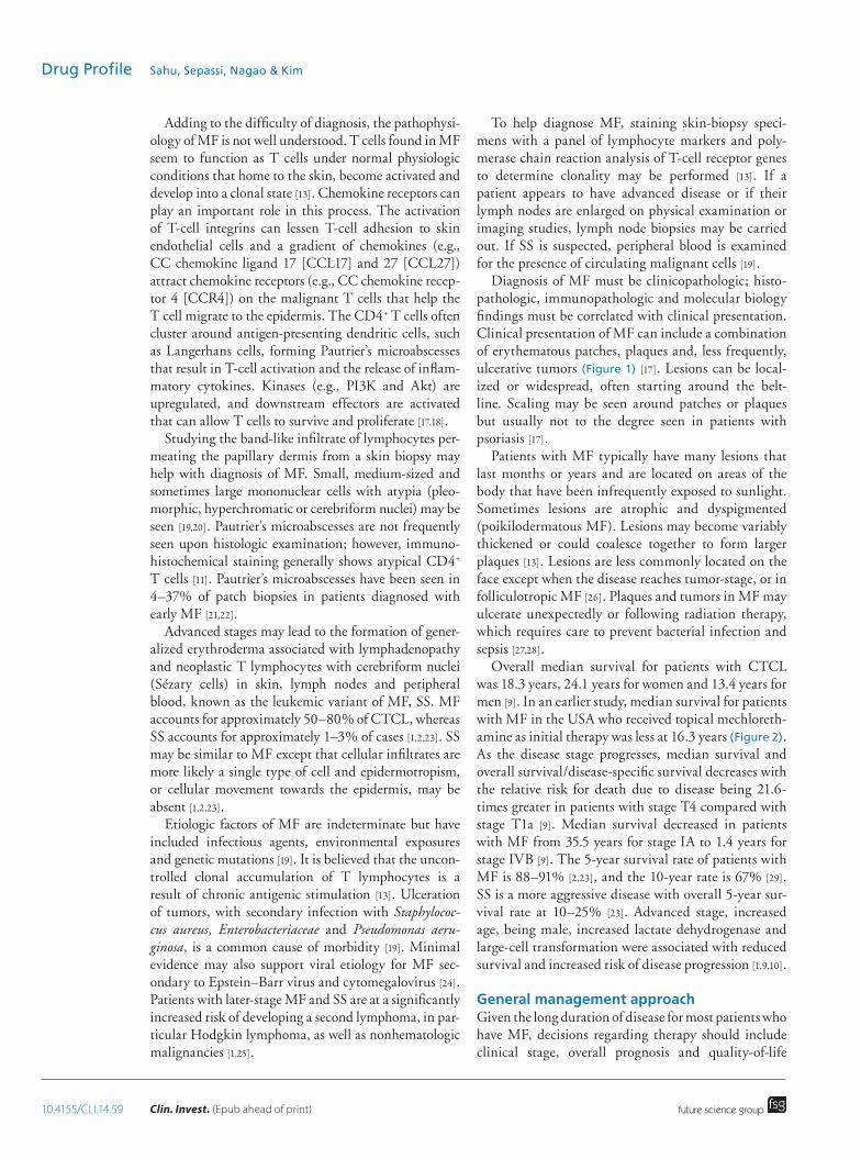
Diagnosis of MF must be clinicopathologic; histo-pathologic, immunopathologic and molecular biology findings must be correlated with clinical presentation. Clinical presentation of MF can include a combination of erythematous patches, plaques and, less frequently, ulcerative tumors (Figure 1) [17]. Lesions can be local-ized or widespread, often starting around the belt-line. Scaling may be seen around patches or plaques but usually not to the degree seen in patients with psoriasis [17].
Patients with MF typically have many lesions that last months or years and are located on areas of the body that have been infrequently exposed to sunlight. Sometimes lesions are atrophic and dyspigmented (poikilodermatous MF). Lesions may become variably thickened or could coalesce together to form larger plaques [13]. Lesions are less commonly located on the face except when the disease reaches tumor-stage, or in folliculotropic MF [26]. Plaques and tumors in MF may ulcerate unexpectedly or following radiation therapy, which requires care to prevent bacterial infection and sepsis [27,28].
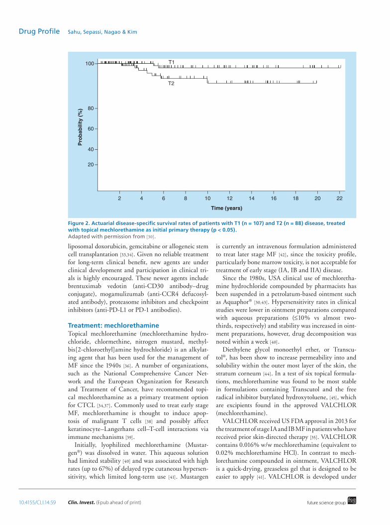
Overall median survival for patients with CTCL was 18.3 years, 24.1 years for women and 13.4 years for men [9]. In an earlier study, median survival for patients with MF in the USA who received topical mechloreth-amine as initial therapy was less at 16.3 years (Figure 2). As the disease stage progresses, median survival and overall survival/disease-specific survival decreases with the relative risk for death due to disease being 21.6-times greater in patients with stage T4 compared with stage T1a [9]. Median survival decreased in patients with MF from 35.5 years for stage IA to 1.4 years for stage IVB [9]. The 5-year survival rate of patients with MF is 88–91% [2,23], and the 10-year rate is 67% [29]. SS is a more aggressive disease with overall 5-year sur-vival rate at 10–25% [23]. Advanced stage, increased age, being male, increased lactate dehydrogenase and large-cell transformation were associated with reduced survival and increased risk of disease progression [1,9,10].
General management approachGiven the long duration of disease for most patients who have MF, decisions regarding therapy should include clinical stage, overall prognosis and quality-of-life
10.4155/CLI.14.59future science group
Recent clinical evidence for topical mechlorethamine in mycosis fungoides Drug Profile
considerations. Patients with MF believe the disease has had a severe impact on their functioning, emotional and social well-being [31].
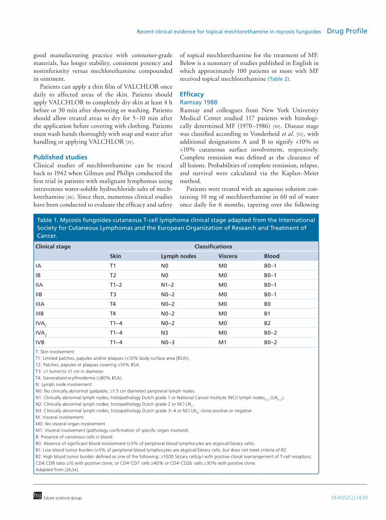
The clinical stage of MF, especially with regard to the degree of skin involvement, is crucial to determine prognosis. Stages of MF have been outlined by a num-ber of organizations [32] and the most recent classifi-cation is summarized in Table 1. Early clinical stage MF (stages IA–IIA) where disease is primarily lim-ited to the skin as patches alone and patches/plaques has a favorable prognosis [33]. Advanced stage MF (stages IIB–IVB), which can also include SS and may include lymph node and peripheral-blood involvement, has a more unfavorable prognosis [20,29,33].
The goals for treating patients with early stage MF are to relieve symptoms and achieve remission,
while avoiding long-term treatment-related toxicity. For patients with early stage MF, therapeutic options include topical corticosteroids, topical mechloretha-mine such as the newly approved VALCHLOR™ [35], local radiation, topical retinoids (bexarotene gel/Targretin® Gel), ultraviolet B therapy, topical imiquimod, topical carmustine (BCNU), psoralen plus ultraviolet A (PUVA) and total skin electron beam therapy [13,28,34].
For patients with advanced-stage disease, treat-ments aimed at reducing tumor burden or delaying disease progression are utilized [33]. Systemic thera-pies may be needed such as oral bexarotene, anti-folates (methotrexate, pralatrexate), extracorporeal photopheresis, IFN-α, histone deacetylase inhibitors inhibitors (vorinostat, romidepsin), alemtuzumab,
Figure 1. Cutaneous lesions of mycosis fungoides and Sézary syndrome. (A) Patch stage T1–2, (B) plaque stage T1–2, (C) tumor stage T3 and (D) erthyroderma stage T4. (A, C & D) reproduced with permission from Youn H Kim. (B) reproduced with permission from Joya Sahu.
A B
C D
10.4155/CLI.14.59 Clin. Invest. (Epub ahead of print) future science group
Drug Profile Sahu, Sepassi, Nagao & Kim
liposomal doxorubicin, gemcitabine or allogeneic stem cell transplantation [33,34]. Given no reliable treatment for long-term clinical benefit, new agents are under clinical development and participation in clinical tri-als is highly encouraged. These newer agents include brentuximab vedotin (anti-CD30 antibody–drug conjugate), mogamulizumab (anti-CCR4 defucosyl-ated antibody), proteasome inhibitors and checkpoint inhibitors (anti-PD-L1 or PD-1 antibodies).
Treatment: mechlorethamineTopical mechlorethamine (mechlorethamine hydro-chloride, chlormethine, nitrogen mustard, methyl-bis[2-chloroethyl]amine hydrochloride) is an alkylat-ing agent that has been used for the management of MF since the 1940s [36]. A number of organizations, such as the National Comprehensive Cancer Net-work and the European Organization for Research and Treatment of Cancer, have recommended topi-cal mechlorethamine as a primary treatment option for CTCL [34,37]. Commonly used to treat early stage MF, mechlorethamine is thought to induce apop-tosis of malignant T cells [38] and possibly affect keratinocyte–Langerhans cell–T-cell interactions via immune mechanisms [39].
Initially, lyophilized mechlorethamine (Mustar-gen®) was dissolved in water. This aqueous solution had limited stability [40] and was associated with high rates (up to 67%) of delayed type cutaneous hypersen-sitivity, which limited long-term use [41]. Mustargen
is currently an intravenous formulation administered to treat later stage MF [42], since the toxicity profile, particularly bone marrow toxicity, is not acceptable for treatment of early stage (IA, IB and IIA) disease.
Since the 1980s, USA clinical use of mechloretha-mine hydrochloride compounded by pharmacists has been suspended in a petrolatum-based ointment such as Aquaphor® [30,43]. Hypersensitivity rates in clinical studies were lower in ointment preparations compared with aqueous preparations (≤10% vs almost two-thirds, respectively) and stability was increased in oint-ment preparations, however, drug decomposition was noted within a week [40].
Diethylene glycol monoethyl ether, or Transcu-tol®, has been show to increase permeability into and solubility within the outer most layer of the skin, the stratum corneum [44]. In a test of six topical formula-tions, mechlorethamine was found to be most stable in formulations containing Transcutol and the free radical inhibitor butylated hydroxytoluene, [45], which are excipients found in the approved VALCHLOR (mechlorethamine).
VALCHLOR received US FDA approval in 2013 for the treatment of stage IA and IB MF in patients who have received prior skin-directed therapy [35]. VALCHLOR contains 0.016% w/w mechlorethamine (equivalent to 0.02% mechlorethamine HCl). In contrast to mech-lorethamine compounded in ointment, VALCHLOR is a quick-drying, greaseless gel that is designed to be easier to apply [41]. VALCHLOR is developed under
Figure 2. Actuarial disease-specific survival rates of patients with T1 (n = 107) and T2 (n = 88) disease, treated with topical mechlorethamine as initial primary therapy (p < 0.05). Adapted with permission from [30].
T1
T2
Time (years)
2 4 6 8 10 12 14 16 18 20 22
Pro
bab
ility
(%
)
20
40
60
80
100
10.4155/CLI.14.59future science group
Recent clinical evidence for topical mechlorethamine in mycosis fungoides Drug Profile
good manufacturing practice with consumer-grade materials, has longer stability, consistent potency and noninferiority versus mechlorethamine compounded in ointment.
Patients can apply a thin film of VALCHLOR once daily to affected areas of the skin. Patients should apply VALCHLOR to completely dry skin at least 4 h before or 30 min after showering or washing. Patients should allow treated areas to dry for 5–10 min after the application before covering with clothing. Patients must wash hands thoroughly with soap and water after handling or applying VALCHLOR [35].
Published studiesClinical studies of mechlorethamine can be traced back to 1942 when Gilman and Philips conducted the first trial in patients with malignant lymphomas using intravenous water-soluble hydrochloride salts of mech-lorethamine [36]. Since then, numerous clinical studies have been conducted to evaluate the efficacy and safety
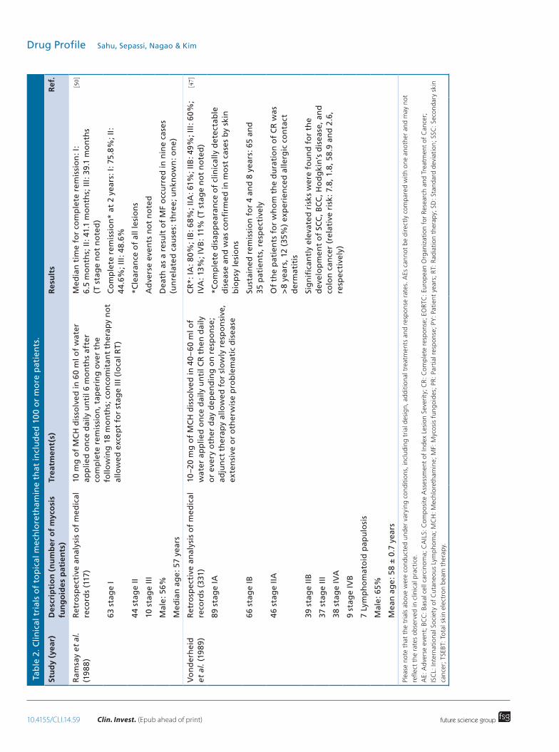
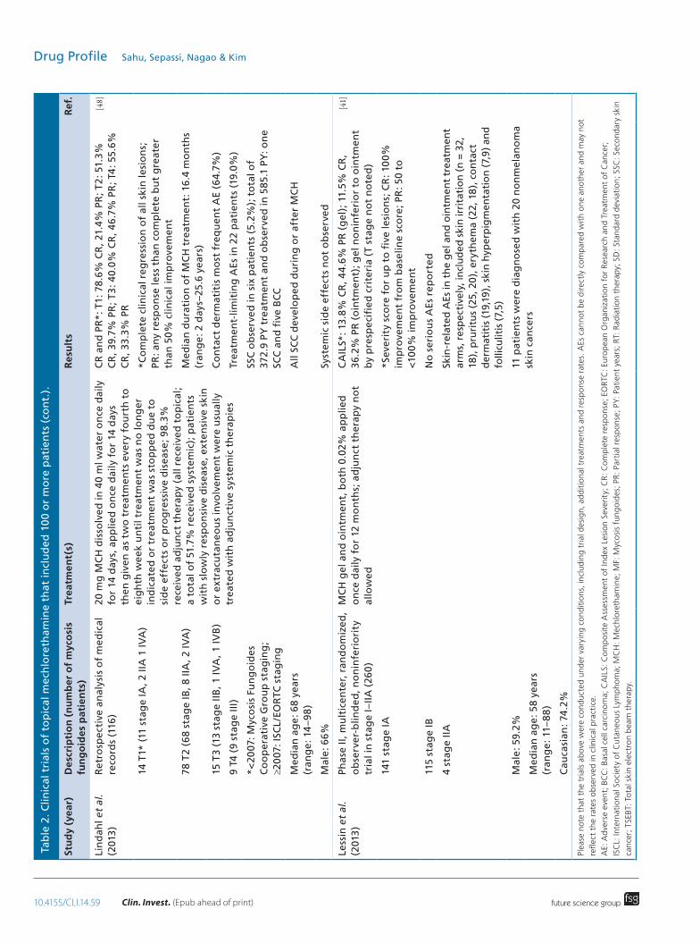
of topical mechlorethamine for the treatment of MF. Below is a summary of studies published in English in which approximately 100 patients or more with MF received topical mechlorethamine (Table 2).
EfficacyRamsay 1988Ramsay and colleagues from New York University Medical Center studied 117 patients with histologi-cally determined MF (1970–1986) [50]. Disease stage was classified according to Vonderheid et al. [51], with additional designations A and B to signify <10% or >10% cutaneous surface involvement, respectively. Complete remission was defined as the clearance of all lesions. Probabilities of complete remission, relapse, and survival were calculated via the Kaplan–Meier method.
Patients were treated with an aqueous solution con-taining 10 mg of mechlorethamine in 60 ml of water once daily for 6 months, tapering over the following
Table 1. Mycosis fungoides-cutaneous T-cell lymphoma clinical stage adapted from the International Society for Cutaneous Lymphomas and the European Organization of Research and Treatment of Cancer.
Clinical stage Classifications
Skin Lymph nodes Viscera Blood
IA T1 N0 M0 B0–1
IB T2 N0 M0 B0–1
IIA T1–2 N1–2 M0 B0–1
IIB T3 N0–2 M0 B0–1
IIIA T4 N0–2 M0 B0
IIIB T4 N0–2 M0 B1
IVA1 T1–4 N0–2 M0 B2
IVA2 T1–4 N3 M0 B0–2
IVB T1–4 N0–3 M1 B0–2
T: Skin involvement:
T1: Limited patches, papules and/or plaques (<10% body surface area [BSA]).
T2: Patches, papules or plaques covering ≥10% BSA.T3: ≥1 tumor(s) ≥1 cm in diameter.T4: Generalized erythroderma (≥80% BSA).N: Lymph node involvement:
N0: No clinically abnormal (palpable; ≥1.5 cm diameter) peripheral lymph nodes.N1: Clinically abnormal lymph nodes; histopathology Dutch grade 1 or National Cancer Institute (NCI) lymph nodes
0–2 (LN
0–2).
N2: Clinically abnormal lymph nodes; histopathology Dutch grade 2 or NCI LN3.
N3: Clinically abnormal lymph nodes; histopathology Dutch grade 3–4 or NCI LN4; clone positive or negative.
M: Visceral involvement:
M0: No visceral organ involvement.
M1: Visceral involvement (pathology confirmation of specific organ involved).
B: Presence of cancerous cells in blood:
B0: Absence of significant blood involvement (≤5% of peripheral blood lymphocytes are atypical/Sézary cells).B1: Low blood tumor burden (>5% of peripheral blood lymphocytes are atypical/Sézary cells, but does not meet criteria of B2.
B2: High blood tumor burden defined as one of the following: ≥1000 Sézary cells/μl with positive clonal rearrangement of T-cell receptors; CD4:CD8 ratio ≥10 with positive clone; or CD4+CD7- cells ≥40% or CD4+CD26- cells ≥30% with positive clone.Adapted from [20,34].
10.4155/CLI.14.59 Clin. Invest. (Epub ahead of print) future science group
Drug Profile Sahu, Sepassi, Nagao & KimTa
ble
2. C
linic
al t
rial
s o
f to
pic
al m
ech
lore
tham
ine
that
incl
ud
ed 1
00
or
mo
re p
atie
nts
.
Stu
dy
(yea
r)D
escr
ipti
on
(n
um
ber
of
myc
osi
s fu
ng
oid
es p
atie
nts
)Tr
eatm
ent(
s)R
esu
lts
Ref
.
Ram
say
et a
l. (1
98
8)
Ret
rosp
ecti
ve a
nal
ysis
of
med
ical
re
cord
s (1
17)
10 m
g o
f M
CH
dis
solv
ed in
60
ml o
f w
ater
ap
plie
d o
nce
dai
ly u
nti
l 6 m
on
ths
afte
r co
mp
lete
rem
issi
on
, tap
erin
g o
ver
the
follo
win
g 1
8 m
on
ths;
co
nco
mit
ant
ther
apy
no
t al
low
ed e
xcep
t fo
r st
age
III (
loca
l RT
)
Med
ian
tim
e fo
r co
mp
lete
rem
issi
on
: I:
6.5
mo
nth
s; II
: 41.
1 m
on
ths;
III:
39.
1 m
on
ths
(T s
tag
e n
ot
no
ted
)
[50]
63 s
tag
e I
Co
mp
lete
rem
issi
on
* at
2 y
ears
: I: 7
5.8%
; II:
4
4.6
%; I
II: 4
8.6
%
44
stag
e II
*Cle
aran
ce o
f al
l les
ion
s
10 s
tag
e II
IA
dve
rse
even
ts n
ot
no
ted
Mal
e: 5
6%D
eath
as
a re
sult
of
MF
occ
urr
ed in
nin
e ca
ses
(un
rela
ted
cau
ses:
th
ree
; un
kno
wn
: on
e)
Med
ian
ag
e: 5
7 ye
ars
Vo
nd
erh
eid
et
al.
(19
89)
Ret
rosp
ecti
ve a
nal
ysis
of
med
ical
re
cord
s (3
31)
10–2
0 m
g o
f M
CH
dis
solv
ed in
40
–60
ml o
f w
ater
ap
plie
d o
nce
dai
ly u
nti
l CR
th
en d
aily
o
r ev
ery
oth
er d
ay d
epen
din
g o
n r
esp
on
se;
adju
nct
th
erap
y al
low
ed f
or
slo
wly
res
po
nsi
ve,
exte
nsi
ve o
r o
ther
wis
e p
rob
lem
atic
dis
ease
CR
*: IA
: 80
%; I
B: 6
8%; I
IA: 6
1%; I
IB: 4
9%
; III
: 60
%;
IVA
: 13%
; IV
B: 1
1% (
T st
age
no
t n
ote
d)
[47]
89 s
tag
e IA
*Co
mp
lete
dis
app
eara
nce
of
clin
ical
ly d
etec
tab
le
dis
ease
an
d w
as c
on
firm
ed in
mo
st c
ases
by
skin
b
iop
sy le
sio
ns
66 s
tag
e IB
Sust
ain
ed r
emis
sio
n f
or
4 an
d 8
yea
rs: 6
5 an
d
35 p
atie
nts
, res
pec
tive
ly
46
stag
e II
AO
f th
e p
atie
nts
fo
r w
ho
m t
he
du
rati
on
of
CR
was
>
8 ye
ars,
12
(35%
) ex
per
ien
ced
alle
rgic
co
nta
ct
der
mat
itis
39 s
tag
e II
BSi
gn
ifica
ntl
y el
evat
ed r
isk
s w
ere
fou
nd
fo
r th
e d
evel
op
men
t o
f SC
C, B
CC
, Ho
dg
kin
’s d
isea
se, a
nd
co
lon
can
cer
(rel
ativ
e ri
sk: 7
.8, 1
.8, 5
8.9
an
d 2
.6,
resp
ecti
vely
)
37 s
tag
e II
I
38
stag
e IV
A
9 st
age
IVB
7 Ly
mp
ho
mat
oid
pap
ulo
sis
Mal
e: 6
5%
Mea
n a
ge
: 58
± 0
.7 y
ears
Please note that the trials above were conducted under varying conditions, including trial design, additional treatments and response rates. AEs cannot be directly compared with one another and may not
reflect the rates observed in clinical practice.
AE: Adverse event; BCC: Basal cell carcinoma; CAILS: Composite Assessment of Index Lesion Severity; CR: Complete response; EORTC: European Organization for Research and Treatment of Cancer;
ISCL: International Society of Cutaneous Lymphoma; MCH: Mechlorethamine; MF: Mycosis fungoides; PR: Partial response; PY: Patient years; RT: Radiation therapy; SD: Standard deviation; SSC: Secondary skin
cancer; TSEBT: Total skin electron beam therapy.
10.4155/CLI.14.59future science group
Recent clinical evidence for topical mechlorethamine in mycosis fungoides Drug ProfileTa
ble
2. C
linic
al t
rial
s o
f to
pic
al m
ech
lore
tham
ine
that
incl
ud
ed 1
00
or
mo
re p
atie
nts
(co
nt.
).
Stu
dy
(yea
r)D
escr
ipti
on
(n
um
ber
of
myc
osi
s fu
ng
oid
es p
atie
nts
)Tr
eatm
ent(
s)R
esu
lts
Ref
.
Kim
et
al.
(20
03)
Ret
rosp
ecti
ve a
nal
ysis
of
med
ical
re
cord
s (2
03)
10–2
0 m
g o
f M
CH
dis
solv
ed in
10
0 m
l of
wat
er
un
til 1
98
0; A
qu
aph
or®
-bas
ed s
ince
19
80,
ap
plie
d o
nce
dai
ly u
nti
l co
mp
lete
clin
ical
cl
eara
nce
ach
ieve
d t
hen
co
nti
nu
ed f
or
6 m
on
ths
to 2
yea
rs a
s m
ain
ten
ance
; slo
w r
esp
on
der
s re
ceiv
ed >
20 m
g/1
00
ml a
t in
terv
als
of
2–3
mo
nth
s; 6
8% r
ecei
ved
MC
H m
on
oth
erap
y;
pat
ien
ts w
ho
rec
eive
d s
ign
ifica
nt
con
curr
ent
or
pre
ced
ing
th
erap
y (r
adia
tio
n, p
ho
toth
erap
y,
syst
emic
) w
ere
excl
ud
ed
CR
an
d P
R*:
T1:
65%
CR
, 28%
PR
; T2
: 34%
CR
, 3
8% P
R; T
3+T
4: 5
0%
CR
, 33%
PR
; ove
rall
resp
on
se
rate
fo
r al
l pat
ien
ts (
PR +
CR
): 8
3%
[39]
107
T1 (
103
stag
e IA
, 4 II
A)
*Co
mp
lete
clin
ical
reg
ress
ion
of
all M
F le
sio
ns;
PR
: an
y re
spo
nse
less
th
an c
om
ple
te b
ut
gre
ater
th
an
50%
clin
ical
imp
rove
men
t
88
T2
(74
stag
e IB
, 15
IIA
)M
edia
n o
vera
ll ti
me
for
CR
: 12
mo
nth
s (T
1: 1
0 m
on
ths;
T2
: 19
mo
nth
s)
4 T
3 (4
sta
ge
IIIB
)M
edia
n t
ime
to r
elap
se: 1
2 m
on
ths
4 T4
(1
stag
e II
IA, 3
IIIB
)M
edia
n s
urv
ival
tim
e: 1
6.3
year
s
Mal
e: 6
1%M
ost
co
mm
on
acu
te A
Es w
ere
irri
tan
t o
r al
lerg
ic
con
tact
der
mat
itis
; nea
rly
all w
ere
man
aged
via
re
du
ced
do
se o
r fr
equ
ency
; mo
st p
atie
nts
wer
e ab
le t
o in
ten
sify
fre
qu
ency
an
d s
tren
gth
of
MC
H
Med
ian
ag
e: 5
6 ye
ars
(ran
ge
: 12–
87)
8/2
03 d
evel
op
ed S
CC
or
BC
C a
fter
beg
inn
ing
MC
H
trea
tmen
t; 6
/8 r
ecei
ved
≥1
trea
tmen
t, in
clu
din
g
TSE
BT
or
ph
oto
ther
apy
afte
r in
itia
l MC
H a
nd
b
efo
re d
evel
op
ing
th
ese
SCC
or
BC
C; t
he
oth
er
two
rec
eive
d M
CH
mo
no
ther
apy;
bo
th d
evel
op
ed
carc
ino
mas
at
site
s u
nre
late
d t
o M
CH
ap
plic
atio
n;
on
e d
evel
op
ed c
uta
neo
us
mel
ano
ma
and
had
h
isto
ry o
f B
CC
pri
or
to M
CH
th
erap
y
Cau
casi
an: 8
6%
Please note that the trials above were conducted under varying conditions, including trial design, additional treatments and response rates. AEs cannot be directly compared with one another and may not
reflect the rates observed in clinical practice.
AE: Adverse event; BCC: Basal cell carcinoma; CAILS: Composite Assessment of Index Lesion Severity; CR: Complete response; EORTC: European Organization for Research and Treatment of Cancer;
ISCL: International Society of Cutaneous Lymphoma; MCH: Mechlorethamine; MF: Mycosis fungoides; PR: Partial response; PY: Patient years; RT: Radiation therapy; SD: Standard deviation; SSC: Secondary skin
cancer; TSEBT: Total skin electron beam therapy.
10.4155/CLI.14.59 Clin. Invest. (Epub ahead of print) future science group
Drug Profile Sahu, Sepassi, Nagao & KimSt
ud
y (y
ear)
Des
crip
tio
n (
nu
mb
er o
f m
yco
sis
fun
go
ides
pat
ien
ts)
Trea
tmen
t(s)
Res
ult
sR
ef.
Lin
dah
l et
al.
(201
3)
Ret
rosp
ecti
ve a
nal
ysis
of
med
ical
re
cord
s (1
16)
20 m
g M
CH
dis
solv
ed in
40
ml w
ater
on
ce d
aily
fo
r 14
day
s, a
pp
lied
on
ce d
aily
fo
r 14
day
s th
en g
iven
as
two
tre
atm
ents
eve
ry f
ou
rth
to
ei
gh
th w
eek
un
til t
reat
men
t w
as n
o lo
ng
er
ind
icat
ed o
r tr
eatm
ent
was
sto
pp
ed d
ue
to
sid
e ef
fect
s o
r p
rog
ress
ive
dis
ease
; 98
.3%
re
ceiv
ed a
dju
nct
th
erap
y (a
ll re
ceiv
ed t
op
ical
; a
tota
l of
51.7
% r
ecei
ved
sys
tem
ic);
pat
ien
ts
wit
h s
low
ly r
esp
on
sive
dis
ease
, ext
ensi
ve s
kin
o
r ex
trac
uta
neo
us
invo
lvem
ent
wer
e u
sual
ly
trea
ted
wit
h a
dju
nct
ive
syst
emic
th
erap
ies
CR
an
d P
R*:
T1:
78
.6%
CR
, 21.
4% P
R; T
2: 5
1.3%
C
R, 3
9.7%
PR
; T3
: 40.
0%
CR
, 46.
7% P
R; T
4: 5
5.6%
C
R, 3
3.3%
PR
[48]
14 T
1* (
11 s
tag
e IA
, 2 II
A 1
IVA
)*C
om
ple
te c
linic
al r
egre
ssio
n o
f al
l ski
n le
sio
ns;
PR
: an
y re
spo
nse
less
th
an c
om
ple
te b
ut
gre
ater
th
an 5
0%
clin
ical
imp
rove
men
t
78 T
2 (6
8 st
age
IB, 8
IIA
, 2 IV
A)
Med
ian
du
rati
on
of
MC
H t
reat
men
t: 1
6.4
mo
nth
s (r
ang
e: 2
day
s–25
.6 y
ears
)
15 T
3 (1
3 st
age
IIB
, 1 IV
A, 1
IVB
)C
on
tact
der
mat
itis
mo
st f
req
uen
t A
E (6
4.7
%)
9 T4
(9
stag
e II
I)Tr
eatm
ent-
limit
ing
AEs
in 2
2 p
atie
nts
(19
.0%
)
*<20
07: M
yco
sis
Fun
go
ides
C
oo
per
ativ
e G
rou
p s
tag
ing
; ≥2
007
: ISC
L/EO
RTC
sta
gin
g
SSC
ob
serv
ed in
six
pat
ien
ts (
5.2%
); t
ota
l of
372.
9 PY
tre
atm
ent
and
ob
serv
ed in
585
.1 P
Y: o
ne
SCC
an
d fi
ve B
CC
Med
ian
ag
e: 6
8 ye
ars
(ran
ge
: 14
–98
)A
ll SC
C d
evel
op
ed d
uri
ng
or
afte
r M
CH
Mal
e: 6
6%Sy
stem
ic s
ide
effe
cts
no
t o
bse
rved
Less
in e
t al
. (2
013
)Ph
ase
II, m
ult
icen
ter,
ran
do
miz
ed,
ob
serv
er-b
lind
ed, n
on
infe
rio
rity
tr
ial i
n s
tag
e I–
IIA
(26
0)
MC
H g
el a
nd
oin
tmen
t, b
oth
0.0
2% a
pp
lied
o
nce
dai
ly f
or
12 m
on
ths;
ad
jun
ct t
her
apy
no
t al
low
ed
CA
ILS*
: 13.
8% C
R, 4
4.6
% P
R (
gel
); 1
1.5%
CR
, 36
.2%
PR
(o
intm
ent)
; gel
no
nin
feri
or
to o
intm
ent
by
pre
spec
ified
cri
teri
a (T
sta
ge
no
t n
ote
d)
[41]
141
stag
e IA
*Sev
erit
y sc
ore
fo
r u
p t
o fi
ve le
sio
ns;
CR
: 10
0%
im
pro
vem
ent
fro
m b
asel
ine
sco
re; P
R: 5
0 to
<1
00
% im
pro
vem
ent
115
stag
e IB
No
ser
iou
s A
Es r
epo
rted
4 st
age
IIA
Skin
-rel
ated
AEs
in t
he
gel
an
d o
intm
ent
trea
tmen
t ar
ms,
res
pec
tive
ly, i
ncl
ud
ed s
kin
irri
tati
on
(n
= 3
2,
18),
pru
ritu
s (2
5, 2
0),
ery
them
a (2
2, 1
8),
co
nta
ct
der
mat
itis
(19,
19),
ski
n h
yper
pig
men
tati
on
(7,
9)
and
fo
llicu
litis
(7,
5)
Mal
e: 5
9.2%
11 p
atie
nts
wer
e d
iag
no
sed
wit
h 2
0 n
on
mel
ano
ma
skin
can
cers
Med
ian
ag
e: 5
8 ye
ars
(ran
ge
: 11–
88
)
Cau
casi
an: 7
4.2
%
Please note that the trials above were conducted under varying conditions, including trial design, additional treatments and response rates. AEs cannot be directly compared with one another and may not
reflect the rates observed in clinical practice.
AE: Adverse event; BCC: Basal cell carcinoma; CAILS: Composite Assessment of Index Lesion Severity; CR: Complete response; EORTC: European Organization for Research and Treatment of Cancer;
ISCL: International Society of Cutaneous Lymphoma; MCH: Mechlorethamine; MF: Mycosis fungoides; PR: Partial response; PY: Patient years; RT: Radiation therapy; SD: Standard deviation; SSC: Secondary skin
cancer; TSEBT: Total skin electron beam therapy.
Tab
le 2
. Clin
ical
tri
als
of
top
ical
mec
hlo
reth
amin
e th
at in
clu
ded
10
0 o
r m
ore
pat
ien
ts (
con
t.).
10.4155/CLI.14.59future science group
Recent clinical evidence for topical mechlorethamine in mycosis fungoides Drug Profile
Stu
dy
(yea
r)D
escr
ipti
on
(n
um
ber
of
myc
osi
s fu
ng
oid
es p
atie
nts
)Tr
eatm
ent(
s)R
esu
lts
Ref
.
Kim
et
al.
(201
4)
6 m
on
th, P
has
e II
, op
en-l
abel
ex
ten
sio
n s
tud
y fo
r p
atie
nts
co
mp
leti
ng
Les
sin
201
3 st
ud
y, b
ut
wh
o d
id n
ot
ach
ieve
a C
R a
fter
12
mo
nth
s (9
8)
MC
H 0
.04%
gel
ap
plie
d o
nce
dai
ly f
or
7 m
on
ths;
ad
jun
ct t
her
apy
no
t al
low
edC
AIL
S*: 6
% C
R, 2
0.4%
PR
(T
stag
e n
ot
no
ted
)[49]
Mal
e: 5
5.1%
*As
defi
ned
in L
essi
n 2
013
Mea
n a
ge
: 53.
4 ye
ars
(SD
: 13.
97)
No
dru
g-r
elat
ed s
ever
e A
Es r
epo
rted
du
rin
g
the
tria
l
Cau
casi
an: 6
8.4
%D
rug
-rel
ated
ski
n a
nd
su
bcu
tan
eou
s A
Es r
epo
rted
b
y 31
pat
ien
ts (
31.6
%);
mo
st c
om
mo
n: s
kin
ir
rita
tio
n (
11.2
%),
ery
them
a (1
0.2%
), p
ruri
tus
(6.1
%),
co
nta
ct d
erm
atit
is (
4.1
%)
and
ski
n
hyp
erp
igm
enta
tio
n (
4.1
%)
No
dea
ths
du
rin
g o
r w
ith
in 3
0 d
ays
of
trea
tmen
t
No
no
nm
elan
om
a sk
in c
ance
rs o
ccu
rred
du
rin
g
op
en-l
abel
stu
dy
Please note that the trials above were conducted under varying conditions, including trial design, additional treatments and response rates. AEs cannot be directly compared with one another and may not
reflect the rates observed in clinical practice.
AE: Adverse event; BCC: Basal cell carcinoma; CAILS: Composite Assessment of Index Lesion Severity; CR: Complete response; EORTC: European Organization for Research and Treatment of Cancer;
ISCL: International Society of Cutaneous Lymphoma; MCH: Mechlorethamine; MF: Mycosis fungoides; PR: Partial response; PY: Patient years; RT: Radiation therapy; SD: Standard deviation; SSC: Secondary skin
cancer; TSEBT: Total skin electron beam therapy.
Tab
le 2
. Clin
ical
tri
als
of
top
ical
mec
hlo
reth
amin
e th
at in
clu
ded
10
0 o
r m
ore
pat
ien
ts (
con
t.).
10.4155/CLI.14.59 Clin. Invest. (Epub ahead of print) future science group
Drug Profile Sahu, Sepassi, Nagao & Kim
18 months. Patients with stage I or II disease received no alternative therapies. Median time for complete remission was higher in patients with later disease stages (6.5, 41.1 and 39.1 months in stages I, II and III, respectively). The probability of achieving complete remission after 2 years was lower in the later disease stages (75.8, 44.6 and 48.6% in stages I, II and III, respectively).
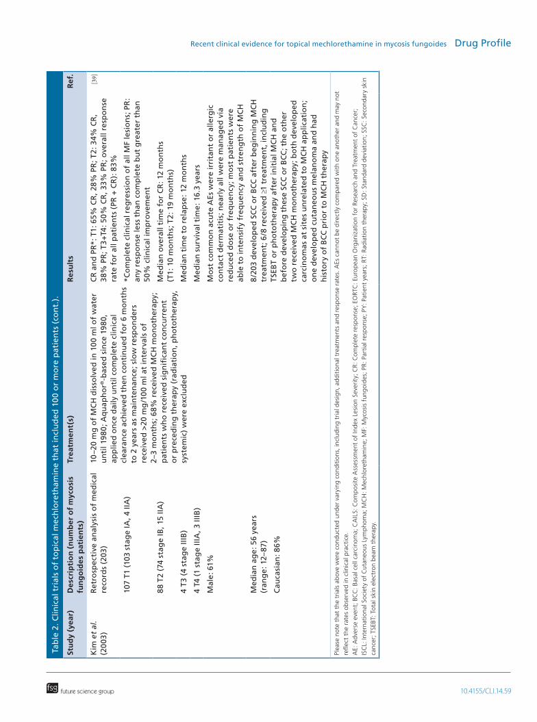
Vonderheid 1989Between 1968 and 1982, Vonderheid and colleagues studied the medical charts of 331 patients with MF [47]. Diagnosis was made based on manifestation of clinical characteristics and conclusive or compatible histopathologic findings for the disease. The T rat-ing and probable stage was recorded for each patient according to the Mycosis Fungoides Cooperative Group recommendations [52]. Stage was determined as ‘probable’ since lymph node biopsy specimens were not obtained routinely. End points were complete response and remission sustained for 4 or 8 years as determined by physician assessment. A complete response was defined as the complete disappearance of clinically detectable disease for at least 2 weeks and confirmed by skin biopsy specimens in most cases.
Patients were treated with an aqueous solution con-taining 10–20 mg of mechlorethamine in 40–60 ml of water once daily. After 2 weeks of treatment, response was noted and patients continued to receive topical mechlorethamine daily or every other day. Patients with advanced disease may have received additional treatments (i.e., local radiation, electron beam radia-tion, PUVA, ultraviolet B and chemotherapy such as intravenous methotrexate and mechlorethamine). Complete response was reported in a higher number of patients who had less severe disease: 71 (80%), 45 (68%), 28 (61%), 19 (49%), 22 (60%), 5 (13%) and 1 (11%) of stages IA, IB, IIA, IIB, III, IVA and IVB, respectively. Of these seven groups, 64 patients had sustained remission for 4 years and 34 patients had sustained remission for 8 years.
Update of the Stanford experience (2003)Of the patients with MF treated at the Stanford Uni-versity Cutaneous Lymphoma Clinic from 1958 to 1999, 203 patients with stage I–III MF who were treated with topical mechlorethamine as initial pri-mary therapy within 60 days of their initial evalua-tion were included in this study [30]. Diagnosis of MF was determined by clinical and histological evaluation and disease staging was classified according to Bunn and Lamberg [52]. Clinical response was defined as complete response (complete clinical regression of all MF lesions), partial response (any response less than
complete but greater than 50% clinical improvement), or no response (less than 50% clinical response to therapy). Progression of disease was defined as wors-ening of disease to a higher T classification or worse clinical stage. Actuarial survival was calculated via the Kaplan–Meier technique. Patients were treated with topical mechlorethamine daily until complete clinical remission was achieved. Prior to 1980, patients were treated with 10–20 mg of mechlorethamine in 100 ml of aqueous solution. After 1980, most patients were treated with an Aquaphor-based ointment. Treatment was continued for 6 months as maintenance therapy after clinical clearance. Patients who received other significant concurrent or preceding therapy, such as irradiation (local and total skin), phototherapy or any systemic therapies were excluded.
The majority of patients in this study (139 patients, 68%) were treated with mechlorethamine alone as initial therapy and throughout their follow-up course. Overall patient response rate was 83% with half of the patients achieving a complete response. Percentages of complete responses were higher in patients with earlier disease: 70 (65%), 30 (34%), 0 (no percentage given) and two (no percentage given) of stages T1, T2, T3 and T4, respectively. Median time to achieve com-plete response was 12 months (10 months for stage T1, 19 months for T2). Median time to relapse was also 12 months. Median survival was 16.3 years, and sur-vival rates at 5, 10 and 20 years were 85, 71 and 40%, respectively.
Lindahl 2013Retrospective data from 116 patients with MF who received mechlorethamine from 1991 to 2009 were analyzed by Lindahl et al. [48]. Diagnosis of MF was verified by histology. Until 2007, disease stage was clas-sified as per the Mycosis Fungoides Cooperative Group staging system [52] and thereafter as per the European Organization for Research and Treatment of Cancer staging system [37]. Clinical response, determined by physical examination, included complete (clinical regression of all skin lesions), partial (any response less than complete but greater than 50% clinical improve-ment), or no response as stable or progressive disease (worsening to a higher T classification or clinical stage). Complete response, relapse and progression event curves were calculated via the Kaplan–Meier method. Patients were treated with an aqueous solu-tion containing 20 mg of mechlorethamine in 40 ml of water daily for 14 days. Maintenance therapy was given as two treatments every fourth to eighth week until treatment was no longer indicated, or treatment was stopped due to side effects or progressive disease. Adjunctive therapies were used by 98.3% of patients
10.4155/CLI.14.59future science group
Recent clinical evidence for topical mechlorethamine in mycosis fungoides Drug Profile
with MF. All these patients received various topical therapies, including corticosteroids and phototherapy. A total of 51.7% received various systemic therapies.
Median duration of mechlorethamine treatment was 16.4 months (range: 2 days–25.6 years).
Although not statistically different, more patients achieved complete responses who had less skin involve-ment (11 [78.6%], 40 [51.3%], 6 [40.0%] and 5 [55.6%] patients in stages T1, T2, T3 and T4, respec-tively). The overall frequency of disease progression observed was 25.0% (T1: 28.6%, T2: 25.6%, T3: 26.7% and T4: 11.1%, respectively).
Lessin 2013The pivotal study conducted by Actelion (Protocol 2005NMMF-201-US, NCT00168064 [53]) was a Phase II, multicenter, randomized, observer-blinded, non-inferiority trial that compared mechlorethamine gel 0.02% (equivalent to 0.016% w/w mechlorethamine, VALCHLOR) versus mechlorethamine Aquaphor (ointment) 0.02% administered daily to 260 patients with stage I or IIA MF in 13 centers in the USA [41]. Histologic criteria [54] and a diagnostic algorithm for defining early MF/CTCL staging [55] were employed.
Primary efficacy end point was ≥50% improve-ment in the baseline Composite Assessment of Index Lesion Severity (CAILS) [32,56]. Secondary efficacy end points included ≥50% improvement in the modi-fied Severity Weighted Assessment Tool (SWAT) [32,57]. Baseline and each study visit CAILS and SWAT scores were calculated for complete response (100% improvement with a score = 0), partial response (≥50 to <100% reduction from baseline) and stable disease (<50% reduction from baseline). Confirmed responses were those observed at equal or greater than 4 weeks. Duration of response was defined as the time from first appearance of confirmed response to first assessment of loss of response (CAILS score <50% improvement from baseline) or progressive disease (CAILS score was ≥25% above baseline). Noninferiority of the gel to the ointment was established if the 95% CI lower bound around the ratio of the response rates (com-plete response and partial response for gel/ointment) was ≥0.75 (Kaplan–Meier methodology for the time to first confirmed response and duration of response curves).
Patients could have received prior therapies (topical corticosteroids, phototherapy, Targretin gel and topical mechlorethamine) but patients were not required to be refractory to or intolerant of prior therapies. Concomi-tant use of topical corticosteroids was not permitted during the study. Treatments were applied once daily to affected skin areas (lesions) or total skin surface (depending on stage) for up to 12 months.
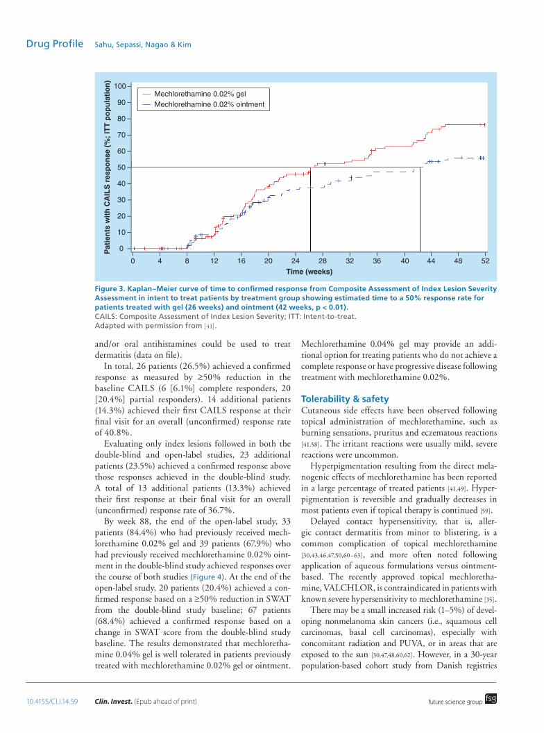
Both primary (CAILS score) and secondary (SWAT) end points met the prespecified criteria for noninferiority. Response rates for mechlorethamine gel and ointment were 58.5 and 47.7% by CAILS, and 46.9 and 46.2% by SWAT, respectively. The esti-mated time to a 50% response rate was significantly earlier for patients who received mechlorethamine gel (26 weeks) than for patients who received mechloreth-amine ointment (42 weeks, p < 0.01, Figure 3). There was no statistically significant difference between the two treatments with respect to duration of response. Analysis of the Kaplan–Meier curves estimated that at least 90% of responses for both gel and ointment will be maintained for >10 months.
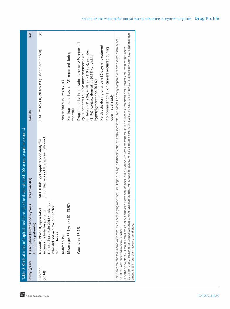
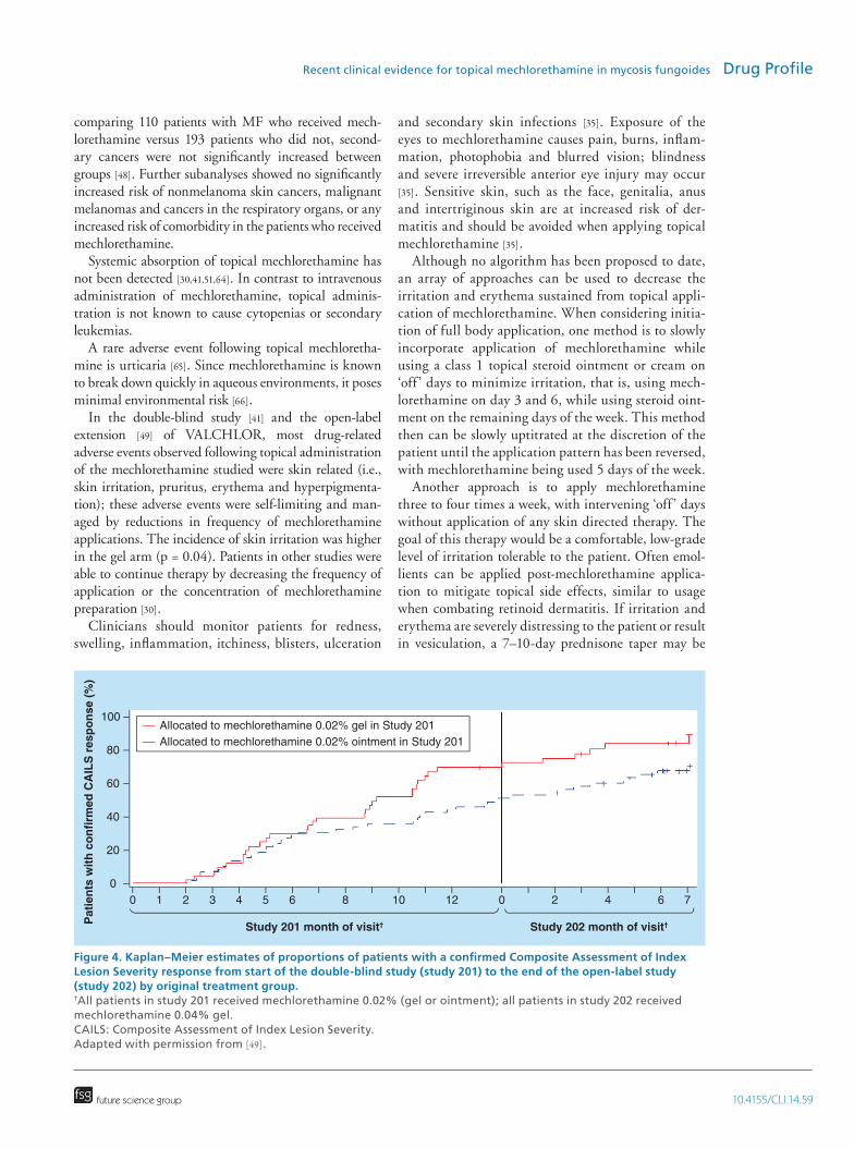
Kim 2014A 6-month, Phase II, open-label extension study (Study 2007NMMF-202-US, NCT00535470 [53]) for patients completing study 2005NMMF-201-US [41] but who did not achieve a complete response (i.e., CAILS score remained >0 as of baseline of study 2007NMMF-202-US) after 12 months, was conducted to evaluate the safety and efficacy of mechlorethamine gel 0.04% in patients with stage I or IIA MF [49]. This represents the first clinical trial to evaluate mechlorethamine 0.04% gel for patients with MF.
Primary end point was the response rate (complete response rate and partial response rate) defined as ≥50% improvement of baseline CAILS score (total severity score of up to five index lesions) of the dou-ble-blind study that was confirmed at the next visit at least 4 weeks later in the open-label study. The index lesions in this open-label study were either the same index lesions that were evaluated during double-blind study that did not have a complete response or, if there were fewer than five original lesions at the start of the double-blind study, additional index lesions could be included if they were present and treated consistently throughout the double-blind study. Complete and partial responses for CAILS were the same as defined in the double-blind study [41]. The secondary efficacy end point, ≥50% reduction in the baseline SWAT score by two or more consecutive observations over at least 4 weeks, was determined by measuring each involved area as a percentage of body surface area and multiplying by a severity-weighting factor (1 = patch, 2 = plaque, 3 = tumor).
In total, 98 patients with MF (stages IA, IB and IIA) applied mechlorethamine, 0.04% gel once daily to affected areas for an additional 7 months after the initial 12-month course in the double-blind study. Use of topical corticosteroids to treat skin adverse events was not allowed during the study. During the study, other therapies to treat MF were prohibited; emollients
10.4155/CLI.14.59 Clin. Invest. (Epub ahead of print) future science group
Drug Profile Sahu, Sepassi, Nagao & Kim
and/or oral antihistamines could be used to treat dermatitis (data on file).
In total, 26 patients (26.5%) achieved a confirmed response as measured by ≥50% reduction in the baseline CAILS (6 [6.1%] complete responders, 20 [20.4%] partial responders). 14 additional patients (14.3%) achieved their first CAILS response at their final visit for an overall (unconfirmed) response rate of 40.8%.
Evaluating only index lesions followed in both the double-blind and open-label studies, 23 additional patients (23.5%) achieved a confirmed response above those responses achieved in the double-blind study. A total of 13 additional patients (13.3%) achieved their first response at their final visit for an overall (unconfirmed) response rate of 36.7%.
By week 88, the end of the open-label study, 33 patients (84.4%) who had previously received mech-lorethamine 0.02% gel and 39 patients (67.9%) who had previously received mechlorethamine 0.02% oint-ment in the double-blind study achieved responses over the course of both studies (Figure 4). At the end of the open-label study, 20 patients (20.4%) achieved a con-firmed response based on a ≥50% reduction in SWAT from the double-blind study baseline; 67 patients (68.4%) achieved a confirmed response based on a change in SWAT score from the double-blind study baseline. The results demonstrated that mechloretha-mine 0.04% gel is well tolerated in patients previously treated with mechlorethamine 0.02% gel or ointment.
Mechlorethamine 0.04% gel may provide an addi-tional option for treating patients who do not achieve a complete response or have progressive disease following treatment with mechlorethamine 0.02%.
Tolerability & safetyCutaneous side effects have been observed following topical administration of mechlorethamine, such as burning sensations, pruritus and eczematous reactions [41,58]. The irritant reactions were usually mild, severe reactions were uncommon.
Hyperpigmentation resulting from the direct mela-nogenic effects of mechlorethamine has been reported in a large percentage of treated patients [41,49]. Hyper-pigmentation is reversible and gradually decreases in most patients even if topical therapy is continued [59].
Delayed contact hypersensitivity, that is, aller-gic contact dermatitis from minor to blistering, is a common complication of topical mechlorethamine [30,43,46,47,50,60–63], and more often noted following application of aqueous formulations versus ointment-based. The recently approved topical mechloretha-mine, VALCHLOR, is contraindicated in patients with known severe hypersensitivity to mechlorethamine [35].
There may be a small increased risk (1–5%) of devel-oping nonmelanoma skin cancers (i.e., squamous cell carcinomas, basal cell carcinomas), especially with concomitant radiation and PUVA, or in areas that are exposed to the sun [30,47,48,60,62]. However, in a 30-year population-based cohort study from Danish registries
Figure 3. Kaplan–Meier curve of time to confirmed response from Composite Assessment of Index Lesion Severity Assessment in intent to treat patients by treatment group showing estimated time to a 50% response rate for patients treated with gel (26 weeks) and ointment (42 weeks, p < 0.01). CAILS: Composite Assessment of Index Lesion Severity; ITT: Intent-to-treat. Adapted with permission from [41].
Pat
ien
ts w
ith
CA
ILS
res
po
nse
(%
; IT
T p
op
ula
tio
n)
0
10
20
30
40
50
60
70
80
90
100
Time (weeks)0 4 8 12 16 20 24 28 32 36 40 44 48 52
Mechlorethamine 0.02% gelMechlorethamine 0.02% ointment
10.4155/CLI.14.59future science group
Recent clinical evidence for topical mechlorethamine in mycosis fungoides Drug Profile
comparing 110 patients with MF who received mech-lorethamine versus 193 patients who did not, second-ary cancers were not significantly increased between groups [48]. Further subanalyses showed no significantly increased risk of nonmelanoma skin cancers, malignant melanomas and cancers in the respiratory organs, or any increased risk of comorbidity in the patients who received mechlorethamine.
Systemic absorption of topical mechlorethamine has not been detected [30,41,51,64]. In contrast to intravenous administration of mechlorethamine, topical adminis-tration is not known to cause cytopenias or secondary leukemias.
A rare adverse event following topical mechloretha-mine is urticaria [65]. Since mechlorethamine is known to break down quickly in aqueous environments, it poses minimal environmental risk [66].
In the double-blind study [41] and the open-label extension [49] of VALCHLOR, most drug-related adverse events observed following topical administration of the mechlorethamine studied were skin related (i.e., skin irritation, pruritus, erythema and hyperpigmenta-tion); these adverse events were self-limiting and man-aged by reductions in frequency of mechlorethamine applications. The incidence of skin irritation was higher in the gel arm (p = 0.04). Patients in other studies were able to continue therapy by decreasing the frequency of application or the concentration of mechlorethamine preparation [30].
Clinicians should monitor patients for redness, swelling, inflammation, itchiness, blisters, ulceration
and secondary skin infections [35]. Exposure of the eyes to mechlorethamine causes pain, burns, inflam-mation, photophobia and blurred vision; blindness and severe irreversible anterior eye injury may occur [35]. Sensitive skin, such as the face, genitalia, anus and intertriginous skin are at increased risk of der-matitis and should be avoided when applying topical mechlorethamine [35].
Although no algorithm has been proposed to date, an array of approaches can be used to decrease the irritation and erythema sustained from topical appli-cation of mechlorethamine. When considering initia-tion of full body application, one method is to slowly incorporate application of mechlorethamine while using a class 1 topical steroid ointment or cream on ‘off ’ days to minimize irritation, that is, using mech-lorethamine on day 3 and 6, while using steroid oint-ment on the remaining days of the week. This method then can be slowly uptitrated at the discretion of the patient until the application pattern has been reversed, with mechlorethamine being used 5 days of the week.
Another approach is to apply mechlorethamine three to four times a week, with intervening ‘off ’ days without application of any skin directed therapy. The goal of this therapy would be a comfortable, low-grade level of irritation tolerable to the patient. Often emol-lients can be applied post-mechlorethamine applica-tion to mitigate topical side effects, similar to usage when combating retinoid dermatitis. If irritation and erythema are severely distressing to the patient or result in vesiculation, a 7–10-day prednisone taper may be
Figure 4. Kaplan–Meier estimates of proportions of patients with a confirmed Composite Assessment of Index Lesion Severity response from start of the double-blind study (study 201) to the end of the open-label study (study 202) by original treatment group. †All patients in study 201 received mechlorethamine 0.02% (gel or ointment); all patients in study 202 received mechlorethamine 0.04% gel. CAILS: Composite Assessment of Index Lesion Severity. Adapted with permission from [49].
Allocated to mechlorethamine 0.02% gel in Study 201Allocated to mechlorethamine 0.02% ointment in Study 201
Study 201 month of visit† Study 202 month of visit†
0 1 2 3 4 5 6 8 10 12 0 2 4 6 70
20
40
60
80
100
Pat
ien
ts w
ith
co
nfi
rmed
CA
ILS
res
po
nse
(%
)
10.4155/CLI.14.59 Clin. Invest. (Epub ahead of print) future science group
Drug Profile Sahu, Sepassi, Nagao & Kim
initiated to obtain relief. Mechlorethamine may also be used in combination with other therapies for more treatment resistant areas such as the fingertips, palms and soles of patients who are receiving ultraviolet light therapy, or on oral Targretin. Lastly, patient education regarding performing a personal patch test prior to ini-tiation of mechlorethamine therapy to reduce contact dermatitis as well as written guidelines and demon-stration of proper application amount per unit body surface area are recommended for successful therapy.
ConclusionTreatment outcomes from published medical literature on MF patients following topical mechlorethamine treatment can be challenging to equate since there may be differences in the institutions’ patient selec-tion, disease staging methods, MF diagnostic criteria, preparation of topical mechlorethamine, specific treat-ment algorithms utilized, duration of maintenance treatment after complete response and various median follow-up time periods. Well-controlled, multi center, prospective studies are needed to elucidate the clini-cal characteristics of topical mechlorethamine. Retro-spective studies that evaluate real-world utilization of topical mechlorethamine are also warranted.
Physicians may employ more of a multimodal approach in treating MF, such as the combination of topical mechlorethamine and corticosteroids. Studies about the interaction of topical mechlorethamine with other agents could help determine the efficacy and safety of combination treatments for MF.
The mechanism of action of topical mechlor-ethamine remains uncertain. Many believe that the effectiveness of mechlorethamine may stem not only from its alkylating properties but also via immune
stimulation or interaction with the epidermal cell–Langerhans cell–T-cell axis [30].
Patients who used topical mechlorethamine as a maintenance regimen had a longer lasting response during maintenance therapy compared to patients who did not [30], suggesting that patients may benefit from a maintenance regimen of mechlorethamine as part of a longer maintenance regimen. Additionally, patients have responded well to topical mechlorethamine fol-lowing relapse with more aggressive therapies; topical mechlorethamine may be used as part of sequential therapy in the future.
A consensus statement about the management of dermatitis should be developed; techniques such as adjusting the frequency of topical mechlorethamine applications and uptitrating the mechlorethamine dose once dermatitis subsides should be addressed. Following this consensus statement, patient educa-tion about the proper amount to apply to the skin and how to perform a personal patch test prior to applying topical mechlorethamine is needed for treatment to be successful.
Mechlorethamine ointment formulations are com-pounded at pharmacies and are not subject to rigorous quality assurance standards. Most health insurance formularies would rarely include compounded medi-cine, or medicines without FDA approval. Addition-ally, petrolatum-based ointments may be difficult to apply and could compromise patient compliance [41]. Given the recent FDA approval, VALCHLOR pro-vides patients with access to a quick-drying, grease-less mechlorethamine gel that has been developed under good manufacturing practices and has a longer stability, consistent potency and noninferiority to compounded ointment.
Executive summary
Mycosis fungoides• Mycosis fungoides is a rare, potentially life-threatening cutaneous T-cell lymphoma characterized by
cutaneous homing of neoplastic T lymphocytes.Treatment: mechlorethamine• Topical mechlorethamine has been used to treat mycosis fungoides since the 1940s in retrospective studies, as
well as a double-blind and open-label studies, leading to the approval of VALCHLOR™.• Mechlorethamine acts as an alkylating agent and mostly likely immune stimulation properties.• With the approval of VALCHLOR [35], patients have access to a quick-drying, greaseless mechlorethamine
gel with longer stability, consistent potency and noninferiority to compounded ointment that has been developed under good manufacturing practice.
Tolerability & safety• Following topical administration of mechlorethamine, dermatitis and hyperpigmentation have been seen as
mild adverse events and a small increased risk (1–5%) of developing nonmelanoma skin cancers, especially with concomitant radiation and psoralen plus ultraviolet A or areas exposed to the sun.
Conclusion• Topical mechlorethamine may be used in the future as part of maintenance regimens, multimodal treatments
and sequential therapy following more aggressive treatments.
10.4155/CLI.14.59future science group
Recent clinical evidence for topical mechlorethamine in mycosis fungoides Drug Profile
Acknowledgements The authors wish to thank H Jeffrey Wilkins from Actelion
Pharmaceuticals US Inc. for medical input.
Financial & competing interests disclosureM Sepassi and M Nagao are both employees of Actelion
Pharmaceuticals US Inc. Y Kim serves on the Advisory Board
and is a consultant for Actelion Pharmaceuticals US Inc.
The authors have no other relevant affiliations or financial
involvement with any organization or entity with a financial
interest in or financial conflict with the subject matter or
materials discussed in the manuscript apart from those
disclosed.
The authors wish to thank Donna Simcoe for medical
writing support, funded by Actelion Pharmaceuticals US Inc.,
for an early draft based on discussions with the authors.
ReferencesPapers of special note have been highlighted as: • of interest; •• of considerable interest
1 Criscione VD, Weinstock MA. Incidence of cutaneous T-cell lymphoma in the United States, 1973–2002. Arch. Dermatol. 143(7), 854–859 (2007).
2 Bradford PT, Devesa SS, Anderson WF et al. Cutaneous lymphoma incidence patterns in the United States: a population-based study of 3884 cases. Blood 113(21), 5064–5073 (2009).
3 Alibert JLM. Descriptions des maladies de la peau observées a l’Hôpital Saint-Louis, et exposition des meilleures méthodes suivies pour leur traitement [in French]. Barrois l’ainé, Paris, France, 286 (1806).
4 National Cancer Institute. Surveillance, Epidemiology, and End Results (SEER) Program. Mycosis Fungoides. http://seer.cancer.gov/seertools/hemelymph/51f6cf57e3e27c3994bd5345
5 Balus L, Manente L, Remotti D et al. Granulomatous slack skin: report of a case and review of the literature. Am. J. Dermatopathol. 18(2), 199–206 (1996).
6 Kempf W, Ostheeren-Michaelis S, Paulli M et al. Granulomatous mycosis fungoides and granulomatous slack skin: a multicenter study of the Cutaneous Lymphoma Histopathology Task Force Group of the European Organization for Research and Treatment of Cancer (EORTC). Arch. Dermatol. 144(12), 1609–1617 (2008).
7 Weinstock MA, Gardstein B. Twenty-year trends in the reported incidence of mycosis fungoides and associated mortality. Am. J. Publ. Health 89(8), 1240–1244 (1999).
8 Kim ST, Sim HJ, Jeon YS et al. Clinicopathological features and T-cell receptor gene rearrangement findings of mycosis fungoides in patients younger than age 20 years. J. Dermatol. 36(7), 392–402 (2009).
9 Agar NS, Wedgeworth E, Crichton S et al. Survival outcomes and prognostic factors in mycosis fungoides/Sezary syndrome: validation of the revised International Society for Cutaneous Lymphomas/European Organisation for Research and Treatment of Cancer staging proposal. J. Clin. Oncol. 28(31), 4730–4739 (2010).
10 Talpur R, Singh L, Daulat S et al. Long-term outcomes of 1,263 patients with mycosis fungoides and Sézary syndrome from 1982 to 2009. Clin. Cancer Res. 18(18), 5051–5060 (2012).
11 Crowley JJ, Nikko A, Varghese A et al. Mycosis fungoides in young patients: clinical characteristics and outcome. J. Am. Acad. Dermatol. 38, 696–701 (1998).
12 Sun G, Berthelot C, Li Y et al. Poor prognosis in non-Caucasian patients with early-onset mycosis fungoides. J. Am. Acad. Dermatol. 60(2), 231–235 (2009).
13 Girardi M, Heald PW, Wilson LD. The pathogenesis of mycosis fungoides. N. Engl. J. Med. 350, 1978–1988 (2004).
14 Smedby KE, Vajdic CM, Falster M et al. Autoimmune disorders and risk of non-Hodgkin lymphoma subtypes: a pooled analysis within the InterLymph Consortium. Blood 111(8), 4029–4038 (2008).
15 Sarantopoulos GP, Palla B, Said J et al. Mimics of cutaneous lymphoma: report of the 2011 Society for Hematopathology/European Association for Haematopathology workshop. Am. J. Clin. Pathol. 139, 536–551 (2013).
16 Zackheim HS, McCalmont TH. Mycosis fungoides: the great imitator. J. Am. Acad. Dermatol. 47, 914–918 (2002).
17 Hwang ST, Janik JE, Jaffe ES et al. Mycosis fungoides and Sézary syndrome. Lancet 371, 945–957 (2008).
18 Edelson RL. Cutaneous T-cell lymphoma: the helping hand of dendritic cells. Ann. NY Acad. Sci. 941, 1–11 (2001).
19 Lutzner M, Edelson R, Schein P et al. Cutaneous T-cell lymphomas: the Sézary syndrome, mycosis fungoides, and related disorders. Ann. Intern. Med. 83, 534–552 (1975).
20 Olsen E, Vonderheid E, Pimpinelli N et al. Revisions to the staging and classification of mycosis fungoides and Sézary syndrome: a proposal of the International Society for Cutaneous Lymphomas (ISCL) and the cutaneous lymphoma task force of the European Organization of Research and Treatment of Cancer (EORTC). Blood 110, 1713–1722 (2007).
21 Santucci M, Biggeri A, Feller AC et al. Efficacy of histologic criteria for diagnosing early mycosis fungoides: an EORTC cutaneous lymphoma study group investigation. Am. J. Surg. Path. 24(1), 40 (2000).
22 Naraghi ZS, Seirafi H, Valikhani M et al. Assessment of histologic criteria in the diagnosis of mycosis fungoides. Int. J. Dermatol. 42(1), 45–52 (2003).
23 Willemze R. Cutaneous T-cell lymphoma: epidemiology, etiology, and classification. Leuk. Lymphoma 44(Suppl. 3), S49–S54 (2003).
24 Herne KL, Talpur R, Breuer-McHam J et al. Cytomegalovirus seropositivity is significantly associated with mycosis fungoides and Sezary syndrome. Blood 101, 2132–2136 (2003).
25 Huang KP, Weinstock MA, Clark CA et al. Second lymphomas and other malignant neoplasms in patients with mycosis fungoides and Sézary syndrome: evidence from population-based and clinical cohorts. Arch. Dermatol. 143(1), 45–50 (2007).
10.4155/CLI.14.59 Clin. Invest. (Epub ahead of print) future science group
Drug Profile Sahu, Sepassi, Nagao & Kim
26 van Doorn R, Scheffer E, Willemze R. Follicular mycosis fungoides, a distinct disease entity with or without associated follicular mucinosis: a clinicopathologic and follow-up study of 51 patients. Arch. Dermatol. 138, 191–198 (2002).
27 Hobbs L, Doughty D. Mycosis fungoides: a wound care challenge. J. Wound Ostomy Continence Nurs. 31, 95–97 (2004).
28 Reavely MM, Wilson LD. Total skin electron beam therapy and cutaneous T-cell lymphoma: a clinical guide for patients and staff. Dermatol. Nurs. 16(1), 36–39 (2004).
29 Zackheim HS, Amin S, Kashani-Sabet M et al. Prognosis in cutaneous T-cell lymphoma by skin stage: long-term survival in 489 patients. J. Am. Acad. Dermatol. 40, 418–425 (1999).
30 Kim YH, Martinez G, Varghese A et al. Topical nitrogen mustard in the management of mycosis fungoides: update of the Stanford experience. Arch. Dermatol. 139, 165–173 (2003).
•• Reportslong-term,retrospectivecohortstudyoftopicalmechlorethamineinthetreatmentofearlydiseasemycosisfungoides.
31 Demierre MF, Gan S, Jones J et al. Significant impact of cutaneous T-cell lymphoma on patients’ quality of life. Cancer 107(10), 2504–2511 (2006).
32 Olsen E, Whittaker S, Kim YH et al. Clinical end points and response criteria in mycosis fungoides and Sézary syndrome: a consensus statement of the International Society for Cutaneous Lymphomas, the United States Cutaneous Lymphoma Consortium, and the Cutaneous Lymphoma Task Force of the European Organisation for Research and Treatment of Cancer. J. Clin. Oncol. 29(18) 2598–2607 (2011).
33 Jawed SI, Myskowski PL, Horwitz S et al. Primary cutaneous T-cell lymphoma (mycosis fungoides and Sezary syndrome). Part II. Prognosis, management, and future directions. J. Am. Acad. Derm. 70(2), 223.e1–223.e17 (2014).
34 National Comprehensive Cancer Network (NCCN) Clinical Practice Guidelines in Oncology (NCCN Guidelines®): Non-Hodgkin’s Lymphomas. Version 1.2014. www.nccn.org/professionals/physician_gls/pdf/nhl.pdf.
• GuidelinesfromtheNationalComprehensiveCancerNetwork,whichincludesdiagnosisandtreatmentofmycosisfungoides.
35 Valchlor™, package insert. Actelion Pharmaceuticals US Inc., South San Francisco, CA, USA.
36 Goodman LS, Wintrobe MM, Dameshek W et al. Nitrogen Mustard therapy: use of methyl-bis(beta-chloroethyl)amine hydrochloride and tris(beta-chloroethyl)amine hydrochloride for Hodgkin’s Disease, lymphosarcoma, leukemia and certain allied and miscellaneous disorders. J. Am. Med. Assoc. 132, 126–132 (1946).
37 Trautinger F, Knobler R, Willemze R et al. EORTC consensus recommendations for the treatment of mycosis fungoides/Sézary syndrome. Eur. J. Cancer 42, 1014–1030 (2006).
38 Kim EJ, Hess S, Richardson SK et al. Immunopathogenesis and therapy of cutaneous T-cell lymphoma. J. Clin. Invest. 115, 798–812 (2005).
39 Kim YH, Liu HL, Mraz-Gernhard S et al. Long-term outcome of 525 patients with mycosis fungoides and Sézary syndrome. Arch. Dermatol. 139, 857–866 (2003).
40 Zhang Y, Trissel LA, Johansen JF et al. Stability of mechlorethamine hydrochloride 0.01% ointment in Aquaphor base. Intern. J. Pharm. Compound. 2, 89–94 (1998).
41 Lessin SR, Duvic M, Guitart J et al. Topical chemotherapy in cutaneous T-cell lymphoma: positive results of a randomized, controlled, multicenter trial testing the efficacy and safety of a novel mechlorethamine, 0.02%, gel in mycosis fungoides. Arch. Dermatol. 149(1), 25–32 (2013).
•• ReportspivotalPhaseIItrialresultsoftopicalmechlorethaminetreatmentofmycosisfungoides.
42 Mustargen®, package insert. Lundbeck, Deerfield, IL, USA.
43 Price NM, Hoppe RT, Deneau DG. Ointment-based mechlorethamine treatment for mycosis fungoides. Cancer 52(12), 2214–2219 (1983).
44 Benson HA. Transdermal drug delivery: penetration enhancement techniques. Curr. Drug Deliv. 2(1), 23–33 (2005).
45 Ritschel WA, Ye W, Buhse L et al. Stability of the nitrogen mustard mechlorethamine in novel formulations for dermatological use. Inter. J. Pharmaceut. 362(1), 67–73 (2008).
46 Ramsay DL, Parnes RE, Dubin N et al. Response of mycosis fungoides to topical chemotherapy with mechlorethamine. Arch. Dermatol. 120(12), 1585–1590 (1984).
47 Vonderheid EC, Tan ET, Kantor AF et al. Long-term efficacy, curative potential, and carcinogenicity of topical mechlorethamine chemotherapy in cutaneous T-cell lymphoma. J. Am. Acad. Dermatol. 20(3), 416–428 (1989).
48 Lindahl L, Fenger-Gron M, Iversen L. Topical nitrogen mustard therapy in patients with mycosis fungoides or parapsoriasis. J. Eu. Acad. Derm. Vener. 27(2), 163–168 (2013).
49 Kim YH, Duvic M, Guitart J et al. Tolerability and efficacy of mechlorethamine 0.04% gel in CTCL (mycosis fungoides) after initial treatment with topical mechlorethamine 0.02% gel. Presented at: The T-cell Lymphoma Forum. San Francisco, CA, USA, 23–25 January 2014.
•• Reports6-monthopen-labelextensionofpivotalPhaseIItrialfortopicalmechlorethaminetreatmentofmycosisfungoides.
50 Ramsay DL, Halperin PS, Zeleniuch-Jacquotte A. Topical mechlorethamine therapy for early stage mycosis fungoides. J. Am. Acad. Dermatol. 19(4), 684–691 (1988).
51 Vonderheid EC, Van Scott EJ, Wallner PE et al. A 10-year experience with topical mechlorethamine for mycosis fungoides: comparison with patients treated by total-skin electron-beam radiation therapy. Cancer Treat. Rep. 63(4), 681–689 (1979).
52 Bunn PA Jr, Lamberg SL. Report of the committee on staging and classification of cutaneous T-cell lymphomas. Cancer Treat. Rep. 63, 725–728 (1979).
53 ClinicalTrials.gov. www.clinicaltrials.gov
10.4155/CLI.14.59future science group
Recent clinical evidence for topical mechlorethamine in mycosis fungoides Drug Profile
54 Duvic M, Olsen EA, Omura GA et al. A Phase III, randomized, double-blind, placebo-controlled study of peldesine (BCX-34) cream as topical therapy for cutaneous T-cell lymphoma. J. Am. Acad. Dermatol. 44(6), 940–947 (2001).
55 Pimpinelli N, Olsen EA, Santucci M et al. Defining early mycosis fungoides. J. Am. Acad. Dermatol. 53(6), 1053–1063 (2005).
56 Heald P, Mehlmauer M, Martin AG et al. Topical bexarotene therapy for patients with refractory or persistent early-stage cutaneous T-cell lymphoma: results of the Phase III clinical trial. J. Am. Acad. Dermatol. 49(5), 801–815 (2003).
57 Stevens SR, Ke MS, Parry EJ et al. Quantifying skin disease burden in mycosis fungoides-type cutaneous T-cell lymphomas: the severity-weighted assessment tool (SWAT). Arch. Dermatol. 138(1), 42–48 (2002).
58 de Quatrebarbes J, Estève E, Bagot M et al. Treatment of early-stage mycosis fungoides with twice-weekly applications of mechlorethamine and topical corticosteroids. Arch. Dermatol. 141, 1117–1120 (2005).
59 Price NM. Topical mechlorethamine. Cutaneous changes in patients with mycosis fungoides after its administration. Arch. Dermatol. 113(10), 1387–1389 (1977).
60 Vonderheid EC, Van Scott EJ, Johnson WC, Grekin DA, Asbell SO. Topical chemotherapy and immunotherapy
of mycosis fungoides: intermediate-term results. Arch. Dermatol. 113(4), 454–462 (1977).
61 Zachariae H, Thestrup-Pedersen K, Sogaard H. Topical nitrogen mustard in early mycosis fungoides: a 12-year experience. Acta Derm. Venereol. 65, 53–58 (1985).
62 Hoppe RT, Abel EA, Deneau DB et al. Mycosis fungoides: management with topical nitrogen mustard. J. Clin. Oncol. 5, 1796–1803 (1987).
63 Estève E, Bagot M, Joly P et al. A prospective study of cutaneous intolerance to topical mechlorethamine therapy in patients with cutaneous T-cell lymphomas. Arch. Dermatol. 135(11), 1349–1353 (1999).
64 Price NM, Deneau DG, Hoppe RT et al. The treatment of mycosis fungoides with ointment-based mechlorethamine. Arch. Dermatol. 118(4), 234–237 (1982).
65 Grunnet E. Contact urticaria and anaphylactoid reaction induced by topical application of nitrogen mustard. Br. J. Dermatol. 94(1), 101–103 (1976).
66 Breneman DL, Nartker AL, Ballman EA et al. Topical mechlorethamine in the treatment of mycosis fungoides. Uniformity of application and potential for environmental contamination. J. Am. Acad. Dermatol. 25(6 Pt 1), 1059–1064 (1991).