Embed Size (px)
Citation preview

Drug Detection Testing
ii
A Review of Drug Detection Testing
and an Examination of Urine, Hair, Saliva
and Sweat
David Rouen, Kate Dolan
and Jo Kimber
NDARC Technical Report No. 120
National Drug and Alcohol Research Centre
University of New South Wales
Sydney Australia
ISBN 0 7334 1790 6
©NDARC 2001

Drug Detection Testing
iii
For further information about this publication please contact: Dr Kate Dolan Senior Lecturer National Drug and Alcohol Research Centre University of New South Wales Sydney NSW 2052 Telephone: +61 (2) 9398 9333 Facsimile: +61 (2) 9399 7143 Email: [email protected] The citation of the report is as follows: Rouen, D., Dolan, K. & Kimber, J. (2001). A review of drug detection testing and an examination of urine, hair, saliva and sweat. Technical Report No. 120, National Drug and Alcohol Research Centre. University of New South Wales, Sydney. For additional copies of this report please see the NDARC web site: http://www.med.unsw.edu.au/ndarc/

Drug Detection Testing
iv
TABLE OF CONTENTS
Acknowledgements ..........................................................................................................v List of Abbreviations ......................................................................................................vi Executive Summary.......................................................................................................vii 1. Introduction............................................................................................................1 2. Methodology of this Review..................................................................................2 3. Idendifying Drug Use.............................................................................................2
3.1 Result Interpretation ...............................................................................................2 4. Methods of Drug Testing .......................................................................................6
4.1 Screening Tests .......................................................................................................6 4.2 Confirmatory Tests .................................................................................................6
5. Biological Indicators of Drug Use.........................................................................7 5.1 Urine Analysis ............................................................................................................7
5.1.1 The Physiology of Urine Production...................................................................7 5.1.2 Incorporation of Drugs into Urine .......................................................................8 5.1.3 Specimen Collection............................................................................................8 5.1.4 Sample Analysis ................................................................................................10 5.1.5 Interpretation of Drug Concentrations in Urine .................................................12 5.1.6 Conclusions ........................................................................................................13
5.2 Hair Analysis............................................................................................................14 5.2.1 Anatomy and Physiology of Hair ......................................................................14 5.2.2 Incorporation of Drugs into Hair .......................................................................15 5.2.3 Specimen Collection..........................................................................................16 5.2.4 Methodological Criteria for Obtaining Hair Test Results .................................17 5.2.5 Analytical Techniques .......................................................................................17 5.2.6 Interpretation of Hair Analyses .........................................................................18 5.2.7 Conclusions ........................................................................................................19
5.3 Saliva Analysis .........................................................................................................20 5.3.1 The Physiology of Saliva ...................................................................................20 5.3.2 Incorporation of Drugs into Saliva ....................................................................21 5.3.3 Specimen Collection..........................................................................................21 5.3.4 Analysis of Saliva ..............................................................................................22 5.3.5 Interpretation of Drug Concentrations in Saliva................................................22 5.3.6 Conclusions ........................................................................................................23
5.4 Sweat Analysis .........................................................................................................23 5.4.1 The Physiology of Sweat ...................................................................................23 5.4.2 Incorporation of Drugs into Sweat ....................................................................24 5.4.3 Specimen Collection..........................................................................................24 5.4.4 Sample Preparation and Analysis ......................................................................25 5.4.5 Interpretation of Drug Concentration in Sweat: ................................................25 5.4.6 Conclusions ........................................................................................................27
6. Tabulated Summary of Issues Related to the Different Biological Matrices.......28 References ......................................................................................................................29 Appendix A: On-Site Urine Screening Devices...........................................................38
DipScan™ 6:...............................................................................................................38
Drug Detection Testing
v
Triage Drugs of Abuse Panel...................................................................................40 Appendix B: Manufactured Saliva Collection Devices ..............................................42
Intercept Oral Fluid Collection Device....................................................................42 Appendix C: On-Site Saliva Screening Devices..........................................................44
ORALscreen™............................................................................................................44 RapiScan ..................................................................................................................46
Appendix D: Manufactured Sweat Collection Devices ..............................................48 PharmChek Sweat Patch .........................................................................................48
Appendix E: On-Site Saliva/Sweat Screening Device.................................................50 Drugwipe™ II .............................................................................................................50
Appendix F: Urine Adulteration Test Strips ...............................................................52 Intect™ 7.....................................................................................................................52
Acknowledgements We would like to thank the Drug Programs Bureau, New South Wales Department of Health and Aged Care for providing funding for this review. Any views expressed in this report do not necessarily represent the views of, and are not necessarily supported by, the Drug Programs Bureau. Thanks also to Penny Adams for her help editing earlier drafts of this manuscript.

Drug Detection Testing
vi
List of Abbreviations
6-MAM 6-Monoacetylmorphine Amphet Amphetamine (Sympathomimetic Amine) BE Benzoylecgonine (Primary metabolite of cocaine) Carboxy-THC Tetrahydrocannabinol Carboxylic Acid (Metabolite of
THC) CO Codeine (analgesic and common impurity of heroin) CO-glucuronide Codeine Glucuronide (Metabolite of Codeine) EDDP Ethylidine Dimethyl Diphenyl Pyrrolidine (Metabolite of
Methadone) EME Ecgonine Methyl Ester (Secondary Metabolite of Cocaine) GC/MS Gas Chromatography/Mass Spectrometry GC/MS/MS Gas Chromatography/Tandem Mass Spectrometry HPLC High Performance Liquid Chromatography LC/MS Liquid Chromatography/Mass Spectrometry Metham Methamphetamine (Sympathomimetic Amine) MO Morphine (Analgesic and Metabolite of Heroin and
Codeine) MO-glucuronide Morphine Glucuronide (Metabolite of Morphine) THC Tetrahydrocannabinol (Psychoactive component of
Cannabis)

Drug Detection Testing
vii
Executive Summary This paper provides a detailed review of drug testing procedures, focusing on the most commonly abused classes of drugs. Four biological specimens that can provide information about human drug exposure are covered. These include urine, hair, saliva and sweat. An overview of the physiology of each matrix and mechanisms of drug incorporation is included followed by a discussion of issues related to their collection, analysis and interpretation. Conclusions regarding the advantages, disadvantages, applicability and usefulness of each matrix for drug detection are provided. The biological detection of drug use is a two-step process. It involves a screening test which, if found to be positive, is followed by a confirmatory test. There are two primary methods of analyzing specimens for drugs: immunoassay and chromatography. Immunoassay is typically used to screen for drugs, as it is quick and comparatively inexpensive. The main limitations of immunoassay screens are low specificity and high cross-reactivity resulting in relatively high rates of false-positive test results. A confirmatory test is conducted to guard against this using a different analytical technique of equal or greater sensitivity. Chromatographic tests such as gas chromatography separation coupled with mass spectrometry detection (GC/MS) are recommended. It is important to note that the determination of drug use through biological analysis is never absolute. Numerous factors associated with the person tested (e.g. metabolism), the drug used (e.g. pharmacokenetic properties, route of administration), the sample taken (e.g. window of detection, biology of drug incorporation), the collection procedure (e.g. testing schedule) and the analytical procedure (e.g. limit of detection, cross-reactivity) all affect the results obtained. Consequently, there are four possible outcomes of a drug test which must be considered: (i) a true-positive result, where a tests correctly identifies the presence of a drug; (ii) a false-positive result, when a drug is detected by a test when, in fact, that drug is not present in the sample; (iii) a true-negative result, where a tests correctly identifies the absence of a drug; and (iv) a false-negative result, when no drug is detected by a test when, in fact, a drug is present in the sample. There is also much information associated with drug use that cannot be determined by biological analysis. For example, conclusions regarding current intoxication, quantity of drug used, frequency of use, and physical or psychological dependency cannot be made. Drug use determination is undertaken through the analysis of a drug’s metabolites as well as the drug itself depending on the sample being examined. This is important for two reasons. Firstly, metabolite(s) are most likely to be detected in some samples, primarily urine, as they often have a longer half- life than the parent compound (drug consumed). Secondly, identification of relative metabolite concentrations is often necessary to determine the drug that has been consumed. Different drugs can metabolise into the same compounds, or an unmetabolised drug may be present in a sample because of passive contamination rather than consumption (as has been shown with hair). Drug use is currently assessed in urine, hair, saliva and sweat. Each biological sample has its own unique advantages and disadvantages stemming from its inherent properties and our current state of knowledge. A summary of these issues can be found in Table 4. Urine is the most widely used matrix. In Australia, analytical facilities and procedures

Drug Detection Testing
viii
for urinalysis are well established, relatively convenient and competitively priced. Urine offers only an intermediate window of detection (1-3 days) thus making test scheduling a significant issue for many applications. Its susceptibility to tampering and adulteration is also a problem and makes appropriate supervision critical. Hair analysis offers the largest window of detection (7-100+ days) and can provide information on historical drug use spanning up to several months. Much research has been undertaken examining hair testing, however incomplete understanding of the mechanisms of drug incorporation has made straightforward result interpretation difficult. The interest in this technology, stemming from its broad range of potential applications, is likely to result in furthe r improvements in the reliability and validity of hair as an alternative test matrix to urine. Saliva analysis is also a developing technology. Currently, there are limited analytical facilities in Australia, however, established United States laboratories are accessible. Sample collection is relatively quick, noninvasive and resistant to tampering although as with urinalysis, adequate supervision is required. Saliva analysis has been shown to be useful in determining very recent drug use (1-36 hours). It is not considered economically viable or practical for continuous drug use monitoring. The analysis of sweat may prove to be the matrix of choice for the medium-term, continuous monitoring of drug use due to recent developments in sweat patch technology. However more naturalistic trials are required. It may also offer an economical alternative to urine, as comparable results can be obtained with fewer analyses. Analytical facilities and expertise is still lacking in Australia but progress is being made. Drug testing has become a faster, more convenient process with the development of point-of-collection (on-site) drug testing devices. This paper concludes with a review of some of the many commercial on-site devices used to screen for drugs of abuse in urine, sweat and saliva. Although improvements are being made, only on-site urine tests are considered adequate at this time. Manufactured devices for the collection of saliva and sweat samples that are analysed by accredited laboratories are reviewed. A test device for the detection of irregularities in urine, and hence possible adulteration, is also reviewed.

Drug Detection Testing
1
1. Introduction Obtaining accurate information on illicit drug use within various populations and contexts has been the subject of much speculation and study. In many contexts, where the aim is reduction of illicit drug usage, drug use measurements are obtained through self-report. The validity of self-reported drug use data is subject to many factors such as the population examined, the types of drugs used and the methods used to elicit information. Generally, accuracy of self- report can be seen as a function of the social, occupational, legal and/or financial cost of admission as perceived by the individual. As the perceived or real costs of reporting increase accuracy tends to decrease. In addition, accurate recall of drug use may be affected by the mental and physical state of an individual, which may, in turn, be affected by drug use. As underreporting of drug use is common in some populations, particularly when real or perceived punitive measures may result from admission, accurate drug use estimates must be obtained from more objective biological drug testing. According to De Angelis (1972), large scale biological testing for illicit drug use was developed during the occupation of Japan by the United States after World War II. The need to develop reasonably inexpensive and accurate urine tests resulted in chromatographic procedures that were able to detect small amounts of opiates in urine. Urine testing was adopted in the first methadone program in New York (De Angelis, 1972) and has since become a familiar component of methadone treatments. The economic costs and other disadvantages involved with urinalysis during opiate replacement therapy has, however, led to a reassessment of its use over recent years. A general harm reduction philosophy, with an increased emphasis on patient retention and a reduced emphasis on the punishment of illicit drug use now dominates in treatment programs in Australia and some European nations. The move away from the clinical use of urinalysis has coincided with a dramatic growth in the use of workplace urinalysis in the United States. The move towards workplace testing in the US dates from the 1980s when concerns began to be expressed regarding the impact of illicit drug use on worker productivity. In September 1986, President Ronald Reagan issued an Executive Order requiring federal agencies to institute urine-testing programs for the purpose of creating “drug-free federal workplaces” (Executive Order 12564). In 1987, when the American Management Association (AMA) began gathering data on corporate drug policies, 21 percent of its members had instituted drug testing programs; 79 percent had not (American Management Association, 1987). A decade later, the percentages were reversed. By January 1996, 81 percent of major U.S. firms tested for illegal or controlled substances. This figure has since fallen back to 1991 levels of around 66 percent (American Management Associa tion, 2000). A greater interest in drug-crime diversion programs in the United States, especially highly supervised drug court programs, has furthered an interest in drug detection from outside the health sector. Criminal justice initiatives such as drug courts use urinalysis to monitor compliance with treatment plans. As these programs are usually abstinence
Drug Detection Testing
2
based and implement penalties for detected incidences of drug use, self-report is considered ineffective. This demand for drug testing products has created a highly competitive market. The resulting technological advances can be seen in the products reviewed in the appendices of this document. Hair, sweat and saliva are showing potential as testing mediums with advantages in detection times, ease of collection and resistance to tampering balancing possible reductions in test accuracy and higher laboratory costs. A very large number of substances can be routinely measured in the different biological matrices. This paper will primarily restrict itself to a discussion of the detection of the five classes of commonly abused drugs. These include opiates (heroin, morphine and codeine), methadone, cocaine, sympathomimetic amines (amphetamine and methamphetamine), cannabis, and benzodiazepines. 2. Methodology of this Review Journal publications and conference presentations on drug detection and on the use of urine, hair, saliva and sweat for the detection of drugs in humans were identified through a comprehensive search of the electronic database Medline. In addition, Australian experts involved in the analysis of urine, hair and sweat were contacted for unpublished reports, policy documents and related information. Key companies and organisations involved in the development, manufacture, evaluation, use and/or analysis of drug testing technologies were also identified through the Internet, telephone directories and word-of-mouth. Requests for unpublished reports, product literature and related information were made. 3. Idendifying Drug Use Typically, identification of drug use by an individual is a two-step process that involves a screening test which, if found to be positive, is followed by a confirmatory test. The screening test is designed to be sensitive to the presence of a class of drug while often sacrificing the ability to specifically identify the particular drug present. For example, screening tests may indicate the presence of an opioid without being able to determine if the opioid is codeine, morphine or heroin. The advantages of screening tests are that they are relatively quick and inexpensive. The confirmatory test, conducted only on a positive sample, is used to identify the specific drug and/or metabolites present and thus to ensure that the sample is truly positive for the targeted drug. 3.1 Result Interpretation 3.1.1 Drug Metabolites The half- life of a drug is defined as the time taken for 50% of the drug to be removed from the body by either metabolism or excretion (Chiang & Hawks, 1986). After a substance is consumed it is broken down, or metabolised, by the body into other chemicals that after excretion, can be detected in the biological specimen. Accurate metabolite identification is important for two main reasons. Firstly, in many cases metabolites have significantly longer half- lives than their parent drug, and are thus more likely to be detected. This is typically the case in urine. For example, cocaine has a half-

Drug Detection Testing
3
life of approximately one and a half hours and therefore the body requires about seven and a half hours to breakdown 97% of the drug. Cocaine’s main metabolite benzoylecgonine (BE) has an average half- life of seven and a half hours and can be detected in urine for up to 48 hours after a single dose. Secondly, accurate identification of relative metabolite concentrations is often essential in the determination of the actual drug used, as different drugs can have the same metabolites. For example, heroin has a half- life of approximately three minutes (Chiang and Hawks, 1986) and is metabolised to 6-monoacetylmorphine (6-MAM) and then to morphine (Jenny, 1989). 6-MAM also has a very short half- life. The half- life of morphine is longer (1.7-4.5 hours) and can usually be detected in urine for up to three days. Codeine, present in many over-the-counter analgesic preparations, is also metabolised to morphine and another substance, norcodeine. Therefore, the presence of morphine in a sample might be due to the ingestion of heroin, codeine, clinical morphine or illicit morphine (Wolff, Farrell, Marsden, et al., 1999). Analysis of relative metabolite concentrations is a useful yet imprecise method of determining the parent compound. For example, the presence of 6-MAM and morphine in urine can indicate very recent heroin use (within 24 to 48 hours); morphine alone can indicate heroin or morphine use; and low concentrations of both morphine and codeine may indicate codeine, morphine or heroin use (as codeine is a common impurity in heroin). However, if the concentration of codeine is greater than morphine then codeine use, rather than morphine or heroin use, is the more likely interpretation (Hawks & Chiang, 1986). A major difference in the analysis of different biological matrices is the relative concentration of parent drug and metabolite(s) expected. Table 1 summarises the relative occurrence of parent drug and metabolites in urine, saliva, sweat and hair. As can be seen from Table 1, the parent compound is more likely to be detected in hair, sweat and saliva than in urine. Thus these alternative matrices has the potential to yield less ambiguous results.
Table 1: The relative occurrence of parent drug and metabolite(s) in urine, saliva, sweat and hair (adapted from Cone, 1997).
Drug Urine Saliva Sweat Hair
Amphetamine Amphet Amphet Amphet Amphet
Methamphetamine Metham > Amphet
Methamphetamine Methamphetamine Metham > Amphet
Cocaine BE > EME > cocaine
Cocaine > BE ≈ EME
Cocaine > EME > BE
Cocaine > BE > EME
Heroin MO-glucuronide > MO
Heroin ≈ 6-MAM > MO
Heroin ≈ 6-MAM > MO²
6-MAM > Heroin ≈ MO
Codeine CO- glucuronide > CO > norcodeine
CO CO CO >MO
Methadone EDDP Methadone Methadone Methadone
Marijuana Carboxy -THC THC THC Carboxy -THC

Drug Detection Testing
4
² Heroin has been observed to hydrolise to 6-MAM during the period of sweat patch wear. Length of patch wear should be considered when interpreting relative drug concentrations in sweat (Cone, Hillsgrove, Jenkins, et al., 1994).
It is noted also that analysis of relative metabolite concentrations is important in the identification of environmental contamination in hair analysis. See Section 5.2.4 for further discussion. 3.1.2 Qualitative Results A qualitative drug test is one that provides a dichotomous result, that is, it indicates whether a sample is positive or negative for a specified drug. However, there are four possible results of a qualitative drug test. Table 2 displays these outcomes. A true-positive result occurs when the test correctly identifies the presence of a drug in the sample taken. A false-positive result is one where the test incorrectly detects the presence of a drug where in fact no drug is present. A true-negative result occurs when the test correctly confirms the absence of a drug. A false-negative result is one where the test fails to detect the presence of a drug when it is in fact present.
Table 2: The four possible results of a qualitative drug test.
Drug Use
Test Results Yes No
Positive True-positive False-positive
Negative False-negative True-negative
3.1.3 Interpreting a Positive Test Result A positive result indicates that the specific drug (or class of drug) is present at or above the designated cut-off level. Typically, the cut-off concentration is set to the lowest concentration the drug can be reliably detected following consumption. It considers environmental and analytical variability caused by such factors as passive contamination/ingestion, technological limits, et cetera. It is important to note that drug presence or absence is never absolute. The cut-offs set by Standards Australia for the immunoassay screening of drugs of abuse in urine (AS4308-1995, Standards Australia, 1995) for example, aim to minimise false-positive rates. This is usually done at a 95 percent confidence level (Jenny, 1989). Based on test validation research, it is expected that environmental noise and analytical variability will cause only 5 tests in every 100 to be classified as positive when they are in fact negative or contain drug concentrations below cut-off. A positive test result cannot reliably determine the amount of drug used, when the drug was consumed, how it was administered, or the degree of impairment (Makkai, 2000). Thus a positive test result raises many questions that it alone cannot answer. Manno (1986) indicates two other unanswered questions:
• Is the person using the drug chronically or intermittently? • Are they physically dependent on the drug?
The only method available to answer these and other potentially relevant questions is to ask the individual concerned. Further, the debate still continues regarding the role of

Drug Detection Testing
5
passive contamination in positive test results, notably in hair tests for smoked drugs such as cannabis and crack cocaine.
3.1.3.1 False-positives A false-positive result can occur when a benign substance in the biological sample mimics the chemical effect of the targeted substance on the test. The test indicates a positive result even though the targeted drug was absent. Such results have reportedly occurred after ingestion of antihistamines, certain anti- inflammatory drugs, cold and flu medications, and poppy seeds (Selavka, 1991). The false-positive rate for particular testing methods is discussed in the relevant chapters below. Although levels are generally low, it does highlight the necessity of appropriate confirmatory testing with metabolite quantification to identify and safeguard against this. 3.1.4 Interpreting a Negative Test Result In the majority of cases a negative result indicates that the drug and its metabolites are absent in the biological sample. It does not mean that the person has not used the substance in the days or weeks prior to testing. The amount of drug present in the sample at the time of sample collection, and thus whether a positive result is obtained, is determined by a number of factors which include: the cut-off level used; the testing schedule employed; the biological sample analysed; when the drug was ingested; the amount of drug ingested; the form in which it was ingested; and physical and pharmacological characteristics of the user.
3.1.4.1 False-negatives It is possible for sub-cut-off levels of a drug to be detected in a sample and for it still to be reported as negative. When an individual ingests a drug and the concentration of the drug in the sample taken is not high enough to exceed the test’s cut-off level it is referred to as a “false-negative”. There are a number of actions an individual can take, depending on the sample being taken, to increase the likelihood of a false-negative result. When providing a urine sample, for example, an individual can adulterate the specimen via dilution by drinking excessive amounts of water (in vivo adulteration), or by adding water or chemicals that will affect the test (in vitro adulteration) (Coleman & Baselt, 1997). Hair testing may be susceptible to excessive washing (Rohrich, Zorntlein, Potsch, et al., 2000), bleaching (Yegles, Marson & Wennig, 2000) and other cosmetic hair treatment (Skopp, Potsch & Moeller, 1997). See Chapter 5 for further discussion of these issues. 3.1.5 Quantitative Results Quantitative drug testing involves the determination of the specific concentrations of a parent drug and/or its metabolite(s) in a sample. Important reasons for the precise quantification of a sample have been given above. A further use of quantitative results applies in situations where multiple specimens are collected, particularly in treatment and rehabilitation. Here quantitative results can provide additional information regarding the quantity and frequency of drug use (Cone, 1997). Since amphetamine can be detected in urine for between 2-4 days, for example, several sequential samples collected within a short period (e.g. daily) may be positive as a result of a single drug use episode. The multiple positive results obtained by qualitative urinalysis would lead to an overestimation of the frequency of amphetamine use as some specimens may be

Drug Detection Testing
6
positive as a result of new amphetamine use, while others simply represent carryover from earlier use. With knowledge of the drug’s pharmacokinetic parameters, including its half- life, an estimate of the frequency of new drug use can be obtained using quantitative analysis (Cone, 1997). See also Huestis and Cone (1998a) for a discussion and practical application of this procedure to cannabis. 4. Methods of Drug Testing 4.1 Screening Tests Immunoassay is the most commonly used method for the screening of illicit drugs in biological samples. Detailed discussion of the characteristics of the commonly used laboratory-based immunoassays is beyond the scope of this review. Interested readers are referred to the review by Liu (1995). Commercially ava ilable immunoassays are sold as kits. Each test kit contains a precise quantity of the drug or metabolite it is measuring. This drug is radioactive, fluorescence or enzyme labelled. The kit also contains a precise quantity of antibodies designed to detect and destroy the drug or drug metabolite for which it is manufactured to assay. When the appropriately prepared specimen is added, the labelled drug from the kit and any drug present in the sample compete to bind with limited antibodies. When specimen drug concentrations exceed the specified cut-off (positive result) some amount of labelled and specimen drug remain unreacted. The unreacted, labelled drug can then be detected indicating a positive sample. When specimen drug concentrations are less than the specified cut-off (negative result) all of the drug present can react with the antibodies and no labelled drug is available to be detected.
The three main limitations of immunoassay screening tests are sensitivity, specificity and cross-reactivity. Sensitivity is a measure of a test’s ability to identify the presence of a drug when it is, in fact, present. Screening tests are designed to be maximally sensitive in order to minimise the possibility of missing a positive sample. The sensitivity of most laboratory-based immunoassays is considered to be good.
Specificity refers to the extent to which the test can discriminate between different drugs. Commonly, screening tests cannot discriminate different drugs or metabolites of the same class. For example, they generally cannot identify the particular kind of opiates, amphetamines or benzodiazepines an individual has consumed.
Cross-reactivity occurs when the test is unable to distinguish between substances that are unrelated but chemically similar. As stated above, use of over-the-counter medications can result in positive amphetamine, benzodiazepines and opiate urine screens. Thus low specificity and high cross-reactivity are a common problem in the determination of the exact cause of a positive test result. Correct testing protocol requires that the individual be asked about recent use of prescription and over-the-counter drugs as an additional way of identifying what drug may have caused a positive result. 4.2 Confirmatory Tests As screening tests have a relatively high probability of a false-positive result, positive specimens should be viewed as presumptive until a confirmatory test is conducted. The confirmation should be made using a different technique of equal or greater sensitivity

Drug Detection Testing
7
with gas chromatography/mass spectrometry (GC/MS) the procedure indicated by Standards Australia (Standards Australia, 1995) for the confirmation of drugs of abuse in urine. A number of variations and modifications of this technology are now also used including liquid chromatography/mass spectrometry (LC/MS) and gas chromatography/ tandem mass spectrometry (GC/MS/MS). In essence, gas chromatography enables the initial separation of the components of a mixture, which are to be analysed by the mass spectrometer. The mass spectrometer relies on the unique fragmentation pattern of a substance to identify minute quantities of it in a mixture. The process involves shattering the drug compound into pieces that form a fragmentation spectrum. Different compounds have characteristic and unique fragment patterns from which they can be identified via their comparison with established analytic standards. Confirmation tests such as GC/MS routinely state results in quantitative terms thus allowing the identification of relative metabolite concentrations. 5. Biological Indicators of Drug Use The presence of drugs can be assessed in a variety of biological matrices. The applicability and usefulness of these matrices, which include urine, blood, hair, saliva and sweat vary depending on the context in which they are applied and the results required. The most significant way in which the matrices differ is the time range or window period for which drug use can be detected. Matrices also differ in their ease and invasiveness of collection, ease and cost of analysis, and the validity and reliability of their results. In addition, the ease with which an individual may manipulate or tamper with a sample, so as to avoid detection, is a consideration. Although some of these factors have changed and will continue to change as relevant facilities and technologies develop, they provide the basis on which to determine which test is most appropriate. The following is a review of these issues for each specimen. It should be noted that blood and plasma analysis is not covered in this review except in the tabulated comparison of specimens in Chapter 6. The primary use of blood analysis is for therapeutic drug monitoring as blood testing can provide discrete information regarding the psychoactive effect or level of intoxication on an individual. In almost all other contexts it is considered too invasive and the risk if disease transmission too great for it to be practical. 5.1 Urine Analysis Urine is the most widely used biological specimen for the analysis of illicit drugs and their metabolites. Despite a number of persistent shortcomings, such as its susceptibility to tampering, urinalysis is a well-researched technology in which most of the problems have been identified and addressed, if not resolved. It offers an intermediate window of detection making test scheduling an important issue in many situations. 5.1.1 The Physiology of Urine Production Urine is produced continuously by the kidneys and is an ultrafiltrate of blood. During urine production the kidneys reabsorb essential substances. Excess water and waste products, such as urea, organic substances and inorganic substances, are eliminated from the body. The daily amount and composition of urine varies widely depending upon many factors such as fluid intake, diet, health, drug effects and environmental

Drug Detection Testing
8
conditions. The volume of urine produced by a healthy adult ranges from 1-2 litres in a 24 hour period but normal values outside these limits are frequently reported (Cone, 1997). 5.1.2 Incorporation of Drugs into Urine When a drug is smoked or injected absorption is nearly instantaneous and excretion in urine begins almost immediately. Absorption is slower when a drug is orally administered and excretion may be delayed for several hours. Generally, a urine specimen will contain the highest concentration of parent drug and metabolite within 6 hours of administration. As drug elimination usually occurs at an exponential rate, for most illicit drugs a dose will be eliminated almost completely within 48 hours. A number of factors influence detection times including the quantity of drug administered, parent drug and metabolite half- life, cut-off level used, and a number of physiological factors. It is also noted that for many of drugs, frequent, multiple dosing over extended periods of time can cause the drug to accumulate in the body resulting in significantly extended detection times. 5.1.3 Specimen Collection The process of urine collection remains possibly the most important practical issue in the application of urinalysis. The ease with which a urine sample can be manipulated to increase the probability of a false-negative result means that significant measures must be taken to ensure the integrity of the specimen. Testing schedules and supervision practices must be carefully considered and consistently applied before valid conclusions may be drawn from test results. They must also be factored into cost estimates. 5.1.3.1 Supervision: The following guidelines are extracted from the recommended practice for the collection, detection and quantitation of drugs of abuse in urine (AS4308-1995; Standards Australia, 1995). To avoid the invalidation of results, chain-of-custody procedures must be maintained. It is recommended that readers refer to the Standard for a more comprehensive discussion of these topics.
1. The collector shall ascertain the positive identity of the donor. If the donor cannot be identified unequivocally, then the collection should not proceed.
2. The collector shall ensure the removal of any unnecessary outer garments of the donor. These include coats, jumpers, jackets etc. that might conceal items or substances that could be used in the substitution or adulteration of the specimen. All personal items should be left outside the collection room.
3. The collector shall ensure the donor has thoroughly washed his/her hands prior to providing a specimen. This is to ensure that no adulterants on the hands can be transferred to the specimen.
4. It is recommended that a colouring agent be added to the toilet water. No other source of water should be available while the specimen is voided. Also, the collector should ensure that the donor does not have access to soap or other cleaning agents while producing the specimen. These steps are to further discourage adulteration.
5. The donor shall provide the specimen in a stall or otherwise partitioned area that allows for individual privacy. There is option for a medical officer to observe the

Drug Detection Testing
9
collection under strictly controlled medical conditions. This is often achieved via one-way glass, video camera or direct observation.
6. The collector is to immediately assess the temperature and colour of the specimen. Acceptable temperature range is between 33-38°C, read via the temperature strip on the sample container within 4 minutes of collection. A urine sample above or below this temperature range may indicate substitution. A very light/clear urine sample may indicate adulteration. Any unusual observations must be documented in writing.
7. The collector shall then transfer the specimen to a labelled urine container and seal it with tamper proof tape in the presence of the donor. NOTE: A minimum of 60 ml of urine, divided equally between two bottles is the preferred sample volume. The second sample can be used for the resolution of disputed results and is labelled the “referee sample”.
8. Each specimen must be labelled. The collector must initial and date the sealing tape. The specimens should not leave the donor’s sight until this has been completed.
9. Any specimen suspected of being adulterated shall be forwarded to the laboratory for testing and another sample is to be taken.
5.1.3.2 Test Schedules: In the context of ongoing urinalysis, the testing schedule is another very important factor to consider. Urinalysis does offer a relatively small window of detection. It is therefore vital that (i) the donor does not know exactly when they will be tested otherwise avoiding detection can be as simple a ceasing drug use 2-3 days prior to this date (for most drugs); or (ii) the donor is tested at a frequency consistent with the window of detection. In a study examining urine testing for the detection of illicit opioid and cocaine use Wasserman, Korcha, Havassy and Hall (1999) examined the results of 166 individuals from four methadone maintenance programs. They tested all patients twice per week on a fixed schedule for 10 weeks. At the same time the programs tested using their standard protocol. One program tested approximately weekly, and three tested approximately every three to four weeks. The researcher’s schedule identified approximately 50% more illicit opioid users and 70% more cocaine users than the less frequent standard schedules. The individuals identified tended to be less frequent users. Given the short elimination half- life of most illicit drugs in urine, the most reliable method of detecting use is to have a daily or near daily schedule of urine testing. However daily or near daily testing is typically not feasible in most testing situations. It is usually prohibitively expensive and very inconvenient for staff and donors. In order to overcome these problems a number of random collection schedules have been devised. The random selection of daily collected samples involves taking daily samples but selecting at random one or two of those samples to test each week. While this lowers cost it does not address the burden to staff and donors. A fixed-interval random schedule is one where each person is tested a specified number of times within a pre-determined time period (Harford & Kleber, 1978). For example, each person is tested once a week on a random schedule during that week. The problem with this is that the person quickly learns the length of the fixed interval. They know that if they are tested early in one week they have a safe period until approximately 24 hours before the start of the next week. A solution to this is to randomise both the length of the interval and the day of testing within that interval. Harford & Kleber (1978) called this random-interval

Drug Detection Testing
10
scheduling as a new testing period begins on the day following the last test. Thus using a weekly testing schedule, if a sample is taken on a Monday the next interval begins on the Tuesday so a minimum of one and a maximum of six days can elapse without testing and the donor has no way of determining this. 5.1.4 Sample Analysis 5.1.4.1 Point-of-Collection Urine Tests: Point-of-collection urine tests can be employed for the initial screening of a urine sample. It is essential that a sample testing positive is sent to a accredited laboratory for appropriate confirmation testing in accord with the Australian Standard (AS4308-1995). It is also recommended that samples testing negative, where some form of tampering or adulteration is suspected, are sent for confirmation if another sample can not be obtained. No action should be taken based on a positive point-of-collection urine test until confirmation is obtained. There are between 15 and 20 on-site or point-of-collection tests manufactured for the qualitative screening of illicit drugs in urine. Different distributors often distribute the same test device under a different name. Generally they can be divided into one of three types. Dip tests are those where the assay device is partially immersed in urine for a specified period of time; pipette tests involve transferring a specified volume of urine to the test with a pipette; and a cup test is one where the assay device is built into the side or top of a cup designed to hold urine. Many manufacturers supply a range of modified tests designed for specific assays: for a single drug, for various combinations of multiple drugs, dip or pipette type tests. These tests are available for the detection of amphetamine, methamphetamine, cannabinoids, cocaine, opiates and phencyclidine. Most devices also test for benzodiazepines and barbiturates, approximately half can test for methadone and a small number can detect tricyclic antidepressants. A few products have separate amphetamine and methamphetamine panels, which in some cases cross-react significantly with some designer amphetamines (e.g. MDMA, MDEA, MBDB, MDA), although specific data is often limited. Most manufacturers of on-site urine tests use the standard cut-offs for drugs indicated by the Australian Standard (AS4308-1995), although some exceptions are observed. Use of these cut-off values is recommended as lower cut-offs increase false-positive rates and higher cut-offs contribute to false-negative rates. The devices can be stored at room temperature (15 – 25 °C). Costs of tests vary between A$5-A$15 for a single parameter test (one drug), to between A$15-A$30 for multiple parameter tests (5-7 drugs). A number of independent reviews and evaluations have been done on many of these instruments (e.g. SAMHSA, 1999; Smith, Shimomura, Summers, et al., 2000; Taylor, Oertli, Wolfgang, et al., 1999; Verstraete, Samyn, Viaene, et al., 1999). These studies indicate that results from the better on-site urine screening kits do compare favorably to laboratory-based immunoassays however they are usually more expensive. Significant variability is observed in the performance of different devices making correct selection important.

Drug Detection Testing
11
One limitation of most of these evaluation studies is that they are conducted in laboratories by trained professional analysts. Accuracy in a more naturalistic setting, such as a drug clinic, is still to be examined. Additional disadvantages are that the results are subjectively interpreted based on the intensity of a coloured line and no permanent record of the test results can be maintained for evidential purposes. See Appendix A for brief reviews of two devices that are available and used in Australia. 5.1.4.2 Initial Laboratory-Based Screening Tests: In accordance with Australian Standards (AS4308-1995) testing methodology should be one of the following:
• Gas chromatography (GC) • Gas chromatography/mass spectrometry (GC/MS) • High pressure liquid chromatography (HPLC) • Immunoassay • Liquid chromatography/mass spectrometry (LC/MS)
If immunoassay procedures are used when performing screening tests, manufacturer’s instructions should be followed. When immunoassay is not suitable, for example due to poor cross-reactivity, an alternative technique should be used. If GC/MS is used as both an initial test and a confirmatory test then a second portion of urine from the original sample should be analysed in the second analytical run. It is also recommended that a different method of ionisation, a different derivative or a different set of chromatographic conditions be used in the second analytical run. It is recommended (AS4308-1995) that determination of urinary creatinine levels be included in testing. Specimens with a creatinine level less than 20mg/dL should be identified as dilute or another specimen obtained (see section 5.1.5.1 for further discussion of this issue). Samples with results equal to, or greater than the specified cut-off values should be subjected to confirmatory testing. A confirmatory test should be performed on such samples before results are issued. Sample results less than the specified cut-off value are reported as “not detected”. 5.1.4.3 Confirmatory Testing: The laboratory performing the initial test should also perform any confirmation tests. Gas chromatography/mass spectroscopy (GC/MS) is the only recommended confirmation method for cocaine, cannabis, opiates, sympathomimetic amines and benzodiazepine metabolites in urine. Quality control measures should be undertaken as stated in the Australian Standard (AS4308-1995). 5.1.4.4 Disputed Results In the event of a disputed result, the referee sample shall be made available and all records of the original test made available for re-examination. Re-testing need only detect the presence of the drug due to the possible degradation of the sample over time (AS4308-1995; Standards Australia, 1995).

Drug Detection Testing
12
5.1.5 Interpretation of Drug Concentrations in Urine Despite wide spread use, interpretation of urinalysis results is still very complex. Parent drugs are often present in urine in very low concentrations or not detected at all. As described above, distinguishing between codeine, heroin and morphine use, for example, can be difficult. Furthermore, inter-subject variations in urine drug concentrations, even after similar dosing, is high. For example, a study by Poklis, Still, Slattum and colleagues (1998) found that in seven healthy male subjects, peak amphetamine concentrations in urine after a single 5 mg dose ranged from 620 to 3160 ng/mL. The time to peak concentration also varied widely, occurring in urine samples 2 to 18 hours post administration. Given that the standard screening cut-off for amphetamine is set at 300 ng/mL for laboratory based immunoassay and usually 1000ng/mL for point-of-collection tests, it can be assumed that at this dose the window of detection for some individuals may only be a couple of hours wide at best. There are three specific actions a urine donor can take that may cause an invalid analysis of drug concentration and result in false-negative results: in vivo adulteration, in vitro adulteration and substitution. 5.1.5.1 In vivo adulteration: Ingestion of large amounts of water, herbal teas or other substances aimed at interfering with drug tests, also referred to as flushing or water loading, can be a way to evade detection as it can lower drug concentrations below cut-off levels. For example, Cone, Lang and Darwin (1998) administered one gallon of water or herbal tea over 4 hours to subjects 22 hours after smoking a marijuana cigarette or snorting cocaine hydrochloride. They found that by the time subjects had consumed half of any fluid they were generally producing false-negative results. Further, negative cannabinoid results rarely returned to positive after excess water was eliminated, however negative cocaine results typically did. A way of detecting in vivo adulteration is through the quantitative analysis of drug/creatinine ratios. Creatinine is a protein by-product of muscle metabolism; it is present in blood at a relatively constant concentration and is excreted into urine. Consequently, the average 24-hour output of creatinine in urine is also constant. For most people, urine creatinine concentrations exceed 20 mg/dL, although concentrations lower than 20 mg/dL are occasionally encountered (Cone, 1997). Creatinine concentrations below 20 mg/dL can be produced by excessive water intake. In fact, ingestion of fluid in the study conducted by Cone, Lang and Darwin (1998) and described above caused creatinine levels to drop below 20 mg/dL. Analysis of drug/creatinine ratios can therefore provide evidence showing that a low creatinine sample (dilute) would test positive if normal water intake had occurred (Cone, 1997). A number of commercially available products taken as capsules or brewed as tea also claim to alter or interfere with drug test results mostly by speeding up the elimination of drugs and their metabolites. The majority of these require the consumption of large amounts of water and it may be suspected that this has the most significant impact. In order to mask the appearance of dilute urine, many also contain Vitamin B-complex, creatine and/or creatinine. The effectiveness of these techniques and preparations to produce false-negative results will probably depend largely on the specific techniques used to identify them and the testing procedures employed.

Drug Detection Testing
13
The use of some over-the-counter medications may also alter drug test results. For example, ibuprofen has been associated with false-negative GC/MS confirmation results for cannabis (Brunk, 1988). 5.1.5.2 In vitro adulteration: Urine can also be diluted after a specimen is voided through the addition of water thus lowering the overall concentration of any drug or metabolite present. The addition of an oxidizing agent such as bleach can cause erroneous test results for some drugs regardless of the method of analysis (Baiker, Serrano, Lindner, 1994). Other substances reportedly used to adulterate specimens are Visine eye drops, vinegar, lemon juice, blood, salt, liquid soaps and detergents (Pearson, Ash, & Urry, 1989; Winecker, & Goldenberger, 1998; Wolff, Farrell, Marsden, et al., 1999). A number of additives are also available through mail-order companies, some of which have been shown to be effective for certain drugs (e.g. Paul, Martin, Maguilo, et al., 2000). 5.1.5.3 Substitution: Substitution is the practice of substituting the donor’s urine specimen with a drug-free specimen. Drug-free urine specimens are often obtained from a family member, partner or friend, or freeze-dried urine can be obtained through mail-order companies through the Internet. Another practice is the use of liquid that resembles urine in colour and consistency such as apple juice or dilute tea. Substituted specimens are often stored in flexible containers such wine cask bladders or condoms and concealed under clothing in the genital, anal or underarm areas. Literature indicates that such storage practices can allow a donor to submit a substituted urine sample within the specified temperature range, measured via the temperature strip on the sample container (Shults & St. Clair, 1995). Donors have also been known to catheterise themselves, placing drug-free urine directly into their bladder. The temperature of the specimen and the collection process appears normal so this practice is nearly impossible to detect. The specimen collection procedures outlined above have been designed to reduce the occurrence of such practices. A further method for the detection of flushing, adulteration and substitution is the use of an adulteration test device. See Appendix F for further discussion of adulteration and a review of a urine adulteration test strip. 5.1.6 Conclusions Advantages of urinalysis:
• Accredited laboratories with facilities and expertise are relatively abundant in Australia.
• Urine is generally available in sufficient quantities to make confirmation testing or sample retesting a simple process.
• Parent drugs and/or metabolites are available in higher concentrations than other matrices making laboratory analysis a simpler process than other mediums.
• Good on-site tests are available, making screening a relatively quick process. Disadvantages of urinalysis:
• Urine has a relatively short window of detection compared to hair and sweat (1 to 3 days for most drug use). For ongoing testing, a schedule of at least 3 urinalyses per week or a well designed, randomised testing schedule is needed.
Drug Detection Testing
14
• Samples are relatively easy to tamper. Collection sites should be appropriately designed and supervised to make adequate observation possible.
• Urine collection is relatively invasive and often reported to be humiliating for the donor and the observer.
• Good on-site tests are often more expensive than laboratory tests. It is pertinent to note that research indicates that the use of urinalysis does not reliably reduce drug use (e.g. Ward, Mattick & Hall, 1998). 5.2 Hair Analysis More than 450 papers on hair analysis for abused and therapeutic drugs have been published in the last 50 years. More than half of these have appeared in the past decade indicating that hair is becoming recognised as a third fundamental biological specimen for drug testing after urine and blood (Nakahara, 1999). The primary advantage of hair analysis in the field of drug testing is its wide window of detection. In contrast to urine, hair may be used to comment on a person’s drug-use history spanning up to several months. Hair analysis has thus found significant application in a wide range of testing situations although debate does continue over the limits to its applicability. In the United States such situations include civil investigations such as workplace testing, neonatal testing, exposure assay and insurance cases. It has also been successfully used in forensic and law enforcement applications including post mortem testing, personnel integrity testing, defendant and victim of crime testing. 5.2.1 Anatomy and Physiology of Hair The biology and physiology of hair is not completely understood. It has a relatively uniform structure and differs between individuals only by colour, texture and amount. Hair is an annex of skin. The hair bulb, 3 to 4 mm below the surface of the skin at the base of the hair follicle, is the region of active cell division. As hair cells are formed they are gradually pushed upward along the follicle where they form the hair shaft. The growing hair follicle may receive nourishment from a number of different sources including: the network of capillaries at the base of the hair bulb; the cutaneous plexus within the dermis layer of the skin; the sebaceous and apocrine glands which secrete directly into the hair shaft; and the eccrine (sweat) gland which secretes onto the surface of the skin (Harkey, 1995). Three regions make up the hair shaft: the cuticle, cortex and central medulla. The outer cuticle protects the hair and anchors it to the follicle. Although the cuticle does form an outer layer it can easily be penetrated by aqueous solutions and damaged by ultraviolet radiation, chemical treatments and mechanical stress. The cortex comprises the majority of the hair shaft and consists of long keratinised cells. The cortex also contains a variety of chemicals including amino acids, proteins, water, lipids and melanin. Melanin gives hair its colour. Medullar cells, found along the centre of the hair shaft, are loosely packed and may be discontinuous or absent in some kinds of hair. They make up only a small percentage of its total volume. There are three types of hair found on the body of humans: terminal, intermediate and vellus. The different types of hair are determined by the different follicles present in the

Drug Detection Testing
15
skin, which react differently to different hormones. Vellus hair is very fine, not pigmented and found in the seemingly hairless parts of the body. Intermediate hair is found on the arms and legs of adults and is intermediate in length and diameter. It does not change after puberty and is unaffected by hormones. Terminal hair is found on the hairy areas of the body such as the head, armpits, eyebrows and pubic area, it is relatively long, course and pigmented and has the largest diameter. Hair does not grow continuously but in phases. The anagen phase is the hair’s growth phase and is a time of increased metabolic activity and cell division in the hair bulb. The catagen phase follows where cell division stops, the hair shaft becomes fully keratinised and the bulb begins to degenerate. The final phase, the telogen or resting phase is the quiescent period where there is no hair growth, the follicle is short and the hair can be easily removed. The resting phase lasts for approximately ten weeks for scalp hair and two to six years for body hair. The actual rate of hair growth varies within and between individuals from 0.5 to 2 cm per month (Saitoh, Usuka, Sakamoto, et al., 1969). The standard rule of thumb however is 1 cm per month. This figure is typically used in hair analysis. The two most important factors found to influence hair growth rate are hair type and anatomical region, however race, sex and age have an effect as well. 5.2.2 Incorporation of Drugs into Hair Hair analysis is historically based on a pharmacologically simple model whereby drugs enter the growing hair follicle by passive diffusion from the capillaries at the base of the hair bulb and are then bound to the hair shaft during keratogenesis (Baumgartner, 1989). According to this model the drug concentrations in hair should be proportional to drug concentrations in blood at the time of hair synthesis. More recently, however, this model has been shown to be inadequate (e.g. Henderson, 1993; Tracqui, Kintz & Mangin, 1995, Wennig, 2000). Firstly, the metabolic profiles and relative concentrations of some drugs when analysed in hair have been found to be quite different to those in blood plasma. For example, heroin and its metabolite 6-MAM are difficult to detect in blood and urine but are readily detected in the hair of heroin users (Goldberger, Caplan, Maguire, et al., 1991). The concentration of cocaine in blood and urine is typically low compared to its metabolite benzoylecgonine, however in hair the reverse is true (Kidwell and Blank, 1995). It has been suggested that this, at least in part, is due to the lipophilicity and/or pH of the drug (Nakahara, Takahashi & Kikura, 1995; Nakahara & Kikura, 1996). Methamphetamine, which is basic in aqueous solution, is incorporated into hair significantly more easily than acetylamphetamine which is more acidic. Similarly, PCP and MDMA are readily incorporated into hair while THC (an acid) is poorly incorporated (Nakahara, Takahashi & Kikura, 1995). Secondly, the literature reports highly variable drug concentrations in hair across individuals receiving similar drug doses. Wilkins, Valdez, Krueger, et al. (1997) administered buprenorphine (BPR) to 12 subjects for up to 180 days with the aim of determining if hair analysis can be used to identify dosing history. They found huge variability: BPR concentrations ranged from no trace in one subject to 123.8 pg/mg in another; the metabolite norbuprenorphine (NBPR) ranged from undetectable to 1517.8 pg/mg. Further, in some subjects, BPR and NBPR were detected in hair segments that

Drug Detection Testing
16
did not correspond to the period of drug ingestion. They interpreted this to indicate drug movement along the hair shaft by diffusion through sweat and other mechanisms. Harkey (1995) reports similar results and conclusions from a series of studies employing controlled administration of a cocaine isomer. Almost no significant correlations were found between the amount of drug in participants’ hair and the dose they received (Harkey, 1995). In a study using methoxymethamphetamine as an amphetamine substitute, Nakahara, Shimamine and Takahashi (1992) found poor correlations between the drug concentrations in the hair of individuals receiving the same dose, but the location of the drugs along the hair shaft was correlated with the time of ingestion. Although not fully understood, a more complex model of drug incorporation into hair has been accepted where drugs are incorporated not only from the capillaries but also from the secretions of the sebaceous glands, apocrine glands, and eccrine (sweat) glands that coat the hair shaft. It is also now accepted that drugs in the environment can be deposited on the hair, absorbed via these secretions into the hair shaft and stored (Cone & Wang, 1995; Kidwell & Blank, 1995; Kidwell & Blank, 1996; Tracqui, Kintz & Mangin, 1995; Wennig, 2000). Attempts have been made to distinguish external contamination from active use, and although progress has been made, results are still mixed and controversial. Inter- individual differences in hair structure and porosity, hair growth (Sachs, 1995), melanin content (Reid, O’Connor, Deakin, et al., 1996), hair hygiene (Rohrich, Zorntlein, Potsch, et al., 2000) and use of cosmetic hair treatments (Pötsch & Skopp, 1996; Skopp, Potsch & Moeller, 1997) and bleaching (Yegles, Marson & Wennig, 2000) have also been shown to have significant effects on the observed concentrations of drugs in hair further increasing the difficulty of inter-individual comparison. It is noted that there is discussion about ways to account for and reduce these biases (Kidwell, Lee & DeLauder, 2000) although more research is required. 5.2.3 Specimen Collection The most important general considerations in hair sampling are: the collection of hair from a location where hairs are relatively uniform; cutting the hair a uniform distance from the scalp (especially when sectional analysis is to be performed); collecting a sufficient sample; preventing contamination during collection; and accurately identifying the sample (Nakahara, 1999). As a result of the first meeting of the Society of Hair Testing (1997) specific guidelines and recommendations were drawn up regarding sample collection. These include:
• Sample collection should be performed by a responsible authority respecting the legal, ethical, and human rights of the person being tested for drugs of abuse;
• Hair samples should be obtained in an environment free of drug contamination; • Hair samples should be collected by an appropriately trained individual, not
necessarily a physician; • Hair should be collected from the posterior vertex region of the scalp; • Hair should be tied together and cut as close to the skin as possible;

Drug Detection Testing
17
• A sufficient amount of hair should be taken so that a repeat analysis or confirmation analysis can be performed by another laboratory if needed. The weight of the specimen should be approximately 200 mg;
• For shipment and storage, the hair sample should be wrapped in aluminium foil to maintain integrity and avoid contamination;
• Specimens can be stored under dry conditions at room temperature. 5.2.4 Methodological Criteria for Obtaining Hair Test Results The society of Hair Testing (1997) recommends the following criteria when employing hair as a specimen in testing for drugs:
1. Standard hair analysis should be performed on a measured segment of hair. 2. All hair samples should undergo a decontamination (washing) procedure. For
example, this should consist of a first wash with an organic solvent, a second wash using water or aqueous buffer, and a third wash with an organic solvent.
3. The washes should be analysed for the drug under investigation, so as to allow comment on external exposure (passive contamination), if necessary.
4. All positive screening tests should be confirmed by alternate methods, for example by chromatography coupled by mass spectrometry or any other technology of comparable or greater specificity and selectivity.
5. In order to evaluate the possibility of passive contamination, evidence from four sources are recommended: • Metabolite identification; • Metabolite-to-parent drug ratio quantification; • Decontamination wash assays; • Comparison to appropriate threshold values.
6. The determination of the following metabolites can be recommended: • Cocaine: Benzoylecgonine and Cocaethylene; • Heroin: 6-monoacetylmorphine and morphine; • Cannabis: Carboxy-tetrahydrocannabinol; • Amphetamines: None.
5.2.5 Analytical Techniques More comprehensive reviews of specific extraction and analytical procedures used in hair analysis can be found elsewhere, for example, Uhl (1997), Sachs and Kintz (1998), United Nations International Drug Control Programme (1998), Nakahara (1999) and Spiehler (2000). What follows is a basic overview of the common methods. 5.2.5.1 Point-of-Collection Hair Tests: Due to the relatively complicated extraction methods and the low drug concentrations found there are no commercially available on-site tests for drugs in hair. 5.2.5.2 Methods of Extracting Drugs from Hair: Preparation: Hair is either left intact, finely cut, powdered or homogenised prior to washing and extraction. Washing: As indicated above, hair should be washed to remove external drug contamination and excess dirt and grease from the surface of the hair. Washing solvents include methanol, ethanol, sodium dodecylsulfate (SDS), other detergents and

Drug Detection Testing
18
dichloromethane (Nakahara, 1999). Hair samples are usually incubated or stirred in these solvents for about 15 minutes. Over-washing needs to be avoided due to indications that drugs incorporated into hair can be removed in this way (Wilkins, Valdez, Krueger, et al., 1997). Extraction: There are three main extraction modes: alkaline digestion, acid extraction and enzymatic treatment (Henderson, Harkey & Jones, 1995; Nakahara, 1999). Alkaline digestion involves incubating the hair sample in sodium hydroxide and is suitable for alkaline stable compounds such as morphine, amphetamines and cannabinoids. It is generally unsuitable for the analysis of cocaine, heroin and 6-MAM (Nakahara, 1999; Polettini, Stramesi, Vignali, et al., 1997). Acidic extraction has been reported using hydrochloric or sulfuric acid. Methanol-trifluoroacetic acid has also been shown to be useful for the extraction of 6- MAM as it minimises hydrolysis and maximises its extraction efficiency (Nakahara, 1999). Enzymatic treatments, for example the use of β-glucuronidase/arysulfatase, have been used to destroy the hair structure. They are expensive, but they can solubilise the hair sample without degrading unstable compounds like cocaine and heroin/6-MAM (Nakahara, 1999). 5.2.5.3 Laboratory-Based Immunoassay Screening Tests: In contrast to urine, analytes found in hair after drug use are generally the parent drug or its lipophilic metabolites. Thus immunoassays developed and used for urine testing are typically not suitable for hair (Spiehler, 2000). Immunoassay methods have, however, been developed and used to detect most drugs of abuse in hair, including, heroin, morphine, methadone, cocaine/benzoylecgonine and amphetamines (Henderson, Harkey & Jones, 1995; Nakahara, 1999). Marijuana detection is still possible though more difficult because most commercial immunoassays are specific for the carboxylic acid metabolite of THC, which is found in hair in concentrations below detection limits (Spiehler, 2000). Immunoassay drug cut-off concentrations with corresponding sensitivity, specificity and predictive values have not yet been reported for hair analysis (Spiehler, 2000) although some research has been done (e.g. Kintz, Ludes & Mangin, 1992). 5.2.5.4 Chromotographic Confirmation Testing: The analytical method most frequently used for hair analysis is gas chromatography / mass spectroscopy. More recently, tandem mass spectroscopy (GC/MS/MS; Uhl, 1997) and liquid chromatography / mass spectroscopy have also been used in routine analysis to increase sensitivity (Spiehler, 2000). Although there is still controversy regarding the interpretation of results, pure analytical work using chromatography has adequately addressed almost all analytical problems (Sachs & Kintz, 1998). Generally, reliable quantitative detection of opiates, cocaine, cannabis, amphetamines and benzodiazepines in hair can be made using chromatography. See for example, Henderson, Harkey & Jones (1995), Sachs and Kintz (1998) and Nakahara (1999) for more comprehensive reviews and discussion. 5.2.6 Interpretation of Hair Analyses One problem in trying to make sense of hair analysis findings is that the evidence for dose/concentration relationships in hair across individuals is very mixed. This variability can most likely be attributed to the complexity of drug incorporation into
Drug Detection Testing
19
hair, particularly the interaction between drug pharmacokinetics and individual differences in factors such as sweating, hair porosity and frequency of washing. The differential liberation of drug analytes from the hair matrix depending on the type of washing and extraction procedure used may also contribute to the inconsistency in published findings (Welch & Sniegoski, 1995). These factors limit the comparability of quantitative results across individuals, different drug types and specimen preparation procedures. Nevertheless, there is some evidence that intra-individual comparisons can serve to control many of these factors. For example, clinically applied antipsychotic agents were found in hair proportional to the given dose with each centimetre of hair analysed reflecting the month-by-month dosage history of the individual (Matsuno, Uematsu & Nakashima, 1990; Sato, 1993). Moreover, in the context of drug treatment and interventions for drug abuse Pepin and Gaillard (1997) found a relationship between how much heroin users reported they had consumed and the concentrations of the heroin metabolite 6-MAM measured in hair. Similarly, with highly motivated and reliable methadone patients, good correlations have been observed between drug levels in hair and self-reports of the amount of drug used (Brewer, 1993). In a series of studies, Nakahara and colleagues (Nakahara, Takahashi, Shimamine, et al., 1990; Nakahara, Takahashi, Takeda, et al., 1990; Nakahara, Kikura & Takahashi, 1994) showed that sectional analysis may be capable of proving self- reported drug histories. Sectional analysis is the procedure of cutting lengths of hair into segments. These segments are analysed separately and the drug concentrations of each segment are compared to allow fluctuations in drug use over time to be observed. Although sample sizes were small they found the drug distribution in hair obtained from sectional analysis agreed well with retrospectively reported amphetamine, methamphetamine and heroin use. 5.2.7 Conclusions The ingestion of most drugs can be detected by the analysis of hair. A clear correlation between drug dose and hair drug concentrations has not been established. More research is required into sources of inter-subject variability and into the utility of hair sample preparation (washing) for reducing the risk of false-positive results through environmental contamination. Further research is also required into the sectional analysis of hair or the use of hair as a ‘calendar’ of drug intake. Variables identified that contribute to inaccuracies in correlating the position of a drug along the hair shaft and the time since drug ingestion include individual differences in hair growth rate, the incorporation of drugs into hair via sweat, measuring hair in different phases of their growth cycle, and variability in alignment of the hair strand prior to cutting. It is concluded however, that this procedure, unique to hair analysis, still holds promise and may prove to be useful for a variety of applications. Advantages of hair analysis:
• Hair analysis has the widest window of drug detection. • Hair may have utility when observing changes in drug use over time within
an individual.
Drug Detection Testing
20
• Hair collection is non- invasive. It is also easy to store and ship specimens. • Very low risk for disease transmission in the handling of samples. • It is generally easily to obtain sufficient hair for confirmation testing or
reanalysis. • Hair is difficult to substitute or adulterate.
Disadvantages include:
• Hair analysis cannot be used to determine levels of drug use. • Hair analysis cannot detect recent drug use (within 7 days) because of its
slow growth rate. • Complexities of drug incorporation and stability of drugs in hair make
accurate and reliable interpretation difficult. • There are a limited number of laboratories offering commercial hair testing
services in Australia. • It is generally not possible to use hair analysis to reliably detect very low
drug use, i.e. 2-3 times per month. 5.3 Saliva Analysis Saliva analysis is also a developing technology with limited analytical facilities in Australia. With the advent of manufactured saliva collection devices saliva collection is relatively controlled, simple and non- invasive. It is also resistant to tampering with moderate supervision. Saliva analysis has proven utility in determining very recent drug use. 5.3.1 The Physiology of Saliva Salivary gland is a term used to include any tissue that normally discharges a secretary product into the oral cavity. Thus, saliva refers to the mixture of fluid so secreted. Saliva is a complex aqueous fluid (99% water) containing electrolytes (principally sodium, potassium, chloride and bicarbonate), proteins (mostly enzymes, including amylase) and mucin (Kidwell, Holland & Athanaselis, 1998). The mucin gives saliva its sticky character. Saliva also contains cell and food debris and oral microorganisms. The composition and production of saliva is determined by the relative contribution of the different glands, which in turn is dependant on a variety of factors including nutritional and emotional state, sex, age, season of the year, time of day, and a variety of diseases and pharmacological agents (Höld, 1996; United Nations, 1998). The three major salivary glands are: (1) the parotid, at the top of the mouth, (2) the submandibular, at the base of the tongue, and (3) the sublingual, at the sides of the oral cavity. The parotid gland, responsible for about 25% of the saliva produced, excretes saliva derived primarily from blood plasma (serous fluid); the submandibular and sublingual glands excrete both serous fluid and mucin and contribute approximately 71% and 4% respectively (Kidwell, Holland & Athanaselis, 1998). The volume of saliva produced by an adult ranges from 500 to 1500 ml per day. Unstimulated saliva has a pH range between 5.6 and 7. Stimulation increases the pH to a maximum of 8 (Kidwell, Holland & Athanaselis, 1998).

Drug Detection Testing
21
5.3.2 Incorporation of Drugs into Saliva To date the process of salivary drug transport is not clearly understood. A thin layer of epithelial cells separates the salivary ducts from the systemic blood circulation (capillaries). The lipid membrane of these cells determines which molecules may be transferred from blood plasma into saliva. Three routes have been identified that may transport a drug across the lipid membrane; these include active transport (secretion), passive diffusion through the membrane across a concentration gradient, and diffusion through pores in the membrane (ultrafiltration) (Höld, de Boer, Zuidema & Maes, 1996, United Nations, 1998). Some molecules with a low molecular mass (i.e. ethanol) may diffuse through the water- filled pores in the membrane. Other small molecules are primarily transported through secretion. For larger molecules of molecular mass greater than 100 Da (most drugs of abuse), passive diffusion across a concentration gradient is thought to be the major factor in transport (Höld, de Boer, Zuidema & Maes, 1996; Huestis & Cone, 1998). For passive diffusion to occur the molecule must be in a lipid soluble form and not bound to protein. For example, compared to plasma, cocaine predominates over its metabolite benzoylecgonine in saliva (and sweat) because cocaine is more lipophilic and thus, can be transported more easily (Cone, 1993; Kidwell, Holland & Athanaselis, 1998). When equilibrium between plasma and saliva is reached (i.e. the electrolyte concentrations are balanced), relative saliva/plasma drug concentrations depend on the pKa of the drug and the pH of the saliva. Salivary stimulation has been shown to alter the pH of saliva and thus the concentration of any drug present (Kato, Hillsgrove, Weinhold, Gorelick, et al., 1993; Höld, de Boer, Zuidema & Maes, 1996). In plasma a large proportion of a drug is bound to proteins. As very little protein is transported across the lipid membrane, drug concentrations in saliva vary with the free fraction of drug in plasma (not bound to proteins) rather than with the total level of drug (Cone, 1993). Once in saliva a drug must then have some water solubility for it to be retained. For most drug compounds ionization allows this solubility and prevents back diffusion (United Nations, 1998). 5.3.3 Specimen Collection There are no published general guidelines for the collection of saliva. Spitting into a container may collect limited amounts of mixed saliva. Typically, the most effective and controlled saliva collection method is via the use of a manufactured saliva collection device. Some devices provide detailed instructions in collection and chain-of-custody procedures. Donors should refrain from eating, drinking and smoking for between 10 and 30 minutes prior to sampling. This is necessary to ensure that the saliva collected is not dilute in any way and the oral cavity is free from food material and other objects that may interfere with the test. 5.3.3.1 Manufactured Saliva Collection Devices: There are currently a number of saliva collection devices on the market. A brief review of the Intercept™ Oral Fluid Collection Device can be found in Appendix B.

Drug Detection Testing
22
5.3.4 Analysis of Saliva 5.3.4.1 Point-of-collection Saliva Tests: The technology and research behind the commercially available on-site saliva screening devices is significantly behind those for urine. A number do exist however. See Appendix C and E for a selective review. 5.3.4.2 Laboratory Testing: The process of saliva preparation and analysis is more complex than for urine. Immunoassay has been used to detect many of the drugs of abuse in saliva (Höld, de Boer, Zuidema & Maes, 1996). Gas chromatography/mass spectroscopy (GC/MS) and high performance liquid chromatography (HPLC) are the most commonly used chromatographic methods for analyzing saliva. It is important to note that laboratory facilities and expertise are lacking in Australia at this time. The Intercept™ Oral Fluid Collection Device, for example, is sold in Australia inclusive of shipment and testing charges to an American laboratory (LabOne). See the review in Appendix B. 5.3.5 Interpretation of Drug Concentrations in Saliva Saliva has been shown to be a suitable matrix for the detection of drugs of abuse, specifically cocaine and benzoylecgonine (e.g. Cone, 1993; Schramm, Craig, Smith, et al., 1993), heroin, 6-MAM and morphine (e.g. Goldberger, Darwin, Grant, et al., 1993), codeine (in Huestis & Cone, 1998b), methadone (e.g. Wolff, 1991) and amphetamines (Cone, 1993). Cannabis use is somewhat more difficult to detect in saliva though it has been shown to be possible (e.g. Menkes, Howard, Spears, et al., 1991). The active component of cannabis is highly protein bound and tends to inhibit salivary excretion. It therefore does not readily pass from blood to saliva. The presence of THC in saliva appears to be due primarily to oral contamination. Detection times tend to range from two to ten hours (Huestis & Cone, 1998b). A similar situation occurs for benzodiazepines. Shallow drug deposits are left in the oral cavity for all drugs that are administered via oral, nasal, and smoking routes (Husteis & Cone, 1998b). This results in elevated saliva drug concentrations for several hours after administration (e.g. Cone, Oyler & Darwin, 1993; O’Neal, Crouch, Rollins, et al., 1999). Thereafter, saliva drug concentrations generally correlate well with the free fraction of drug in blood when saliva is collected under controlled conditions (e.g. Cone, 1993; Kidwell, Holland & Athanaselis, 1998). The ratio of saliva drug concentrations to plasma drug concentrations (S/P ratio) is highly variable for different drugs depending on their protein binding and pKa. Theoretical and actual S/P ratios have been published elsewhere (Cone, 1993). Like the other biological matrices reviewed, large inter-subject variability is observed in dose/concentration relationships. For example, Jenkins, Olyer and Cone (1995) reported peak saliva concentrations after intravenous administration of cocaine hydrochloride of 428 to 1927 ng/mL, and 300 to 2000 ng/mL after smoked heroin base. Most drugs disappear from saliva (and blood) within 12 to 24 hours after administration. Some studies, for example with cocaine, have shown that chronic drug use can extend this time significantly (Cone & Weddington, 1989; Moolchan, Cone, Wstadik, et al., 2000).

Drug Detection Testing
23
5.3.6 Conclusions Saliva can be used to provide both qualitative and quantitative information on the drug status of an individual undergoing testing for all drugs of abuse reviewed (Cone, 1993). Much research into saliva testing has examined its utility as an alternative test matrix to blood. The major advantages of saliva analysis are:
• The collection process is less invasive and less objectionable than urine collection and a sample can always be obtained.
• Little training is required in the collection and handling of saliva when using a commercial saliva collection device.
• The parent drug is usually present in higher concentrations compared to urine, often allowing more confident drug identification.
• The correlation between saliva and plasma free drug concentrations is high for many drugs allowing results to be related to psychoactive effect. Consequently it is useful for detecting possible drug related impairment.
• Saliva is useful when information is required only about recent use. For example, saliva testing has utility prior to safety-sensitive activities or activities where drug related impairment may be an issue such as psychological drug treatment.
The major disadvantages include:
• The use of saliva drug concentrations to predict blood concentrations is limited by the possibility of oral contamination by the use of oral, intranasal or smoking routes of drug administration. Qualitative identification of recent use is still valid however.
• The window of drug detection is quite small compared to other specimens. Saliva is therefore inappropriate for the detection of historical drug use, and usually prohibitively expensive for reliable detection of ongoing drug use.
• Adequate supervision of the donor is required for at least 10 minutes prior to sampling to ensure against adulteration via drinking, rinsing or adding substances to the mouth.
5.4 Sweat Analysis Sweat may prove to be a useful matrix for continuous drug use monitoring due to developments in sweat patch technology. It may also offer an economical alternative, as comparable results to urine can be obtained with fewer analyses. Analytical facilities and expertise is still lacking in Australia although progress is being made. 5.4.1 The Physiology of Sweat The secretion of sweat is an important homeostatic mechanism for maintaining a constant core body temperature. It is secreted onto the skin surface and, through evaporation, causes convectional body heat loss. Sweat also plays a role in immunological protection and hydration of the skin. It is composed of 99% hypertonic aqueous solution, as well as lactate, urea, ammonium ions and some enzymes. Between 300-700 ml of insensible sweat is produced per day over the whole body. With rigorous exercise sweat production can increase to 2-4 litres per hour for short periods (Lenter, 1981). The average pH of sweat in resting individuals is 5.8 (Lenter, 1981). With

Drug Detection Testing
24
increased flow rate, sweat pH can increase to between 6.1 and 6.7 (Huestis, Oyler, Cone, et al., 1999). Two types of sweat glands have been classified: eccrine and apocrine. The apocrine glands are larger and are primarily located in the axillae, pubic and mammary areas. Eccrine glands are distributed over almost the entire body and are particularly abundant on the palms, soles, forehead and chest. Both types of glands originate deep within the skin dermis and terminate in secretory ducts that empty onto the skin surface and into hair follicles. Besides aqueous secretions, the skin is also bathed with sebaceous secretions, especially on the face and scalp. These secretions are made up primarily of lipids. Capillaries nourish sweat glands in a similar way to hair follicles and salivary glands (Kidwell, Holland & Athanaselis, 1998). 5.4.2 Incorporation of Drugs into Sweat The secretion of exogenous and endogenous chemicals in sweat has been studied for a number of decades yet is still not fully understood. Nearly identical considerations are thought to apply to the excretion of drugs in sweat as apply to the excretion of drugs in saliva (Kidwell, Holland & Athanaselis, 1998) with passive diffusion being the primary transport mechanism. Sweat does, however, have a lower average pH than saliva, which is likely to have an effect on drug transportation and retention. Sweat collection methods used in almost all studies obtain a mixture of sweat and sebum, which is incorrectly referred to as sweat (Kidwell, Holland & Athanaselis, 1998). The method of drug transport in sebum has not been thoroughly examined but it is likely that fat soluble drugs will appear in higher concentrations than in saliva due to the sweat/sebum mixture. In a recent paper Levisky, Bowerman, Jenkins and Karch (2000) proposed an additional mechanism besides transportation in sweat for the presence of drugs in sweat patches: that of drug movement through subcutaneous tissue. They analysed samples of dermis and adipose tissue from individuals who experienced drug-related deaths. The adipose layer is the layer beneath the dermis and consists of lobules of fat and connective tissue. They found drug concentrations that were in some cases higher than those in blood, suggesting that drugs had been accumulated and stored in these layers. This was found to apply to drugs not considered lipophilic such as heroin, cocaine and their metabolites, and could not be explained by the presence of penetrating blood vessels and blood contamination. Levisky and colleagues (2000) concluded that if drugs move slowly from these layers to the skin surface then their clearance would be significantly delayed. They speculated that this might occur in large enough quantities to cause a sweat patch to erroneously indicate that new drug use had occurred. Research indicating that this does in fact occur, and under what conditions, is still required. 5.4.3 Specimen Collection 5.4.3.1 Sweat Collection Devices: A variety of methods have been used to collect drugs in sweat however the most widely used and researched device is the PharmChek Sweat Patch (Pharmchem Laboratories, Inc.). Worn over a period of 1 to 14 days, this absorbent pad covered by a semipermeable membrane accumulates drugs and metabolites while allowing water, oxygen and carbon dioxide to pass through. A review of the PharmChek Sweat Patch can be found in Appendix D.

Drug Detection Testing
25
Another sweat collection device recently developed by Sudormed Corporation (Santa Ana, CA) is the Fast Patch. These Fast Patches require 30 minutes for sweat collection and use heat to stimulate sweat production. In one recent controlled cocaine and codeine administration study conducted by Huestis, Oyler, Cone and colleagues (1999) the Fast Patches was evaluated and compared to the PharmChek Sweat Patch. Fast Patches were either applied to the torso (flank) or palm. The researchers found that both cocaine and codeine could be detected for at least 48 hours after administration using the Fast Patch. Peak concentrations were observed between 4.5 to 24 hours after dosing. Codeine and cocaine were the primary analytes detected in sweat. Generally, concentrations of cocaine and codeine were highest for the Hand-held Fast Patch followed by the Torso Fast Patch. These concentrations were considerably higher than those reported for the PharmChek Sweat Patch by Kintz, Tracqui, Jamey and Mangin (1996). Similar to other studies using the PharmChek Sweat Patch including Kintz, Tracqui, Jamey and Mangin (1996), no clear relationship was evident between drug dose and patch drug concentrations in the Fast Patches due to large inter-subject variability. 5.4.3.2 Sweat Patch Application and Removal: Sweat patches can be applied to the upper arm, lower back or flank. The process of application and removal is quite simple. Manufacturers instructions should be followed to avoid the possibility of environmental contamination. The area to which the pad is applied is washed using isopropanol. See Appendix D for an overview. 5.4.4 Sample Preparation and Analysis 5.4.4.1 Point-of-collection Sweat Tests: The Drugwipe II (Securetec GmbH) is the only on-site test commercially available for the screening of sweat. Although it is used by German police in roadside drug testing its utility and reliability has been questioned. See Appendix E for a review. 5.4.4.2 Laboratory Testing: After removal of the patch it is stored in a plastic tube at –20 °C until it is analysed. Drugs present are washed from the patch into an extraction solvent and tested by assays similar to those used for testing urine samples. Laboratory bases immunoassay (ELISA or RIA) is used for the screening test and gas chromatography (GC/MS) is used for the confirmation test. 5.4.5 Interpretation of Drug Concentration in Sweat: The application of sweat patches to drug testing is still a developing area. Studies have been published that examine the detection of cocaine (e.g. Burns & Baselt, 1995; Cone, Hillsgrove, Jenkins, et al., 1994; Huestis, Oyler, Cone, et al., 1999; Preston, Huestis, Wong, et al., 1999; Spiehler, Fay, Fogerson, et al., 1996), opiates (e.g. Cone, Hillsgrove, Jenkins, et al., 1994; Fogerson, Schoendorfer, Fay, et al., 1997; Huestis, Cone, Wong, et al., 2000; Kintz, Tracqui, Jamey, et al., 1996; Kintz, Tracqui, Mangin, et al., 1996; Kintz, Tracqui, Marzullo, et al., 1998; Skopp, Potsch, Eser, et al., 1996), benzodiazepines, cannabis and amphetamines (e.g. Kintz, Tracqui, Mangin, et al., 1996). The general conclusion of these studies was that sweat testing has utility as a method for obtaining a cumulative estimate of drug exposure over several days. In a controlled drug administration study Burns and Baselt (1995) found that a single episode of cocaine hydrochloride use (50 mg) may be detected for up to seven days

Drug Detection Testing
26
when monitored with the PharmChek Sweat Patch. Cocaine was found to be the primary analyte collected in the patches with benzoylecgonine detected at approximately 10% concentration. Cocaine was detected in most patches although inter-dose and inter-subject variability precluded determination of drug dose or time of drug consumption. In a somewhat similar study Kintz, Brenneisen, Bundeli and Mangin (1997) examined sweat analysis using PharmChek Sweat Patches in a heroin maintenance program. Subjects received two or three doses of heroin hydrochloride totalling 80 to 100 mg per day. Sweat patches applied for 24 hours were analysed by GC/MS. Heroin was identified as the major drug present in sweat in all but one case with concentrations ranging from 2.1 to 96.3 ng/patch. No significant correlations were observed between heroin dose and concentrations of heroin or metabolites measured in sweat. Cone, Hillsgrove, Jenkins and colleagues (1994) conducted a serie s of studies examining the characteristics of the excretion of heroin and cocaine in sweat under controlled dosing conditions. They found that cocaine appeared in sweat 1-2 hours after administration and peaked in a dose-dependent manner within 24 hours. Similar to Burns and Baselt (1995), cocaine was the major analyte and inter-subject variability in excretion was high. Analysis of duplicate adjacent patches did however indicate that intra-subject variability was relatively low. An interesting finding reported was that 6-MAM rapidly appeared in the patch after administration and continued to increase in concentration while heroin decreased. They concluded that heroin underwent hydrolysis in the sweat patch. This finding is significant and will need to be considered when interpreting relative analyte concentrations in patches worn for different lengths of time. Studies comparing the effectiveness of monitoring drug use using the PharmChek Sweat Patch and urinalysis have generally shown sweat to provide a satisfactory alternative. Taylor, Watson, Tames and colleagues (1998) examined concurrent validity using 48 patients attending an outpatient methadone maintenance clinic. Despite a number of methodological problems, they reported an agreement between qualitative urinalysis and sweat analysis of 100% for methadone, 97% for morphine (heroin metabolite) and 77% for benzoylecgonine (cocaine metabolite). At a detoxification centre Kintz, Tracqui, Mangin and Edel (1996) tested 20 known heroin users using sweat patches and urinalysis. They identified a number of drugs using GC/MS and LC-MS including heroin (eight cases), codeine (four cases), cocaine (one case), THC (nine cases), MDEA (one case) and benzodiazepines. In all cases urine tests were consistent with sweat findings. The authors commented however that to identify drug use in the 5-day period of the study two urine specimens were required whereas they only required one sweat specimen. They concluded that sweat patches may provide the advantage of being a continuous, cumulative monitor of drug exposure, a conclusion echoed in the review by Swan (1995). Thus it appears that multiple mechanisms are operating in determining the amount of drug and metabolite secreted in sweat including passive diffusion from blood into sweat glands and outward transdermal migration of the drug. Additional important factors are the physico-chemical properties of the drug analyte, specific characteristics of the sweat

Drug Detection Testing
27
collection device, site of sweat collection and, as the Fast Patch demonstrates, use of heat to increase the amount of drug secreted. 5.4.6 Conclusions Sweat testing using sweat patch devices may find useful application for qualitative drug testing in certain contexts, specifically when medium term continuous monitoring is required. Advantages of sweat analysis using sweat patches include:
• Continuous drug testing can be undertaken over a longer period (up to 7-14 days) than urine or saliva.
• Sweat testing has the potential to be cost effective: approximately 1 analysis per week for sweat is equivalent to 2/3 analyses for urine.
• Most donors report the process to be less invasive, and less embarrassing than urine collection (particularly females).
• The sweat patch does appear to be quite tamper resistant and tamper evident. • Quick application and removal of patch is possible. Little training is required. • Visual awareness of the patch may remind wearer that they will be tested and
may deter drug use (there is little/no empirical evidence of this). Disadvantages include:
• There is limited facilities and expertise in Australian for the analysis of sweat. One Australian laboratory is currently attempting to gain accreditation.
• Concentrations of drugs in the patch are comparatively low making repeated testing (confirmation/re-testing) more difficult than urinalysis.
• Possibility of environmental contamination of patch before application or after removal.
• Risk of accidental or deliberate removal of patch during monitoring period. • The effects of vigorous or prolonged exercise on the transfer of drugs into sweat
and/or the deposition of these drugs onto the patch are unknown.

Drug Detection Testing
28
6. Tabulated Summary of Issues Related to the Different Biological Matrices Issues Blood Urine Saliva Sweat Hair
Method of drug disposition in sample:
Incremental Incremental Incremental Cumulative Cumulative
Level of invasiveness: Very High High Low Low Low Detection period: 1 1-48 hours 1-3 days 1-36 hours 1-14 days 7-100+ days Risk of false-positives: 2 Low Low Low Moderate High Risk of false-negatives: 3 High High High Low Low Collection problems:
⇒ in vivo adulteration 4 ⇒ in vitro adulteration 5 ⇒ substitution 6
Low Low Low
High High High
Low/Moderate
Low Low
Undetermined
Low Low
Low
Moderate Low
Technological development:
⇒ on-site assays ⇒ screening assays ⇒ confirmation assays ⇒ cut-offs ⇒ control material
Needed
Established Established
Needed Plentiful
Established
Plentiful Plentiful
Established Plentiful
Needed Needed Needed Needed Needed
Needed Needed Needed Needed Needed
N/A
Needed Needed Needed Needed
Australian testing facilities:
Yes Yes No Yes but minimal
Yes but minimal
Cost per unit test: ⇒ on-site test ⇒ screening test ⇒ confirmation test
N/A N/A N/A
$20-$27
$20 $20
$10 per drug
$90 inclusive
$10 per drug
$35 $65
N/A
$30-$50 inclusive
1The detection periods listed typically apply to drugs other than cannabis. Cannabis can be detected in urine, for example, for 1-3 days for casual use and up to 30 days for chronic use. Note that other drugs have also been shown to accumulate when used chronically for long periods, thus resulting in detection times somewhat greater than those stated. 2False-positives resulting from environmental contamination due to passive drug exposure. 3False-negatives resulting from the “window” of drug detection or collection difficulties. 4In vivo adulteration by means of ingestion of substances, primarily large quantities of water, to avoid a positive result. 5In vitro adulteration by means of addition of substance to sample to avoid positive result. 6Substitution by means of sample replacement.
Drug Detection Testing
29
References American Management Association (1987). Drug Abuse: The Workplace Issues, p. 13. AMA Press. American Management Association (2000). Workplace Testing: Medical Testing. Summary of key findings available at http://www.amanet.org/research/summ.htm. Baiker, C., Serrano, L. & Lindner, B. (1994). Hypochlorite adulteration of urine causing decreased concentration of delta-9-THC-COOH by GC/MS. Journal of Analytical Toxicology, 18: 101-103. Baumgartner, W. A. & Hill, V. A. (1996). Hair analysis for organic analytes: Methodology, reliability issues, and field studies. In: Kintz, P. (ed.), Drug Testing in Hair, pp. 223-265. Boca Raton: CRC Press. Brewer, C. (1993). Hair ana lysis: Perspectives and limits of a proposed forensic method of proof: A review. Forensic Science International, 63: 31-42. Brunk, S. D. (1988). False negative GC/MS assay for carboxy THC due to ibuprofen interference. Journal of Analytical Toxicology, 12: 290-291. Buchan, B. J., Walsh, J. M. & Leaverton, P. E. (1998). Evaluation of the accuracy of on-site multi-analyte drug testing devices in the determination of the prevalence of illicit drugs in drivers. Journal of Forensic Science, 43: 395-399. Buechler, K. F., Moi, S., Noar, B., McGrath, D., Villela, J., Clancy, M., Shenhav, A., Colleymore, A., Valkirs, G., Lee, T., Bruni, J.F., Walsh, M., Hoffman, R., Ahmuty, F., Nowakowski, M., Buechler, J., Mitchell, M., Boyd, D., Stiso, N. & Anderson, R. (1992). Simultaneous detection of seven drugs of abuse by the Triage panel for drugs of abuse. Clinical Chemistry, 38: 1678-1684. Burns, M. & Baselt, R. C. (1995). Monitoring drug use with the sweat patch: An experiment with cocaine. Journal of Analytical Toxicology, 19: 41-48. Coleman, D. E. & Baselt, R. C. (1997). Efficacy of two commercial products for altering urine drug test results. Journal of Toxicology - Clinical Toxicology, 35: 637-642. Cone, E.J. (1993). Saliva testing for drugs of abuse. Annals of the New York Academy of Sciences, 694: 91-127. Cone, E. J. (1997). New developments in biological measures of drug prevalence. NIDA Monograph No. 167: 108-129. Cone, E. J., Hillsgrove, M. J., Jenkins, A. J. Keenan, R. M. & Darwin, W.D. (1994). Sweat testing for heroin, cocaine and metabolites. Journal of Analytical Toxicology, 18: 298-305.

Drug Detection Testing
30
Cone, E. J., Lang, R. & Darwin, W. D. (1998). In vivo adulteration: Excess fluid ingestion causes false-negative marijuana and cocaine urine test results. Journal of Analytical Toxicology, 22: 460-473. Cone, E.J., Olyer, J. & Darwin, W.D. (1997). Cocaine disposition in saliva following intravenous, intranasal and smoked administration. Journal of Analytical Toxicology, 21: 465-475. Cone, E. J. & Wang, W. L. (1995). How environmental exposure can affect hair testing for drugs of abuse. In Cone, E. J., Welch, M. J. & Babecki, M. (eds.), Hair Testing for Drugs of Abuse: International Research on Standards and Technology. pp. 19-90. National Institute of Health Publication No. 95-3727. Cone, E. J. & Weddington, W. W. (1989). Prolonged occurrence of cocaine in human saliva and urine after chronic use. Journal of Analytical Toxicology, 13: 65-72. Chiang, C. N. & Hawks, R. (1986). Implications of drug levels in body fluids: Basic concepts. In: Hawks, R. & Chiang, C. N. (eds.), Urine Testing for Drugs of Abuse. National Institute on Drug Abuse Research Monograph 73, Department of Health and Human Services, Public Health Service and Alcohol, Drug Abuse, and Mental Health Administration, Maryland. De Angelis, G. G. (1972). Testing for drugs: Advantages and disadvantages. International Journal of the Addictions, 7: 365-385. Fitzgerald, R. L., Rexin, D. A. & Herold, D. A. (1994). Detecting benzodiazepines: immunoassays compared with negative chemical ionization gas chromatography/mass spectrometry. Clinical Chemistry, 40: 373-380. Fogerson, R., Schoendorfer, D., Fay, J. & Spiehler, V. (1997). Qualitative detection of opiates in sweat by EIA and GC-MS. Journal of Analytical Toxicology, 21: 451-458. Godberger, B. A., Caplan, Y. H., Maguire, T. & Cone, E. J. (1991) Testing human hair for drugs of abuse: III Identification of heroin and 6-monoacetylmorphine as indicators of heroin use. Journal of Analytical Toxicology, 15: 226-231. Harford, R. J. & Kleber, H. D. (1978). Comparative validity of random-interval and fixed interval urinalysis schedules. Archives of General Psychiatry, 35: 356-359. Harkey, M. R. (1995). Technical issues concerning hair analysis for drugs of abuse. NIDA Research Monograph No. 154: 218-234. Rockville, MD: U.S. Department of Health and Human Services, National Institute on Drug Abuse. Hawks, R. & Chiang, C. N. (1986). Examples of specific drug assays. In Urine Testing for Drugs of Abuse, Hawks, R. & Chiang, C. N. (eds.), National Institute on Drug Abuse Research Monograph No. 73, Department of Health and Human Services, Public Health Service and Alcohol, Drug Abuse, and Mental Health Administration, Maryland. Henderson, G. L. (1993). Mechanisms of drug incorporation into hair. Forensic Science

Drug Detection Testing
31
International, 63: 19-29. Henderson, G.L., Harkey, M.R. & Jones, R.T. (1995). Analysis of hair for cocaine. In Cone, E.J., Welch, M.J. & Babecki, M.B.G. (eds.), Hair Testing for Drugs of Abuse: International Research on Standards and Technology: 91-120. National Institute of Health Publication No. 95-3727. Höld, K. M., de Boer, D., Zuidema, J. & Maes, R. A. A. (1996), Saliva as an analytical tool in toxicology. International Journal of Drug Testing, 1: 1-29. Available at website: http://big.stpt.usf.edu/~journal/volume1.html Huestis, M. A. & Cone, E. J. (1998a). Differentiating new marijuana use from residual drug excretion in occasional marijuana users. Journal of Analytical Toxicology, 22: 445-454. Huestis, M. A. & Cone, E. J. (1998b). Alternative testing matrices. In: Karch, S. B. (ed.), Drug Abuse Handbook, pp. 799- 857. CRC Press: New York. Huestis, M. A., Cone, E. J., Wong, C. J., Umbricht, A. & Preston, K. L. (2000). Monitoring opiate use in substance abuse treatment patients with sweat and urine drug testing. Journal of Analytical Toxicology, 24: 509-521. Huestis, M. A., Oyler, J. M., Cone, E. J., Wstadik, A. T., Schoendorfer, D. & Joseph, R. E. Jr. (1999). Sweat testing for cocaine, codeine and metabolites by gas chromatography - mass spectrometry. Journal of Chromatography B, 733: 247-264. Jenkins, A. J., Olyer, J. M. 7 Cone, E. J. (1995). Comparison of heroin and cocaine concentrations in saliva with concentrations in blood and plasma. Journal of Analytical Toxicology, 19: 359-366. Jenny, R. (1989). Quality assurance. In: Deutsch, D. (ed.), Analytical Aspects of Drug Testing. John Wiley & Sons: New York. Kato, K., Hillsgrove, M., Weinhold, L., Gorelick, D. A., Darwin, W. D. & Cone, E. J. (1993). Cocaine and metabolite excretion in saliva under stimulated and nonstimulated conditions. Journal of Analytical Toxicology, 17: 338-341. Kidwell, D. A. & Blank, D. L. (1995). Mechanisms of incorporation of drugs into hair and the interpretation of hair analysis data. In Cone, E. J., Welch, M. J. & Babecki, M. (eds.), Hair Testing for Drugs of Abuse: International Research on Standards and Technology. pp. 19-90, National Institute of Health Publication No. 95-3727. Kidwell, D. A. & Blank, D. L. (1996). Environmental exposure: The stumbling block of hair testing. In: Kintz, P. (ed.), Drug Testing in Hair, pp. 17-68. Boca Raton: CRC Press. Kidwell, D. A., Holland, J. C. & Athanaselis, S. (1998). Testing for drugs of abuse in saliva and sweat. Journal of Chromatography B, 713: 111-135.

Drug Detection Testing
32
Kidwell, D. A., Lee, E. H. & DeLauder, S. F. (2000). Evidence for bias in hair testing and procedures to correct bias. Forensic Science International, 107: 39-61. Kintz, P., Brenneisen, R., Bundeli, P. & Mangin, P. (1997). Sweat testing for heroin and metabolites in a heroin maintenance program. Clinical Chemistry, 43: 736-739. Kintz, P., Ludes, B. & Mangin, P. (1992). Detection of drugs in human hair using Abbott Adx with confirmation by GC/MS. Journal of Forensic Science, 37: 328-331. Kintz, P., Tracqui, A., Jamey, C. & Mangin, P. (1996). Detection of codeine and phenobarbital in sweat with a sweat patch. Journal of Analytical Toxicology, 20: 197-201. Kintz, P., Tracqui, A., Mangin, P. & Edel, Y. (1996). Sweat testing in opioid users with a sweat patch. Journal of Analytical Toxicology, 20: 393-397. Kintz, P., Tracqui, A., Marzullo, C., Darreye, A., Tremeau, F., Greth, P. & Ludes, B. (1998). Enantioselective analysis of methadone in sweat as monitored by liquid chromatography/ion spray-mass spectrometry. Therapeutic Drug Monitoring, 20: 35-40. Koch, T. R., Raglin, R. L., Scheree, K. and Bruni, J. F. (1994). Improved screening for benzodiazepine metabolites in urine using the Triage Panel for Drugs of Abuse. Journal of Analytical Toxicology, 18: 168-172. Lenter, C. (ed.) (1981-1992). Geigy Scientific Tables, 8th ed., Vol. 1. Basle: Ciba- Geigy. Levisky, J. A., Bowerman, D. L., Jenkins, W. W. & Karch, S. B. (2000). Drug deposition in adipose tissue and skin: Evidence for an alternative source of positive sweat patch tests. Forensic Science International, 110: 35-46. Liu, R. H. (1995). Evaluation of commercial immunoassay kits for effective workplace drug testing. In: Liu, R. H. & Goldberger, B. A. (eds.), Handbook of Workplace Drug Testing. AACC Press: Washington. Makkai, T. (2000). Drug use monitoring in Australia (DUMA): Drug detection testing. Australian Institute of Criminology Research and Public Policy Series, No. 25. Australian Institute of Criminology: Canberra. Manno, J. (1986). Interpretation of urinalysis results. In: Hawks, R. & Chiang, C. N. (eds.), Urine Testing for Drugs of Abuse. National Institute on Drug Abuse Research Monograph 73. Department of Health and Human Services, Public Health Service and Alcohol, Drug Abuse, and Mental Health Administration, Maryland. Matsuno, H., Uematsu T. & Nakashima M. (1990) The measurement of haloperidol and reduced haloperidol in hair as an index of dosage history. British Journal of Clinical Pharmacology, 29: 187-194. Menkes, D. B., Howard, R. C., Spears, G. F. S. & Cairns, E. R. (1991). Salivary THC

Drug Detection Testing
33
following cannabis smoking correlates with subjective intoxication and heart rate. Psychopharmacology, 103: 277-285. Mieczkowski, T. (1991). Concordance of three measures of cocaine use in an arrestee population: Hair, urine and self-report. Journal of Psychoactive Drugs, 23: 241-249. Moolchan, E. T., Cone, E. J., Wstadik, A., Huestis, M. A. & Preston, K. L. (2000). Cocaine and metabolite elimination patterns in chronic cocaine users during cessation: Plasma and saliva analysis. Journal of Analytical Toxicology, 24: 458-466. Nakahara, Y. (1999). Hair analysis for abused and therapeutic drugs. Journal of Chromatography B, 733: 161-180. Nakahara, Y. & Kikura, R. (1996). Hair analysis for drugs of abuse. XIII Effect of structural factors on incorporation of drugs into hair: the incorporation rates of amphetamine analogs. Archives of Toxicology, 70: 841-849. Nakahara, Y., Kikura, R. & Takahashi, K. (1994). Hair analysis for drugs of abuse, VIII. Effective extraction and determination of 6-acetylmorphine and morphine in hair with trifluoroacetic acid-methanol for the confirmation of retrospective heroin use by gas chromatography/mass spectrometry. Journal of Chromatography B: Biomedical Applications, 657: 93-101. Nakahara Y., Shimamine, M. & Takahashi, K. (1992). Hair analysis for drugs of abuse. III. Movement and stability of methoxyphenamine (as a model compound of methamphetamine) along hair shaft with hair growth. Journal of Analytical Toxicology, 16: 253-257. Nakahara, Y., Takahashi, K. & Kikura, R. (1995). Hair analysis for drugs of abuse. X Effects of physicochemical properties of drugs on the incorporation rates into hair. Biological and Pharmaceutical Bulletin, 18: 1223-1227. Nakahara, Y., Takahashi, K., Shimamine, M. & Takeda, Y. (1990). Hair analysis for drug abuse. I. Determination of methamphetamine and amphetamine in hair by stable isotope dilution gas chromatography/mass spectrometry method. Journal of Forensic Sciences, 36: 70-78. Nakahara, Y., Takahashi, K., Takeda, Y., Konuma, K., Fukui, S. & Tokui, T. (1990). Hair analysis for drugs of abuse. II Hair analysis for monitoring of methamphetamine abuse by isotope dilution gas chromatography/mass spectroscopy. Forensic Science International, 46: 243-254. O’Neal, C. L., Crouch, D. J., Rollins, D. E., Fatah, A. & Cheever, M. L. (1999). Correlation of saliva codeine concentrations with plasma concentrations after oral codeine administration. Journal of Analytical Toxicology, 23: 452-459. Paul, B. D., Martin, K. K., Maguilo, J. & Smith, M. L. (2000). Effects of pyridinium chlorochromate adulterant (Urine Luck) on testing for drugs of abuse and a method for quantitative detection of chromium (VI) in urine. Journal of Analytical Toxicology, 24: 233-237.

Drug Detection Testing
34
Pearson, S. D., Ash, K. O. & Urry, F. M. (1989). Mechanism of false-negative urine cannabinoid immunoassay screens by Visine eyedrops. Clinical Chemistry, 35: 636-638. Pepin, G. & Gaillard, Y. (1997). Concordance between self- reported drug use and findings in hair about cocaine and heroin. Forensic Science International, 84: 37-41. Poklis, A., Still, J., Slattum, P. W., Edinboro, L. F., Saady, J. J. & Costantino, A. (1998). Urinary excretion of d-amphetamine following oral doses in humans: Implications for urine drug testing in humans. Journal of Analytical Toxicology, 22: 481-487. Polettini, A., Stramesi, C., Vignali, C. & Montagna, M. (1997). Determination of opiates in hair. Effects of extraction methods on recovery and on stability of analytes. Forensic Science International, 84: 259-269. Pötsch, L. & Skopp, G. (1996). Stability of opiates in hair fibers after exposure to cosmetic treatment. Forensic Science International, 81: 95-102. Preston, K. L., Huestis, M. A., Wong, C. J., Umbricht, A., Goldberger, B. A. & Cone, E. J. (1999). Monitoring cocaine use in substance abuse treatment patients by sweat and urine testing. Journal of Analytical Toxicology, 23: 313-322. Reid, R. W., O’Connor, F. L., Deakin, A. G., Ivery, D. M. & Crayton, J. W. (1996). Cocaine and metabolites in human graying hair: Pigmentary relationship. Journal of Toxicology: Clinical Toxicology, 34: 685-690. Rohrich, J., Zorntlein, S., Potsch, L, Skopp, G. & Becker, J. (2000). Effect of the shampoo Ultra Clean on drug concentrations in hair. International Journal of Legal Medicine, 113: 102-106. Sachs, H. (1995). Theoretical limits of the evaluation of drug concentrations in hair due to irregular hair growth. Forensic Science International, 70: 53-61. Sachs, H. & Kintz, P. (1998). Testing for drugs in hair: Critical review of chromatographic procedures since 1992. Journal of Chromatography B, 713: 147-161. Saitoh, M., Usuka, M., Sakamoto, M. & Korobi, T. (1969). Rate of hair growth. In: Montagna, W. & Dobson, R.L. (eds.), Advances in the Biology of the Skin. Hair Growth. Oxford: Pergamon. Sato, I. (1993). Chlorpramzine in human scalp hair as an index of dosage history: Comparison with simultaneously measured halperidol. European Journal of Clinical Pharmacology, 44: 439-444. Schramm, W., Craig, P, A., Smith, R. H. & Berger, G. E. (1993). Cocaine and benzoylecgonine in saliva, serum and urine. Clinical Chemistry, 39: 481-487. Selavka, C. M. (1991). Poppy seed ingestion as a contributing factor to opiate-positive

Drug Detection Testing
35
urinalysis results: The Pacific perspective. Journal of Forensic Sciences. 36: 685-696. Shults, T. F. & St. Clair, S. (1995). The Medical Review Officer Handbook, Sixth Edition, Quadrangle Research, LLC, Research Triangle Park, NC. Skopp, G., Potsch, L. Eser, H-P. & Moller, M. R. (1996). Preliminary practical findings on drug monitoring by a transcutaneous collection device. Journal of Forensic Science, 41: 933-937. Skopp, G., Potsch, L. & Moeller, M. R. (1997). On cosmetically treated hair- aspects and pitfalls of interpretation. Forensic Science International, 84: 43-52. Smith, M. L., Shimomura, E. T., Summers, J., Paul, B. D., Nichols, D. & Shippee, R. (2000). Detection times and analytical performance of commercial urine opiate immunoassays following heroin administration. Journal of Analytical Toxicology, 24: 522-529. Society of Hair Testing. (1997). Forensic Science International, 84: 3-6. Spiehler, V. (2000). Hair analysis by immunological methods from the beginning to 2000. Forensic Science International, 107: 249-259. Spiehler, V., Fay, J., Fogerson, R., Schoendorfer, D. & Niedbala, R. S. (1996). Enzyme immunoassay validation for qualitative detection of cocaine in sweat. Clinical Chemistry, 42: 34-38. Standards Australia (1995). AS4308-1995 Australian Standard: Recommended Practice for the Collection, Detection and Quantitation of Drugs of Abuse in Urine. Standards Australia: Sydney. Substance Abuse and Mental Health Services Administration Center for Substance Abuse Prevention Division of Workplace Programs (SAMHSA) (1999). An Evaluation of Non-Instrumented Drug Test Devices. A printed copy of this report may be obtained by contacting the Division of Workplace Programs, 5600 Fishers Lane, Rockwall II, Suite 815, Rockville, Maryland 20857, Phone: 301-443-6014, Fax: 301-443-3031, [email protected] Swan, N. (1995). Sweat testing may prove useful in drug-use surveillance. NIDA Notes, 10: 12-13. Taylor, E. H., Oertli, E. H. Wolfgang, J. W. & Mueller, E. (1999). Accuracy of five on-site immunoassay drugs-of-abuse testing devices. Journal of Analytical Toxicology, 23: 119-124. Taylor, J. R., Watson, I. D., Tames, F. J. & Lowe, D. (1998). Detection of drug use in a methadone maintenance clinic: Sweat patches versus urine testing. Addiction, 93: 847-853. Tracqui, A., Kintz, P. & Mangin, P. (1995). Hair analysis: A worthless tool for

Drug Detection Testing
36
therapeutic compliance monitoring. Forensic Science International, 70: 183-189. Uhl, M. (1997). Determination of drugs in hair using GC/MS/MS. Forensic Science International, 84: 281-294. United Nations International Drug Control Programme (1998). Guidelines for Testing Drugs Under International Control in Hair, Sweat and Saliva. United Nations: New York. Verstraete, A., Samyn, N., Viaene, B. & Vandevenne, L. (1999). Inventory of state-of-the-art road side drug testing equipment. Available from website: http://www.rosita.com Ward, J., Mattick, R. P. & Hall, W. (1998). The use of urinalysis during opioid replacement therapy. In: Ward, J., Mattick, R. P. & Hall, W. (eds.), Methadone Maintenance Treatment and Other Opioid Replacement Therapies. The Netherlands: Harwood Academic Publishers. Wasserman, D. A., Korcha, R., Havassy, B. E. & Hall, S. M. (1999). Detection of illicit opioid and cocaine use in methadone maintenance treatment. American Journal of Drug and Alcohol Abuse, 25: 561-571. Welch, M. J. & Sniegoski, L. T. (1995). Interlaboratory Studies on the analysis of hair for drugs of abuse: Results from three exercises. In: Cone, E. J., Welch, M. J. & Babecki, M. (eds.), Hair Testing for Drugs of Abuse: International Research on Standards and Technology, pp. 133-147. National Institute of Health Publication No. 95-3727. Wennig, R. (2000). Potential problems with the interpretation of hair analysis results. Forensic Science International, 107: 5-12. Wilkins, D. G., Valdez, G. G., Krueger, D. E. & Rollins, J. (1997). Quantitative analysis of l-α-acetylmethadol, l-α-acetyl-N, N-dinormethadol in human hair by positive- ion chemical ionization-mass spectrometry. Journal of Analytical Toxicology, 21: 420-426. Winecker, R. E. & Goldenberger, B. A. (1998). Urine specimen suitability for drug testing. In: Karch, S. B. (ed.), Drug Abuse Handbook. New York: CRC Press. Wolff, K. (1991). Methadone in saliva [letter]. Clinical Chemistry, 37: 1297-1298. Wolff, K., Farrell, M., Marsden, J., Monteiro, M. G., Ali, R., Welch, S. & Strang, J. (1999). A review of biological indicators of illicit drug use: Practical considerations and clinical usefulness. Addiction, 94:1279-1298. Wu, A. H. B., Wong, S. S., Johnson, K. G., Callies, J., Shu, D. X., Dunn, W. E. & Wong, S. H. Y. (1993). Evaluation of the Triage system for emergency drugs-of-abuse testing in urine. Journal of Analytical Toxicology, 17: 241-245. Yegles, M., Marson, Y. & Wennig, R. (2000). Influence of bleaching on the stability of benzodiazepines in hair. Forensic Science International, 107: 87-92.

Drug Detection Testing
37
APPENDICES

Drug Detection Testing
38
Appendix A: On-Site Urine Screening Devices DipScan™ 6: (Point of Care Diagnostics Pty. Ltd.) The DipScan™ 6 on-site drug test (Syntron Bioresearch Inc.) is a dip style test distributed in Australia by Point of Care Diagnostics Pty. Ltd. It is a competitive binding immunoassay in which drug and drug metabolites in a urine sample compete with chemically labelled drug compounds for limited antibody binding sites. In the assay procedure, urine mixes with labelled antibody-dye conjugate and migrates along a porous membrane. When the concentration of a given drug is below the cut-off value of the test, unbound antibody-dye conjugate binds to antigen conjugate immobilised on the membrane, producing a rose-pink colour band in the appropriate ‘test zone’ (marked by the ‘T’ in Figure 2) for that drug. When the drug level is at or above the detection limit, free drug competes with the immobilised antigen conjugate on the membrane by binding to antibody-dye conjugate, forming an antigen-antibody complex, preventing the development of a rose-pink colour band.
Figure 2: Picture of a DipScan™ The device has a control zone (marked by the ‘C’ in Figure 2) where, regardless of the drug level in the sample, a rose-pink band is produced to verify that the reagents are chemically active. Drugs tested and cut-off concentrations:
Test Zone Drug Class Cut-off Conc. (ng/mL) 1 Marijuana/THC 50 2 Opiates/Morphine 300 3 Cocaine/Benzoylecgonine 300 4 Amphetamine 1000 5 Benzodiazepine 300 6 Methamphetamine 500
Assay Procedure:
1. Bring urine sample and kit components to room temperature (15-28°C) 2. Remove Test Cassette from foil pouch and remove protective cap from Test
Cassette.

Drug Detection Testing
39
3. Place the revealed strips into the urine sample. Do not allow urine level to touch the plastic device.
4. Read the results at 5 minutes. Interpretation of Results: An invalid result is indicated when colour bands are not present in each of the control zones (one for each drug strip). A positive result is indicated by a rose-pink band visible in each control zone and no colour band in one or more test zone. The absence of the band indicates a positive result for the drug corresponding to that specific test zone. A negative result is indicated by rose-pink bands visible in each control zone and each test zone (regardless of intensity). Cost: $A19.95 + GST. (Discounts for orders over 500) Comments and Conclusions: The DipScan 6 is a simple, easy to use device. It has a built- in control line to verify activity of chemical reagents. On-site performance evaluations are very limited however. One small performance study found a relatively high false-positive rate for opiates. The cut-off concentration for amphetamine is higher than that recommended by the Australian Standard (AS4308-1995) potentially causing an increase in false-negatives for this drug.
Manufacturer: Australian Distributor: Syntron Bioresearch Inc. Point of Care Diagnostics Pty. Ltd. 2774 Loker Ave. West PO Box 930 Carlsbad CA 92008 USA Epping NSW 2121 (ph) +1 760 930 2200 (ph) 1800 640 075 (fax) +1 760 930 2212 (fax) (02) 9437 1339
http://www.syntron.net

Drug Detection Testing
40
Triage Drugs of Abuse Panel (Medtec Products Australia) The Triage Drugs of Abuse Panel (Biosite Diagnostics) is a ‘pipette’ style of on-site test distributed in Australia by Medtec Products Australia. According to Wu, Wong, Johnson, et al. (1993) the Triage DOA Panel uses an immunochemical technique known as ASCEND MultImmunoassay. The monoclonal antibody system simultaneously analyses multiple analytes in a competitive binding mode, using a solution-phase reaction followed by a solid-phase reaction. The solution-phase reaction incorporates three pre-dispensed reagent beads (one each for the antibodies, drug conjugates, and buffer) that are reconstituted by the addition of a urine specimen to the reaction well. The antibody bead contains monoclonal antibodies for the targeted drug classes. The conjugate bead contains a representative drug of each class that is conjugated to a colloidal gold particle. When a drug free urine sample is added to the beads all of the antibodies will bind to the drug conjugates leaving no exposed conjugated drugs. If a urine sample contains one or more drugs at concentrations above the threshold limit, the antibodies bind to both conjugated and free drug leaving some drug conjugates unbound to antibodies. Figure 3: Picture of Triage Drugs of Abuse Panel
After the incubation time (10 minutes), the reaction mixture is transferred to the detection area containing a nylon membrane with monoclonal antibodies immobilised in discrete detection zones corresponding to each of the drugs being screened. Samples that have no exposed drug conjugates (negative samples) pass through the membrane and do not bind to the monoclonal antibodies immobilised on the membrane. Samples that have exposed drug conjugates (positive samples) are captured by the second immobilised antibody producing a distinct colour bar at the zone corresponding to each positive drug. After a wash solution has passed through the membrane, the presence of colour bars is read visually. Samples are considered positive as long as the built- in “test valid” colour bar appears and a colour bar is absent in the “test invalid” zone. Drugs tested and cut-off concentrations:
Drug Class Cut-off Conc. (ng/mL) Marijuana/THC 50 Opiates (Morphine) 300 Methadone 300 Cocaine (Benzoylecgonine) 300 Amphetamine 1000 Methamphetamine 500

Drug Detection Testing
41
Benzodiazepine 300 Barbiturates 300 Tricyclic Antidepressants 1000
Assay Procedure:
1. Slide the cap from the reaction cup. Using the pipette provided, pipette the urine sample (140µl) into the reaction cup and incubate for 10 minutes at room temperature.
2. Using the pipette, transfer the reaction mixture from the cup to the point in the Detection Area. Allow the contents to soak through completely.
3. Add three drops of Wash Solution onto the center of the detection area. Results may be read anytime within 5 minutes of completion.
Interpretation of Results: A valid result is indicated by the absence of a colour bar in the Control Negative zone and the presence of a colour bar in the Control Positive zone. A positive result is indicated by the presence of a colour bar on the membrane adjacent to the name of the specified drug designated on the panel. A negative result is indicated by the absence of a colour bar on the membrane adjacent to the name of the specified drug designated on the panel. Cost: $A30 + GST (discounts for orders over 500) Comments and Conclusions: There have been a number of published evaluation studies done on the Triage Drugs of Abuse Panel that show that it is a very accurate device (e.g. Buchan, Walsh & Leaverton, 1998; Buechler, Moi, Noar, et al., 1992; Fitzgerald, Rexin & Herold, 1994; Koch, Raglin, Scheree, et al., 1994; Wu, Wong, Johnson, et al., 1993). It has good positive and negative controls and the results are intuitive and easy to read. It is one of the relatively few point-of-collection devices that test for both methadone (distinguished from other opiates) and tricyclic antidepressants. The inclusion of methadone, tricyclic antidepressant and barbiturate panels may however be irrelevant in most contexts. The assay process is relatively complicated and the 10-minute time period must be accurately adhered to. Like the Dipscan, the Triage cut-off for amphetamines is higher than that recommended by the Australian Standard (AS4308-1995). It is also a relatively expensive device.
Manufacturer: Australian Distributor: Biosite Diagnostics Medtec Products Australia 11030 Roselle St. PO Box 5188 San Diego CA 92121 USA Alphington Vic. 3078 (ph) +1 888 246 7483 (ph) 419 406 508 (fax) +1 619 455 4815 (fax) 1800 127 436
http://www.biosite.com http://www.medtec.co.nz

Drug Detection Testing
42
Appendix B: Manufactured Saliva Collection Devices Intercept Oral Fluid Collection Device (LabOne Inc.)
Figure 4: Picture of the Intercept™ Oral Fluid Collection Device Intercept™ is a laboratory based oral fluid (saliva) drug test. It is the only saliva test which has been cleared by the US Federal Drugs Administration for the detection of marijuana, opiates, cocaine and amphetamines. It is sold as a package that includes appropriate screening and confirmatory testing at LabOne Inc. Drugs tested and cut-off concentrations:
Drug Class Screening Cut-off Conc. (ng/mL)
Confirmation Cut-off Conc. (ng/mL)
Marijuana/THC 1 0.5 Opiates 10 10 Cocaine (Benzoylecgonine) 5 2 Amphetamine 40 40 Methamphetamine 40 40 Benzodiazepine (1) (1)
Collection Procedure:
1. Ensure donor has not had anything in their mouth (including water) for at least 10 minutes prior to providing the sample.
2. Open outer packaging of collection device containing the specimen pad and the specimen vial.
3. Instruct donor to grasp the handle of the collection device and remove it from the packaging sleeve.
4. Instruct donor to place the collection pad between the lower cheek and gums and gently rub the pad back and forth along gum line until the pad is moist. Once moist, leave the collection pad between cheek and gums for a full two minutes.
5. Open specimen vial and hand it to donor. Instruct donor to push collection pad into the specimen vial as far as it will go.
6. Instruct the donor to snap the collection wand at the scored line and replace the cap on the vial.
7. Instruct donor to seal cap with tamper evident tape and to date and initial tape.
8. Place Intercept ™ specimen vial and chain-of-custody form into shipping bag and send to laboratory for testing.

Drug Detection Testing
43
Assay Procedure: LabOne Inc. employs enzyme-linked immunosorbent assay (ELISA) for the screen and gas chromatography/tandem mass spectroscopy (GC/MS/MS) for confirmation of positive results. LabOne Inc. is considered to be one of the leading oral fluid testing laboratories in the US. It has been SAMHSA accredited since 1994. Once the sample is received by LabOne test results are typically available within 24 hours for a negative results and 72 hours when confirmation of a positive screen is required. Lab reports can be retrieved via the Internet. Interpretation of Results: Laboratory report. Quantitative confirmation. Oral fluid is suitable fo r the detection of recently used drugs (12-48 hours). The sensitivity and specificity of results reported by LabOne Inc. are generally comparable to that for urinalysis. Cost: 10 tests $A900 plus GST. This includes chain of custody procedures and all necessary testing at LabOne Inc. Note: price may vary depending on the proportion of positive screens. Manufacturer: Testing Laboratory: Australian Distributor: Epitope Inc LabOne Inc Drug Testing Australia Ltd 8505 Creekside Pl 10101 Renner Blvd GPO Box 2003 Beaverton OG 97008 Lenexa KS 66219 Sydney NSW 1043 (ph) 800 234 3786 (ph) 800 728 4064 (ph) (02) 9299 9255 (fax) 913 888 1692 (fax) (02) 9299 9277 http://www.orasure.com http://www.LabOne.com [email protected]

Drug Detection Testing
44
Appendix C: On-Site Saliva Screening Devices ORALscreen™ (Avitar Technologies Inc.) The ORALscreen is a relatively simple on-site instrument for saliva screening. It is packaged as a three (opiates, cocaine, cannabis) or four panel (opiates, cocaine, cannabis, methamphetamine/MDMA) device.
Figure 5: Picture of the ORALscreen™ Drugs tested and cut-off concentrations:
Drug Class Cut-off Conc. (ng/mL) Opiates (Morphine) 25 Cocaine (Benzoylecgonine) ? Methamphetamine ? Cannabis/THC ?
Assay Procedure:
1. Collect an "oral fluid" sample using Accusorb™ foam collection device: Slide the plastic hood back to reveal foam; place the foam end in the mouth; move foam around for 30 to 60 seconds until sufficient oral fluid enters the foam.
2. Remove Accusorb device from mouth and slide the plastic hood forward to cover the foam. Squeeze the hood between the fingers to expel 4 drops of oral fluid into the sample well.
3. Read the result after 10 minutes. Interpretation of Results: A valid result is indicated by the presence of the control line. A positive result is indicated by the presence of the control line and the absence of the test line. A negative result is indicated by the presence of both the control and test lines. Cost: ORALscreen 3: Approx. $A40 for THC, COC, OPI ORALscreen 4: Approx. $A55 for THC, COC, OPI, MET

Drug Detection Testing
45
Conclusions: The Oral Screen is a relatively simple device based on a simple testing principle. Further development of the test is required. It can be used to test for opiates, cocaine, methamphetamine (which cross-reacts significantly with MDMA) and cannabis. Very little research has been done to validate the test. It is not FDA approved. Avitar Inc. and Sun Biomedical Labs are working together on the further development of this technique. Manufacturer: Avitar, Inc. 65 Dan Road Canton, MA 02021 (ph) 781 821 2440 (fax) 781 821 4458 http://www.avitarinc.com

Drug Detection Testing
46
RapiScan (Cozart Bioscience Limited) The RapiScan is an electronic device for the detection of a variety of drugs of abuse in saliva. A variety of cartridges can be purchased that simultaneously test for amphetamines, benzodiazepines, cannabis (THC), cocaine and opiates; amphetamines, benzodiazepines, methadone, cocaine and opiates; opiates and methadone; or cannabis (THC).
Figure 6: Picture of the Rapiscan™. Drugs tested and cut-off concentrations:
Drug Class Cut-off Conc. (ng/mL) Amphetamine 30 MBDB 30 000 MDA 30 MDEA 30 000 MDMA 3 000 Methamphetamine 3 000 Marijuana (THC) 600 Cocaine 150 Opiates (Morphine) 30
Assay Procedure:
1. Collect a saliva sample with the Cozart Rapiscan Saliva Collection pack (based on the Omni-Sal saliva sampler which is not reviewed)
2. Pipette required volume of oral fluid into the cartridge using a disposable pipette tip.
3. Place the cartridge, which houses the immunoassays, into the instrument. An incubation period of 10 min is activated.
4. A digital read-out for each drug tested is provided and results are saved by the device.
Interpretation of Results: Digital readout. Cost: Approx. $A25 for Cozart Rapiscan Saliva Collection pack and cartridge.
Price of the Cozart Rapiscan instrument is not known but reported to be expensive ($A3000-$A8000).
Conclusions: The Clozart Rapiscan provides an objective qualitative result, and storage of results is possible. Training of the operator is absolutely necessary. Some good results for opiates, benzodiazepines and methadone have been obtained in laboratory studies however there

Drug Detection Testing
47
is no data for other drugs at this time. The cut-off concentration for cannabinoids is significantly higher than those typically used for saliva analysis. Overall, more validation research is required. The device is not FDA approved and is relatively expensive. Manufacturer: Cozart Bioscience Limited 45 Milton Park Abingdon Oxfordshire OX14 4RU (ph) 44 (0)1235 861483 (fax) 44 (0)1235 835607 http://www.cozart.co.uk/drugtest/rapiscan.html

Drug Detection Testing
48
Appendix D: Manufactured Sweat Collection Devices PharmChek Sweat Patch (Pharmchem Laboratories Inc.) The PharmChek Sweat Patch is worn on the outer portion of the upper-arm or back, after the skin has been cleaned with an isopropyl alcohol swab. It consists of an adhesive layer on a thin transparent film of semi-permeable surgical dressing to which a special absorbent pad 14cm2 is attached. The patch can be worn for between 1-14 days. During patch wear, non-volatile substances cannot penetrate the outer film yet water, carbon dioxide and oxygen can pass freely leaving the skin underneath healthy. Periods of wear of (usually) 7 days are reported to be tolerated well (Cone, Hillsgrove, Jenkins, et al., 1994; Taylor, Watson, Tames, et al., 1998) with some cases of mild irritation or swelling. Over the period of wear sweat saturates the pad. At least 300 µL of insensible sweat is collected per day. Sweat solutes are thus concentrated in the device while the aqueous component evaporates. The device is designed so that attempts to remove it prematurely or tamper with it are readily visible and each pad displays a unique serial number. It has been approved by the Federal Drugs Administration in the United States for the detection of drugs in sweat.
Figure 6: Picture of the PharmChek Sweat Patch In a study examining adulteration of the PharmChek Sweat Patch Fogerson, Schoendorfer, Fay and colleagues (1997) examined the effects of 18 chemicals that could be injected into or under the sweat patch. They found tile cleaner and detergent could cause false-positive results on a laboratory-based immunoassay analysis and Visine™ eye drops and Ben Gray ointment could cause false-negative results. The frequency and visibility of such tampering methods is still to be determined in a naturalistic setting. In another study looking at the physical properties of the PharmChek Sweat Patch Skopp, Potsch, Eser and colleagues (1996) examined whether aqueous solutions could pass through the outer membrane into the sweat patch. They applied Rhodamine B on

Drug Detection Testing
49
the outside of the patch and incubated it over night at ambient temperature. No visible contamination of the inner cellulose pad was observed. Drugs tested and cut-off concentrations:
Drug Class Cut-off Conc. (ng/mL) Cocaine 10 Opiates 10 Amphetamines 10 Marijuana (THC) 1.5
Assay Procedure: After removal of the patch it is stored in a plastic tube at –20 °C until it is analysed. Drugs present are washed from the patch into an extraction solvent and tested by assays similar to those used for testing urine samples. Laboratory bases immunoassay (ELISA or RIA) is used for the screening test and gas chromatography (GC/MS) is used for the confirmation test. Cost: $A35 for sweat patch and initial screening analysis (includes alcohol swabs,
chain-of-custody forms, tamper evident sealing tape and evidence bag). $A65 (approx.) for confirmatory test for each drug found positive on screen. Bulk prices will be available.
Manufacturer: Pharmchem Laboratories Inc. 1505A O'Brien Dr Menlo Park, CA 94025 USA (ph1) 650 328 6200 (ph2) 800 446 5177 (fax) 650 463 7500 http://www.pharmchem.com/pharmchek.htm
NOTE: There is an Australian Distributor and Laboratory currently undergoing accreditation for sweat patch analysis. Enquiries should be made to:
Trakpharm (ph) (08) 9299 6128 (fax) (08) 9299 7021 [email protected]
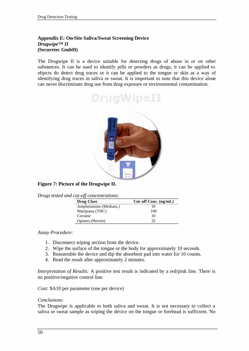
Drug Detection Testing
50
Appendix E: On-Site Saliva/Sweat Screening Device Drugwipe™ II (Securetec GmbH) The Drugwipe II is a device suitable for detecting drugs of abuse in or on other substances. It can be used to identify pills or powders as drugs, it can be applied to objects do detect drug traces or it can be applied to the tongue or skin as a way of identifying drug traces in saliva or sweat. It is important to note that this device alone can never discriminate drug use from drug exposure or environmental contamination.
Figure 7: Picture of the Drugwipe II. Drugs tested and cut-off concentrations:
Drug Class Cut-off Conc. (ng/mL) Amphetamine (Metham.) 10 Marijuana (THC) 100 Cocaine 10 Opiates (Heroin) 25
Assay Procedure:
1. Disconnect wiping section from the device. 2. Wipe the surface of the tongue or the body for approximately 10 seconds. 3. Reassemble the device and dip the absorbent pad into water for 10 counts. 4. Read the result after approximately 2 minutes.
Interpretation of Results: A positive test result is indicated by a red/pink line. There is no positive/negative control line. Cost: $A10 per parameter (one per device) Conclusions: The Drugwipe is applicable to both saliva and sweat. It is not necessary to collect a saliva or sweat sample as wiping the device on the tongue or forehead is sufficient. No

Drug Detection Testing
51
drug panel (simultaneous testing of multiple drugs) is available making testing relatively slow and expensive if a number of drugs are to be tested. The device is not FDA approved and has not been fully evaluated for saliva and sweat testing. A result reader (similar to the Rapiscan) may be introduced for the Drugwipe by the manufacturer. Analytical Conclusions of the Drugwipe (from Verstraete, 1999): Saliva: Good detection of recent abuse of cocaine, amphetamine and designer amphetamines. Good detection of recent abuse of opiates but with some false-negative results when the on-site Drugwipe result is compared to GC/MS. The results of the cannabis test are considered unreliable. Sweat: Results for cocaine and amphetamines are considered good. Results for opiates are reported to be more questionable. Surfaces: Large-scale trial showed the Drugwipe to be reliable for the detection of drugs on surfaces. Manufacturer: SecureTec Detektions-Systeme AG Rosenheimer Landstraße 129 85521 Ottobrunn / München Phone: +49 89 6072 9501 Fax: +49 89 6072 9182 http://www.securetec.net/english/home/home_frames.html American Distributor: SecureTec Contraband Detection & Identification Inc. 101 Phillips Park Drive South Williamsport, PA 17702, USA Ph: (570) 327 6112 Fax: (570) 322 1966

Drug Detection Testing
52
Appendix F: Urine Adulteration Test Strips Intect™ 7 (Branan Medical Corporation) The Intect™ 7 urine adulteration test strip is a plastic strip affixed with 7 chemically treated pads for assessing the integrity of the urine sample prior to testing. By visual color examination of the pads after dipping the strip into the urine sample, semi-quantitative values of creatinine, nitrite, glutaraldehyde, Ph, specific gravity, bleach and pyridinium chlorochromate can be determined.
Figure 7: Picture of the Intect™ 7 container and strips. What the Strip can test for: The following information can be obtained which may be useful in assessing the integrity of the urine sample:
1. Whether the sample is possibly diluted with water or other liquids. This is indicated by the creatinine and specific gravity tests.
2. Whether the sample may contain commercially available adulterants including nitrite ("Klear"), glutaraldehyde ("Instant Clean Add-it- ive" and "Urinaid"), bleach, pyridinium chlorochromate ("Urine Luck") and other oxidizing agents ("Stealth").
3. Whether the sample is possibly contaminated by acidic (vinegar) or basic (ammonia solution) adulterants. This is indicated by the pH test.
Test Procedure:
1. In order to avoid contamination of the whole urine sample, aliquot a small portion of the urine sample into another container for testing. Do not dip Intect™ 7 directly into the primary collection container.
2. Dip the numbered reagent end of the test strip in urine sample and remove immediately.
3. Blot the strip to remove excess urine. 4. Read and compare the numbered test areas with corresponding panels in the
color chart in one minute. Interpretation of Results: Results are obtained by visually comparing the color of each numbered pad with the

Drug Detection Testing
53
corresponding test color block pictured on the container label. No equipment is required. Cost: Approx. $A1.00 per strip. Conclusions: Some compounds or conditions that may affect test strip results are listed below. Medications that discolor the urine may also cause abnormal results due to masking of the reactions of the reagents on the test pads. Pad #1 Creatinine: Daily creatinine excretion, related to the muscle mass of the human body, is usually constant. A urine specimen with a creatinine level less than 20 mg/dl is typically considered indicative of adulteration. Pad #2 Nitrite: Although nitrite is not a normal component of urine, nitrite levels of up to 3.6 mg/dl may be found in some urine specimens due to urinary tract infections, bacterial contamination or improper storage. In the Intect™ 7 test strip, nitrite level above 7.5 mg/dl is considered abnormal. Pad #3 Glutaraldehyde: Glutaraldehyde is not a normal component of urine. Hence, the detection of glutaraldehyde in the urine sample indicates the possibility of adulteration. However, in ketoacidosis, starvation or other metabolic abnormalities, ketone bodies may appear in urine, interact with the glutaraldehyde pad and provide a false result. Pad #4 pH: Normal urine pH ranges from 4.5 to 8.0. Values below pH 4.0 or above pH 9.0 are indicative of adulteration. Pad #5 Specific Gravity: Fresh urine samples should have a range of 1.002 to 1.030. However, high protein concentration in the urine may elevate the specific gravity value. Urine specimens with a specific gravity of less than 1.003 is indicative of adulteration. Specific gravity and creatinine values should be considered together to provide a better picture of whether the sample is adulterated. Pad #6 Bleach: The presence of bleach in the urine is indicative of adulteration since bleach is not a normal constituent of urine. The formation of a brown or blackish blue pad color may also indicate the presence of other oxidative adulterants. Pad #7 Pyridinium Chlorochromate: The presence of pyridinium is also not a normal constituent of urine. The formation of a blue pad color may also indicate the presence of other oxidative adulterants.
Manufacturer: Australian Distributor: Branan Medical Corporation Microgenics Diagnostics Pty Ltd
Irvine, CA, USA PO Box 1213 Auburn NSW 1825 (ph) 949 598 7166 (ph) 02 9649 9599 (fax) 949 598 7167 (ph) 02 9649 9788 http://www.brananmedical.com/ [email protected]


![Drug testing[1]](https://img.pdfslide.us/doc/110x75/559235e01a28ab3f158b45cf/drug-testing1.jpg)