Embed Size (px)
Citation preview

Dr.Otman Siregar SpOT.(K)SpineDr.Otman Siregar SpOT.(K)Spine
April 2009

Limb Threatening InjuriesCan be caused by:
� MVA
� Occupational accident
Domestic accident� Domestic accident
� Open injury
� Closed injury

Limb Threatening Injuries� Is an emergency
situation
� Need accurate diagnosis and prompt treatmentand prompt treatment

Limb Threatening Injuries� Fracture
- Open fracture
- Closed Fracture
Vascular Injury� Vascular Injury
� Compartment syndrome
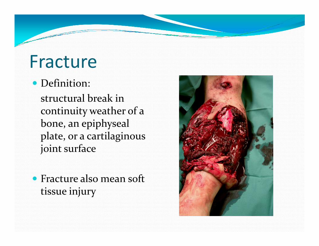
Fracture� Definition:
structural break in continuity weather of a bone, an epiphyseal bone, an epiphyseal plate, or a cartilaginous joint surface
� Fracture also mean soft tissue injury

Fracture� The causative force that produces a fracture may be :
- Direct injury
- Indirect injury

Fracture DiagnosisPatient History, ask about
� Pain
� Deformity
Time of injury
� Mechanism of injury:
- Fall
- Direct blow
- Road accident� Time of injury - Road accident
- Gun Shot Wound
- Often lack of detail

Fracture Diagnosis� Always do Primary Survey (ABC)
� General condition
� Local Condition:
- Look- Look
-Feel
-Move
� Principle: DO NO FURTHER HARM!

Fracture DiagnosisLook:
� Local swelling
� Deformity ( angulations, rotation, discrepancy)rotation, discrepancy)
� Discoloration of the skin
� Open wound (size, margin, depth, contamination)

Fracture DiagnosisFeel
� Sharply localized tenderness
� Aggravation of pain and muscle spasm
Crepitation� not necessary� Crepitation� not necessary
� Neurovascular Condition is important
� Always look and feel for other less apparent injuries

Fracture DiagnosisMove
� Not necessary if the deformity is obvious
� Abnormal movement
Usually ROM limited due to pain� Usually ROM limited due to pain
Fracture DiagnosisSpecial Test and measurement
� Allen test: vascular patency in forearm
� True, apparent, and anatomical length
Drawer test ( is better to do it under anesthesia)� Drawer test ( is better to do it under anesthesia)

Diagnostic Imaging� Immobilized the limb before being
subjected to imaging examination
� Plain X ray
CT Scan� CT Scan
� MRI
� angiography

Diagnostic ImagingX ray : Rules of two
� 2 joint
� 2 projection
2 extremities (paediatric)� 2 extremities (paediatric)
� 2 densities (able to differ hard and soft tissue)
Special projection may be necessary

Diagnostic Imaging� CT Scan and MRI can
provide useful additional data especially for pelvis and spinal injuryand spinal injury
� Angiography is performed if vascular injury is suspected
� Doppler duplex sonogram

Descriptive Term Pertaining to
Fractures� Site
-diaphyseal, metaphyseal, epiphyseal or intraarticular
� Extent
- Complete or incomplete- Complete or incomplete
� Configuration
-transverse, oblique or spiral
-comminuted or segmental
� Relationship of the fracture fragments to each other
-translated,angulated,rotated,distracted,overriding, impacted

Descriptive Term Pertaining to
Fractures� Relationship of the
fracture to the external environment
-open or closed-open or closed
� Complications
-uncomplicated or complicated

Complications of Musculoskeletal
InjuriesClassified as :
� Initial (immediate) complications
- Local and Remote
Early� Early
-Local and remote
� Late complications
-Local and remote

Complications of Musculoskeletal
InjuriesInitial Complication:
� Local complication
-Skin injuries (from within or without)
-vascular injuries (artery or vein, division, contusion or -vascular injuries (artery or vein, division, contusion or spasm)
-neurological injuries (brain, spinal cord, peripheral nerve)
-muscular
-visceral
� Remote complication
-multiple injuries and hemorrhagic shock

Complications of Musculoskeletal
InjuriesEarly Complication
� Local complication
-Skin necrosis, gangrene, compartment syndrome, compartment syndrome, etc
-Joint complication (septic arthritis)
-Bony complications (Osteomyelitis or avascular necrosis)

Complications of Musculoskeletal
InjuriesEarly
� Remote Complications
-Fat embolism
-Pulmonary embolism-Pulmonary embolism
-Pneumonia
-Tetanus
-Delirium Tremens

Complications of Musculoskeletal
InjuriesLate Complications
� Local Complication
-Joint: stiffness, degenerative arthritis
-Bony: abnormal fr healing, growth disturbance, chronic -Bony: abnormal fr healing, growth disturbance, chronic osteomyelitis
-Muscular :myositis ossificans, late rupture tendon
-Neurological : Tardy nerve plasy

Complications of Musculoskeletal
InjuriesLate
� Remote complications
-Renal calculi
-accident neurosis-accident neurosis


An open An open
fracture fracture
indicates …indicates …
… a communications
between the fracture between the fracture
and the external
environment …

Classification
• Gustillo / Anderson 1976
• Oestern & Tscherne 1984
Open Fractures
AO Courses Jakarta 2008
• Oestern & Tscherne 1984
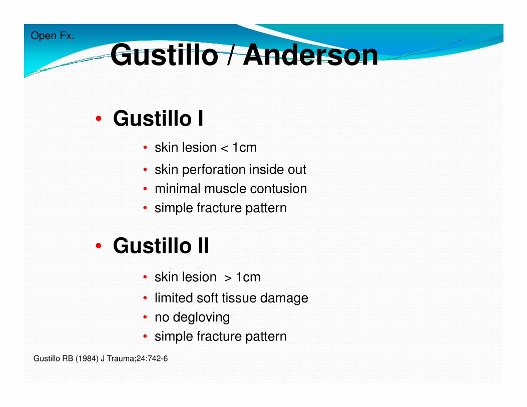
Gustillo / AndersonOpen Fx.
• Gustillo I
• skin lesion < 1cm
• skin perforation inside out
• minimal muscle contusion
• simple fracture pattern• simple fracture pattern
• Gustillo II
• skin lesion > 1cm
• limited soft tissue damage
• no degloving
• simple fracture pattern
Gustillo RB (1984) J Trauma;24:742-6

Gustillo / AndersonOpen Fx.
• Gustillo III A• Extensive soft tissue damage (skin, muscles,
neurovascular strucures) with still sufficient
bone coverage (periosteum)
• Gustillo III B• Gustillo III B• Extensive soft tissue damage with periosteal
detachment and exposed bone
• Massive contomination of the wound
• Gustillo III C
• Vascular injury to be reconstructed

Gustillo I
AO Courses Jakarta 2008

Gustillo III A/B
AO Courses Jakarta 2008

Gustillo III C
AO Courses Jakarta 2008

Erfurt algorithm
• remove wound dressing only in OR
• foto documentation
• debridement
management of open fx.
• fracture fixation (FixEx)
• leave the wound open or
• temporary wound coverage by
� skin substitute or
� vacuum therapy


Mechanisms of Vascular Injury in
the Extremities
� Gunshot wound – 54%
� Stab wound – 15%
� Shotgun wound – 12%� Shotgun wound – 12%
� Blunt trauma – 15%
� Iatrogenic – 3%

Presentation of Vascular Injury
� First priority is hemorrhage control followed by control followed by appropriate diagnostic work-up

Presentation of Vascular Injury� Dislocations and
displaced or angulated fractures: realigned realigned immediately if vascularity is compromised

Evaluation for Vascular Injury� Physical Examination� Doppler Flowmeter� Duplex Ultrasonography� Arteriogram� Local wound exploration should not be � Local wound exploration should not be
done in an uncontrolled setting� Close coordination with a general or
vascular surgeon recommended

Physical Examination
Hard Signs
� Absent or diminished distal pulses
� Active hemorrhage
� Large, expanding or pulsatile hematomaLarge, expanding or pulsatile hematoma
� Bruit or thrill
� Distal ischemia (pain, pallor, paralysis, paresthesias, coolness)

Physical Examination
Soft Signs
� Small, stable hematoma
� Injury to anatomically related nerveInjury to anatomically related nerve
� Unexplained hypotension
� History of hemorrhage no longer present
� Proximity of injury to major vessel

Doppler Examination
� Non-invasive adjunct to physical examination� Small, hand-held (non-directional) Doppler
flowmeter provides for subjective interpretation of flowmeter provides for subjective interpretation of audible signal
� Useful as modality for determining the Ankle-Brachial Index (ABI)

Arteriography� Gold standard for evaluation of peripheral vascular
injuries
� Formal arteriograms done in radiology may cause critical delays in diagnosis or interventioncritical delays in diagnosis or intervention
� Single-shot arteriograms done in the emergency room or operating room should be considered in cases where arteriography is indicated.

Indications for Arteriography� Multiple potential sites of injury (shotgun wounds)� Missile track parallels vessel over long distance� Blunt trauma with signs of vascular trauma� Chronic vascular disease� Extensive bone or soft tissue injury� Extensive bone or soft tissue injury� Thoracic outlet wounds� Evaluation of equivocal results from non-invasive
tests� Proximity (gsw, knife wound) (controversial)� ABI < .9

Single-shot Arteriogram in the
Emergency or Operating Room


Compartment SyndromeDefinition
� Elevated tissue pressure within a closed fascial space
� Reduces tissue perfusion� Reduces tissue perfusion
� Results in cell death
� Pathogenesis
� Too much inflow (edema, hemorrhage)
� Decreased outflow (venous obstruction, tight dressing/cast)
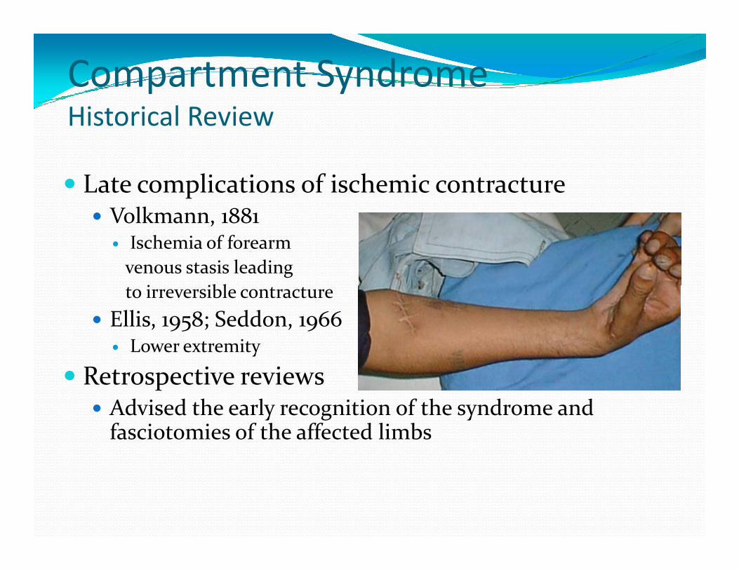
Compartment SyndromeHistorical Review
� Late complications of ischemic contracture� Volkmann, 1881
� Ischemia of forearm
venous stasis leading
to irreversible contracture to irreversible contracture
� Ellis, 1958; Seddon, 1966� Lower extremity
� Retrospective reviews � Advised the early recognition of the syndrome and
fasciotomies of the affected limbs

Compartment SyndromeTissue Survival
� Muscle� 3-4 hours - reversible changes
� 6 hours - variable damage� 6 hours - variable damage
� 8 hours - irreversible changes
� Nerve � 2 hours - looses nerve conduction
� 4 hours - neuropraxia
� 8 hours - irreversible changes

Compartment SyndromeEtiology
� Fractures-closed and open
� Blunt trauma
� Exertional states
� GSW
� Temp vascular occlusion
� Cast/dressing
� Closure of fascial defects
� Burns/electrical
� IV/A-lines
� Hemophiliac/coag
� Intraosseous IV(infant)
� Snake bite
� Arterial injury

Compartment SyndromeDiagnosis
� Pain out of proportion
� Palpably tense compartment
� Pain with passive stretch� Pain with passive stretch
� Paresthesia/hypoesthesia
� Paralysis
� Pulselessness/pallor

Compartment SyndromeDifferential diagnosis
� Arterial occlusion
� Peripheral nerve injury
� Muscle rupture

Compartment SyndromeEmergent Treatment
� Remove cast or dressing
� Place at level of heart
(DO NOT ELEVATE to optimize (DO NOT ELEVATE to optimize perfusion)
� Alert OR and Anesthesia
� Bedside procedure
� Medical treatment

Compartment SyndromeSurgical Treatment
� Fasciotomy - prophylactic release of pressure before permanent damage occurs. Will not reverse injury from trauma.
� Fracture care – rigidstabilization
� Ex-fix
� IM Nail


![ACUTE ABDOMEN -EM K 27.ppt [Read-Only] - ocw.usu.ac.idocw.usu.ac.id/course/download/1110000130-emergency-medicine/emd… · usually related to inflammation or ... • Review anatomy](https://img.pdfslide.us/doc/110x75/5b55e6ef7f8b9ac31e8bc12a/acute-abdomen-em-k-27ppt-read-only-ocwusuacidocwusuacidcoursedownload1110000130-emergency-medicineemd.jpg)


![Bowel Elimination Si.ppt [Read-Only] - ocw.usu.ac.idocw.usu.ac.id/.../kdm_slide_bowel_elimination.pdfPrimary organ of bowel elimination ... Small bowel series Barium enema. ... Sigmoid](https://img.pdfslide.us/doc/110x75/5adf17e77f8b9ac0428bbfc8/bowel-elimination-sippt-read-only-ocwusuacidocwusuacidkdmslidebowel.jpg)


![2. ATOPIC DERMATITIS.ppt [Read-Only] - ocw.usu.ac.idocw.usu.ac.id/.../mk_aia_slide_atopic_dermatitis.pdf · Atopic dermatitis Definition An inflammatory skin disorder 2 characterized](https://img.pdfslide.us/doc/110x75/5d63d77f88c993635d8b7207/2-atopic-read-only-ocwusuacidocwusuacidmkaiaslideatopicdermatitispdf.jpg)


![KULIAH TUMOR MARKER S.ppt [Read-Only] - ocw.usu.ac.idocw.usu.ac.id/.../elo173_slide_tumor_markers.pdf · TUMOR MARKER’S Prof. Adi Koesoema Aman . ... Klasifikasi Penanda Tumor](https://img.pdfslide.us/doc/110x75/5a77bc1f7f8b9a93088e0a67/kuliah-tumor-marker-sppt-read-only-ocwusuacidocwusuacidelo173slidetumor.jpg)

