Embed Size (px)
Citation preview
Drosophila PTB promotes formation ofhigh-order RNP particles and repressesoskar translation
Florence Besse,1,2 Sonia Lopez de Quinto,1,3 Virginie Marchand, Alvar Trucco, and Anne Ephrussi4
Developmental Biology Unit, European Molecular Biology Laboratory, 69117 Heidelberg, Germany
Local translation of asymmetrically enriched mRNAs is a powerful mechanism for functional polarization of thecell. In Drosophila, exclusive accumulation of Oskar protein at the posterior pole of the oocyte is essential fordevelopment of the future embryo. This is achieved by the formation of a dynamic oskar ribonucleoprotein (RNP)complex regulating the transport of oskar mRNA, its translational repression while unlocalized, and its translationalactivation upon arrival at the posterior pole. We identified the nucleo–cytoplasmic shuttling protein PTB(polypyrimidine tract-binding protein)/hnRNP I as a new factor associating with the oskar RNP in vivo. While PTBfunction is largely dispensable for oskar mRNA transport, it is necessary for translational repression of the localizingmRNA. Unexpectedly, a cytoplasmic form of PTB can associate with oskar mRNA and repress its translation,suggesting that nuclear recruitment of PTB to oskar complexes is not required for its regulatory function.Furthermore, PTB binds directly to multiple sites along the oskar 39 untranslated region and mediates assembly ofhigh-order complexes containing multiple oskar RNA molecules in vivo. Thus, PTB is a key structural componentof oskar RNP complexes that dually controls formation of high-order RNP particles and translational silencing.
[Keywords: Drosophila; PTB; oskar; translation; RNP assembly; hnRNP]
Supplemental material is available at http://www.genesdev.org.
Received September 5, 2008; revised version accepted November 20, 2008.
In eukaryotic cells, transcription represents the first step ofgene expression. However, a variety of nuclear and cyto-plasmic post-transcriptional events determine the finalfate of RNAs and thus regulate gene product diversity aswell as the spatio–temporal pattern of gene expression.
In recent years, subcellular targeting of mRNAs, cou-pled to localized translation, has emerged as a powerfulmechanism to spatially and temporally restrict proteinsynthesis. Furthermore, a genome-wide in situ hybridiza-tion analysis in Drosophila embryos suggests that RNAlocalization could represent a general mechanism for theestablishment of cell polarity (Lecuyer et al. 2007).Consistent with this, functional studies have shown thatlocal translation of asymmetrically enriched mRNAs isused by differentiated cells to generate functionallydistinct compartments, or by developing organisms topartition cell fate determinants (St Johnston 2005; Du
et al. 2007). In several species, the asymmetric distribu-tion in unfertilized eggs of maternal RNAs encodingcytoplasmic determinants controls embryonic body axisspecification. In Drosophila, oskar mRNA encodes theposterior determinant and is transported to the posteriorpole of the oocyte, where it is specifically translated. Thisprecise spatio–temporal control is critical for embryonicpatterning, as mutant oocytes in which oskar is notexpressed at the posterior pole develop into embryoslacking abdominal structures and germ cells (Ephrussiet al. 1991; Kim-Ha et al. 1991). Conversely, ectopictranslation of unlocalized oskar causes patterning defectscharacterized by a loss of anterior structures, and in ex-treme cases, duplication of posterior structures (Ephrussiand Lehmann 1992; Kim-Ha et al. 1995).
RNA localization relies on the formation of functionalribonucleoprotein (RNP) complexes that precisely con-trol and coordinate multiple steps including motor-basedtransport of the mRNA along the cytoskeleton and trans-lational repression of the localizing mRNA, as well as itstranslation activation and anchoring upon arrival at thefinal destination (St Johnston 2005). These complexescontain different RNA-associated proteins, including het-erogeneous nuclear RNPs (hnRNPs) that regulate variousRNA processing events (Dreyfuss et al. 2002; Glisovicet al. 2008). Furthermore, RNPs seem to assemble in vivo
1These authors contributed equally to this work.Present addresses: 2Institute of Developmental Biology and Cancer/UMR6543, University Nice-Sophia Antipolis, Parc Valrose, 06108 NiceCedex 2, France.3Cardiff University, School of Biosciences, Museum Avenue, CardiffCF10 3AX, Wales, UK.4Corresponding author.E-MAIL [email protected]; FAX 49-6221-387-8166.Article published online ahead of print. Article and publication date areonline at http://www.genesdev.org/cgi/doi/10.1101/gad.505709.
GENES & DEVELOPMENT 23:195–207 � 2009 by Cold Spring Harbor Laboratory Press ISSN 0890-9369/09; www.genesdev.org 195
Cold Spring Harbor Laboratory Press on January 30, 2018 - Published by genesdev.cshlp.orgDownloaded from
into large particles containing several RNA moleculesand associated proteins, as evidenced by light microscopyvisualization or by biochemical sedimentation techniques(Kiebler and Bassell 2006; Lange et al. 2008). Although thein vivo functional relevance of such high-order structuresremains unknown, their formation has been proposed torepresent a crucial step in the establishment of transport-competent RNP complexes.
The composition, architecture, and dynamics of RNPcomplexes dictate the specific behavior of target RNAswithin the cell. Importantly, RNP complexes undergo anextensive remodeling upon export into the cytoplasm, yettheir cytoplasmic fate is highly connected to their nuclearhistory (Kress et al. 2004; St Johnston 2005; Giorgi andMoore 2007; Lewis and Mowry 2007). For example, in thecase of oskar mRNA, nuclear recruitment of the exon–junctioncomplexupon splicing isnecessaryforoskar mRNAtransport to the posterior pole of the oocyte (Hachet andEphrussi 2001, 2004). Furthermore, oskar translationalrepression is controlled by nucleo–cytoplasmic shuttlingRNA-binding proteins such as Bruno and Hrp48 (Kim-Haet al. 1995; Yano et al. 2004; Snee et al. 2008). While theseproteins likely associate with the RNA in the nucleus, howimportant the nuclear recruitment of these proteins to theirtarget RNA is remains to be functionally tested.
In a visual protein-trap screen, we identified Drosoph-ila polypyrimidine tract-binding protein (PTB) as a proteincolocalizing with oskar mRNA. PTB belongs to thehnRNP family of RNA-binding proteins and regulatesvarious nuclear and cytoplasmic RNA processing eventsin vertebrates (Auweter and Allain 2008). Specifically,PTB plays a predominant role in the regulation of alter-native splicing (Valcarcel and Gebauer 1997; Spellmanet al. 2005), and has also been shown to regulate internalribosome entry site (IRES)-dependent translation initia-tion (Stoneley and Willis 2004; Jang 2006; Semler andWaterman 2008) and mRNA localization (Cote et al.1999; Ma et al. 2007). In our in vivo study, we show thatDrosophila PTB is a new component of the oskar RNPcomplex and that PTB regulates translational repressionof oskar mRNA. Surprisingly, while PTB strongly accu-mulates in germ cell nuclei and thus, potentially, asso-ciates with oskar in this compartment, its recruitment tooskar RNP in the germ cell cytoplasm is sufficient forefficient repression. Consistent with a direct role in oskartranslation regulation, PTB binds to multiple bindingsites in the oskar 39 untranslated region (UTR). Finally,we show that PTB is a trans-acting factor required in vivofor oskar mRNA oligomerization. Our study thus revealsthat Drosophila PTB is a key structural component ofoskar RNP complexes and suggests a functional linkbetween the assembly of high-order RNP particles andthe mechanism of oskar translational repression.
Results
PTB colocalizes with oskar mRNA duringDrosophila oogenesis
To identify new proteins involved in oskar mRNAregulation, we performed a protein-trap screen that relies
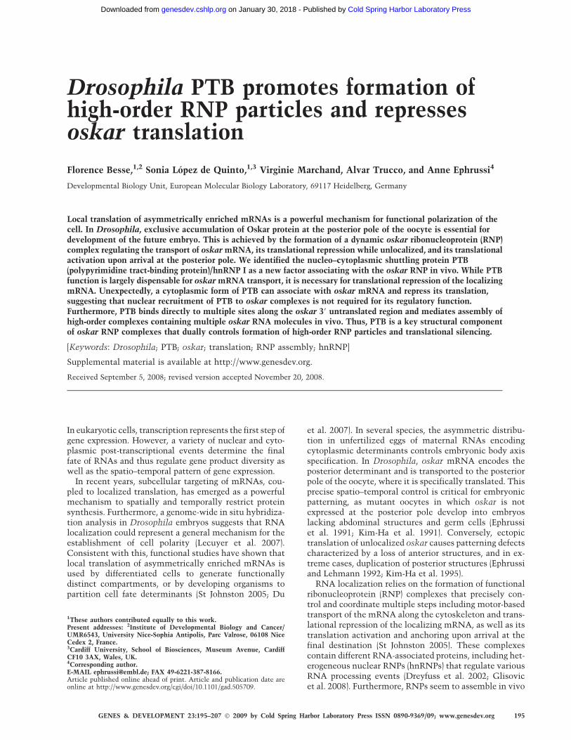
on GFP-tagging of proteins by random mobilization ofa GFP-containing transposable element in the genome(Morin et al. 2001; Bonin and Mann 2004). Among therecovered GFP fusion proteins colocalizing with oskarmRNA, one showed an enrichment in the oocyte cyto-plasm during early oogenesis, followed by an exclusiveaccumulation at the posterior pole from mid-oogenesisonward (Fig. 1A,B; see Spradling 1993 for a detaileddescription of ovarian cell types and developmentalstages). Additionally, the GFP-tagged protein localizesto the nuclei of both somatic and germ cells, as well asin the cytoplasm of the follicular epithelial cells sur-rounding the oocyte, at late stages of oogenesis.
Using inverse PCR, we mapped the insertion site of theprotein-trap cassette to the hephaestus locus (CG31000),encoding the unique Drosophila homolog of mammalianPTB, also known as hnRNP-I (Dansereau et al. 2002;Davis et al. 2002). Similar to its mammalian ortholog,Drosophila PTB contains a putative N-terminal bipartitenuclear localization signal (NLS) and four conserved RNArecognition motifs (RRM1–4) (Fig. 1C). Western-blotanalysis using anti-PTB antibody raised against the Dro-sophila protein revealed a 65-kDa doublet in wild-typeovarian extracts (Fig. 1D), which is shifted to 95 kDa inthe protein-trap line as a result of the GFP insertionupstream of RRM1 (Fig. 1C).
Staining of wild-type ovaries with anti-PTB antibodiesrevealed a localization pattern of endogenous PTB iden-tical to that of GFP-PTB fusions (Fig. 1E–G). Similarly tothe oskar-associated protein Staufen, PTB accumulates inthe cytoplasm of young oocytes as early as in thegermarium (Fig. 1E–G0; data not shown), transiently inthe center of the oocyte at stage 7/8 (Fig. 1F–F0), and at theposterior pole of the oocyte from stage 9 onward (Fig. 1G–G0; see Supplemental Fig. S1C for a specificity control).The perfect colocalization of PTB with Staufen in theoocyte cytoplasm throughout oogenesis suggested thatPTB is transported together with oskar mRNA to theposterior pole of the oocyte.
PTB is a component of the oskar RNP complex
To confirm the in vivo association of PTB with oskarmRNA, we tested if the distribution of PTB in the oocytedepends on oskar mRNA. Strikingly, the cytoplasmicenrichment of PTB in young oocytes is no longer detectedin egg chambers lacking oskar mRNA (oskA87/Df(3R)pXT03) (Fig. 2B), indicating that the initial transport of PTBinto the oocyte cytoplasm requires the presence of oskarmRNA. As oskar mRNA-null egg chambers do not de-velop past stage 7 (Jenny and Hachet et al. 2006), weanalyzed the oskar dependence of PTB localization duringmid-oogenesis, following the distribution of PTB ingurken (grk) and staufen mutant oocytes. In grk2E12/grk2B6 females, microtubule (MT) polarity is altered suchthat MT plus ends are enriched in the center of the oocyteinstead of at the posterior pole (Gonzalez-Reyes et al.1995). In these mutants, oskar mRNA and Staufen aredetected in the center of the oocyte, together with PTB(Fig. 2D–D0). Furthermore, PTB fails to localize at theposterior pole in stauD3 oocytes (Fig. 2E), which appear to
Besse et al.
196 GENES & DEVELOPMENT
Cold Spring Harbor Laboratory Press on January 30, 2018 - Published by genesdev.cshlp.orgDownloaded from
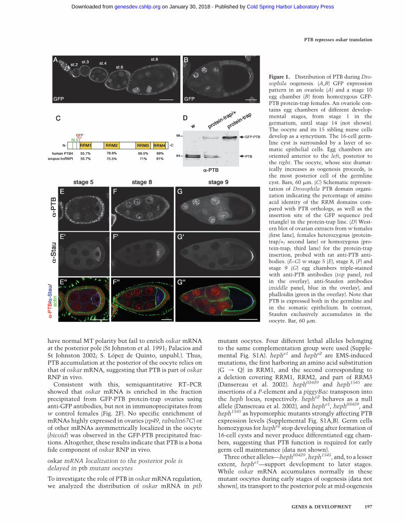
have normal MT polarity but fail to enrich oskar mRNAat the posterior pole (St Johnston et al. 1991; Palacios andSt Johnston 2002; S. Lopez de Quinto, unpubl.). Thus,PTB accumulation at the posterior of the oocyte relies onthat of oskar mRNA, suggesting that PTB is part of oskarRNP in vivo.
Consistent with this, semiquantitative RT–PCRshowed that oskar mRNA is enriched in the fractionprecipitated from GFP-PTB protein-trap ovaries usinganti-GFP antibodies, but not in immunoprecipitates fromw control females (Fig. 2F). No specific enrichment ofmRNAs highly expressed in ovaries (rp49, tubulin67C) orof other mRNAs asymmetrically localized in the oocyte(bicoid) was observed in the GFP-PTB precipitated frac-tions. Altogether, these results indicate that PTB is a bonafide component of oskar RNP in vivo.
oskar mRNA localization to the posterior pole isdelayed in ptb mutant oocytes
To investigate the role of PTB in oskar mRNA regulation,we analyzed the distribution of oskar mRNA in ptb
mutant oocytes. Four different lethal alleles belongingto the same complementation group were used (Supple-mental Fig. S1A). hephe1 and hephe2 are EMS-inducedmutations, the first harboring an amino acid substitution(G / Q) in RRM1, and the second corresponding toa deletion covering RRM1, RRM2, and part of RRM3(Dansereau et al. 2002). heph03429 and heph1545 areinsertions of a P-element and a piggyBac transposon intothe heph locus, respectively. hephe2 behaves as a nullallele (Dansereau et al. 2002), and hephe1, heph03429, andheph1545 as hypomorphic mutants strongly affecting PTBexpression levels (Supplemental Fig. S1A,B). Germ cellshomozygous for hephe2 stop developing after formation of16-cell cysts and never produce differentiated egg cham-bers, suggesting that PTB function is required for earlygerm cell maintenance (data not shown).
Three other alleles—heph03429, heph1545, and, to a lesserextent, hephe1—support development to later stages.While oskar mRNA accumulates normally in thesemutant oocytes during early stages of oogenesis (data notshown), its transport to the posterior pole at mid-oogenesis
Figure 1. Distribution of PTB during Dro-
sophila oogenesis. (A,B) GFP expressionpattern in an ovariole (A) and a stage 10egg chamber (B) from homozygous GFP-PTB protein-trap females. An ovariole con-tains egg chambers of different develop-mental stages, from stage 1 in thegermarium, until stage 14 (not shown).The oocyte and its 15 sibling nurse cellsdevelop as a syncytium. The 16-cell germ-line cyst is surrounded by a layer of so-matic epithelial cells. Egg chambers areoriented anterior to the left, posterior tothe right. The oocyte, whose size dramat-ically increases as oogenesis proceeds, isthe most posterior cell of the germlinecyst. Bars, 60 mm. (C) Schematic represen-tation of Drosophila PTB domain organi-zation indicating the percentage of aminoacid identity of the RRM domains com-pared with PTB orthologs, as well as theinsertion site of the GFP sequence (redtriangle) in the protein-trap line. (D) West-ern blot of ovarian extracts from w females(first lane), females heterozygous (protein-trap/+; second lane) or homozygous (pro-tein-trap; third lane) for the protein-trapinsertion, probed with rat anti-PTB anti-bodies. (E–G) w stage 5 (E), stage 8, (F) andstage 9 (G) egg chambers triple-stainedwith anti-PTB antibodies (top panel, redin the overlay), anti-Staufen antibodies(middle panel, blue in the overlay), andphalloidin (green in the overlay). Note thatPTB is expressed both in the germline andin the somatic epithelium. In contrast,Staufen exclusively accumulates in theoocyte. Bar, 60 mm.
PTB represses oskar translation
GENES & DEVELOPMENT 197
Cold Spring Harbor Laboratory Press on January 30, 2018 - Published by genesdev.cshlp.orgDownloaded from
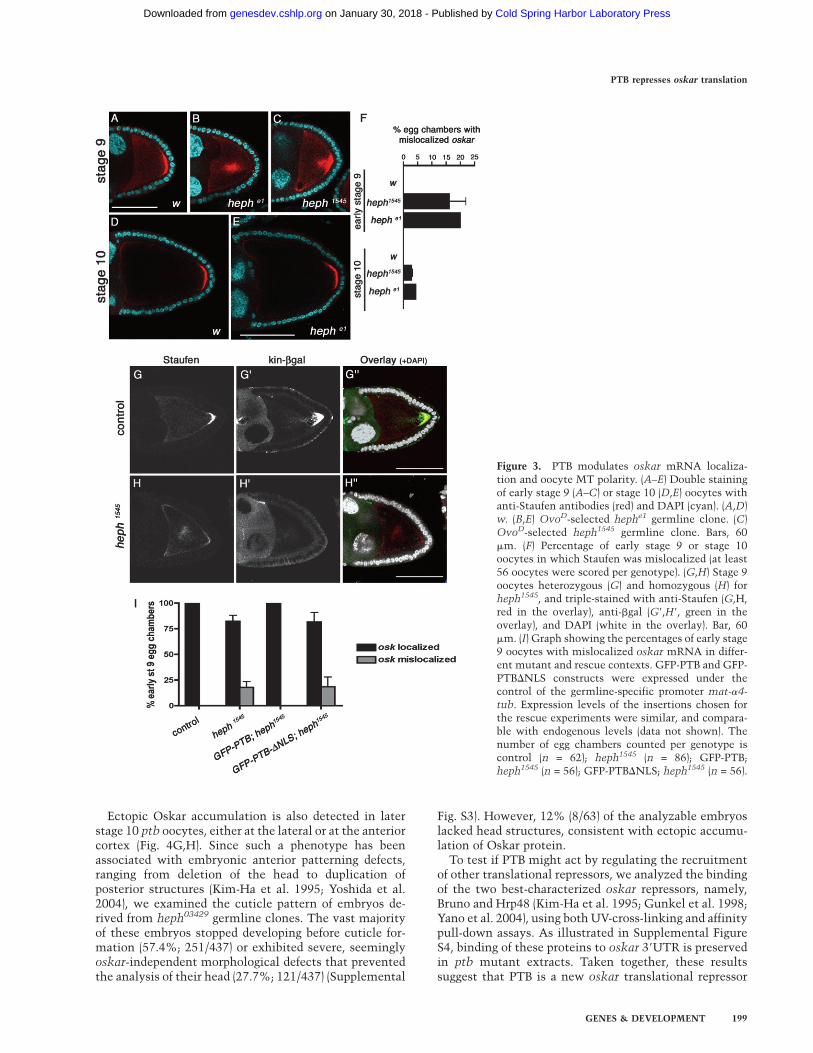
is affected (Fig. 3B,C). In wild-type oocytes, oskar mRNAand Staufen transiently localize in the center of theoocyte at stages 7/8 and accumulate as a tight posterior
crescent from early stage 9 onward (Figs.1E–G, 3A; datanot shown). In contrast, oskar mRNA persists in thecenter of the oocyte or as a cloud near the posteriorcortex, in ;20% of early stage 9 ptb oocytes (Fig. 3B,C,F).This phenotype seems to reflect a transient delay in oskarmRNA transport to the posterior pole, as it is rarelyobserved in late stage 9 or stage 10 oocytes (Fig. 3E,F; datanot shown). Since oskar mRNA localization relies on thepolarity of the MT cytoskeleton, we assessed the distri-bution of the MT plus-end marker kin-b-gal (Clark et al.1994) in ptb mutant oocytes at stage 9. In these oocytes,oskar mRNA mislocalization correlates with a strongreduction in the posterior accumulation of kin-b-gal (Fig.3H–H9), suggesting that oskar mRNA localization defectsmay result from a delay in focusing the MT plus-ends atthe posterior pole.
As shown in Figure 3I, the oskar localization phenotypewas rescued upon expression of a wild-type GFP-PTBfusion in ptb mutant germ cells. Interestingly, a mutantversion of PTB unable to enter the nucleus, but still ableto associate with oskar mRNA in the cytoplasm (GFP-PTB-DNLS) (Supplemental Fig. S2), failed to rescue thisphenotype (Fig. 3I). Our results thus indicate that PTBfunction is required in germ cell nuclei for efficient estab-lishment of MT polarity at stages 8–9 of oogenesis, ulti-mately controlling localization of oskar mRNA. Giventhat mammalian PTB has been extensively implicatedin the regulation of alternative splicing (Valcarceland Gebauer 1997; Spellman et al. 2005), it is possiblethat nuclear Drosophila PTB regulates the splicing ofgenes involved in establishment of MT polarity in theoocyte.
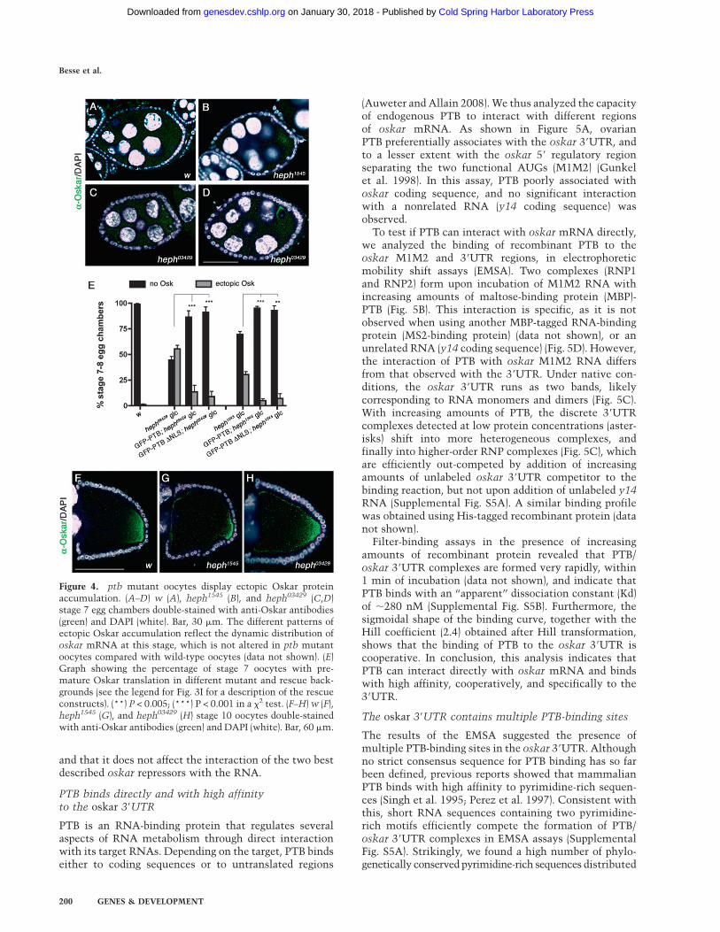
oskar is prematurely translated in ptb mutant oocytes
In wild-type females, oskar translation is repressed inearly egg chambers and is activated only once the mRNAreaches the posterior pole of the oocyte (Markussen et al.1995; Rongo et al. 1995). Thus, Oskar protein is notdetected before late stage 8 (Fig. 4A) and accumulatesexclusively at the oocyte posterior pole in later stages(Fig. 4F). Interestingly, oskar is prematurely translated inptb mutant females, and ectopic Oskar is detected asearly as in stages 5–6 ptb oocytes (data not shown). Atstage 7, ;50% of heph03429 and 30% of heph1545 mutantoocytes display ectopic Oskar expression (Fig. 4B–E), theprotein accumulating as a poorly defined cloud in thecenter of the oocyte (Fig. 4B), more rarely as cytoplasmicaggregates (Fig. 4C), or close to the oocyte nucleus at theanterior-dorsal corner (Fig. 4D). Notably, premature oskartranslation is suppressed by germline expression of wild-type GFP-PTB, as well as by expression of the GFP-PTB-DNLS mutant protein (Fig. 4E). These results indicatethat PTB represses oskar mRNA translation during earlyoogenesis and that assembly of translationally silencedoskar RNP complexes does not require the recruitmentof PTB in the nuclei of germ cells. Furthermore, thedifferential phenotypic rescue obtained when expressingGFP-PTB-DNLS rules out that the premature translationof oskar observed in ptb mutant oocytes is a consequenceof MT polarity defects.
Figure 2. PTB colocalizes and associates with oskar mRNA.(A,B) Stage 5 egg chambers from oskA87/+ (A) and oskA87/
Df(3R)pXT03 (B) females expressing GFP-PTB constructs underthe control of the mat-a4-tub promoter, and triple-stained forGFP (green in the overlay), DNA (blue, DAPI), and the oocytecytoplasm marker Orb (red). Bar, 15 mm. (C–E) Distribution ofthe GFP-PTB protein-trap fusion in wild-type (C), grk2E12/grk2B6
(D), and stauD3 (E) oocytes. (Left) GFP signal. (Middle) Staufenprotein. (Right) Overlay. Bar, 60 mm. (F, left panel) RT–PCRamplification of mRNAs recovered in fractions immunoprecipi-tated from GFP-PTB protein-trap or w control ovarian extractsusing anti-GFP antibodies. (Right panel) Amplifications fromunbound fractions are used as controls.
Besse et al.
198 GENES & DEVELOPMENT
Cold Spring Harbor Laboratory Press on January 30, 2018 - Published by genesdev.cshlp.orgDownloaded from
Ectopic Oskar accumulation is also detected in laterstage 10 ptb oocytes, either at the lateral or at the anteriorcortex (Fig. 4G,H). Since such a phenotype has beenassociated with embryonic anterior patterning defects,ranging from deletion of the head to duplication ofposterior structures (Kim-Ha et al. 1995; Yoshida et al.2004), we examined the cuticle pattern of embryos de-rived from heph03429 germline clones. The vast majorityof these embryos stopped developing before cuticle for-mation (57.4%; 251/437) or exhibited severe, seeminglyoskar-independent morphological defects that preventedthe analysis of their head (27.7%; 121/437) (Supplemental
Fig. S3). However, 12% (8/63) of the analyzable embryoslacked head structures, consistent with ectopic accumu-lation of Oskar protein.
To test if PTB might act by regulating the recruitmentof other translational repressors, we analyzed the bindingof the two best-characterized oskar repressors, namely,Bruno and Hrp48 (Kim-Ha et al. 1995; Gunkel et al. 1998;Yano et al. 2004), using both UV-cross-linking and affinitypull-down assays. As illustrated in Supplemental FigureS4, binding of these proteins to oskar 39UTR is preservedin ptb mutant extracts. Taken together, these resultssuggest that PTB is a new oskar translational repressor
Figure 3. PTB modulates oskar mRNA localiza-tion and oocyte MT polarity. (A–E) Double stainingof early stage 9 (A–C) or stage 10 (D,E) oocytes withanti-Staufen antibodies (red) and DAPI (cyan). (A,D)w. (B,E) OvoD-selected hephe1 germline clone. (C)OvoD-selected heph1545 germline clone. Bars, 60mm. (F) Percentage of early stage 9 or stage 10oocytes in which Staufen was mislocalized (at least56 oocytes were scored per genotype). (G,H) Stage 9oocytes heterozygous (G) and homozygous (H) forheph1545, and triple-stained with anti-Staufen (G,H,red in the overlay), anti-bgal (G9,H9, green in theoverlay), and DAPI (white in the overlay). Bar, 60mm. (I) Graph showing the percentages of early stage9 oocytes with mislocalized oskar mRNA in differ-ent mutant and rescue contexts. GFP-PTB and GFP-PTBDNLS constructs were expressed under thecontrol of the germline-specific promoter mat-a4-tub. Expression levels of the insertions chosen forthe rescue experiments were similar, and compara-ble with endogenous levels (data not shown). Thenumber of egg chambers counted per genotype iscontrol (n = 62); heph1545 (n = 86); GFP-PTB;heph1545 (n = 56); GFP-PTBDNLS; heph1545 (n = 56).
PTB represses oskar translation
GENES & DEVELOPMENT 199
Cold Spring Harbor Laboratory Press on January 30, 2018 - Published by genesdev.cshlp.orgDownloaded from
and that it does not affect the interaction of the two bestdescribed oskar repressors with the RNA.
PTB binds directly and with high affinityto the oskar 39UTR
PTB is an RNA-binding protein that regulates severalaspects of RNA metabolism through direct interactionwith its target RNAs. Depending on the target, PTB bindseither to coding sequences or to untranslated regions
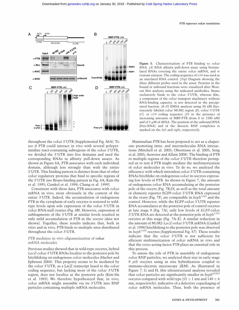
(Auweter and Allain 2008). We thus analyzed the capacityof endogenous PTB to interact with different regionsof oskar mRNA. As shown in Figure 5A, ovarianPTB preferentially associates with the oskar 39UTR, andto a lesser extent with the oskar 59 regulatory regionseparating the two functional AUGs (M1M2) (Gunkelet al. 1998). In this assay, PTB poorly associated withoskar coding sequence, and no significant interactionwith a nonrelated RNA (y14 coding sequence) wasobserved.
To test if PTB can interact with oskar mRNA directly,we analyzed the binding of recombinant PTB to theoskar M1M2 and 39UTR regions, in electrophoreticmobility shift assays (EMSA). Two complexes (RNP1and RNP2) form upon incubation of M1M2 RNA withincreasing amounts of maltose-binding protein (MBP)-PTB (Fig. 5B). This interaction is specific, as it is notobserved when using another MBP-tagged RNA-bindingprotein (MS2-binding protein) (data not shown), or anunrelated RNA (y14 coding sequence) (Fig. 5D). However,the interaction of PTB with oskar M1M2 RNA differsfrom that observed with the 39UTR. Under native con-ditions, the oskar 39UTR runs as two bands, likelycorresponding to RNA monomers and dimers (Fig. 5C).With increasing amounts of PTB, the discrete 39UTRcomplexes detected at low protein concentrations (aster-isks) shift into more heterogeneous complexes, andfinally into higher-order RNP complexes (Fig. 5C), whichare efficiently out-competed by addition of increasingamounts of unlabeled oskar 39UTR competitor to thebinding reaction, but not upon addition of unlabeled y14RNA (Supplemental Fig. S5A). A similar binding profilewas obtained using His-tagged recombinant protein (datanot shown).
Filter-binding assays in the presence of increasingamounts of recombinant protein revealed that PTB/oskar 39UTR complexes are formed very rapidly, within1 min of incubation (data not shown), and indicate thatPTB binds with an ‘‘apparent’’ dissociation constant (Kd)of ;280 nM (Supplemental Fig. S5B). Furthermore, thesigmoidal shape of the binding curve, together with theHill coefficient (2.4) obtained after Hill transformation,shows that the binding of PTB to the oskar 39UTR iscooperative. In conclusion, this analysis indicates thatPTB can interact directly with oskar mRNA and bindswith high affinity, cooperatively, and specifically to the39UTR.
The oskar 39UTR contains multiple PTB-binding sites
The results of the EMSA suggested the presence ofmultiple PTB-binding sites in the oskar 39UTR. Althoughno strict consensus sequence for PTB binding has so farbeen defined, previous reports showed that mammalianPTB binds with high affinity to pyrimidine-rich sequen-ces (Singh et al. 1995; Perez et al. 1997). Consistent withthis, short RNA sequences containing two pyrimidine-rich motifs efficiently compete the formation of PTB/oskar 39UTR complexes in EMSA assays (SupplementalFig. S5A). Strikingly, we found a high number of phylo-genetically conserved pyrimidine-rich sequences distributed
Figure 4. ptb mutant oocytes display ectopic Oskar proteinaccumulation. (A–D) w (A), heph1545 (B), and heph03429 (C,D)stage 7 egg chambers double-stained with anti-Oskar antibodies(green) and DAPI (white). Bar, 30 mm. The different patterns ofectopic Oskar accumulation reflect the dynamic distribution ofoskar mRNA at this stage, which is not altered in ptb mutantoocytes compared with wild-type oocytes (data not shown). (E)Graph showing the percentage of stage 7 oocytes with pre-mature Oskar translation in different mutant and rescue back-grounds (see the legend for Fig. 3I for a description of the rescueconstructs). (**) P < 0.005; (***) P < 0.001 in a x2 test. (F–H) w (F),heph1545 (G), and heph03429 (H) stage 10 oocytes double-stainedwith anti-Oskar antibodies (green) and DAPI (white). Bar, 60 mm.
Besse et al.
200 GENES & DEVELOPMENT
Cold Spring Harbor Laboratory Press on January 30, 2018 - Published by genesdev.cshlp.orgDownloaded from
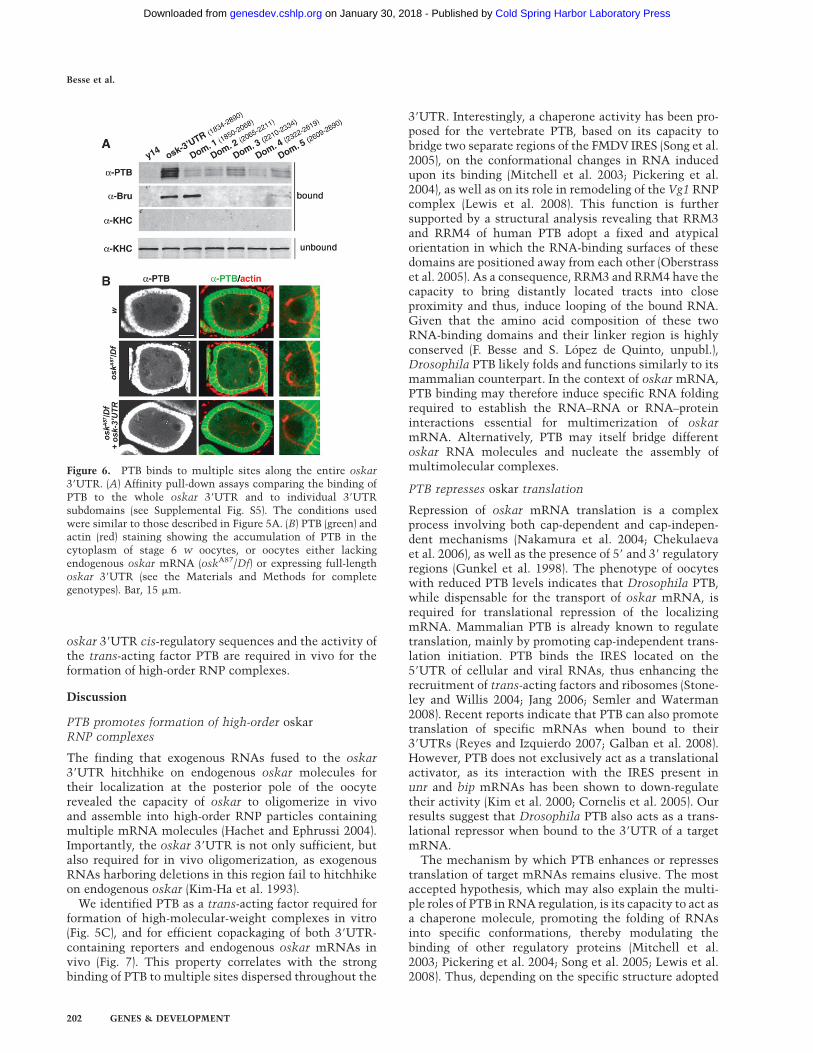
throughout the oskar 39UTR (Supplemental Fig. S6A). Tosee if PTB could interact in vivo with several polypyr-imidine tract-containing subregions of the oskar 39UTR,we divided the 39UTR into five domains and used thecorresponding RNAs in affinity pull-down assays. Asshown in Figure 6A, PTB associates with each individualdomain, although less strongly than with the entire39UTR. This binding pattern is distinct from that of otheroskar regulatory proteins that bind to specific regions ofthe 39UTR (see Bruno-binding pattern in Fig. 6A; Kim-Haet al. 1995; Gunkel et al. 1998; Chang et al. 1999).
Consistent with these data, PTB associates with oskarmRNA in vivo, most obviously in the context of theentire 39UTR. Indeed, the accumulation of endogenousPTB in the cytoplasm of early oocytes is restored to wild-type levels upon sole expression of the oskar 39UTR inoskar RNA-null ovaries (Fig. 6B). However, expression ofsubfragments of the 39UTR at similar levels resulted inonly mild accumulation of PTB in the oocyte (data notshown). Together, these results indicate that, both invitro and in vivo, PTB binds to multiple sites distributedthroughout the oskar 39UTR.
PTB mediates in vivo oligomerization of oskarmRNA molecules
Previous studies showed that in wild-type oocytes, hybridLacZ-oskar 39UTR RNAs localize to the posterior pole byhitchhiking on endogenous oskar molecules (Hachet andEphrussi 2004). This property seems to be mediated bythe oskar 39UTR, as a LacZ transcript fused to the oskarcoding sequence, but lacking most of the oskar 39UTRregion, does not localize at the posterior pole (Kim-Haet al. 1993). We therefore hypothesized that, in vivo,oskar mRNA might assemble via its 39UTR into RNPparticles containing multiple mRNA molecules.
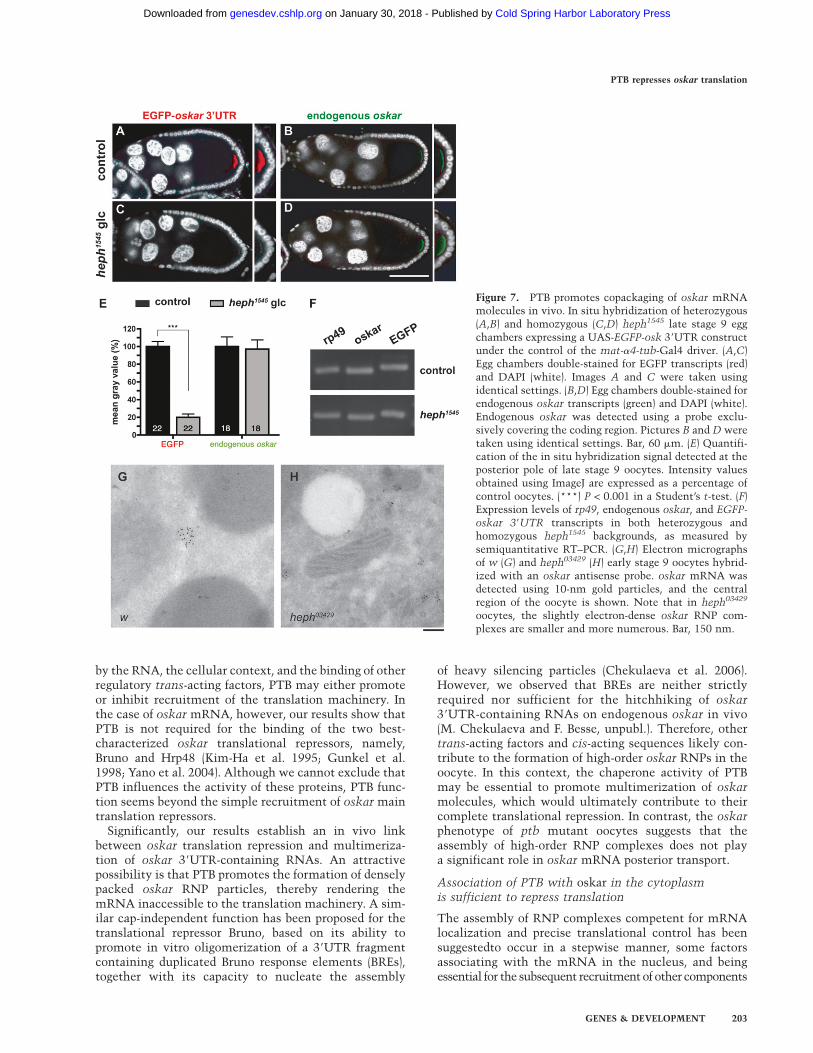
Mammalian PTB has been proposed to act as a chaper-one promoting intra- and intermolecular RNA interac-tions (Mitchell et al. 2005; Oberstrass et al. 2005; Songet al. 2005; Auweter and Allain 2008). The binding of PTBto multiple regions of the oskar 39UTR therefore promp-ted us to test if PTB might mediate the multimerizationof oskar molecules in vivo. To do so, we analyzed theefficiency with which intronless oskar 39UTR-containingRNAs hitchhike on endogenous oskar in oocytes express-ing low levels of PTB. As shown in Figure 7, the amountof endogenous oskar RNA accumulating at the posteriorpole of the oocyte (Fig. 7B,D), as well as the total amountof chimeric reporter EGFP-oskar 39UTR RNA expressedin the ovary (Fig. 7F), are comparable in heph1545 and thecontrol. However, while the EGFP-oskar 39UTR reporterRNA accumulates at the posterior pole of control oocytesat late stage 9 (Fig. 7A), only low levels of EGFP-oskar39UTR RNA are detected at the posterior pole of heph1545
oocytes at this stage (Fig. 7A–E). A similar reduction inthe amount of M1M2-LacZ-oskar 39UTR mRNA (Gunkelet al. 1998) hitchhiking to the posterior pole was observedin heph1545 oocytes (Supplemental Fig. S7). These resultsindicate that the oskar 39UTR is not sufficient forefficient multimerization of oskar mRNA in vivo andthat the trans-acting factor PTB plays an essential role inthis process.
To assess the role of PTB in assembly of endogenousoskar RNP particles, we analyzed their size in early stage9 ptb oocytes using in situ hybridization coupled toimmuno-electron microscopy (IEM). As illustrated inFigure 7, G and H, this ultrastructural analysis revealedthat oskar particles are significantly smaller in heph03429
oocytes compared with wild type (52 6 1 nm and 149 6 6nm, respectively), indicative of a defective copackaging ofoskar mRNA molecules. Thus, both the presence of
Figure 5. Characterization of PTB binding to oskarRNA. (A) RNA affinity pull-down assay using biotiny-lated RNAs covering the entire oskar mRNA, and w
ovarian extracts. The coding sequence of y14 was used asan unrelated RNA control. (Top) Diagram showing thethree different probes used in the assay. Proteins in thebound or unbound fractions were visualized after West-ern blot analysis using the indicated antibodies. Brunoexclusively binds to the oskar 39UTR, whereas Khc,a component of the oskar transport machinery withoutRNA-binding capacity, is not detected in the precipi-tated fraction. (B–D) EMSA analysis using 50 nM fluo-rescently labeled oskar M1M2 region (B), oskar 39UTR(C), or y14 coding sequence (D) in the presence ofincreasing amounts of MBP-PTB (from 0 to 1500 nM)and of 5 mM of tRNA. The position of the unbound RNA(Free-RNA) and of the discrete RNP complexes ismarked on the left and right, respectively.
PTB represses oskar translation
GENES & DEVELOPMENT 201
Cold Spring Harbor Laboratory Press on January 30, 2018 - Published by genesdev.cshlp.orgDownloaded from
oskar 39UTR cis-regulatory sequences and the activity ofthe trans-acting factor PTB are required in vivo for theformation of high-order RNP complexes.
Discussion
PTB promotes formation of high-order oskarRNP complexes
The finding that exogenous RNAs fused to the oskar39UTR hitchhike on endogenous oskar molecules fortheir localization at the posterior pole of the oocyterevealed the capacity of oskar to oligomerize in vivoand assemble into high-order RNP particles containingmultiple mRNA molecules (Hachet and Ephrussi 2004).Importantly, the oskar 39UTR is not only sufficient, butalso required for in vivo oligomerization, as exogenousRNAs harboring deletions in this region fail to hitchhikeon endogenous oskar (Kim-Ha et al. 1993).
We identified PTB as a trans-acting factor required forformation of high-molecular-weight complexes in vitro(Fig. 5C), and for efficient copackaging of both 39UTR-containing reporters and endogenous oskar mRNAs invivo (Fig. 7). This property correlates with the strongbinding of PTB to multiple sites dispersed throughout the
39UTR. Interestingly, a chaperone activity has been pro-posed for the vertebrate PTB, based on its capacity tobridge two separate regions of the FMDV IRES (Song et al.2005), on the conformational changes in RNA inducedupon its binding (Mitchell et al. 2003; Pickering et al.2004), as well as on its role in remodeling of the Vg1 RNPcomplex (Lewis et al. 2008). This function is furthersupported by a structural analysis revealing that RRM3and RRM4 of human PTB adopt a fixed and atypicalorientation in which the RNA-binding surfaces of thesedomains are positioned away from each other (Oberstrasset al. 2005). As a consequence, RRM3 and RRM4 have thecapacity to bring distantly located tracts into closeproximity and thus, induce looping of the bound RNA.Given that the amino acid composition of these twoRNA-binding domains and their linker region is highlyconserved (F. Besse and S. Lopez de Quinto, unpubl.),Drosophila PTB likely folds and functions similarly to itsmammalian counterpart. In the context of oskar mRNA,PTB binding may therefore induce specific RNA foldingrequired to establish the RNA–RNA or RNA–proteininteractions essential for multimerization of oskarmRNA. Alternatively, PTB may itself bridge differentoskar RNA molecules and nucleate the assembly ofmultimolecular complexes.
PTB represses oskar translation
Repression of oskar mRNA translation is a complexprocess involving both cap-dependent and cap-indepen-dent mechanisms (Nakamura et al. 2004; Chekulaevaet al. 2006), as well as the presence of 59 and 39 regulatoryregions (Gunkel et al. 1998). The phenotype of oocyteswith reduced PTB levels indicates that Drosophila PTB,while dispensable for the transport of oskar mRNA, isrequired for translational repression of the localizingmRNA. Mammalian PTB is already known to regulatetranslation, mainly by promoting cap-independent trans-lation initiation. PTB binds the IRES located on the59UTR of cellular and viral RNAs, thus enhancing therecruitment of trans-acting factors and ribosomes (Stone-ley and Willis 2004; Jang 2006; Semler and Waterman2008). Recent reports indicate that PTB can also promotetranslation of specific mRNAs when bound to their39UTRs (Reyes and Izquierdo 2007; Galban et al. 2008).However, PTB does not exclusively act as a translationalactivator, as its interaction with the IRES present inunr and bip mRNAs has been shown to down-regulatetheir activity (Kim et al. 2000; Cornelis et al. 2005). Ourresults suggest that Drosophila PTB also acts as a trans-lational repressor when bound to the 39UTR of a targetmRNA.
The mechanism by which PTB enhances or repressestranslation of target mRNAs remains elusive. The mostaccepted hypothesis, which may also explain the multi-ple roles of PTB in RNA regulation, is its capacity to act asa chaperone molecule, promoting the folding of RNAsinto specific conformations, thereby modulating thebinding of other regulatory proteins (Mitchell et al.2003; Pickering et al. 2004; Song et al. 2005; Lewis et al.2008). Thus, depending on the specific structure adopted
Figure 6. PTB binds to multiple sites along the entire oskar39UTR. (A) Affinity pull-down assays comparing the binding ofPTB to the whole oskar 39UTR and to individual 39UTRsubdomains (see Supplemental Fig. S5). The conditions usedwere similar to those described in Figure 5A. (B) PTB (green) andactin (red) staining showing the accumulation of PTB in thecytoplasm of stage 6 w oocytes, or oocytes either lackingendogenous oskar mRNA (oskA87/Df) or expressing full-lengthoskar 39UTR (see the Materials and Methods for completegenotypes). Bar, 15 mm.
Besse et al.
202 GENES & DEVELOPMENT
Cold Spring Harbor Laboratory Press on January 30, 2018 - Published by genesdev.cshlp.orgDownloaded from
by the RNA, the cellular context, and the binding of otherregulatory trans-acting factors, PTB may either promoteor inhibit recruitment of the translation machinery. Inthe case of oskar mRNA, however, our results show thatPTB is not required for the binding of the two best-characterized oskar translational repressors, namely,Bruno and Hrp48 (Kim-Ha et al. 1995; Gunkel et al.1998; Yano et al. 2004). Although we cannot exclude thatPTB influences the activity of these proteins, PTB func-tion seems beyond the simple recruitment of oskar maintranslation repressors.
Significantly, our results establish an in vivo linkbetween oskar translation repression and multimeriza-tion of oskar 39UTR-containing RNAs. An attractivepossibility is that PTB promotes the formation of denselypacked oskar RNP particles, thereby rendering themRNA inaccessible to the translation machinery. A sim-ilar cap-independent function has been proposed for thetranslational repressor Bruno, based on its ability topromote in vitro oligomerization of a 39UTR fragmentcontaining duplicated Bruno response elements (BREs),together with its capacity to nucleate the assembly
of heavy silencing particles (Chekulaeva et al. 2006).However, we observed that BREs are neither strictlyrequired nor sufficient for the hitchhiking of oskar39UTR-containing RNAs on endogenous oskar in vivo(M. Chekulaeva and F. Besse, unpubl.). Therefore, othertrans-acting factors and cis-acting sequences likely con-tribute to the formation of high-order oskar RNPs in theoocyte. In this context, the chaperone activity of PTBmay be essential to promote multimerization of oskarmolecules, which would ultimately contribute to theircomplete translational repression. In contrast, the oskarphenotype of ptb mutant oocytes suggests that theassembly of high-order RNP complexes does not playa significant role in oskar mRNA posterior transport.
Association of PTB with oskar in the cytoplasmis sufficient to repress translation
The assembly of RNP complexes competent for mRNAlocalization and precise translational control has beensuggestedto occur in a stepwise manner, some factorsassociating with the mRNA in the nucleus, and beingessential for the subsequent recruitment of other components
Figure 7. PTB promotes copackaging of oskar mRNAmolecules in vivo. In situ hybridization of heterozygous(A,B) and homozygous (C,D) heph1545 late stage 9 eggchambers expressing a UAS-EGFP-osk 39UTR constructunder the control of the mat-a4-tub-Gal4 driver. (A,C)Egg chambers double-stained for EGFP transcripts (red)and DAPI (white). Images A and C were taken usingidentical settings. (B,D) Egg chambers double-stained forendogenous oskar transcripts (green) and DAPI (white).Endogenous oskar was detected using a probe exclu-sively covering the coding region. Pictures B and D weretaken using identical settings. Bar, 60 mm. (E) Quantifi-cation of the in situ hybridization signal detected at theposterior pole of late stage 9 oocytes. Intensity valuesobtained using ImageJ are expressed as a percentage ofcontrol oocytes. (***) P < 0.001 in a Student’s t-test. (F)Expression levels of rp49, endogenous oskar, and EGFP-
oskar 39UTR transcripts in both heterozygous andhomozygous heph1545 backgrounds, as measured bysemiquantitative RT–PCR. (G,H) Electron micrographsof w (G) and heph03429 (H) early stage 9 oocytes hybrid-ized with an oskar antisense probe. oskar mRNA wasdetected using 10-nm gold particles, and the centralregion of the oocyte is shown. Note that in heph03429
oocytes, the slightly electron-dense oskar RNP com-plexes are smaller and more numerous. Bar, 150 nm.
PTB represses oskar translation
GENES & DEVELOPMENT 203
Cold Spring Harbor Laboratory Press on January 30, 2018 - Published by genesdev.cshlp.orgDownloaded from
in the cell cytoplasm (Hachet and Ephrussi 2001, 2004;Huynh et al. 2004; Kress et al. 2004; St Johnston 2005;Giorgi and Moore 2007; Lewis and Mowry 2007; Sneeet al. 2008). For example, nucleolar association of theRNA-binding protein She2p with its mRNA target ash1was recently proposed to be an essential step in theassembly of translationally silenced localizing ash1RNP complexes in yeast (Du et al. 2008). Some oskartranslation repressors have been shown to localize both inthe nucleus and in the cytoplasm of germ cells (Huynhet al. 2004; Yano et al. 2004; Snee et al. 2008). However,whether these regulators are recruited to the oskarcomplex in the nucleus and whether nuclear associationof these factors is required for subsequent translationsilencing have not been tested so far.
PTB belongs to the hnRNP family of nucleo–cytoplas-mic shuttling RNA-binding proteins, which regulatedifferent aspects of RNA metabolism both in the nucleusand in the cytoplasm of eukaryotic cells (Dreyfuss et al.2002; Glisovic et al. 2008). Consistent with this, weobserved that Drosophila PTB not only colocalizes withoskar in the oocyte cytoplasm, but also strongly accu-mulates in the nuclei of germ cells. Given that thenuclear association of PTB with Vg1 mRNA has beenproposed to control the subsequent localization of thesetranscripts in the cytoplasm of Xenopus oocytes (Kresset al. 2004), we tested if the association of Drosophila PTBwith oskar mRNA in the nuclei of germ cells is requiredfor its translation repression activity. Notably, we foundthat a cytoplasmic version of PTB localizes to theposterior pole of wild-type and ptb mutant oocytes andthat this localization is oskar-dependent (SupplementalFig. S2; data not shown), strongly suggesting that it is stillable to associate with oskar mRNA. More importantly,the mutant GFP-PTB-DNLS is competent in oskar trans-lation repression (Fig. 4E). Although it is possible thatendogenous PTB is loaded onto oskar RNP complexesin the nucleus of germ cells, our data suggest that nu-clear recruitment of PTB is not a prerequisite for theformation of translationally silenced complexes. Ouranalysis supports a model in which the complex behaviorof RNP particles is controlled by the independent associ-ation of specific protein modules in different cell com-partments. It also provides further evidence for thereorganization of RNP complexes upon translocation intothe cytoplasm.
Materials and methods
Identification of GFP-PTB protein-trap line
The GFP protein-trap screen and inverse PCR mapping weredescribed previously (Besse et al. 2007). In the PTB protein-trapline, the piggyBac transposon is inserted at position 27,699,986in the genomic scaffold AE014297. Females homozygous for theprotein-trap insertion are homozygous viable and do not showoskar-related phenotypes.
Fly stocks
w1118 flies (w) served as wild-type controls. The P(PZ}heph[03429] (Bloomington Stock Center #11589) and PBac(WH}-
heph[f01545] (Exelixis Collection, Harvard Medical School) wererecombined onto the FRT82B chromosome. The FRT82B-recom-bined EMS mutant alleles hephe1 and hephe2 were characterizedby Dansereau et al. (2002). Germline clone analysis was carriedout using the dominant female sterile technique (Chou et al.1993). Other mutant stocks used were oskA87 and Df(3R)pXT03
(Jenny and Hachet et al. 2006), grk2B6 (Schupbach 1987), grk2E12
(Neuman-Silberberg and Schupbach 1993), and stauD3 (Schupbachand Wieschaus 1986). The MT polarity marker kin-b-gal wasobtained from the stock y w KZ32; Ly/TM3Sb (Clark et al. 1994).Rescue of the oskar RNA-null phenotype using an UAS-oskar
39UTR transgene was performed as described (Jenny and Hachetet al. 2006). For the hitchhiking assay described in Figure 7,a mat-a4-tub-Gal4VP16 insertion (Bloomington stock center#7062) was recombined with a UASp-EGFP-oskar 39UTR trans-gene (H. Jambor and A. Ephrussi, unpubl.).
Transgenes
For rescue analyses, a full-length clone (RE56755) was N-termi-nal-tagged with EGFP and expressed using the germline-specifictubulin67c promoter. Briefly, the PTB coding sequence was PCR-amplified using the PTB-sense and PTB-antisense primers (Sup-plemental Table 1) and ligated into pCR II-TOPO (Invitrogen). Togenerate the PTBDNLS protein, lysines at positions 56, 57, and 59were mutated into glutamic acid using a nested-PCR site-di-rected mutagenesis approach with the mutagenic primer Mut-NLS-II together with PTB-sense and Internal-as as externalprimers (Supplemental Table 1). The SspI–NruI fragment cover-ing the mutated positions was exchanged in the wild-type pCR II-TOPO-PTB construct, and both wild-type and DNLS-mutantpCR II-TOPO-PTB clones were fully sequenced. To generateEGFP fusions of PTB and PTBDNLS, the 1.45-kb BamHI–XhoIfragment was introduced into a GFP-containing BamHI–XhoIbackbone of GFP:par-1(N1S) plasmid (Shulman et al. 2000).
Generation of Drosophila anti-PTB antiseraand Western blot analysis
The BDGP clone LD03185 was PCR-amplified using PTB-GST-sense and PTB-GST-as primers (Supplemental Table 1). The PCRfragment was subsequently ligated into pCR II-TOPO (Invitro-gen) and fully sequenced, and the ;1.4 BamHI–XhoI fragmentwas inserted into the pGEX5-5X-2 plasmid (GenBank accessionno. U13857) resulting in the N-terminal fusion of GST to PTBcoding sequence. The recombinant protein was expressed inEscherichia coli, purified using standard conditions, and injectedinto rabbits and rats.
Immunofluorescence
Ovaries from females mated for 1–2 d at 25°C on fresh food weredissected in PBS and processed as described (Vanzo and Ephrussi2002). Samples were incubated overnight at 4°C with mouseanti-bGal (Promega; 1:2000), rabbit anti-Staufen (St Johnstonet al. 1991), rabbit anti-Oskar (Vanzo and Ephrussi 2002), ratanti-PTB (1:1000), and rabbit anti-Bruno (1:5000) (R. Matthiesenand A. Ephrussi, unpubl.). Alexa-conjugated secondary antibod-ies (1:750; Invitrogen) were used. For detection of ectopic Oskar,we used the protocol for in situ hybridization coupled to immu-nofluorescence described already (Vanzo and Ephrussi 2002)in combination with preadsorbed anti-Oskar and Alexa488-conjugated antibodies. Ovaries were stained with phalloidin tolabel F-actin (Invitrogen) and/or with 3 mg/mL DAPI to labelnuclei. Images were captured by a confocal microscope (LeicaTCS SP2 AOBS) using a 403 PL APO oil-immersion lens (N.A.1.25) and processed with Adobe Photoshop.
Besse et al.
204 GENES & DEVELOPMENT
Cold Spring Harbor Laboratory Press on January 30, 2018 - Published by genesdev.cshlp.orgDownloaded from
RNA in situ hybridization
RNA in situ hybridization was performed as described previously(Vanzo and Ephrussi 2002). Probes corresponding to either oskar
or EGFP coding sequences were detected using sheep HRP-conjugated anti-DIG antibody (1:200; Roche) followed by Cy3-tyramide signal amplification reaction (Perkin Elmer).
Immunoprecipitation and RT–PCR analysis
The protocol was adapted from Munro et al. (2006) with thefollowing modifications: protein extracts were precleared withprotein A-agarose beads (Roche) and incubated without priorcross-linking with 1.5 mg of mouse anti-GFP antibodies (Molec-ular Probes) coupled to protein A-agarose beads (Roche) for 3 h at4°C. RNAs were released in 100 mL of extraction buffer byincubation for 20 min at 65°C. Total RNA was extracted usingTrizol (Invitrogen), DNase-treated, and used as template for RT.All RNA recovered from the bound fraction and 2.5% of that inthe unbound fraction was used for cDNA synthesis in combina-tion with Superscript III (Invitrogen) and Oligo(dT). One percentof the RT product was then PCR-amplified. Twenty-two cycleswere carried out at an annealing temperature of 55°C usingspecific primers (Supplemental Table 1).
Affinity pull-down assays
Ovaries of w1118 females were dissected in cold PBS and extractedusing a pestle in 20 mL of hypotonic buffer [10 mM HEPES at pH7.4, 10 mM KOAc, 1.5 mM Mg(OAc)2, 2.5 mM DTT]. Typically,20 pairs of ovaries were used per condition. The protein extractwas cleared by centrifugation at 12,000 rpm for 5 min, and thesupernatant was incubated with magnetic streptavidin beads(Roche) previously coupled to equimolecular amounts of in vitrotranscribed UTP-biotinylated-RNA probes (ranging from 0.5 to2 mg) in binding buffer (10 mM HEPES-KOH at pH 7.9, 3 mMMgCl2, 40 mM KCl, 5 mM EDTA, 5% glycerol, 2 mM DTT, 0.5%IGEPAL, 3 mg/mL Heparin, and 0.5 mg/mL tRNA) during 1 h at4°C. After two washes in binding buffer followed by two morewashes in binding buffer containing 150 mM KCl, the boundproteins were eluted in SDS-PAGE loading buffer and subjectedto electrophoresis and Western blotting using rabbit anti-PTB(1:2000), rat anti-Bruno (1:4000) (R. Matthiesen and A. Ephrussi)and rabbit anti-Khc (1:10.000; Cytoskeleton).
Subcloning of different oskar mRNA regions
All oskar regions were cloned into pBS-II-KS+ (Stratagene)downstream from the T7 promoter. Nucleotides flanking eachfragment described below have been numbered based on theirposition in the oskar cDNA sequence (osk-RA; Flybase), and allprimer sequences are specified in Supplemental Table 1. Usinga genomic oskar clone as template (Ephrussi et al. 1991), theM1M2 (nucleotides 1–434) and 39UTR (nucleotides 1834–2890)regions were PCR-amplified with primers M1M2-sense, andM1M2-as, or 3UTR-s and 3UTR-as, respectively, and subse-quently inserted into the SacI–HindIII pBS fragment. A similarstrategy was used for subcloning the individual 39UTRdomains with the following pair of primers: Domain1 (nucleo-tides 1850–2068), Dom1-sense and Dom1-as; Domain2 (nucleo-tides 2065–2211), Dom2-Long-s and Dom2-as; Domain3 (nucleotides2210–2334), Dom3-Long-s and Dom3-as; Domain4 (nucleotides2322–2619), Dom4-sense and Dom4-as and Domain5 (nucleotides2609–2890), Dom5-sense and 3UTR-as. The region correspondingto Short Oskar coding sequence (nucleotides 427–1838) was PCR-amplified using ShortOsk-Not and ShortOsk-as as primers and an
oskar cDNA clone as template (Ephrussi et al. 1991). The PCRfragment was ligated into pCR II-TOPO (Invitrogen), and theEcoRI fragment was inserted into EcoRI-linearized pBS-II-SK+
(Stratagene).
Recombinant PTB protein
Full-length PTB was N-terminal-tagged by MBP via PCR ampli-fication of the DGC clone RE56755 with GW-PTB-sense andGW-PTB-as primers (Supplemental Table 1). The PCR productwas ligated into pENTRY/D-TOPO vector (Invitrogen), fullysequenced, and subsequently recombined into the Gatewaydestination vector pETG-40A (A. Geerlof). The protein wasexpressed in E. coli (BL21 DE3) and purified under standardconditions. The MS2-binding protein—MBP-expressing plasmid(Zhou et al. 2002) was a gift from Reinhard Luhrmann.
EMSA
RNA transcripts were fluorescently labeled with ATTO-680Aminoallyl-UTP (Jena Bioscience) via in vitro transcription (T7megascript kit; Ambion). Full-length RNA molecules were gel-purified and incubated with different amounts of recombinantMBP-PTB in the presence of 20 mM Tris-HCl (pH 8.0), 60 mMNaCl, 3 mM MgCl2, 1 mM DTT, 8% glycerol, 5 mM tRNA during15 min at 4°C. Complexes were resolved in native 0.8% agarosegels containing 0.53 TBE buffer and 5% (v/v) glycerol, run at 100V constant at 4°C. Gels were visualized and quantitated usingthe Odyssey Infrared Imaging System (LI-COR).
IEM
Ovaries were fixed in 4% paraformaldehyde and processed asdescribed (Delanoue and Herpers et al. 2007), with minormodifications. Digoxygenin-labeled oskar antisense probes weredetected using sheep anti-Dig antibody (1:650; Roche), followedby rabbit anti-sheep secondary antibody (1:500; Dako) and pro-tein A-coupled gold particles (10 nm). For quantification, thelargest diameter of electron-dense oskar-containing particlespresent in the center of the oocyte was measured using ImageJ. At least 200 particles from three different oocytes wereanalyzed for each genotype.
Acknowledgments
We thank Helena Jambor, William Brook, Daniel St Johnston,Reinhard Luhrmann, and Paul McDonald for flies and reagents.We also thank the EMBL Laboratory Animal Resources forantibody production and the EMBL Electron Microscopy CoreFacility for technical support. We thank Eva Loser and AnnaCyrklaff for excellent technical assistance. We are grateful tomembers of the Ephrussi group for discussions and to VladimirRybin for advice. F.B. was supported by fellowships from theFederation of European Biochemical Societies (FEBS) and theHuman Frontier Science Program Organization. S.L.Q. wassupported by a Long Term EMBO Fellowship, a Marie CurieIntra-European Fellowship, and a RCUK Fellowship in Trans-lational Medicine. V.M. was supported by a fellowship from theFondation pour la Recherche Medicale and A.T. was supportedby a FEBS fellowship.
References
Auweter, S.D. and Allain, F.H. 2008. Structure–function rela-tionships of the polypyrimidine tract binding protein. Cell.
Mol. Life Sci. 65: 516–527.
PTB represses oskar translation
GENES & DEVELOPMENT 205
Cold Spring Harbor Laboratory Press on January 30, 2018 - Published by genesdev.cshlp.orgDownloaded from
Besse, F., Mertel, S., Kittel, R.J., Wichmann, C., Rasse, T.M.,Sigrist, S.J., and Ephrussi, A. 2007. The Ig cell adhesionmolecule Basigin controls compartmentalization and vesiclerelease at Drosophila melanogaster synapses. J. Cell Biol. 177:843–855.
Bonin, C.P. and Mann, R.S. 2004. A piggyBac transposon genetrap for the analysis of gene expression and function inDrosophila. Genetics 167: 1801–1811.
Chang, J.S., Tan, L., and Schedl, P. 1999. The Drosophila CPEBhomolog, Orb, is required for Oskar protein expression inoocytes. Dev. Biol. 215: 91–106.
Chekulaeva, M., Hentze, M.W., and Ephrussi, A. 2006. Brunoacts as a dual repressor of oskar translation, promotingmRNA oligomerization and formation of silencing particles.Cell 124: 521–533.
Chou, T.-B., Noll, E., and Perrimon, N. 1993. AutosomalP[ovoD1] dominant female-sterile insertions in Drosophilaand their use in generating germ-line chimeras. Develop-
ment 119: 1359–1369.Clark, I., Giniger, E., Ruohola-Baker, H., Jan, L.Y., and Jan, Y.N.
1994. Transient posterior localization of a kinesin fusionprotein reflects anteroposterior polarity of the Drosophila
oocyte. Curr. Biol. 4: 289–300.Cornelis, S., Tinton, S.A., Schepens, B., Bruynooghe, Y., and
Beyaert, R. 2005. UNR translation can be driven by an IRESelement that is negatively regulated by polypyrimidine tractbinding protein. Nucleic Acids Res. 33: 3095–3108.
Cote, C.A., Gautreau, D., Denegre, J.M., Kress, T.L., Terry, N.A.,and Mowry, K.L. 1999. A Xenopus protein related to hnRNPIhas a role in cytoplasmic RNA localization. Mol. Cell 4: 431–437.
Dansereau, D.A., Lunke, M.D., Finkielsztein, A., Russell, M.A.,and Brook, W.J. 2002. Hephaestus encodes a polypyrimidinetract binding protein that regulates Notch signalling duringwing development in Drosophila melanogaster. Develop-
ment 129: 5553–5566.Davis, M.B., Sun, W., and Standiford, D.M. 2002. Lineage-
specific expression of polypyrimidine tract binding protein(PTB) in Drosophila embryos. Mech. Dev. 111: 143–147.
Delanoue, R., Herpers, B., Soetaert, J., Davis, I., and Rabouille, C.2007. Drosophila Squid/hnRNP helps Dynein switch froma gurken mRNA transport motor to an ultrastructural staticanchor in sponge bodies. Dev. Cell 13: 523–538.
Dreyfuss, G., Kim, V.N., and Kataoka, N. 2002. Messenger-RNA-binding proteins and the messages they carry. Nat. Rev.Mol. Cell Biol. 3: 195–205.
Du, T.G., Schmid, M., and Jansen, R.P. 2007. Why cells movemessages: The biological functions of mRNA localization.Semin. Cell Dev. Biol. 18: 171–177.
Du, T.G., Jellbauer, S., Muller, M., Schmid, M., Niessing, D., andJansen, R.P. 2008. Nuclear transit of the RNA-binding pro-tein She2 is required for translational control of localizedASH1 mRNA. EMBO Rep. 9: 781–787.
Ephrussi, A. and Lehmann, R. 1992. Induction of germ cellformation by oskar. Nature 358: 387–392.
Ephrussi, A., Dickinson, L.K., and Lehmann, R. 1991. oskarorganizes the germ plasm and directs localization of theposterior determinant nanos. Cell 66: 37–50.
Galban, S., Kuwano, Y., Pullmann Jr., R., Martindale, J.L., Kim,H.H., Lal, A., Abdelmohsen, K., Yang, X., Dang, Y., Liu, J.O.,et al. 2008. RNA-binding proteins HuR and PTB promote thetranslation of hypoxia-inducible factor 1a. Mol. Cell. Biol.
28: 93–107.Giorgi, C. and Moore, M.J. 2007. The nuclear nurture and
cytoplasmic nature of localized mRNPs. Semin. Cell Dev.
Biol. 18: 186–193.
Glisovic, T., Bachorik, J.L., Yong, J., and Dreyfuss, G. 2008.RNA-binding proteins and post-transcriptional gene regula-
tion. FEBS Lett. 582: 1977–1986.Gonzalez-Reyes, A., Elliott, H., and St Johnston, D. 1995.
Polarization of both major body axes in Drosophila by
gurken–torpedo signalling. Nature 375: 654–658.Gunkel, N., Yano, T., Markussen, F.-H., Olsen, L.C., and
Ephrussi, A. 1998. Localization-dependent translationrequires a functional interaction between the 59 and 39 ends
of oskar mRNA. Genes & Dev. 12: 1652–1664.Hachet, O. and Ephrussi, A. 2001. Drosophila Y14 shuttles to
the posterior of the oocyte and is required for oskar mRNA
transport. Curr. Biol. 11: 1666–1674.Hachet, O. and Ephrussi, A. 2004. Splicing of oskar RNA in the
nucleus is coupled to its cytoplasmic localization. Nature
428: 959–963.Huynh, J.R., Munro, T.P., Smith-Litiere, K., Lepesant, J.A., and St
Johnston, D. 2004. The Drosophila hnRNPA/B homolog,
Hrp48, is specifically required for a distinct step in osk
mRNA localization. Dev. Cell 6: 625–635.Jang, S.K. 2006. Internal initiation: IRES elements of picornavi-
ruses and hepatitis c virus. Virus Res. 119: 2–15.Jenny, A., Hachet, O., Zavorszky, P., Cyrklaff, A., Weston, M.D.,
Johnston, D.S., Erdelyi, M., and Ephrussi, A. 2006. A trans-
lation-independent role of oskar RNA in early Drosophila
oogenesis. Development 133: 2827–2833.Kiebler, M.A., and Bassell, G.J. 2006. Neuronal RNA granules:
Movers and makers. Neuron 51: 685–690.Kim, Y.K., Hahm, B., and Jang, S.K. 2000. Polypyrimidine tract-
binding protein inhibits translation of bip mRNA. J. Mol.
Biol. 304: 119–133.Kim-Ha, J., Smith, J.L., and Macdonald, P.M. 1991. oskar mRNA
is localized to the posterior pole of the Drosophila oocyte.
Cell 66: 23–35.Kim-Ha, J., Webster, P.J., Smith, J.L., and Macdonald, P.M. 1993.
Multiple RNA regulatory elements mediate distinct steps inlocalization of oskar mRNA. Development 119: 169–
178.Kim-Ha, J., Kerr, K., and Macdonald, P.M. 1995. Translational
regulation of oskar mRNA by bruno, an ovarian RNA-
binding protein, is essential. Cell 81: 403–412.Kress, T.L., Yoon, Y.J., and Mowry, K.L. 2004. Nuclear RNP
complex assembly initiates cytoplasmic RNA localization. J.
Cell Biol. 165: 203–211.Lange, S., Katayama, Y., Schmid, M., Burkacky, O., Brauchle, C.,
Lamb, D.C., and Jansen, R.P. 2008. Simultaneous transport of
different localized mRNA species revealed by live-cell imag-
ing. Traffic 9: 1256–1267.Lecuyer, E., Yoshida, H., Parthasarathy, N., Alm, C., Babak, T.,
Cerovina, T., Hughes, T.R., Tomancak, P., and Krause, H.M.
2007. Global analysis of mRNA localization reveals a prom-inent role in organizing cellular architecture and function.
Cell 131: 174–187.Lewis, R.A., and Mowry, K.L. 2007. Ribonucleoprotein remodel-
ing during RNA localization. Differentiation 75: 507–518.Lewis, R.A., Gagnon, J.A., and Mowry, K.L. 2008. PTB/hnRNP I
is required for RNP remodeling during RNA localization inXenopus oocytes. Mol. Cell. Biol. 28: 678–686.
Ma, S., Liu, G., Sun, Y., and Xie, J. 2007. Relocalization of the
polypyrimidine tract-binding protein during PKA-inducedneurite growth. Biochim. Biophys. Acta 1773: 912–923.
Markussen, F.-H., Michon, A.-M., Breitwieser, W., and Ephrussi,
A. 1995. Translational control of oskar generates Short OSK,the isoform that induces pole plasm assembly. Development
121: 3723–3732.
Besse et al.
206 GENES & DEVELOPMENT
Cold Spring Harbor Laboratory Press on January 30, 2018 - Published by genesdev.cshlp.orgDownloaded from
Mitchell, S.A., Spriggs, K.A., Coldwell, M.J., Jackson, R.J., andWillis, A.E. 2003. The Apaf-1 internal ribosome entry seg-
ment attains the correct structural conformation for func-tion via interactions with PTB and unr. Mol. Cell 11: 757–771.
Mitchell, S.A., Spriggs, K.A., Bushell, M., Evans, J.R., Stoneley,
M., Le Quesne, J.P., Spriggs, R.V., and Willis, A.E. 2005.Identification of a motif that mediates polypyrimidine tract-binding protein-dependent internal ribosome entry. Genes &
Dev. 19: 1556–1571.Morin, X., Daneman, R., Zavortink, M., and Chia, W. 2001. A
protein trap strategy to detect GFP-tagged proteins expressedfrom their endogenous loci in Drosophila. Proc. Natl. Acad.
Sci. 98: 15050–15055.Munro, T.P., Kwon, S., Schnapp, B.J., and St Johnston, D. 2006. A
repeated IMP-binding motif controls oskar mRNA trans-lation and anchoring independently of Drosophila mela-
nogaster IMP. J. Cell Biol. 172: 577–588.Nakamura, A., Sato, K., and Hanyu-Nakamura, K. 2004. Dro-
sophila cup is an eIF4E binding protein that associates withBruno and regulates oskar mRNA translation in oogenesis.
Dev. Cell 6: 69–78.Neuman-Silberberg, F.S. and Schupbach, T. 1993. The Drosophila
dorsoventral patterning gene gurken produces a dorsally local-ized RNA and encodes a TGFa-like protein. Cell 75: 165–174.
Oberstrass, F.C., Auweter, S.D., Erat, M., Hargous, Y., Henning,A., Wenter, P., Reymond, L., Amir-Ahmady, B., Pitsch, S.,
Black, D.L., et al. 2005. Structure of PTB bound to RNA:Specific binding and implications for splicing regulation.
Science 309: 2054–2057.Palacios, I.M., and St Johnston, D. 2002. Kinesin light chain-
independent function of the Kinesin heavy chain in cytoplas-mic streaming and posterior localisation in the Drosophila
oocyte. Development 129: 5473–5485.Perez, I., Lin, C.H., McAfee, J.G., and Patton, J.G. 1997.
Mutation of PTB binding sites causes misregulation ofalternative 39 splice site selection in vivo. RNA 3: 764–778.
Pickering, B.M., Mitchell, S.A., Spriggs, K.A., Stoneley, M., andWillis, A.E. 2004. Bag-1 internal ribosome entry segment
activity is promoted by structural changes mediated bypoly(rC) binding protein 1 and recruitment of polypyrimidinetract binding protein 1. Mol. Cell. Biol. 24: 5595–5605.
Reyes, R., and Izquierdo, J.M. 2007. The RNA-binding protein
PTB exerts translational control on 39-untranslated region ofthe mRNA for the ATP synthase b-subunit. Biochem. Bio-
phys. Res. Commun. 357: 1107–1112.Rongo, C., Gavis, E.R., and Lehmann, R. 1995. Localization of
oskar RNA regulates oskar translation and requires Oskarprotein. Development 121: 2737–2746.
Schupbach, T. 1987. Germ line and soma cooperate duringoogenesis to establish the dorsoventral pattern of egg shell
and embryo in Drosophila melanogaster. Cell 49: 699–707.Schupbach, T., and Wieschaus, E. 1986. Germline autonomy of
maternal-effect mutations altering the embryonic body pat-tern of Drosophila. Dev. Biol. 113: 443–448.
Semler, B.L., and Waterman, M.L. 2008. IRES-mediated path-ways to polysomes: Nuclear versus cytoplasmic routes.
Trends Microbiol. 16: 1–5.Shulman, J., Benton, R., and St Johnston, D. 2000. The Dro-
sophila homolog of C.elegans PAR-1 organizes the oocytecytoskeleton and directs oskar mRNA localization to the
posterior pole. Cell 101: 377–388.Singh, R., Valcarcel, J., and Green, M.R. 1995. Distinct binding
specificities and functions of higher eukaryotic polypyrimi-dine tract-binding proteins. Science 268: 1173–1176.
Snee, M., Benz, D., Jen, J., and Macdonald, P.M. 2008. Twodistinct domains of Bruno bind specifically to the oskarmRNA. RNA Biol. 5: 1–9.
Song, Y., Tzima, E., Ochs, K., Bassili, G., Trusheim, H., Linder,M., Preissner, K.T., and Niepmann, M. 2005. Evidence for anRNA chaperone function of polypyrimidine tract-bindingprotein in picornavirus translation. RNA 11: 1809–1824.
Spellman, R., Rideau, A., Matlin, A., Gooding, C., Robinson, F.,McGlincy, N., Grellscheid, S.N., Southby, J., Wollerton, M.,and Smith, C.W. 2005. Regulation of alternative splicing byPTB and associated factors. Biochem. Soc. Trans. 33: 457–460.
Spradling, A.C. 1993. Developmental genetics of oogenesis. InThe development of Drosophila melanogaster (eds. M. Bateand A. Martinez-Arias), pp. 1–70. Cold Spring Harbor Labo-ratory Press, Cold Spring Harbor, NY.
St Johnston, D. 2005. Moving messages: The intracellularlocalization of mRNAs. Nat. Rev. Mol. Cell Biol. 6: 363–375.
St Johnston, D., Beuchle, D., and Nusslein-Vorhard, C. 1991.staufen, a gene required to localize maternal RNAs inDrosophila eggs. Cell 66: 51–63.
Stoneley, M., and Willis, A.E. 2004. Cellular internal ribosomeentry segments: Structures, trans-acting factors and regula-tion of gene expression. Oncogene 23: 3200–3207.
Valcarcel, J. and Gebauer, F. 1997. Post-transcriptional regula-tion: The dawn of PTB. Curr. Biol. 7: R705–R70810.1016/S0960-9822(06)00361-7.
Vanzo, N.F., and Ephrussi, A. 2002. Oskar anchoring restrictspole plasm formation to the posterior of the Drosophila
oocyte. Development 129: 3705–3714.Yano, T., Lopez de Quinto, S., Matsui, Y., Shevchenko, A., and
Ephrussi, A. 2004. Hrp48, a Drosophila hnRNPA/B homolog,binds and regulates translation of oskar mRNA. Dev. Cell 6:637–648.
Yoshida, S., Muller, H.A., Wodarz, A., and Ephrussi, A. 2004.PKA-R1 spatially restricts Oskar expression for Drosophila
embryonic patterning. Development 131: 1401–1410.Zhou, Z., Sim, J., Griffith, J., and Reed, R. 2002. Purification and
electron microscopic visualization of functional humanspliceosomes. Proc. Natl. Acad. Sci. 99: 12203–12207.
PTB represses oskar translation
GENES & DEVELOPMENT 207
Cold Spring Harbor Laboratory Press on January 30, 2018 - Published by genesdev.cshlp.orgDownloaded from
10.1101/gad.505709Access the most recent version at doi: originally published online January 8, 200923:2009, Genes Dev.
Florence Besse, Sonia López de Quinto, Virginie Marchand, et al.
translation oskarrepresses PTB promotes formation of high-order RNP particles andDrosophila
Material
Supplemental
http://genesdev.cshlp.org/content/suppl/2009/01/08/gad.505709.DC1
Related Content
Genes Dev. January , 2009 23: 133-137
Robin P. WhartonA splicer that represses (translation)
References
http://genesdev.cshlp.org/content/23/2/195.full.html#related-urls
Articles cited in:
http://genesdev.cshlp.org/content/23/2/195.full.html#ref-list-1This article cites 67 articles, 23 of which can be accessed free at:
License
ServiceEmail Alerting
click here.right corner of the article or
Receive free email alerts when new articles cite this article - sign up in the box at the top
Copyright © 2009 by Cold Spring Harbor Laboratory Press
Cold Spring Harbor Laboratory Press on January 30, 2018 - Published by genesdev.cshlp.orgDownloaded from