Embed Size (px)
Citation preview
Available online at www.sciencedirect.com
12 (2007) 203–216www.elsevier.com/developmentalbiology
Developmental Biology 3
Drosophila glypican Dally-like acts in FGF-receiving cells to modulate FGFsignaling during tracheal morphogenesis
Dong Yan, Xinhua Lin ⁎
Division of Developmental Biology, Cincinnati Children’s Hospital Medical Center, Cincinnati, OH 45229, USAThe Graduate Program in Molecular and Developmental Biology, University of Cincinnati College of Medicine, Cincinnati, OH 45229, USA
Received for publication 15 May 2007; revised 9 September 2007; accepted 12 September 2007Available online 20 September 2007
Abstract
Previous studies inDrosophila have shown that heparan sulfate proteoglycans (HSPGs) are involved in both breathless (btl)- and heartless (htl)-mediated FGF signaling during embryogenesis. However, the mechanism(s) by which HSPGs control Btl and Htl signaling is unknown. Here weshow that dally-like (dlp, a Drosophila glypican) mutant embryos exhibit severe defects in tracheal morphogenesis and show a reduction in btl-mediated FGF signaling activity. However, htl-dependent mesodermal cell migration is not affected in dlpmutant embryos. Furthermore, expressionof Dlp, but not other Drosophila HSPGs, can restore effectively the tracheal morphogenesis in dlp embryos. Rescue experiments in dlp embryosdemonstrate that Dlp functions only in Bnl/FGF receiving cells in a cell-autonomous manner, but is not essential for Bnl/FGF expression cells. Tofurther dissect the mechanism(s) of Dlp in Btl signaling, we analyzed the role of Dlp in Btl-mediated air sac tracheoblast formation in wing discs.Mosaic analysis experiments show that removal of HSPG activity in FGF-producing or other surrounding cells does not affect tracheoblastsmigration, while HSPGmutant tracheoblast cells fail to receive FGF signaling. Together, our results argue strongly that HSPGs regulate Btl signalingexclusively in FGF-receiving cells as co-receptors, but are not essential for the secretion and distribution of the FGF ligand. This mechanism isdistinct from HSPG functions in morphogen distribution, and is likely a general paradigm for HSPG functions in FGF signaling in Drosophila.© 2007 Elsevier Inc. All rights reserved.
Keywords: FGF; Heparan sulfate proteoglycans; Dally; Dally-like (Dlp); Trachea; Air sac tracheoblast; Signaling; Morphogen
Introduction
During metazoan development, the formation of complexbody pattern is controlled by a handful of secreted signalingmolecules, including members of the Wnt, Hedgehog (Hh),transforming growth factor-β (TGFβ) and fibroblast growthfactor (FGF) families. FGFs comprise a large family of proteinsthat participate in numerous developmental and physiologicalprocesses including patterning, cell migration and proliferation(Coumoul and Deng, 2003; Itoh and Ornitz, 2004; Ornitz andItoh, 2001; Thisse and Thisse, 2005). In Drosophila, two FGFreceptors, Heartless (Htl) and Breathless (Btl), are expressed indistinct patterns and mediate different developmental events
⁎ Corresponding author. Fax: +1 513 6364317.E-mail address: [email protected] (X. Lin).
0012-1606/$ - see front matter © 2007 Elsevier Inc. All rights reserved.doi:10.1016/j.ydbio.2007.09.015
during embryogenesis. Htl is expressed in the early embryonicmesoderm where it is required for the dorsolateral migration ofmesoderm cells following gastrulation (Beiman et al., 1996;Gisselbrecht et al., 1996; Michelson et al., 1998; Shishido et al.,1993, 1997). Both pyramus and thisbe are identified as genesencoding the FGF ligands for Htl (Gryzik and Muller, 2004;Stathopoulos et al., 2004). btl is specifically expressed intracheal cells (Klambt et al., 1992; Shishido et al., 1993), whichmigrate toward clusters of cells expressing the FGF ligandBranchless (Bnl). Bnl functions as a motogen and guidancemolecule for tracheal cell migration during embryogenesis(Sutherland et al., 1996). It is currently unknown whether Bnlforms a gradient and whether such a presumptive gradient isessential for guiding the migration of tracheal cells (Affolter andWeijer, 2005).
Recently, Sato and Kornberg has characterized another Bnl/Btl-mediated event, the development of the air sac of the dorsal
204 D. Yan, X. Lin / Developmental Biology 312 (2007) 203–216
thorax (Sato and Kornberg, 2002). The air sac precursor cells,“the tracheoblasts”, develop just before metamorphosis and willultimately serve the adult organism. In this system, Bnl is ex-pressed in the columnar epithelia of wing imaginal discs, whereit acts as a chemoattractant to guide the migration of the air sactracheoblasts on the top of columnar epithelia (Sato andKornberg, 2002). Further study showed that FGF signaling isessential for the leading tracheal cells, as cells defective in FGFsignaling are excluded from the tip of the air sac (Cabernard andAffolter, 2005). Consistent with this, recent genetic mosaicanalysis showed that Btl activity is also required for guiding themigration of the leading tracheal cells during larva trachealdevelopment (Ghabrial and Krasnow, 2006). Drosophilaembryonic tracheal branching occurs only after cell divisionceases. However, air sac tracheoblasts develop and proliferate atthe same time. Since mosaic clonal analysis can be usedeffectively in the imaginal discs, the air sac in the wing discprovides an excellent model system to dissect further themechanism(s) of Bnl/Btl mediated tracheoblasts formation(Cabernard et al., 2004).
It has long been appreciated that FGF signaling is facilitatedby heparan sulfate proteoglycans (HSPGs) (Ornitz, 2000).HSPGs are cell-surface and extracellular matrix (ECM) mole-cules composed of a protein core to which heparan sulfate (HS)glycosaminoglycan (GAG) chains are attached (Bernfield et al.,1999; Esko and Selleck, 2002; Hacker et al., 2005; Lin, 2004).HSPGs are implicated in many developmental signaling path-ways both in Drosophila and in vertebrates (Lin, 2004).However, the mechanistic functions of HSPGs in these signalingpathways are not well-understood. In Drosophila, there are twoglypicans (Division abnormally delayed (Dally) and Dally-like(Dlp)) (Baeg et al., 2001; Khare and Baumgartner, 2000; Nakatoet al., 1995), one syndecan (Sdc) (Spring et al., 1994) and oneperlecan (Terribly reduced optic lobes (Trol)) (Datta and Kankel,1992; Voigt et al., 2002). Glypicans and syndecans are linked tothe plasma membrane by a glycosylphosphatidylinositol (GPI)anchor or a transmembrane domain, respectively. Perlecans aresecreted HSPGs that are mainly distributed in the ECM.Previously, we have shown that both Htl- and Btl-mediatedsignaling events are defective in embryos mutant for sugarless(sgl) and sulfateless (sfl), which encode two enzymes requiredfor the biosynthesis of HSPG GAG chains (Hacker et al., 1997;Lin et al., 1999). This study provides the first in vivo evidencefor a role of HSPGs in FGF signaling during development.However, two important questions remained to be addressed.First, which HSPG core protein is involved in Htl- and Btl-mediated FGF signaling? Among four Drosophila HSPGs, theglypicans Dally and Dlp are best characterized and are essentialfor signaling activities of morphogens includingWingless (Wg),Hh and Decapentaplegic (Dpp) (Hacker et al., 2005; Lin, 2004).It is currently unknown whether they are also required for FGFsignaling. Second, as HSPGs control Wg, Hh and Dpp signalingmainly by regulating the distribution of these secretedmolecules, it is important to determine whether HSPGs controlFGF-dependent processes by similar mechanisms. This is aparticularly interesting question as FGF functions as anextracellularly diffusible and/or matrix-bound guidance cues
whose gradient might be essential for the directionality oftracheal morphogenesis.
In this study, we have further defined the molecular mech-anisms of HSPG-mediated FGF signaling in Drosophila. Ouranalyses demonstrate that Dlp is essential for Btl-mediatedtracheal development, but is not critical for Htl signaling. Wefurther explore the mechanism of Dlp function in FGF signalingby rescue experiments in dlp mutant embryos and by mosaicanalysis in air sac system. To our surprise, we found that Dlpcontrols Btl signaling mainly in FGF-receiving cells, but not inFGF-producing cells or its surrounding tissues. This mechanismof HSPG activity in FGF signaling differs from its role inmorphogen signaling. Our new findings favour a model inwhich HSPGs act as co-receptors to facilitate FGF/FGFR inter-action and/or stabilization in FGF-receiving cells.
Materials and methods
Drosophila strains
The followingDrosophilamutant strains were used: dally80, dallyP2, dlpA187,htlAB42, sfll(3)03844, wgIG22, btlLG19, bnlP1, bnlP2, 1-eve-1. dally80, dlpA187 (Han etal., 2004b), htlAB42 (Gisselbrecht et al., 1996), sfll(3)03844 (Lin and Perrimon,1999), wgIG22 (van den Heuvel et al., 1993), btlLG19 (Klambt et al., 1992), bnlP1
(Sutherland et al., 1996) are null alleles. dallyP2 and bnlP2 are enhancer-trap lacZlines used to visualize dally and bnl expression (Lin and Perrimon, 1999;Sutherland et al., 1996). 1-eve-1 is a trachealess enhancer-trap line used tovisualize tracheal cells. The following UAS and Gal4 lines were used for ectopicexpression: UAS-dlp (Baeg et al., 2001), UAS-dally (Franch-Marro et al., 2005),UAS-syndecan (Johnson et al., 2004), UAS-perlecan (Voigt et al., 2002), UAS-dlp-GFP (Han et al., 2004b), UAS-btl-GFP (Sato and Kornberg, 2002), UAS-GFP (flybase), UAS-CD8-GFP (Lee and Luo, 1999); btl-Gal4 (Shiga et al.,1996), bnl-Gal4 (Kamimura et al., 2006), 69B-Gal4 (Brand and Perrimon, 1993),sim-Gal4 (Golembo et al., 1996), paired-Gal4 (Yoffe et al., 1995), tubP-Gal80(Lee and Luo, 1999).
Generation of germline clones and marked disc clones
Females with germline clones were generated by the autosomal FLP-DFStechnique (Chou and Perrimon, 1996). Negatively marked clones of mutantcells in the wing disc were generated by the FLP-FRT method (Golic, 1991; Xuand Rubin, 1993) and induced by subjecting first- or second-instar larvae to aheat shock at 37 °C for 1 h. Mutation in Minute on chromosome 3L was usedto generate large clones of cells mutant for dlp, dally-dlp and sfl in disccolumnar epithelial cells (Han et al., 2005). Positively marked clones of mutantcells in trachea were generated by the MARCM system (Lee and Luo, 1999).Mutant alleles were crossed to the MARCM strain (y w hsp70-flp; btl-gal4,UAS-CD8-GFP/CyO; tubP-gal80 FRT2A/TM6B); 2- to 10-h-old embryos wereheat shocked at 37 °C for 1.5 h. Below, we list the genotypes used in ouranalysis.
Germline clones (Fig. 1)dlpA187 germline clones: y w hsp70-flp; dlpA187 FRT2A/P[OvoD1]FRT2A
x dlpA187 FRT2A/TM6Bdally80dlpA187 germline clones: y w hsp70-flp; dally80 dlpA187 FRT2A/P[OvoD1]FRT2A x dally80dlpA187 FRT2A/TM6Btrachealess expression in dlpA187 mutant embryo: y w hsp70-flp; dlpA187
FRT2A/P[OvoD1]FRT2A x dlpA187 FRT2A 1-eve-1/TM6BdlpA187genetic interaction with bnlP1: y w hsp70-flp; dlpA187 FRT2A/P[OvoD1]FRT2A x bnlP1/TM3bnl expression in dlpA187mutant embryo: y w hsp70-flp; dlpA187 FRT2A/P[OvoD1]FRT2A x dlpA187 FRT2A bnlP2/TM6BOverexpression of bnl in dlpA187mutant embryo: y w hsp70-flp; dlpA187
btl-Gal4 FRT2A/P[OvoD1]FRT2A x dlpA187 FRT2A UAS-bnl/TM6B

205D. Yan, X. Lin / Developmental Biology 312 (2007) 203–216
For rescue experiments (Fig 2):btlLG19prd-Gal4/TM6B x btlLG19 UAS-btl-GFP/TM6By w hsp70-flp; dlpA187 btl-Gal4 FRT2A/P[OvoD1]FRT2A x dlpA187 UAS-dlpFRT2A/TM6BAZy w hsp70-flp; dlpA187 bnl-Gal4 FRT2A/P[OvoD1]FRT2A x dlpA187 UAS-dlpFRT2A/TM6BAZy w hsp70-flp; dlpA187 bnl-Gal4 FRT2A/P[OvoD1]FRT2A x btl-Gal4-UAS-CD8-GFP/+; dlpA187 UAS-dlpFRT2A/+y w hsp70-flp; dlpA187 UAS-dlp FRT2A/P[OvoD1]FRT2A x dlpA187 69B-Gal4FRT2A/prd-Gal4 UAS-GFPy w hsp70-flp; dlpA187FRT2A/P[OvoD1]FRT2A x sim-Gal4-UAS-dlp/+;dlpA187UAS-eGFP/+y w hsp70-flp; dlpA187 UAS-dlp FRT2A/P[OvoD1]FRT2A x dlpA187 prd-Gal4FRT2A/TM6B
Fig. 1. Dlp is required for bnl/btl-dependent tracheal cell migration during embryogestage 15 embryos immunostained with 2A12 antibody. Insets show higher magnifimaternal/zygotic null dlp embryo (B), virtually no tracheal branching occurs. This defwith dlp. One copy of bnlmutation can lead to strong tracheal defects combined withGal4 generates masses of fine branches (H). This phenotype is markedly suppressedembryos stained with the diphospho-MAPK-specific antibody. The strong expressionpositions in maternal/zygotic null dlp embryos (F). (G) β-Gal antibody staining of staform normally with respect to size and position in these mutant embryos. (J–M) Venmutant embryos (K), these Twi-positive mesodermal cells migrate normally in mate
MAPK staining: y w hsp70-flp; dlpA187 prd-Gal4 FRT2A/P[OvoD1]FRT2A xUAS-dlp/+; dlpA187 FRT2A 1-eve-1/+y w hsp70-flp; dlpA187 prd-Gal4 FRT2A/P[OvoD1]FRT2A x UAS-dallyflag/+;dlpA187 UAS-eGFP FRT2A/+y w hsp70-flp; dlpA187 prd-Gal4 FRT2A/P[OvoD1]FRT2A x UAS-syndecan/+; dlpA187 UAS-eGFP FRT2A/+y w hsp70-flp; dlpA187 prd-Gal4 FRT2A/P[OvoD1]FRT2A x UAS-perlecan/Y; dlpA187 UAS-eGFP FRT2A/+
Wing disc clones mutant for dally-dlp or sfl marked by absence of GFP (Fig. 4)y w hsp70-flp/y w hsp70-flp; hsp70-Myc-GFP M(3)i55 FRT2A/dally80dlpA187FRT2AbnlP2
y w hsp70-flp/y w hsp70-flp; hsp70-Myc-GFP M(3)i55 FRT2A/sfll(3)03844FRT2AbnlP2
nesis. All embryos are oriented anterior to the left. (A–D, H–I) Lateral views ofcation of a single typical tracheal metamere. (A) Wild-type tracheal pattern. Inect is completely paternally rescued (C). (D) bnl shows strong genetic interactiondlpmaternal mutation. In wild-type embryos, overexpression of UAS-bnl by btl-in maternal/zygotic dlp mutant background (I). (E–F) Lateral views of stage 11observed in wild-type (E) tracheal pits is markedly reduced in the correspondingge 11 maternal/zygotic dlpmutant embryos, which contain 1-eve-1. Tracheal pitstral views of stage 9 embryos immunostained for Twi expression. Unlike in htlrnal/zygotic null dlp (L) or dally-dlp (M) mutant embryos.
206 D. Yan, X. Lin / Developmental Biology 312 (2007) 203–216
Tracheal clones mutant for dlp, dally-dlp, sfl or wild type clones marked by thepresence of GFP (Fig. 5)
y w hsp70-flp/y w hsp70-flp; btl-gal4 UAS-CD8-GFP/+; tubP-gal80 FRT2A/FRT2A
y w hsp70-flp/y w hsp70-flp; btl-gal4 UAS-CD8-GFP/+; tubP-gal80 FRT2A/dlpA187FRT2A
y w hsp70-flp/y w hsp70-flp; btl-gal4 UAS-CD8-GFP/+; tubgal80 FRT2A/dally80FRT2A
y w hsp70-flp/y w hsp70-flp; btl-gal4 UAS-CD8-GFP/+; tubP-gal80 FRT2A/dally80dlpA187FRT2A
y w hsp70-flp/y w hsp70-flp; btl-gal4 UAS-CD8-GFP/UAS-dlp; tubP-gal80FRT2A/dally80dlpA187FRT2A
y w hsp70-flp/y w hsp70-flp; btl-gal4 UAS-CD8-GFP/+; tubP-gal80 FRT2A/sfll(3)03844FRT2A
Immunostainings
Fixation of embryos and imaginal discs as well as antibody stainingprocedure were performed as described (Belenkaya et al., 2002; Hacker et al.,1997; Han et al., 2004a). Primary antibodies were used at the followingdilutions: mouse anti-tracheal lumen antibody mAb2A12 (1:5) (Iowa Develop-mental Studies Hybridoma Bank; IDSHB), mouse anti-diphospho-MAPK(1:200) (Sigma), rabbit anti-Twist (1:1000) (Michelson et al., 1998), rabbit anti-Dlp (1:200) (Baeg et al., 2001), rat anti-E-Cadherin (1:5) (IDSHB), rabbit anti-β-Gal (1:500) (Cappel), mouse anti-β-Gal (1:3000) (Roche), chicken anti-β-Gal(1:1000) (Abcam), rabbit anti-GFP Alexa Fluor 488 (1:1000) (MolecularProbes), mouse anti-GFP (1:200) (Chemicon), mouse anti-Dlp (1:50)(Lum etal., 2003), rabbit anti-Sal (1:150)(Belenkaya et al., 2004). The primaryantibodies were detected by fluorescent-conjugated secondary antibodies fromJackson ImmunoResearch Laboratories or ABC kit from Vectastain.
Results
Dlp is required for Btl-dependent tracheal development duringembryogenesis
To dissect the molecular mechanisms by which HSPGscontrol FGF signaling in Drosophila, we first asked whichHSPG is involved in Htl- and Btl-mediated signaling. As dlpmutant embryos are embryonic lethal (Han et al., 2004b), wesuspected that Dlp may be involved in FGF signaling duringembyogenesis. Therefore, we analyzed tracheal branching andmesodermal migration phenotypes associated with dlp nullmutant embryos. dlp homozygous mutant embryos derived fromfemales lacking maternal dlp activity (referred to as dlp embryoshereafter) were generated by “FLP-DFS” technique (Chou andPerrimon, 1996). To visualize Drosophila embryonic trachealsystem, we used a monoclonal antibody 2A12 which recognizestracheal lumen. We observed severe tracheal branching defectsin dlp embryos (Fig. 1B). This defect is very similar to those
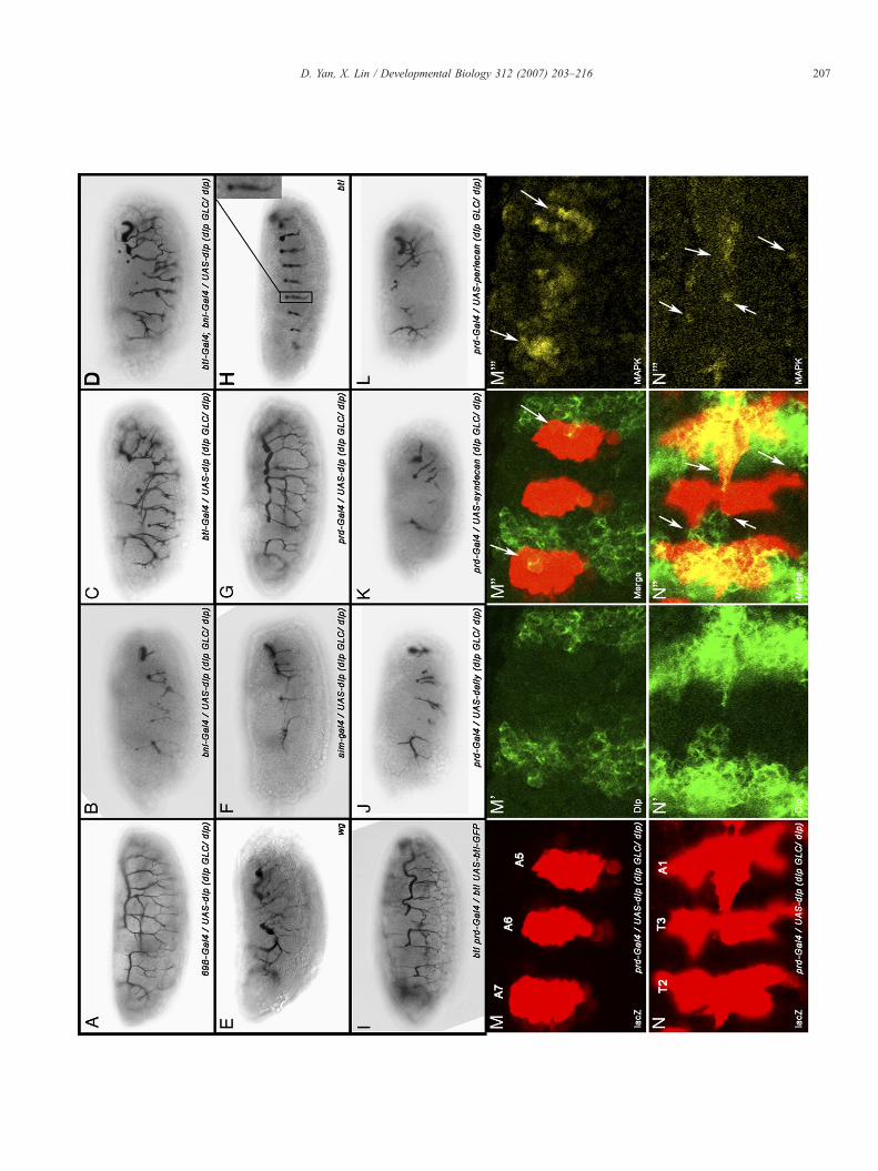
Fig. 2. Rescue of dlpmutant embryos by ectopic expression of UAS-dlp in different dstained with the tracheal lumenal antibody 2A12. (A–D, F–G) dlpmutant embryos ar(A), FGF expression cells (bnl-Gal4) (B), tracheal cells (btl-Gal4) (C), both FGF exprevery other segment (prd-Gal4) (G). Ectopic expression in whole ectoderm can almventral midline cells fail to rescue (B, F). btl-Gal4 rescued embryos develop an extensegmentation defect associated with other signaling pathways. In fact, this phenotypeand bnl-Gal4 are similar to those rescued by btl-Gal4 alone (D). Ectopic expressionalternative truncations in dlp embryos (G). This phenotype is very similar to that iembryos ectopically expressing UAS-dally (J), UAS-syndecan (K) and UAS-perlecembryos compared to Dlp expression (G). (M–N′′′) Stage 11 (M–M′′′) and 13 (N–NN), Dlp (M′, N′) and diphospho-MAPK (M′′′, N′′′). The first two channels are me
observed in embryos mutant for bnl, btl (Fig. 2H), sgl or sfl(Klambt et al., 1992; Lin et al., 1999; Sutherland et al., 1996),suggesting that Dlp is the main HSPG required for Btl signalingduring tracheal development. The following three lines ofevidence further support our hypothesis. First, tracheal defectassociated with dlp embryos is completely paternally rescuableas one copy of paternal wild-type dlp can rescue the trachealdefect associated with dlp embryos (Fig. 1C). However, whenthe paternal chromosome contains a bnl mutation, multipletruncations and branching defects were observed in the tracheaof these embryos, suggesting that dlp shows strong geneticinteraction with bnl (Fig. 1D). Second, Btl signaling is requiredfor activation of MAPK in the tracheal pits at stage 11 (Gabay etal., 1997a, b). Btl-dependent MAPK activity visualized bydiphospho-MAPK-specific antibody staining is shown in 10tracheal pits (T1-A7) in a wild-type embryo at stage 11 (Fig. 1E).This Btl-dependent MAPK activity is strikingly reduced in dlpembryos (Fig. 1F). The reduced Btl-dependentMAPK activity isnot due to lack of tracheal anlagen, as tracheal anlagendetermined by the enhancer trap fly line 1-eve-1 is normal indlp embryos (Fig. 1G) (Perrimon et al., 1991). Finally, inwild-type embryos, ectopic expression of bnl in tracheal cellshyperactivates Btl signaling, leading to a marked increase in finetracheal branching (Fig. 1H). This effect is suppressed in dlpembryos (Fig. 1I). This data also suggests that Dlp is required inFGF-receiving cells. It is worthwhile to note that Bnl expressionis not defective in dlp embryos (see Figure S1 in thesupplemental materials). In addition, dlp embryos exhibitedmore severe morphological defects than btl or bnl mutantembryos. This is due to patterning defects associated with Wgand Hh signalling as Dlp is required for Hh and Wg signallingduring embryogenesis (Baeg et al., 2001; Desbordes andSanson, 2003; Franch-Marro et al., 2005; Han et al., 2004b;Kirkpatrick et al., 2004). Taken together, our results argue thatDlp is essential for Bnl/Btl-dependent FGF signaling duringembryonic tracheal development.
We further asked whether Dlp is involved in mesodermal cellmigration controlled by Htl which requires HSPGs for itssignaling (Lin et al., 1999). Mesodermal cell migration can bevisualized by Twist (Twi) staining. After invagination throughthe ventral furrow at gastrulation, Twi-positive mesodermal cellsmigrate along the ectoderm in a dorsolateral direction (Fig. 1J).In htl mutant embryos, mesoderm migration does not occurproperly, resulting in an irregular pattern of Twi-positive cells inthe dorsal margin (Fig. 1K) (Beiman et al., 1996; Gisselbrecht etal., 1996; Michelson et al., 1998). Interestingly, Twi-positive
omains or by other HSPG core proteins. (A–L) Lateral view of stage 15 embryose rescued by ectopic expression of UAS-dlp in whole ectoderm cells (69B-Gal4)ession and tracheal cells (D), ventral midline cells (sim-Gal4) (F), or ectoderm inost completely rescue dlp embryos (A). Expressions in FGF expression cells orsive tracheal network which has an abnormal pattern (C). This is possibly due tois similar to that in Wg mutant embryos (E). Embryos rescued by both btl-Gal4in ectoderm of every other segment can rescue most of the tracheal defect withn btl mutant embryos rescued by prd-Gal4/UAS-btl-GFP (I). (J–L) dlp mutantan (L) by prd-Gal4. None of these HSPG core proteins is able to rescue dlp′′′) dlp embryos rescued by prd-Gal4/UAS-dlp stained for trachealess-lacZ (M,rged in panels M′′ and N′′ to indicate overlapping region (arrows).
207D. Yan, X. Lin / Developmental Biology 312 (2007) 203–216
208 D. Yan, X. Lin / Developmental Biology 312 (2007) 203–216
cells have an even distribution of migrating margins, suggestingthat mesodermal cell migration is normal in dlp embryos(Fig. 1L). Similarly, mesodermal cell migration is normal indally-dlp double mutant embryos, which are derived fromfemales with germline clones (Fig. 1M). These data argue thatneither Dally nor Dlp is required in Htl-dependent mesodermalcell migration. Alternatively, Dally and Dlp may be redundantwith Syndecan or Perlecan in Htl signaling.
Mechanism of Dlp function in FGF signaling duringembryonic tracheal development
To further determine the mechanism and specificity of Dlp inFGF signaling, we performed three series of rescue experiments.First, we asked which cells require Dlp in FGF signaling. Toaddress this question, we ectopically expressed a transgene dlpin different domains to rescue dlp embryos by the Gal4–UASsystem (Brand and Perrimon, 1993).WhenUAS-dlp is expressedin the whole embryonic ectoderm by 69B-Gal4 (for all Gal4expression pattern, see Figure S2 in the supplemental materials)in dlp embryos, the tracheal defect is almost completely rescued(Fig. 2A). However, when UAS-dlp is induced in FGFexpression cells by bnl-Gal4, almost no rescue was observedcompared to dlp embryos (Fig. 2B). Interestingly, when UAS-dlp is only expressed by btl-Gal4 in FGF receiving cells, thetracheal cells, we observed significant rescue of the trachealdefect (Fig. 2C). Furthermore, when we provide UAS-dlp inboth FGF expression and receiving cells by a combination ofbnl-Gal4 and btl-Gal4, the tracheal phenotype appears verysimilar to that rescued by btl-Gal4 alone (Fig. 2D). These datatogether argue that Dlp is only required in FGF receiving cells,but not in FGF producing cells. The rescue is not completeprobably due to patterning defect in these embryos, as Dlp is alsorequired for signaling activities ofWg andHh (Baeg et al., 2001;Desbordes and Sanson, 2003; Franch-Marro et al., 2005; Han etal., 2004b; Kirkpatrick et al., 2004). In fact, the btl-Gal4 rescuedtracheal phenotype is similar to that in a Wg mutant embryo(Fig. 2E). In particular, the dorsal trunk tracheal cells are lessrescued in these embryos (Figs. 2C, D). This is likely due to areduced Spalt (Sal) expression in these embryos (see Figure S3in the supplemental materials) as Sal expression is regulated byWg signaling and is required for dorsal trunk trachealdevelopment (Kuhnlein and Schuh, 1996; Llimargas, 2000).
Above experiments suggest a role of Dlp in tracheal cells forFGF signaling. Next, we asked whether Dlp functions cell-autonomously in FGF signaling as Dlp acts non-autonomouslyover a long range in Hh signaling in embryo (unpublished data).The following three experiments argue that Dlp is required
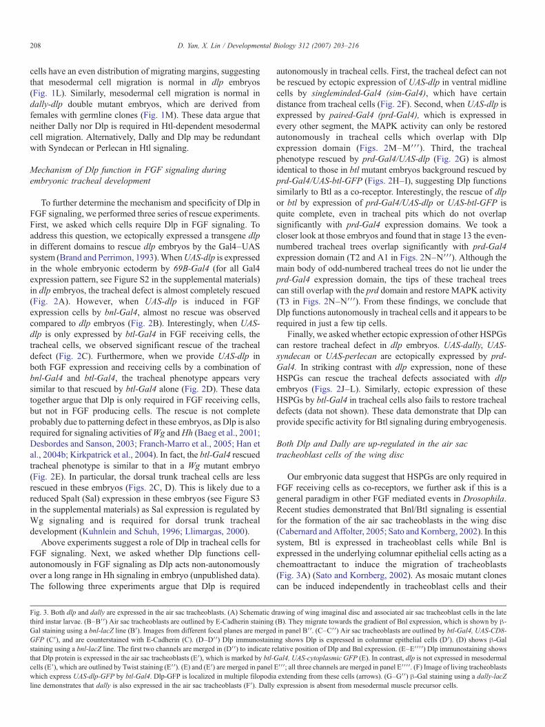
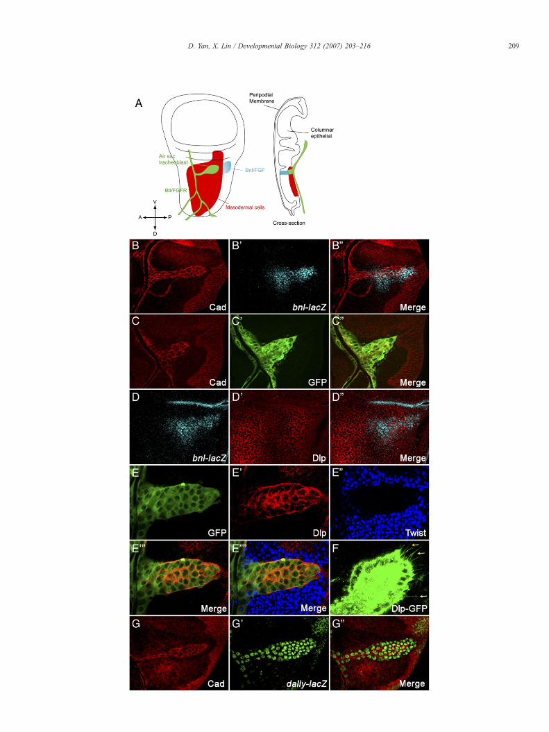
Fig. 3. Both dlp and dally are expressed in the air sac tracheoblasts. (A) Schematic dthird instar larvae. (B–B′′) Air sac tracheoblasts are outlined by E-Cadherin stainingGal staining using a bnl-lacZ line (B′). Images from different focal planes are mergedGFP (C′), and are counterstained with E-Cadherin (C). (D–D′′) Dlp immunostainistaining using a bnl-lacZ line. The first two channels are merged in (D′′) to indicate rthat Dlp protein is expressed in the air sac tracheoblasts (E′), which is marked by btl-Gcells (E'), which are outlined by Twist staining (E′′). (E) and (E′) are merged in panel Ewhich express UAS-dlp-GFP by btl-Gal4. Dlp-GFP is localized in multiple filopodialine demonstrates that dally is also expressed in the air sac tracheoblasts (F′). Dally
autonomously in tracheal cells. First, the tracheal defect can notbe rescued by ectopic expression of UAS-dlp in ventral midlinecells by singleminded-Gal4 (sim-Gal4), which have certaindistance from tracheal cells (Fig. 2F). Second, when UAS-dlp isexpressed by paired-Gal4 (prd-Gal4), which is expressed inevery other segment, the MAPK activity can only be restoredautonomously in tracheal cells which overlap with Dlpexpression domain (Figs. 2M–M′′′). Third, the trachealphenotype rescued by prd-Gal4/UAS-dlp (Fig. 2G) is almostidentical to those in btl mutant embryos background rescued byprd-Gal4/UAS-btl-GFP (Figs. 2H–I), suggesting Dlp functionssimilarly to Btl as a co-receptor. Interestingly, the rescue of dlpor btl by expression of prd-Gal4/UAS-dlp or UAS-btl-GFP isquite complete, even in tracheal pits which do not overlapsignificantly with prd-Gal4 expression domains. We took acloser look at those embryos and found that in stage 13 the even-numbered tracheal trees overlap significantly with prd-Gal4expression domain (T2 and A1 in Figs. 2N–N′′′). Although themain body of odd-numbered tracheal trees do not lie under theprd-Gal4 expression domain, the tips of these tracheal treescan still overlap with the prd domain and restore MAPK activity(T3 in Figs. 2N–N′′′). From these findings, we conclude thatDlp functions autonomously in tracheal cells and it appears to berequired in just a few tip cells.
Finally, we asked whether ectopic expression of other HSPGscan restore tracheal defect in dlp embryos. UAS-dally, UAS-syndecan or UAS-perlecan are ectopically expressed by prd-Gal4. In striking contrast with dlp expression, none of theseHSPGs can rescue the tracheal defects associated with dlpembryos (Figs. 2J–L). Similarly, ectopic expression of theseHSPGs by btl-Gal4 in tracheal cells also fails to restore trachealdefects (data not shown). These data demonstrate that Dlp canprovide specific activity for Btl signaling during embryogenesis.
Both Dlp and Dally are up-regulated in the air sactracheoblast cells of the wing disc
Our embryonic data suggest that HSPGs are only required inFGF receiving cells as co-receptors, we further ask if this is ageneral paradigm in other FGF mediated events in Drosophila.Recent studies demonstrated that Bnl/Btl signaling is essentialfor the formation of the air sac tracheoblasts in the wing disc(Cabernard andAffolter, 2005; Sato andKornberg, 2002). In thissystem, Btl is expressed in tracheoblast cells while Bnl isexpressed in the underlying columnar epithelial cells acting as achemoattractant to induce the migration of tracheoblasts(Fig. 3A) (Sato and Kornberg, 2002). As mosaic mutant clonescan be induced independently in tracheoblast cells and their
rawing of wing imaginal disc and associated air sac tracheoblast cells in the late(B). They migrate towards the gradient of Bnl expression, which is shown by β-in panel B′′. (C–C′′) Air sac tracheoblasts are outlined by btl-Gal4, UAS-CD8-ng shows Dlp is expressed in columnar epithelial cells (D′). (D) shows β-Galelative position of Dlp and Bnl expression. (E–E′′′′) Dlp immunostaining showsal4, UAS-cytoplasmic GFP (E). In contrast, dlp is not expressed in mesodermal′′′; all three channels are merged in panel E′′′′. (F) Image of living tracheoblastsextending from these cells (arrows). (G–G′′) β-Gal staining using a dally-lacZexpression is absent from mesodermal muscle precursor cells.
209D. Yan, X. Lin / Developmental Biology 312 (2007) 203–216
210 D. Yan, X. Lin / Developmental Biology 312 (2007) 203–216
underlying columnar epithelial cells, the wing air sac systemprovides an excellent model system to further define thefunctions of HSPGs in tracheal cells or in underlying columnarepithelial cells.
We marked the air sac tracheoblasts using UAS-CD8-GFPdriven by the btl-Gal4 (Fig. 3C′). We also stained thetracheoblasts by E-Cadherin, the adherent junction marker(Figs. 3B, C). It is worthwhile to note that although these cellsare changing their morphology dynamically, theymaintain intactpolarity as demonstrated by E-Cadherin staining. The expressionof Bnl was visualized by LacZ staining utilizing a bnl-LacZ line(Sutherland et al., 1996) (Figs. 3B′, D). From the compositeimage of E-Cadherin and LacZ staining in two cell layers(Fig. 3Bʺ), we observed Bnl expression in underlying epitheliumand tracheoblast formation toward the direction of Bnlexpression cells.
As a first step to determine the role of Dlp in this process, weexamined dlp expression using an anti-Dlp antibody (Lum et al.,2003). We found that Dlp is expressed in the plasma membraneof the tracheoblast cells (Figs. 3E–E′′′). Importantly, a Dlp–GFP fusion protein is distributed in the multiple filopodiaextending from the leading air sac cells (Fig. 3F, arrows). Theexpression pattern of Dlp suggests its possible involvement inreception of FGF. Dlp is also expressed abundantly in thecolumnar epithelial layer including the Bnl expression cells(Figs. 3D′, Dʺ). In contrast, Dlp is not expressed in Htl-positivemuscle precursor cells (Figs. 3E′–E′′′′) which surround trachealcells in the notum region of the wing discs.
We also examined Dally expression using dallyP2, a dallyenhancer trap lacZ line. Dally is also highly expressed in thetracheoblast cells (Figs. 3G′, Gʺ). Interestingly, Dally expres-sion is also absent in muscle cells (Figs. 3G′, Gʺ). The absenceof both Dlp and Dally in Htl-expressing cells suggests that theyare not involved in Htl signaling in the wing disc. This result isconsistent with the embryonic data that Dlp and Dally are notessential for Htl signaling during mesodermal cell migration(Figs. 1J–M).
Removal of HSPGs in FGF producing cells and othersurrounding columnar epithelia cells does not affecttracheoblast formation
The wing disc air sac can be used to determine whetherHSPGs are required for FGF signaling in tracheal cells (btlexpressing cells) or/and their underlying columnar epithelialcells which produce Bnl (Fig. 3). We and others havepreviously shown that HSPGs are essential for the distributionof HSPG-binding morphogen molecules including Wg, Hh andDpp (Baeg et al., 2004; Belenkaya et al., 2004; Bornemann etal., 2004; Franch-Marro et al., 2005; Han et al., 2004a,b, 2005;Kirkpatrick et al., 2004; Kreuger et al., 2004; Takei et al.,2004). HSPGs might be essential for the distribution of Bnland possibly the gradient formation of Bnl in the columnarepithelial cells. Alternatively, HSPGs might be only requiredfor Bnl/Btl signaling in FGF receiving cells, the tracheoblastcells. To distinguish these possibilities, we generated bigHSPG mutant clones which cover completely the Bnl ex-
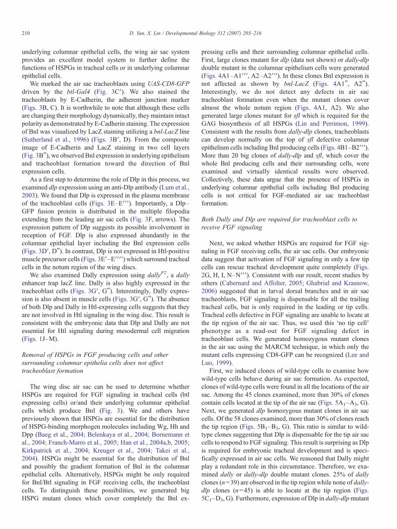
pressing cells and their surrounding columnar epithelial cells.First, large clones mutant for dlp (data not shown) or dally-dlpdouble mutant in the columnar epithelium cells were generated(Figs. 4A1–A1′′′, A2–A2′′′). In these clones Bnl expression isnot affected as shown by bnl-LacZ (Figs. 4A1ʺ, A2ʺ).Interestingly, we do not detect any defects in air sactracheoblast formation even when the mutant clones coveralmost the whole notum region (Figs. 4A1, A2). We alsogenerated large clones mutant for sfl which is required for theGAG biosynthesis of all HSPGs (Lin and Perrimon, 1999).Consistent with the results from dally-dlp clones, tracheoblastscan develop normally on the top of sfl defective columnarepithelium cells including Bnl producing cells (Figs. 4B1–B2′′′).More than 20 big clones of dally-dlp and sfl, which cover thewhole Bnl producing cells and their surrounding cells, wereexamined and virtually identical results were observed.Collectively, these data argue that the presence of HSPGs inunderlying columnar epithelial cells including Bnl producingcells is not critical for FGF-mediated air sac tracheoblastformation.
Both Dally and Dlp are required for tracheoblast cells toreceive FGF signaling
Next, we asked whether HSPGs are required for FGF sig-naling in FGF receiving cells, the air sac cells. Our embryonicdata suggest that activation of FGF signaling in only a few tipcells can rescue tracheal development quite completely (Figs.2G, H, I, N–N′′′). Consistent with our result, recent studies byothers (Cabernard and Affolter, 2005; Ghabrial and Krasnow,2006) suggested that in larval dorsal branches and in air sactracheoblasts, FGF signaling is dispensable for all the trailingtracheal cells, but is only required in the leading or tip cells.Tracheal cells defective in FGF signaling are unable to locate atthe tip region of the air sac. Thus, we used this ‘no tip cell'phenotype as a read-out for FGF signaling defect intracheoblast cells. We generated homozygous mutant clonesin the air sac using the MARCM technique, in which only themutant cells expressing CD8-GFP can be recognized (Lee andLuo, 1999).
First, we induced clones of wild-type cells to examine howwild-type cells behave during air sac formation. As expected,clones of wild-type cells were found in all the locations of the airsac. Among the 45 clones examined, more than 30% of clonescontain cells located at the tip of the air sac (Figs. 5A1–A3, G).Next, we generated dlp homozygous mutant clones in air saccells. Of the 58 clones examined, more than 30% of clones reachthe tip region (Figs. 5B1–B3, G). This ratio is similar to wild-type clones suggesting that Dlp is dispensable for the tip air saccells to respond to FGF signaling. This result is surprising as Dlpis required for embryonic tracheal development and is speci-fically expressed in air sac cells. We reasoned that Dally mightplay a redundant role in this circumstance. Therefore, we exa-mined dally or dally-dlp double mutant clones. 25% of dallyclones (n=39) are observed in the tip region while none of dally-dlp clones (n=45) is able to locate at the tip region (Figs.5C1–D3, G). Furthermore, expression of Dlp in dally-dlpmutant
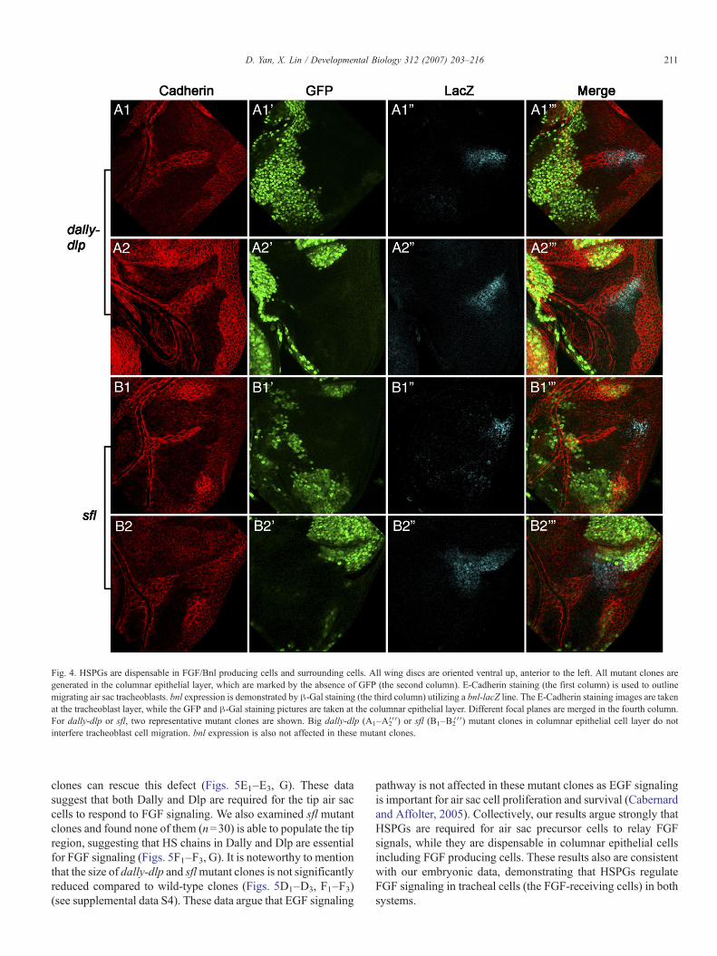
Fig. 4. HSPGs are dispensable in FGF/Bnl producing cells and surrounding cells. All wing discs are oriented ventral up, anterior to the left. All mutant clones aregenerated in the columnar epithelial layer, which are marked by the absence of GFP (the second column). E-Cadherin staining (the first column) is used to outlinemigrating air sac tracheoblasts. bnl expression is demonstrated by β-Gal staining (the third column) utilizing a bnl-lacZ line. The E-Cadherin staining images are takenat the tracheoblast layer, while the GFP and β-Gal staining pictures are taken at the columnar epithelial layer. Different focal planes are merged in the fourth column.For dally-dlp or sfl, two representative mutant clones are shown. Big dally-dlp (A1–A2′′′) or sfl (B1–B2′′′) mutant clones in columnar epithelial cell layer do notinterfere tracheoblast cell migration. bnl expression is also not affected in these mutant clones.
211D. Yan, X. Lin / Developmental Biology 312 (2007) 203–216
clones can rescue this defect (Figs. 5E1–E3, G). These datasuggest that both Dally and Dlp are required for the tip air saccells to respond to FGF signaling. We also examined sfl mutantclones and found none of them (n=30) is able to populate the tipregion, suggesting that HS chains in Dally and Dlp are essentialfor FGF signaling (Figs. 5F1–F3, G). It is noteworthy to mentionthat the size of dally-dlp and sflmutant clones is not significantlyreduced compared to wild-type clones (Figs. 5D1–D3, F1–F3)(see supplemental data S4). These data argue that EGF signaling
pathway is not affected in these mutant clones as EGF signalingis important for air sac cell proliferation and survival (Cabernardand Affolter, 2005). Collectively, our results argue strongly thatHSPGs are required for air sac precursor cells to relay FGFsignals, while they are dispensable in columnar epithelial cellsincluding FGF producing cells. These results also are consistentwith our embryonic data, demonstrating that HSPGs regulateFGF signaling in tracheal cells (the FGF-receiving cells) in bothsystems.
212 D. Yan, X. Lin / Developmental Biology 312 (2007) 203–216
213D. Yan, X. Lin / Developmental Biology 312 (2007) 203–216
Discussion
There are three main important findings in this work. First,we have identified Dlp as an essential molecule required fortracheal development. Dlp is required for Btl-mediated trachealbranching during embryogenesis while both Dlp and Dally areinvolved in the formation of air sac tracheoblasts in the wingdisc. Second, our data show that other HSPGs cannot replaceDlp for Btl signaling during embryogenesis and that both Dlpand Dally are not essential for Htl-mediated mesodermal cellmigration. These data demonstrate that different FGFs mayrequire different HSPGs to execute their effective signalingactivities during development. Third and most importantly, weprovide strong evidence that Dlp controls Btl signaling only inFGF-receiving cells in both embryonic and larval trachealsystems. This mechanism of HSPG activity in FGF signaling isvery different from its roles in regulating the signaling activitiesof morphogens including Wnt, Hh and Dpp. Together, our newfindings further define novel mechanisms and the specificitiesof HSPGs in FGF signaling during development.
Dlp is the major HSPG involved in Btl-mediated FGFsignaling
Extensive biochemical and cell culture studies suggest thatHSPGs are the part of the FGF/FGFR signaling complex(Ornitz, 2000; Ornitz and Itoh, 2001). However, the mechanismsof HSPGs in FGF signaling during development are less known.Previously, we have shown that embryos mutant for two HSPGbiosynthesis enzymes, sgl and sfl, exhibit defects in both Btl-and Htl-mediated FGF signaling (Lin et al., 1999). An importantissue remaining to be solved is which HSPG core proteins areinvolved in these signaling events. The data in this work providestrong evidence for the first time that Dlp is the key moleculerequired for Btl signaling during embryonic tracheal develop-ment, while both Dlp and Dally are involved in the Btl mediatedair sac tracheoblasts formation in the wing disc. Our resultsprovide several novel insights into the specificity of individualHSPG in FGF signaling. First, Dlp is involved in Btl signaling,but not in Htl signaling. These findings indicate that differentFGF/FGFR complexes may require different HSPGs for theirsignaling activities. Second, Dlp is highly active and specific forBtl signaling; overexpression of the other three DrosophilaHSPGs fail to rescue tracheal defects in dlp embryos. Thespecific activity of Dlp in Btl signaling could be due to the Dlpprotein core or the HS GAG chains attached to the Dlp coreprotein. In this regard, it is especially surprising that Dally,which has 22% identity with Dlp and also bears a GPI anchor,
Fig. 5. Both dally and dlp are essential in FGF/Bnl receiving cells. All mutant clones aCD8-GFP; the entire air sac is outlined with E-Cadherin staining. (A1–A3) Three retracheoblasts (the most distal part of air sac). (B1–B3) Three representative dlp murepresentative dallymutant clones located in the tip region of migrating tracheoblasts.the tip of air sac. (E1–E3) Three representative dally-dlp mutant clones rescued by UThree representative sfl mutant clones. These clones never reach the tip of air sac. (Among 45 Wild-type clones, 33% contribute to the tip region. Among 58 dlp mutancontribute to the tip region. Among 45 dally-dlp or 30 sfl mutant clones, none of the24% contribute to the tip region.
cannot rescue tracheal phenotypes associated with dlp embryos.As Dlp is involved in several other signaling pathways such asHh (Desbordes and Sanson, 2003; Han et al., 2004b), Wg (Baeget al., 2001, 2004; Franch-Marro et al., 2005; Han et al., 2005;Kirkpatrick et al., 2004; Kreuger et al., 2004), and Dpp(Belenkaya et al., 2004), it is unlikely that Dlp core proteininteracts with the ligands directly. In this regard, it is worthwhileto note that ectopic expression of Dally also fails to rescue Hhsignaling in dlp embryos (data not shown). We propose that Dlpmay have unique HS GAG chains that might provide high andspecific activity for ligands such as Bnl and Hh.
The biosynthesis of HS GAG chains is determined by theHSPG protein core in which the GAG attachment sites and otherprotein parts such as the N-terminal cystenine-rich domaincontrol both quantity and quality of the attached GAG chains(Esko and Selleck, 2002; Lin, 2004). Detailed structure andfunctional studies of Dlp will further help to define specificrequirements of the core protein or GAG attachment sites in FGFsignaling. Furthermore, the unique GAG chains may bemodified by specific enzymes. In this regard, it is particularlyimportant to note that 6-O sulfation of HS is critical for Btlsignaling, as Drosophila heparan sulfate 6-O-sulfotransferaseis specifically expressed in embryonic tracheal system and isrequired for Btl signaling during embryogenesis (Kamimura etal., 2001). More recent study showed that the overall sulfationlevel is more important than strictly defined HS fine structuresfor FGF signaling in some developmental contexts (Kamimuraet al., 2006). In this regard, we suggest that Dlp may be theoptimal substrate for sulfation enzymes during embryogenesis.Therefore, the activity of Dlp in FGF signaling during embryo-genesis cannot be replaced by other HSPGs including Dally,Syndecan and Perlecan.
Although Dlp is essential for Btl signaling during embryo-genesis, both Dally and Dlp are involved in Btl signaling in airsac tracheoblast cells. Similarly, our previous studies showedthat both Dally and Dlp are involved in regulating Wg, Hh andDpp distribution in the wing disc (Belenkaya et al., 2004; Han etal., 2004b, 2005). The different functions of the same HSPG inembryos and discs may reflect temporal and developmentalstage dependent regulation of HSPG functions (Allen andRapraeger, 2003).
Mechanism(s) of HSPG function in FGF signaling
While it is well established that HSPGs can regulate FGFsignaling by facilitating FGF/FGFR interaction (Ornitz, 2000), itis unknown whether HSPGs can also control FGF distribution,thereby modulating FGF signaling. This is a particularly
re generated in the air sac tracheoblast cells. The clones are positively marked bypresentative wild-type clones, which are located in the tip region of migratingtant clones located in the tip region of migrating tracheoblasts. (C1–C3) Three(D1–D3) Three representative dally-dlpmutant clones. These clones never reachAS-dlp. They can localize in the tip region of migrating tracheoblasts. (F1–F3)G) Statistic data demonstrate ratio of clones that contribute to the tip of air sac.t clones, 38% contribute to the tip region. Among 39 dally mutant clones, 25%m reach the tip region. Among 38 dally-dlp mutant clones rescued by UAS-dlp,
214 D. Yan, X. Lin / Developmental Biology 312 (2007) 203–216
important issue as in many developmental contexts FGF ligandis produced in one type of cell and acts on other cells to initiate itsbiological activity (Thisse and Thisse, 2005; Zhang et al., 2006).One important finding of this work is that HSPGs controltracheal morphogenesis by regulating FGF signaling only inFGF-receiving cells, but not by regulating the secretion ordistribution of FGF ligand in its producing cells and surroundingcells. Several important results support our conclusion: 1. dlpmutant embryos can suppress the phenotype of overexpressingBnl in the tracheal cells. 2. Ectopic expression of Dlp in trachealcells, rather than FGF expression cells, can effectively restoretracheal defects associated with dlp embryos. 3. Embryosrescued by prd-Gal4/UAS-dlp in dlp backbround is very similarto btl mutant embryos rescued by prd-Gal4/UAS-btl-GFP. 4.HSPGs are required for FGF signaling in its receiving cells in theair sac, but are dispensable in the columnar epithelial layer whichincludes FGF producing cells and other surrounding cells. Ourdetailed analyses thus demonstrate for the first time the specificand distinct requirement of HSPGs in FGF signaling duringtracheal development. Moreover, our embryonic and larval datatogether suggest this is likely a general mechanism for HSPGfunction in FGF signaling in Drosophila.
Twomajor models are proposed for the role of HSPGs in FGFsignaling (Lin, 2004; Ornitz, 2000). In one model, low affinityHS/GAG chains on the cell surface limit the diffusion of FGFligand, thereby increasing its local concentration and the pro-bability that it will interact with high-affinity FGFRs. In thesecond model, HSPGs facilitate the dimerization or oligomer-ization of FGF ligands thereby inducing receptor clustering andsignal transduction. Our experimental data cannot exclude eitherof these mechanisms. However, our results are in favour of thesecond case as we show that HSPGs are not required in FGFconcentration gradient in FGF producing cells, but are essentialin FGF-receiving cells. Finally, a recent study showed that dy-namin-mediated vesicle internalization is a crucial step toregulate FGF signaling in Drosophila tracheal system (Dammaiet al., 2003). Mutants in awd (abnormal wing disc) or shi(shibire), which encodes for a nucleoside diphosphate kinaseand Drosophila dynamin, respectively, have increased levels ofBtl in tracheal cell surface, increased FGF signaling activity andectopic tracheal branching. In this regard, HSPGs may controlFGF signaling by stabilizing the FGF/FGFR complex fromdegradation or internalization in FGF receiving cells. Furtherexperiments using HSPG and awd/shi double mutant are neededto test this possibility.
Comparison of HSPG functions in FGF and morphogensignaling
Over the past several years, extensive studies in Drosophilaand other model systems have established the essential roles ofHSPGs in developmental signaling pathways including Wg, Hhand Dpp (Hacker et al., 2005; Lin, 2004). In Drosophila embryoand wing imaginal disc, HSPGs are involved in the transport ofmorphogens including Wg, Hh and Dpp by a restricted diffusionmechanism (Belenkaya et al., 2004; Han et al., 2004b, 2005; Theet al., 1999). Narrow stripes of clones mutant for HSPGs can
impede the movement of morphogens to further cells. However,in all of these cases, the first mutant cells adjacent to themorphogen source can still transduce signals arguing that HSPGsare not essential for morphogen signaling activity, but rathercontrol the distributions or local concentrations of morphogens(Belenkaya et al., 2004; Han et al., 2004b, 2005). Our novelresults from this work point out a major difference for a role ofHSPGs in FGF signaling from their roles inmorphogen signaling,as removal of HSPGs (dally-dlp or sfl) from FGF receiving cellscan effectively block FGF signaling. Although the graded FGFactivity may play an essential role in tracheal morphogenesis(Affolter andWeijer, 2005), our data from this work argue that themain function of HSPGs in FGF signaling is not to regulate thedistribution of FGF ligand. Consistent with the different roles ofHSPGs in FGF and morphogen signaling, we found that Dlp actscell-autonomously in FGF signaling while it functions non-autonomously in Hh signaling in embryos (unpublished data).Our results suggest that Bnl transportation may be different frommorphogen movement in the epithelial cells of the wing pouch.Indeed, morphogen molecules diffuse through the same layer ofcells, columnar epithelial cells, while FGF is transported betweendifferent layers of tissues, from columnar epithelia to tracheo-blasts. Moreover, leading air sac cells are always in closeproximity with underlying columnar epithelia. They also extendmultiple filopodia toward ligand gradient and presumablyactively pursue the FGF ligands (Sato and Kornberg, 2002)while wing disc morphogens including Wg, Hh and Dpp need totransport many cell diameters from their sources to reach theirreceiving cells. Studies in vertebrate also suggest that a gradeddistribution of FGF8 protein can be generated by the decay of fgf8mRNA and this RNA gradient is translated into a protein gradient(Dubrulle and Pourquie, 2004). In this case, no active transportmechanism is required to form a FGF gradient. In mammalianlimb and lung development different FGFs are often expressedin different layers of cells, such as epithelium and mesenchyme,and signal through each other (Thisse and Thisse, 2005; Zhanget al., 2006). It is interesting to determine whether HSPGsfunction similarly in these systems as in Drosophila.
Acknowledgments
We thank M. Krasnow, S. Cohen, J. Vincent, S. Hou,T. Kornberg, M. Sato, H. Nakato and the Bloomington StockCenter for Drosophila stocks; P. Beachy, A. Michelson and theIowa Developmental Studies Hybridoma Bank (IDSHB) forantibodies; H. Liu for technical assistance. We thank TanyaBelenkaya and Caitlin Maynard for comments on the manu-script. This work was supported partially by NIH grant2R01GM063891, Research Scholar Grant RSG-07-051 fromthe American Cancer Society, March of Dimes foundation toX. L. D. Y is an Albert J. Ryan fellow and is supported by apredoctoral fellowship from the American Heart Association.
Appendix A. Supplementary data
Supplementary data associated with this article can be found,in the online version, at doi:10.1016/j.ydbio.2007.09.015.

215D. Yan, X. Lin / Developmental Biology 312 (2007) 203–216
References
Affolter, M., Weijer, C.J., 2005. Signaling to cytoskeletal dynamics duringchemotaxis. Dev. Cell 9, 19–34.
Allen, B.L., Rapraeger, A.C., 2003. Spatial and temporal expression of heparansulfate in mouse development regulates FGF and FGF receptor assembly.J. Cell Biol. 163, 637–648.
Baeg, G.H., et al., 2001. Heparan sulfate proteoglycans are critical for theorganization of the extracellular distribution of Wingless. Development 128,87–94.
Baeg, G.H., et al., 2004. The Wingless morphogen gradient is established by thecooperative action of Frizzled and Heparan Sulfate Proteoglycan receptors.Dev. Biol. 276, 89–100.
Beiman, M., et al., 1996. Heartless, a Drosophila FGF receptor homolog, isessential for cell migration and establishment of several mesodermallineages. Genes Dev. 10, 2993–3002.
Belenkaya, T.Y., et al., 2002. Pygopus encodes a nuclear protein essential forwingless/Wnt signaling. Development 129, 4089–4101.
Belenkaya, T.Y., et al., 2004. Drosophila Dpp morphogen movement isindependent of dynamin-mediated endocytosis but regulated by the glypicanmembers of heparan sulfate proteoglycans. Cell 119, 231–244.
Bernfield, M., et al., 1999. Functions of cell surface heparan sulfateproteoglycans. Annu. Rev. Biochem. 68, 729–777.
Bornemann, D.J., et al., 2004. Abrogation of heparan sulfate synthesis inDrosophila disrupts the Wingless, Hedgehog and Decapentaplegic signalingpathways. Development 131, 1927–1938.
Brand, A.H., Perrimon, N., 1993. Targeted gene expression as a means ofaltering cell fates and generating dominant phenotypes. Development 118,401–415.
Cabernard, C., Affolter, M., 2005. Distinct roles for two receptor tyrosinekinases in epithelial branching morphogenesis in Drosophila. Dev. Cell 9,831–842.
Cabernard, C., et al., 2004. Cellular and molecular mechanisms involved inbranching morphogenesis of the Drosophila tracheal system. J. Appl.Physiol. 97, 2347–2353.
Chou, T.B., Perrimon, N., 1996. The autosomal FLP-DFS technique forgenerating germline mosaics in Drosophila melanogaster. Genetics 144,1673–1679.
Coumoul, X., Deng, C.X., 2003. Roles of FGF receptors in mammaliandevelopment and congenital diseases. Birth Defects Res., C Embryo Today69, 286–304.
Dammai, V., et al., 2003. Drosophila awd, the homolog of human nm23,regulates FGF receptor levels and functions synergistically with shi/dynaminduring tracheal development. Genes Dev. 17, 2812–2824.
Datta, S., Kankel, D.R., 1992. l(1)trol and l(1)devl, loci affecting thedevelopment of the adult central nervous system in Drosophila melanoga-ster. Genetics 130, 523–537.
Desbordes, S.C., Sanson, B., 2003. The glypican Dally-like is required forHedgehog signalling in the embryonic epidermis of Drosophila. Develop-ment 130, 6245–6255.
Dubrulle, J., Pourquie, O., 2004. fgf8 mRNA decay establishes a gradient thatcouples axial elongation to patterning in the vertebrate embryo. Nature 427,419–422.
Esko, J.D., Selleck, S.B., 2002. Order out of chaos: assembly of ligand bindingsites in heparan sulfate. Annu. Rev. Biochem. 71, 435–471.
Franch-Marro, X., et al., 2005. Glypicans shunt the Wingless signal betweenlocal signalling and further transport. Development 132, 659–666.
Gabay, L., et al., 1997a. In situ activation pattern of Drosophila EGF receptorpathway during development. Science 277, 1103–1106.
Gabay, L., et al., 1997b. MAP kinase in situ activation atlas during Drosophilaembryogenesis. Development 124, 3535–3541.
Ghabrial, A.S., Krasnow, M.A., 2006. Social interactions among epithelial cellsduring tracheal branching morphogenesis. Nature 441, 746–749.
Gisselbrecht, S., et al., 1996. heartless encodes a fibroblast growth factorreceptor (DFR1/DFGF-R2) involved in the directional migration ofearly mesodermal cells in the Drosophila embryo. Genes Dev. 10,3003–3017.
Golembo, M., et al., 1996. The Drosophila embryonic midline is the site of Spitz
processing, and induces activation of the EGF receptor in the ventralectoderm. Development 122, 3363–3370.
Golic, K.G., 1991. Site-specific recombination between homologous chromo-somes in Drosophila. Science 252, 958–961.
Gryzik, T., Muller, H.A., 2004. FGF8-like1 and FGF8-like2 encode putativeligands of the FGF receptor Htl and are required for mesoderm migration inthe Drosophila gastrula. Curr. Biol. 14, 659–667.
Hacker, U., et al., 1997. The Drosophila sugarless gene modulates Winglesssignaling and encodes an enzyme involved in polysaccharide biosynthesis.Development 124, 3565–3573.
Hacker, U., et al., 2005. Heparan sulphate proteoglycans: the sweet side ofdevelopment. Nat. Rev., Mol. Cell Biol. 6, 530–541.
Han, C., et al., 2004a. Distinct and collaborative roles of Drosophila EXT familyproteins in morphogen signalling and gradient formation. Development 131,1563–1575.
Han, C., et al., 2004b. Drosophila glypicans control the cell-to-cell movementof Hedgehog by a dynamin-independent process. Development 131,601–611.
Han, C., et al., 2005. Drosophila glypicans Dally and Dally-like shape theextracellular Wingless morphogen gradient in the wing disc. Development132, 667–679.
Itoh, N., Ornitz, D.M., 2004. Evolution of the Fgf and Fgfr gene families. TrendsGenet. 20, 563–569.
Johnson, K.G., et al., 2004. Axonal heparan sulfate proteoglycans regulate thedistribution and efficiency of the repellent slit during midline axon guidance.Curr. Biol. 14, 499–504.
Kamimura, K., et al., 2001. Drosophila heparan sulfate 6-O-sulfotransferase(dHS6ST) gene. Structure, expression, and function in the formation of thetracheal system. J. Biol. Chem. 276, 17014–17021.
Kamimura, K., et al., 2006. Specific and flexible roles of heparan sulfatemodifications in Drosophila FGF signaling. J. Cell Biol. 174, 773–778.
Khare, N., Baumgartner, S., 2000. Dally-like protein, a new Drosophila glypicanwith expression overlapping with wingless. Mech. Dev. 99, 199–202.
Kirkpatrick, C.A., et al., 2004. Spatial regulation of Wingless morphogendistribution and signaling by Dally-like protein. Dev. Cell 7, 513–523.
Klambt, C., et al., 1992. breathless, a Drosophila FGF receptor homolog, isessential for migration of tracheal and specific midline glial cells. GenesDev. 6, 1668–1678.
Kreuger, J., et al., 2004. Opposing activities of Dally-like glypican at high andlow levels of Wingless morphogen activity. Dev. Cell 7, 503–512.
Kuhnlein, R.P., Schuh, R., 1996. Dual function of the region-specific homeoticgene spalt during Drosophila tracheal system development. Development122, 2215–2223.
Lee, T., Luo, L., 1999. Mosaic analysis with a repressible cell marker for studiesof gene function in neuronal morphogenesis. Neuron 22, 451–461.
Lin, X., 2004. Functions of heparan sulfate proteoglycans in cell signalingduring development. Development 131, 6009–6021.
Lin, X., Perrimon, N., 1999. Dally cooperates with Drosophila Frizzled 2 totransduce Wingless signalling. Nature 400, 281–284.
Lin, X., et al., 1999. Heparan sulfate proteoglycans are essential for FGFreceptor signaling during Drosophila embryonic development. Development126, 3715–3723.
Llimargas, M., 2000. Wingless and its signalling pathway have common andseparable functions during tracheal development. Development 127,4407–4417.
Lum, L., et al., 2003. Identification of Hedgehog pathway components by RNAiin Drosophila cultured cells. Science 299, 2039–2045.
Michelson, A.M., et al., 1998. Dual functions of the heartless fibroblast growthfactor receptor in development of the Drosophila embryonic mesoderm.Dev. Genet. 22, 212–229.
Nakato, H., et al., 1995. The division abnormally delayed (dally) gene: aputative integral membrane proteoglycan required for cell divisionpatterning during postembryonic development of the nervous system inDrosophila. Development 121, 3687–3702.
Ornitz, D.M., 2000. FGFs, heparan sulfate and FGFRs: complex interactionsessential for development. BioEssays 22, 108–112.
Ornitz, D.M., Itoh, N., 2001. Fibroblast growth factors. Genome Biol. 2(REVIEWS3005).
216 D. Yan, X. Lin / Developmental Biology 312 (2007) 203–216
Perrimon, N., et al., 1991. Generating lineage-specific markers to studyDrosophila development. Dev. Genet. 12, 238–252.
Sato, M., Kornberg, T.B., 2002. FGF is an essential mitogen and chemoat-tractant for the air sacs of the drosophila tracheal system. Dev. Cell 3,195–207.
Shiga, Y., Tanaka-Matakatsu, M., Hayashi, S., 1996. A nuclear GFP beta-galactosidase fusion protein as a marker for morphogenesis in livingDrosophila. Dev. Growth Differ. 38, 99–106.
Shishido, E., et al., 1993. Two FGF-receptor homologues of Drosophila: one isexpressed in mesodermal primordium in early embryos. Development 117,751–761.
Shishido, E., et al., 1997. Requirements of DFR1/Heartless, a mesoderm-specific Drosophila FGF-receptor, for the formation of heart, visceral andsomatic muscles, and ensheathing of longitudinal axon tracts in CNS.Development 124, 2119–2128.
Spring, J., et al., 1994. Drosophila syndecan: conservation of a cell-surfaceheparan sulfate proteoglycan. Proc. Natl. Acad. Sci. U. S. A. 91, 3334–3338.
Stathopoulos, A., et al., 2004. pyramus and thisbe: FGF genes that pattern themesoderm of Drosophila embryos. Genes Dev.' 18, 687–699.
Sutherland, D., et al., 1996. branchless encodes a Drosophila FGF homolog that
controls tracheal cell migration and the pattern of branching. Cell 87,1091–1101.
Takei, Y., et al., 2004. Three Drosophila EXT genes shape morphogen gradientsthrough synthesis of heparan sulfate proteoglycans. Development 131,73–82.
The, I., et al., 1999. Hedgehog movement is regulated through tout velu-dependent synthesis of a heparan sulfate proteoglycanMol. Cell 4, 633–639.
Thisse, B., Thisse, C., 2005. Functions and regulations of fibroblast growthfactor signaling during embryonic development. Dev. Biol. 287, 390–402.
van den Heuvel, M., et al., 1993. Mutations in the segment polarity geneswingless and porcupine impair secretion of the wingless protein. EMBO J.12, 5293–5302.
Voigt, A., et al., 2002. Perlecan participates in proliferation activation ofquiescent Drosophila neuroblasts. Dev. Dyn. 224, 403–412.
Xu, T., Rubin, G.M., 1993. Analysis of genetic mosaics in developing and adultDrosophila tissues. Development 117, 1223–1237.
Yoffe, K.B., et al., 1995. Evidence for engrailed-independent winglessautoregulation in Drosophila. Dev. Biol. 170, 636–650.
Zhang, X., et al., 2006. Reciprocal epithelial–mesenchymal FGF signaling isrequired for cecal development. Development 133, 173–180.


![Rizzoli.and.Isles.s01e03.Hdtv.xvid Xii.eng[Ragbear]Fgf](https://img.pdfslide.us/doc/110x75/577d36f21a28ab3a6b9464d0/rizzoliandisless01e03hdtvxvid-xiiengragbearfgf.jpg)