Embed Size (px)
Citation preview

1
A rhesus macaque model of Asian lineage Zika virus infection 1
2
Dawn M. Dudley1*, Matthew T. Aliota2*, Emma Mohr3*, Andrea M. Weiler4, Gabrielle Lehrer-3
Brey4, Kim L. Weisgrau4, Mariel S. Mohns1, Meghan E. Breitbach1, Mustafa N. Rasheed1, 4
Christina M. Newman1, Dane D. Gellerup4, Louise H. Moncla1,2, Jennifer Post4, Nancy Schultz-5
Darken4, Michele L. Schotkzo4, Jennifer M. Hayes4, Josh A. Eudailey5, M. Anthony Moody5, 6
Sallie R. Permar5, Shelby L. O’Connor1, Eva G. Rakasz4, Heather A. Simmons4, Saverio 7
Capuano III4, Thaddeus G. Golos4,6, Jorge E. Osorio2, Thomas C. Friedrich2,4, and David H. 8
O’Connor1,4 9
10
1Department of Pathology and Laboratory Medicine, University of Wisconsin-Madison; 11
2Department of Pathobiological Sciences, University of Wisconsin-Madison;3Department of 12
Pediatrics, School of Medicine and Public Health, University of Wisconsin-Madison; 4Wisconsin 13
National Primate Research Center, University of Wisconsin-Madison; 5Department of Pediatrics 14
and Human Vaccine Institute, Duke University Medical Center; 6Department of Comparative 15
Biosciences and Obstetrics and Gynecology, University of Wisconsin-Madison. 16
*These authors contributed equally to this work. 17
18
Infection with Asian lineage Zika virus has been associated with Guillain-Barré 19
syndrome and fetal abnormalities 1-4, but the mechanisms and risk factors for these 20
outcomes remain unknown. Here we show that rhesus macaques are susceptible to 21
infection by an Asian-lineage virus closely related to strains currently circulating in the 22
Americas. Following subcutaneous inoculation, Zika virus RNA was detected in plasma 23
one day post-infection (dpi) in all animals (N = 8, including 2 animals infected during the 24
first trimester of pregnancy). Plasma viral loads peaked above 1 x 10⁵ viral RNA 25
.CC-BY-ND 4.0 International licensecertified by peer review) is the author/funder. It is made available under aThe copyright holder for this preprint (which was notthis version posted May 10, 2016. . https://doi.org/10.1101/046334doi: bioRxiv preprint

2
copies/mL in seven of eight animals. Viral RNA was also present in saliva, urine, and 26
cerebrospinal fluid (CSF), consistent with case reports from infected humans. Viral RNA 27
was cleared from plasma and urine by 21 dpi in non-pregnant animals. In contrast, both 28
pregnant animals remained viremic longer, up to 57 days. In all animals, infection was 29
associated with transient increases in proliferating natural killer cells, CD8+ T cells, CD4+ 30
T cells, and plasmablasts. Neutralizing antibodies were detected in all animals by 21 dpi. 31
Rechallenge of three non-pregnant animals with the Asian-lineage Zika virus 10 weeks 32
after the initial challenge resulted in no detectable virus replication, suggesting that 33
primary Zika virus infection elicits protective immunity against homologous virus strains. 34
These data establish that Asian-lineage Zika virus infection of rhesus macaques provides 35
a relevant animal model for studying pathogenesis in pregnant and non-pregnant 36
individuals and evaluating potential interventions against human infection, including 37
during pregnancy. 38
Zika virus (ZIKV) is a mosquito-borne flavivirus first identified in 19475. Little was known 39
about ZIKV when fetal abnormalities and Guillain-Barré syndrome were reported coincident with 40
epidemic spread of Asian-lineage ZIKV in South America6,7,1. Animal models are essential for 41
quickly understanding ZIKV transmission and pathogenesis, as well as for evaluating candidate 42
vaccines and therapeutics. ZIKV infects immunocompromised mice8 but such mice do not mimic 43
key attributes of human infection and fetal development. 44
In contrast, immunocompetent macaque monkeys are widely used in both infectious 45
disease and obstetric research. To determine whether a phyisiologically relevant dose and route 46
of Asian lineage ZIKV infects immunocompetent pregnant and non-pregnant macaques, we 47
inoculated eight Indian-origin rhesus macaques (Macaca mulatta) subcutaneously with ZIKV 48
derived from a French Polynesian virus isolate (Zika virus/H.sapiens-49
tc/FRA/2013/FrenchPolynesia-01_v1c1). The eight animals were divided into three cohorts as 50
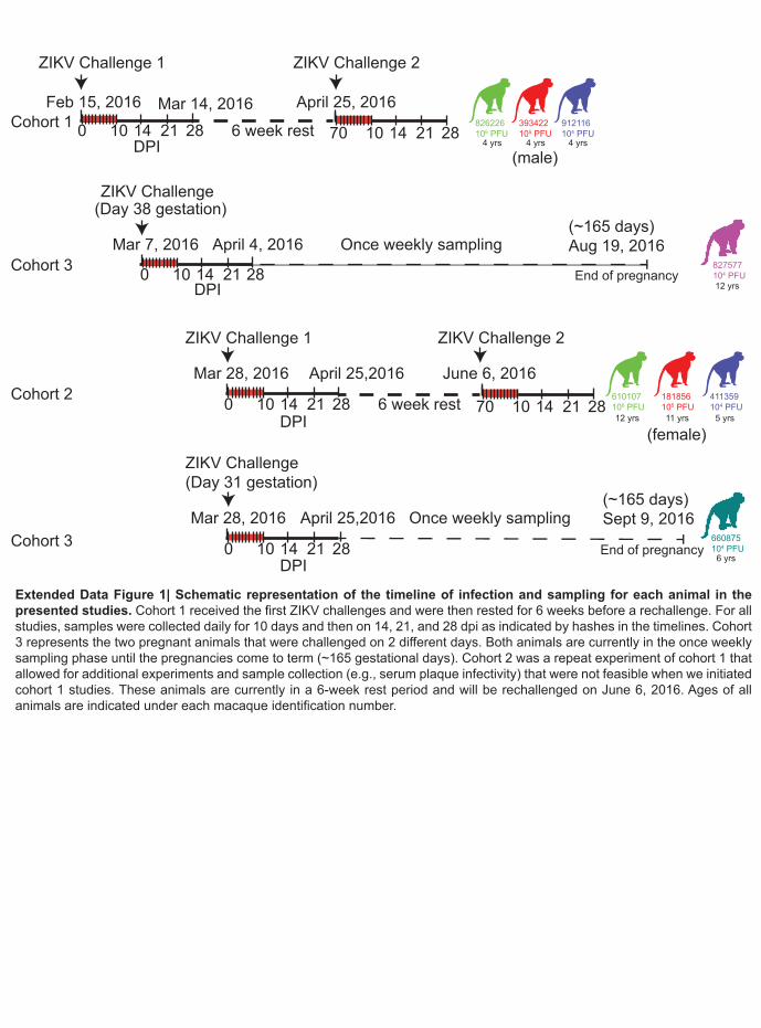
shown in Extended Data Fig. 1. 51
.CC-BY-ND 4.0 International licensecertified by peer review) is the author/funder. It is made available under aThe copyright holder for this preprint (which was notthis version posted May 10, 2016. . https://doi.org/10.1101/046334doi: bioRxiv preprint

3
To define the minimal dosage necessary to establish infection, two macaques per group 51
were infected with 1 x 10⁶, 1 x 10⁵, or 1 x 10⁴ PFU ZIKV (cohorts 1 and 2) (Fig. 1a). This dose 52
range of inocula is based on previous work in related flaviviruses such as West Nile virus (WNV) 53
and DENV, where it was estimated that mosquitoes delivered 1 x 10⁴- 1x 10⁶ PFU of virus9,10. 54
This is also the range found in mosquito saliva in a recent publication specifically for Brazilian 55
Zika virus 11. None of the animals developed significant clinical disease (see Extended Data Fig. 56
2 and SI Data) 57
Blood was sampled daily for 10-11 days post-infection (dpi) and every 3-7 days 58
thereafter. Viral RNA (vRNA) was quantified by qRT-PCR from plasma12 and was detected in all 59
six animals at 1 dpi (Fig. 1b). Peak plasma viremia occurred between 2 and 6 dpi, and ranged 60
from 8.2 x 10⁴ to 2.5 x 10⁶ vRNA copies/mL. These results resemble findings in humans in 61
Colombia where the mean serum viral load was 2.6 x10⁵ copies/ml ± 10 copies/ml in acutely 62
infected individuals (n=10, range=537 - 6.9 x 10⁵ copies/ml) (manuscript submitted13). Infectious 63
titers, measured from serum in cohort 2 animals, were 500-1000-fold less than copies of vRNA 64
detected from plasma at the same time points (Fig. 1c). Copies of vRNA detected in the serum 65
and plasma were very similar as shown in Extended Data Fig. 3. The estimated doubling time 66
for plasma viremia averaged 7.7 hours (range = 4.8-10.2 hours) and was independent of the 67
infecting dose and sex of the macaque. By 10 dpi, plasma viral loads were undetectable (<100 68
vRNA copies/mL) in all six animals, although intermittent low-level detection (<550 vRNA 69
copies/mL) continued sporadically through 17 dpi. Thereafter viral RNA remained undetectable 70
in all fluids throughout follow-up (longest follow-up 70 dpi; Fig. 1b insets). 71
We also measured ZIKV vRNA by qRT-PCR in other body fluids including urine, saliva, 72
CSF and vaginal fluid. Viruria was detected starting at 2-5 dpi and as late as 17 dpi, in urine 73
passively collected from cage pans (Fig. 1b). Despite possible degradation of virus between the 74
time of urination and sample collection and processing, 1 x 10³-1 x 10⁴ vRNA copies/mL urine 75
were detected at multiple timepoints. Virus was also detected in oral swabs collected from all six 76
.CC-BY-ND 4.0 International licensecertified by peer review) is the author/funder. It is made available under aThe copyright holder for this preprint (which was notthis version posted May 10, 2016. . https://doi.org/10.1101/046334doi: bioRxiv preprint
4
animals, peaking at over 1 x 10³ vRNA copies per sample in 3 of 6 animals (Fig. 1b). Notably, 77
as with urine, the kinetics of virus detection in saliva occurred after peak plasma viremia. 78
Cisterna magna punctures were performed at 4 and 14 dpi to quantify viral RNA in CSF; vRNA 79
was detectable at 4 dpi in 3 out of 5 animals from which CSF could be obtained (Fig. 1d). 80
Vaginal swabs collected from the 3 female animals in cohort 2 had detectable vRNA starting at 81
1 and/or 7 dpi, but were undetectable at 14, 21, and 28 dpi (Fig. 1e). 82
We next characterized the immune response to infection by staining peripheral blood 83
mononuclear cells (PBMC) for multiple lineage and activation markers. Proliferating (Ki-67+) NK 84
cells, CD8+ T cells, and CD4+ T cells expanded significantly above baseline levels by 6 dpi 85
(Fig. 2a,b). NK and CD8+ T cell expansion increased as plasma vRNA loads decreased starting 86
at 6 dpi. We also enumerated circulating plasmablasts, defined as CD3-/20-/14-/16-/11c-/123- 87
and CD80+/HLA-DR+ cells, on 0, 3, 7, 11 and 14 dpi (Fig. 2c)14. The peak plasmablast 88
expansion occurred between 7 and 10 dpi in 5/6 animals. Serum neutralizing antibody 89
responses were also measured by plaque reduction neutralization tests (PRNT90). All animals 90
exhibited high neutralizing antibody (nAb) titers as early as 14 dpi (Fig. 2d), the earliest time 91
point tested. Cohort 1 animals were tested at 64 dpi and cohort 2 animals were tested at 14 and 92
28 dpi. Together these data suggest that peak activation of the adaptive immune response and 93
antibody production occurs 5-7 dpi and may both be important to control viral replication as 94
evidenced by reducing vRNA loads in the plasma at these time points. 95
To determine whether activation of T cells correlated with the appearance of Zika virus-96
specific responses, we performed interferon-gamma ELISPOT on PBMCs collected at 4, 10 and 97
14 dpi for cohort 2 animals. Cells were stimulated with pools of 15mer peptides collectively 98
representing the amino acid sequence of the Asian-lineage NS5 protein (GenBank: KU321639). 99
We detected specific IFN-gamma secretion in response to 12 of 16 peptide pools in at least one 100
animal (Extended Data Fig. 4a). Overall, this data supports that there are ZIKV-specific T cell 101
responses in all animals tested. 102
.CC-BY-ND 4.0 International licensecertified by peer review) is the author/funder. It is made available under aThe copyright holder for this preprint (which was notthis version posted May 10, 2016. . https://doi.org/10.1101/046334doi: bioRxiv preprint

5
To determine whether the immune responses that we detected following primary 103
challenge were protective against homotypic rechallenge, we rechallenged the three animals in 104
cohort 1 ten weeks after primary infection with 1 x 10⁴ PFU of a homologous virus (Fig. 1b inset 105
and Extended Data Fig. 1). Plasma, urine and saliva vRNA loads remain negative to at least 9 106
dpi (as of 5/4/2016), indicating complete protection against ZIKV re-infection. 107
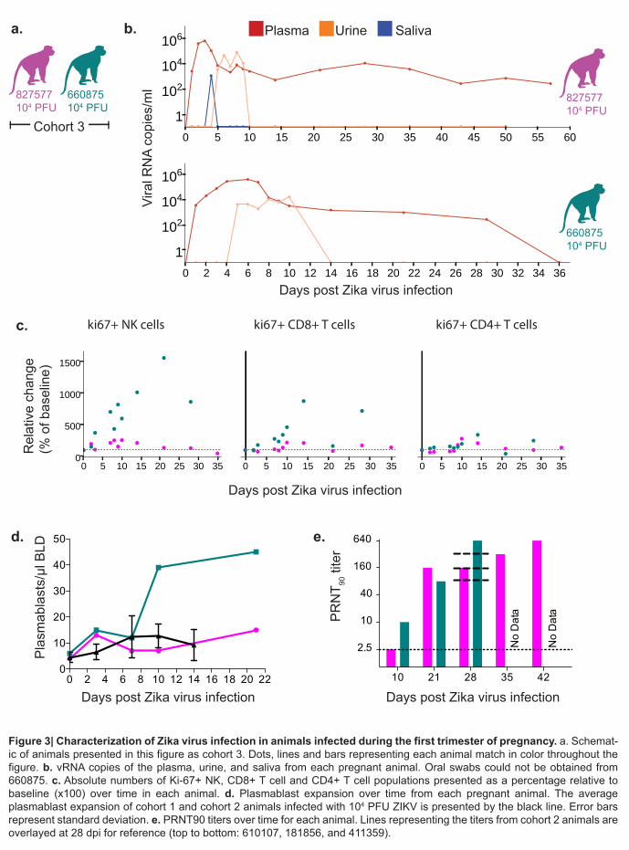
We also challenged two time-mated rhesus macaques at approximately gestation day 31 108
and 38 (mid-first trimester) with 1 x 10⁴ PFU of ZIKV (cohort 3; see Extended Data Fig. 1 and 109
Fig. 3a). Both animals were viremic by 1 dpi and exhibited peak plasma viral loads of > 4 x 10⁵ 110
vRNA copies/ml by 3 or 6 dpi (Fig. 3b). Infectious virus was also quantitated by plaque assay 111
from the serum of 660875 (Extended Data Fig. 5). In contrast to their non-pregnant 112
counterparts, both animals maintained persistent plasma viremia (vRNA copies/ml) to 57+ and 113
29 dpi (Fig. 3b). This is similar to a case described by Driggers et. al., where a pregnant mother 114
had persistent ZIKV vRNA detected from 35 to 70 dpi that did not resolve until termination of 115
pregnancy7. The fetus was found to have 2x10⁸ copies/ml of virus in brain tissue and it is 116
speculated that the fetus may have been the source of the prolonged maternal plasma viremia. 117
We will continue to monitor these pregnant animals for vRNA in the blood and amniotic fluid and 118
will determine the infection status of the fetus upon termination of the pregnancy either at full 119
term or earlier if necessary for the health and safety of the mother. Amniocentesis using 120
ultrasound guidance was performed at 43 dpi for 827577 and 36 dpi for 660875 and both were 121
negative for ZIKV RNA. 122
Both animals generated similar activation of NK, CD8+ T cell and CD4+ T cell responses 123
above baseline as non-pregnant animals (Fig. 3c). Expansion of plasmablast cells was also 124
observed by 10-21 dpi with one animal expanding more than, and one animal expanding less 125
than, the average non-pregnant animal (Fig. 3d). Neutralizing antibodies were detected by 21 126
dpi for 827577 and 10 dpi for 660875 and were similar to the cohort 2 non-pregnant animals at 127
28 dpi (Fig. 3e). One pregnant animal exhibited an increase in CK above RI and above levels 128
.CC-BY-ND 4.0 International licensecertified by peer review) is the author/funder. It is made available under aThe copyright holder for this preprint (which was notthis version posted May 10, 2016. . https://doi.org/10.1101/046334doi: bioRxiv preprint

6
expected with repeated ketamine sedation and blood collection. The same pregnant animal also 129
developed persistent regenerative anemia characterized by circulating nucleated erythrocytes. 130
Updates to the cohort 3 experiments are available in real-time at goo.gl/rmNCqf. 131
Altogether, our study shows the persistence of ZIKV RNA in the plasma of rhesus 132
macaques for approximately 10 days, similar to other vector-borne flaviviruses that cause acute, 133
typically self-limiting infections in humans. This work also shows that natural ZIKV infection 134
elicits a robust immune response including ZIKV-specific T cell response and neutralizing 135
antibody responses that confers protection against reinfection. However, the prolonged 136
detection of vRNA in urine and saliva after apparent clearance from the blood, detection of virus 137
in the CSF, and occasional plasma “blips” after initial clearance, suggest that ZIKV may persist 138
longer, at low levels, in certain tissues. Future work in rhesus macaques will seek to determine if 139
and where these reservoirs may exist and whether they seed virus into fluids that might allow for 140
human-to-human transmission. 141
Our study establishes immunocompetent rhesus macaques infected with physiologically 142
relevant ZIKV as a relevant translational model for infection and pathogenesis. The large 143
immunological toolset available for rhesus macaques will enable investigations of immunity and 144
potential vaccines. Pregnancy, the maternal-fetal interface, and fetal development have been 145
described in detail in rhesus macaques, so this model will also enable assessments of the 146
impact of maternal ZIKV infection on the developing fetus. We have established persistent 147
viremia in pregnant macaques despite activation of NK cells and T cells as well as development 148
of a neutralizing antibody response. We continue to follow these pregnant animals and will 149
establish whether fetal infection and/or abnormalities have occurred through serial ultrasound 150
assessments of the fetus and placenta as well as tissue analysis at pregnancy termination. 151
152
Methods 153
Study design 154
.CC-BY-ND 4.0 International licensecertified by peer review) is the author/funder. It is made available under aThe copyright holder for this preprint (which was notthis version posted May 10, 2016. . https://doi.org/10.1101/046334doi: bioRxiv preprint
7
This was a proof-of-concept study designed to establish the infectivity and viral dynamics of 155
Asian lineage ZIKV. Because nothing is known about ZIKV dosing in macaques, one male and 156
one female rhesus macaque of Indian ancestry were each challenged with the following ZIKV 157
doses: 1x10⁶, 1x10⁵, and 1x10⁴ PFU ZIKV. Two pregnant macaques at 31 and 38 days of 158
gestation were infected with 1x10⁴ PFU ZIKV. We selected 2 animals per inoculum dose and 2 159
pregnant animals as a minimum number of animals for this pilot study to provide proof-of-160
concept and design larger studies necessary to place statistical significance on the findings. All 161
macaques utilized in the study were free of Macacine herpesvirus 1, Simian Retrovirus Type D, 162
Simian T-lymphotropic virus Type 1, and Simian Immunodeficiency Virus. Primary data from the 163
study is available at goo.gl/rmNCqf. 164
165
Care and use of macaques at the Wisconsin National Primate Research Center 166
All macaque monkeys used in this study were cared for by the staff at the Wisconsin National 167
Primate Research Center (WNPRC) in accordance with the regulations and guidelines outlined 168
in the Animal Welfare Act and the Guide for the Care and Use of Laboratory Animals and the 169
recommendations of the Weatherall report. This study was approved by the University of 170
Wisconsin-Madison Graduate School Institutional Animal Care and Use Committee (Animal 171
Care and Use Protocol Number G005401). For all procedures (i.e., physical examinations, virus 172
inoculations, ultrasound examinations, blood and swab collection), animals were anesthetized 173
with an intramuscular dose of ketamine (10 mL/kg). Blood samples were obtained using a 174
vacutainer system or needle and syringe from the femoral or saphenous vein. 175
176
Inoculations 177
ZIKV strain H/PF/2013 (GenBank: KJ776791), originally isolated from a 51-year-old female in 178
France returning from French Polynesia with a single round of amplification on Vero cells, was 179
.CC-BY-ND 4.0 International licensecertified by peer review) is the author/funder. It is made available under aThe copyright holder for this preprint (which was notthis version posted May 10, 2016. . https://doi.org/10.1101/046334doi: bioRxiv preprint

8
obtained from Xavier de Lamballerie (European Virus Archive, Marseille France). We deep 180
sequenced the challenge stock to verify the expected origin. The ZIKV challenge stock 181
consensus sequence matched the Genbank sequence (KJ776791) of the parental virus, but 182
there were 8 sites where between 5-40% of sequences contained variants that appear to be 183
authentic (6/8 were non-synonymous changes) (Extended Data Fig. 6). Sequences have been 184
deposited in the Sequence Read Archive (SRA) under accession number SRP072852 . 185
186 Virus stocks were prepared by inoculation onto a confluent monolayer of C6/36 mosquito 187
cells. These cell lines were obtained from ATCC, were not further authenticated and were not 188
specifically tested for mycoplasma. A single harvest of virus with a titer of 1.26 x 10⁶ PFU/mL 189
(equivalent to 1.43 x 10⁹ vRNA copies/mL) was used for all 8 challenges. The stock was 190
thawed, diluted in PBS to the appropriate concentration for each challenge, and loaded into a 1 191
mL syringe that was kept on ice until challenge. Animals were anesthetized as described above, 192
and 1 mL of inocula was administered subcutaneously over the cranial dorsum. Post-193
inoculation, animals were closely monitored by veterinary and animal care staff for adverse 194
reactions and signs of disease. 195
196
Viral RNA isolation from plasma 197
Fresh plasma and PBMC were isolated from EDTA-treated whole blood by Ficoll density 198
centrifugation at 1860 rcf for 30min. The plasma layer was collected and centrifuged for an 199
additional 8 min at 670 rcf to remove residual cells. RNA was extracted from 300 µl of plasma 200
using the Viral Total Nucleic Acid Purification Kit (Promega, Madison, WI) on a Maxwell 16 MDx 201
instrument. The RNA was then quantified by quantitative RT-PCR. 202
203
Viral RNA isolation from urine 204
.CC-BY-ND 4.0 International licensecertified by peer review) is the author/funder. It is made available under aThe copyright holder for this preprint (which was notthis version posted May 10, 2016. . https://doi.org/10.1101/046334doi: bioRxiv preprint

9
Urine was collected from a pan beneath the animal’s cage. Urine was centrifuged for 5 min at 205
500 rcf to remove cells and other debris. RNA was isolated from 300 µl urine using the Viral 206
Total Nucleic Acid Purification Kit (Promega, Madison, WI) on a Maxwell 16 MDx instrument. 207
208
Viral RNA isolation from oral swabs 209
Oral swab samples were collected from infected animals while anesthetized by gently running a 210
sterile swab under the animal’s tongue. Swabs were placed immediately into either RNAlater or 211
viral transport medium (tissue culture medium 199 supplemented with 0.5% FBS and 1% 212
antibiotic/antimycotic) for 60-90 minutes. Samples were vortexed vigorously, then centrifuged 213
for 5 min at 500 rcf before removing the swabs. Samples were stored at either -20°C (RNAlater 214
samples) or -80°C (viral transport medium) until processing. Prior to extraction, virus was 215
pelleted by centrifugation for 1 hour at 4°C at 14000 rpm. Supernatant was removed, leaving the 216
virus in 200 µl media. Viral RNA was extracted from these samples using the Qiamp MinElute 217
Virus Spin kit (Qiagen, Germantown, Maryland) with all optional washes. Viral load data from 218
oral swabs are expressed as vRNA copies/mL eluate. 219
220
Quantitative reverse transcription PCR (qRT-PCR) 221
Viral RNA isolated from plasma, urine, or oral swabs was quantified by qRT-PCR using the 222
primers and probe designed by Lanciotti et al. 12.The RT-PCR was performed using the 223
SuperScript III Platinum one-step quantitative RT-PCR system (Invitrogen, Carlsbad, CA) on the 224
LightCycler 480 instrument (Roche Diagnostics, Indianapolis, IN). Primers and probe were used 225
at final concentrations of 600 nm and 100 nm respectively, along with 150 ng random primers 226
(Promega, Madison, WI). Cycling conditions were as follows: 37°C for 15 min, 50°C for 30 min 227
and 95°C for 2 min, followed by 50 cycles of 95°C for 15 sec and 60°C for 1 min. Virus 228
concentration was determined by interpolation onto an internal standard curve composed of 229
.CC-BY-ND 4.0 International licensecertified by peer review) is the author/funder. It is made available under aThe copyright holder for this preprint (which was notthis version posted May 10, 2016. . https://doi.org/10.1101/046334doi: bioRxiv preprint

10
seven 10-fold serial dilutions of a synthetic ZIKV RNA fragment based on the Asian-lineage 230
(ZIKV strain H/PF/2013). 231
232
Viral quantification by plaque assay 233
Titrations for replication competent virus quantification of the challenge stock as well as from 234
serum collected at multiple time points from cohort 2 animals were completed by plaque assay 235
on Vero cell cultures. Vero cells were obtained from ATCC, were not further authenticated and 236
were not specifically tested for mycoplasma. Duplicate wells were infected with 0.1 mL aliquots 237
from serial 10-fold dilutions in growth media and virus was adsorbed for one hour. Following 238
incubation, the inoculum was removed, and monolayers were overlaid with 3 ml containing a 1:1 239
mixture of 1.2% oxoid agar and 2X DMEM (Gibco, Carlsbad, CA) with 10% (vol/vol) FBS and 240
2% (vol/vol) penicillin/streptomycin. Cells were incubated at 37°C in 5% CO2 for four days for 241
plaque development. Cell monolayers then were stained with 3 mL of overlay containing a 1:1 242
mixture of 1.2% oxoid agar and 2X DMEM with 2% (vol/vol) FBS, 2% (vol/vol) 243
penicillin/streptomycin, and 0.33% neutral red (Gibco). Cells were incubated overnight at 37°C 244
and plaques were counted. Titers of virus detected from the serum of cohort 2 animals were 245
compared to plasma and serum viral load assays. For both the challenge stock and the virus 246
isolated from macaque serum, the level of infectious virus detected by plaque assay was ~500-247
1000-fold less than the number of viral RNA particles detected by qRT-PCR in either the plasma 248
or serum. This was true throughout the duration of viremia where plaque assay titers were 249
detectable. 250
251
Plaque reduction neutralization test (PRNT90) 252
Macaque serum samples were screened for ZIKV neutralizing antibody utilizing a plaque 253
reduction neutralization test (PRNT). Endpoint titrations of reactive sera, utilizing a 90% cutoff 254
(PRNT90) were performed as described15 against ZIKV strain H/PF/2013. 255
.CC-BY-ND 4.0 International licensecertified by peer review) is the author/funder. It is made available under aThe copyright holder for this preprint (which was notthis version posted May 10, 2016. . https://doi.org/10.1101/046334doi: bioRxiv preprint

11
256
Immunophenotyping 257
The amount of activated/proliferating NK cells were quantified using a modified version of our 258
protocol detailed step-by step in OMIP-2816. Briefly 0.1 mL of EDTA-anticoagulated whole blood 259
samples were incubated for 15 min at room temperature in the presence of a mastermix of 260
antibodies against CD45 (clone D058-1283, Brilliant Violet 786 conjugate), CD3 (clone SP34-2 261
Alexa Fluor 700 conjugate), CD8 (clone SK2, Brilliant Violet 510), NKG2A/C (clone Z199, PE-262
Cy7 conjugate), CD16 (clone 3G8, Pacific Blue conjugate), CD69 (clone TP1.55.3, ECD 263
conjugate), HLA-DR (clone 1D11, Brilliant Violet 650 conjugate), CD4 (clone SK3, Brilliant Violet 264
711 conjugate), CCR7 (clone 150503, Fluorescein conjugate), CD28 (clone CD28.2, PE 265
conjugate), and CD95 (clone DX2, PE-Cy5 conjugate) antigens. All antibodies were obtained 266
from BD BioSciences, except the NKG2A/C-specific antibody, which was purchased from 267
Beckman Coulter, and the CCR7 antibody that was purchased from R&D Systems. Red blood 268
cells were lysed using BD Pharm Lyse, after which they were washed twice in media and fixed 269
with 0.125 ml of 2% paraformaldehyde for 15 min. After an additional wash the cells were 270
permeabilized using Life Technology’s Bulk Permeabilization Reagent. The cells were stained 271
for 15 min. with Ki-67 (clone B56, Alexa Fluor 647 conjugate) while the permeabilizer was 272
present. The cells were then washed twice in media and resuspended in 0.125 ml of 2% 273
paraformaldehyde until they were run on a BD LSRII Flow Cytometer. Flow data were analyzed 274
using Flowjo version 9.8.2. 275
276
Interferon-gamma ELISPOT assay 277
Peripheral blood mononuclear cells (PBMCs) were isolated from EDTA-treated whole blood by 278
using Ficoll-Paque Plus (GE Health Sciences) density centrifugation. Enzyme-linked 279
immunosorbent spot (ELISPOT) assays were conducted according to the manufacturer’s 280
.CC-BY-ND 4.0 International licensecertified by peer review) is the author/funder. It is made available under aThe copyright holder for this preprint (which was notthis version posted May 10, 2016. . https://doi.org/10.1101/046334doi: bioRxiv preprint
12
protocol. Briefly, 1 x 10⁵ cells in 100ul of R10 medium were added to pre-coated monkey 281
gamma interferon (IFNγ) ELISpot-PLUS plates (Mabtech Inc., Mariemont, OH) with peptide at a 282
final concentration of 1uM. Full proteome peptides derived from the ZIKV NS5 sequence 283
(GenBank: KU321639.1) used in this study were synthesized by GenScript (Piscataway, NJ). 284
Pools were created using 10 overlapping 15mer peptides, each at a working concentration of 285
1mM. Concanavalin A (10uM) was used as a positive control. Assays of all samples were 286
repeated in duplicate or triplicate. Cells alone in the absence of stimulant were used as a 287
negative control. Wells were imaged by using an AID ELISPOT reader, and spots were counted 288
by using an automated program with parameters including size, intensity, and gradient. The limit 289
of detection was set at 100 spot-forming cells per million PBMCs. 290
Plasmablast detection 291
Peripheral blood mononuclear cells (PBMCs) isolated from three ZIKV-infected rhesus monkeys 292
at 3, 7, 11, and 14 dpi were stained with the following panel of fluorescently labeled antibodies 293
(Abs) specific for the following surface markers: CD20 FITC (L27), CD80 PE(L307.4), CD123 294
PE-Cy7(7G3), CD3 APC-Cy7 (SP34-2), IgG BV605(G18-145) (all from BD Biosciences, San 295
Jose, CA), CD14 AF700 (M5E2), CD11c BV421 (3.9), CD16 BV570 (3G8), CD27 BV650(O323) 296
(all from BioLegend, San Diego, CA), IgD AF647 (polyclonal)(Southern Biotech, Birmingham, 297
AL), and HLA-DR PE-TxRed (TÜ36) (Invitrogen, Carlsbad, CA). LIVE/DEAD Fixable Aqua Dead 298
Cell Stain Kit (Invitrogen, Carlsbad, CA) was used to discriminate live cells. Briefly, cells were 299
resuspended in 1X PBS/1%BSA and stained with the full panel of surface Abs for 30 min in the 300
dark at 4°C, washed once with 1X PBS, stained for 30 min with LIVE/DEAD Fixable Aqua Dead 301
Cell Stain Kit in the dark at 4°C, washed once with 1X PBS, washed again with 1X 302
PBS/1%BSA, and resuspended in 2% PFA Solution. Stained PBMCs were acquired on a LSRII 303
Flow Analyzer (BD Biosciences, San Jose, CA) and the data was analyzed using FlowJo 304
software v9.7.6 (TreeStar, Ashland, OR). Plasmablasts were defined similarly to the method 305
.CC-BY-ND 4.0 International licensecertified by peer review) is the author/funder. It is made available under aThe copyright holder for this preprint (which was notthis version posted May 10, 2016. . https://doi.org/10.1101/046334doi: bioRxiv preprint

13
previously described14 excluding lineage cells (CD14+, CD16+, CD3+, CD20+, CD11c+, 306
CD123+), and selecting CD80+ and HLA-DR+ cells (known to be expressed on rhesus 307
plasmablasts and their human counterpart17). 308
309
Estimation of plasma viremia doubling time 310
The doubling time of plasma viremia was estimated in R version 3.2.3 (The R Foundation for 311
Statistical Computing, http://www.R-project.org). For each animal, the slope of the linear portion 312
of the line (between 1 and 2 dpi for the animals treated with 1x10⁶ and 1x10⁵ PFU and between 313
1, 2, and 3 dpi for the animal treated with 1x10⁴ PFU) was generated by plotting the log of the 314
plasma viral loads. The linear portion represents the exponential growth phase and has been 315
used to estimate doubling time in other systems18. The slopes were then used in the equation: 316
log(2)/slope. Each result was then multiplied by 24 hours to produce a simple estimate of 317
doubling time in hours. 318
319
CBC and blood chemistry panels 320
CBCs were performed on EDTA-anticoagulated whole blood samples on a Sysmex XS-1000i 321
automated hematology analyzer (Sysmex Corporation, Kobe, Japan). Blood smears were 322
prepared and stained with Wright-Giemsa stain (Wescor Aerospray Hematology Slide Stainer; 323
Wescor Inc, Logan, UT). Manual slide evaluations were performed on samples as appropriate 324
when laboratory-defined criteria were met (including the presence of increased total white blood 325
cell counts, increased monocyte, eosinophil, and basophil percentages, decreased hemoglobin, 326
hematocrit, and platelet values, and unreported automated differential values). Individuals 327
performing manual slide evaluations screened both white blood cells (WBC) and red blood cells 328
(RBC) for cellular maturity, toxic change, and morphologic abnormalities. 329
.CC-BY-ND 4.0 International licensecertified by peer review) is the author/funder. It is made available under aThe copyright holder for this preprint (which was notthis version posted May 10, 2016. . https://doi.org/10.1101/046334doi: bioRxiv preprint

14
Whole blood was collected into serum separator tubes (Becton, Dickinson and 330
Company, Franklin Lakes, NJ) for blood chemistry analysis and processed per manufacturer’s 331
instructions. Blood chemistry panels were performed on the serum using a Cobas 6000 332
analyzer (Roche Diagnostics, Risch-Rotkreuz, Switzerland). Results from CBC and blood 333
chemistry panels were reported with species, age, and sex-specific reference ranges. 334
335
Zika virus deep sequencing of the challenge stock 336
A vial of the same ZIKV strain H/PF/2013 virus stock that infected macaques was deep 337
sequenced by preparing libraries of fragmented double-stranded cDNA using methods similar to 338
those previously described19. Briefly, the sample was centrifuged at 5000 rcf for 5 min. The 339
supernatant was then filtered through a 0.45-µm filter. The Qiagen QiAmp Minelute viral RNA 340
isolation kit (omitting carrier RNA) was used to isolate vRNA. The eluted RNA was then treated 341
with DNAse I. Double stranded DNA was prepared with the Superscript double stranded cDNA 342
synthesis kit (Invitrogen) and priming with random hexamers. Agencourt Ampure XP beads 343
were used to purify double stranded DNA. The purified DNA was fragmented with the Nextera 344
XT kit (Illumina), tagged with Illumina-compatible primers, and then purified with Agencourt 345
Ampure XP beads. Purified libraries were then sequenced with 2 x 300 bp kits on an Illumina 346
MiSeq. Of note, challenge stock viral loads were 1.43x10⁹ vRNA copies/ml. This results in an 347
input of 7.15 x10⁸ RNA copies into the sequencing reactions. This far exceeds the average 348
depth of coverage of 11,877 (+/- 4658) sequences per nucleotide site indicating little resampling 349
effects in our data analysis. 350
351
References 352
1. Brasil, P. et al. Zika Virus Infection in Pregnant Women in Rio de Janeiro - Preliminary 353
Report. N Engl J Med (2016). 354
.CC-BY-ND 4.0 International licensecertified by peer review) is the author/funder. It is made available under aThe copyright holder for this preprint (which was notthis version posted May 10, 2016. . https://doi.org/10.1101/046334doi: bioRxiv preprint

15
2. Cao-Lormeau, V. M. et al. Guillain-Barré Syndrome outbreak associated with Zika virus 355
infection in French Polynesia: a case-control study. Lancet (2016). 356
3. Calvet, G. et al. Detection and sequencing of Zika virus from amniotic fluid of fetuses with 357
microcephaly in Brazil: a case study. Lancet Infect Dis (2016). 358
4. Carteaux, G. et al. Zika Virus Associated with Meningoencephalitis. N Engl J Med (2016). 359
5. Musso, D. & Gubler, D. J. Zika Virus. Clin Microbiol Rev 29, 487-524 (2016). 360
6. Sarno, M. et al. Zika Virus Infection and Stillbirths: A Case of Hydrops Fetalis, 361
Hydranencephaly and Fetal Demise. PLoS Negl Trop Dis 10, e0004517 (2016). 362
7. Driggers, R. W. et al. Zika Virus Infection with Prolonged Maternal Viremia and Fetal Brain 363
Abnormalities. N Engl J Med (2016). 364
8. Rossi, S. L. et al. Characterization of a Novel Murine Model to Study Zika Virus. Am J Trop 365
Med Hyg (2016). 366
9. Styer, L. M. et al. Mosquitoes inoculate high doses of West Nile virus as they probe and 367
feed on live hosts. PLoS Pathog 3, 1262-1270 (2007). 368
10. Cox, J., Mota, J., Sukupolvi-Petty, S., Diamond, M. S. & Rico-Hesse, R. Mosquito bite 369
delivery of dengue virus enhances immunogenicity and pathogenesis in humanized mice. 370
J Virol 86, 7637-7649 (2012). 371
11. Dutra, H. L. et al. Wolbachia Blocks Currently Circulating Zika Virus Isolates in Brazilian 372
Aedes aegypti Mosquitoes. Cell Host Microbe (2016). 373
12. Lanciotti, R. S. et al. Genetic and serologic properties of Zika virus associated with an 374
epidemic, Yap State, Micronesia, 2007. Emerg Infect Dis 14, 1232-1239 (2008). 375
13. Aliota MT, P. S. A., Dario Velez I, and Osorio JE. The wMel strain of Wolbachia reduces 376
transmission of Zika virus by Aedes aegypti. Scientific reports, in review (2016). 377
14. Silveira, E. L. et al. Vaccine-induced plasmablast responses in rhesus macaques: 378
phenotypic characterization and a source for generating antigen-specific monoclonal 379
antibodies. J Immunol Methods 416, 69-83 (2015). 380
.CC-BY-ND 4.0 International licensecertified by peer review) is the author/funder. It is made available under aThe copyright holder for this preprint (which was notthis version posted May 10, 2016. . https://doi.org/10.1101/046334doi: bioRxiv preprint

16
15. Lindsey, H. S., Calisher, C. H. & Mathews, J. H. Serum dilution neutralization test for 381
California group virus identification and serology. J Clin Microbiol 4, 503-510 (1976). 382
16. Pomplun, N., Weisgrau, K. L., Evans, D. T. & Rakasz, E. G. OMIP-028: activation panel 383
for Rhesus macaque NK cell subsets. Cytometry A 87, 890-893 (2015). 384
17. Wrammert, J. et al. Rapid cloning of high-affinity human monoclonal antibodies against 385
influenza virus. Nature 453, 667-671 (2008). 386
18. Staprans, S. I. et al. Simian immunodeficiency virus disease course is predicted by the 387
extent of virus replication during primary infection. J Virol 73, 4829-4839 (1999). 388
19. Lauck, M. et al. Discovery and full genome characterization of two highly divergent simian 389
immunodeficiency viruses infecting black-and-white colobus monkeys (Colobus guereza) 390
in Kibale National Park, Uganda. Retrovirology 10, 107 (2013). 391
392
Supplementary Information is linked to the online version of the paper at 393
www.nature.com/nature. 394
395
Acknowledgements We thank the Veterinary, Animal Care, Scientific Protocol Implementation, 396
and the Pathology staff at the Wisconsin National Primate Research Center (WNPRC) for their 397
contribution to this study. We thank the DHHS/PHS/NIH (R01Al116382-01A1 to D.H.O.), 398
(R01Al107157-01A1 to T.G.G.) and (DP2HD075699 to S.R.P.) for funding. We also thank the 399
P51OD011106 awarded to the WNPRC, Madison-Wisconsin. This research was conducted in 400
part at a facility constructed with support from Research Facilities Improvement Program grants 401
RR15459-01 and RR020141-01. The publication’s contents are solely the responsibility of the 402
authors and do not necessarily represent the official views of NCRR or NIH. 403
404
Author contributions D.H.O., T.C.F., J.E.O., M.T.A., E.M., T.G.G. and D.M.D. designed the 405
experiments. D.H.O., D.M.D., M.T.A., E. M., T.C.F., and L.H.M. drafted the manuscript. M.T.A., 406
.CC-BY-ND 4.0 International licensecertified by peer review) is the author/funder. It is made available under aThe copyright holder for this preprint (which was notthis version posted May 10, 2016. . https://doi.org/10.1101/046334doi: bioRxiv preprint

17
and J.E.O. provided and prepared viral stocks and performed plaque assays. A.M.W., G.L-B., 407
and T.C.F. developed and performed viral load assays. K.L.W. and E.G.R. performed 408
immunophenotyping assays. M.S.M., M.E.B., M.N.R., C.M.N., and D.M.D. coordinated and 409
processed macaque samples for distribution. D.D.G., S.L.O., and D.M.D. designed and 410
performed the sequencing experiments. L.H.M. and T.C.F. performed nucleotide diversity 411
calculations. J.P., N.S-D., H.A.S., S.C., and J.M.H. coordinated the macaque infections, 412
sampling, and performed blood chemistries and CBC analysis. M.L.S. coordinated experiments 413
and helped perform ultrasounds on the pregnant macaques. J.A.E., M.A.M., and S.R.P. 414
performed the plasmablast experiments. 415
416
Author information: Sequences have been deposited in the Sequence Read Archive (SRA) 417
under accession number SRP072852. All data from these studies are available at 418
zika.labkey.com. Reprints and permissions information is available at www.nature.com/reprints. 419
The authors declare no competing interests. Correspondence and requests for materials should 420
be addressed to [email protected]. 421
422
Figure Legends 423
Figure 1. Animal cohort definitions and ZIKV viral load from rhesus macaque fluids. a. 424
Animals included in this study and the ZIKV doses used to infect them. Solid lines and bars 425
throughout the figure represent cohort 1 animals by color while stripped bars and dotted lines 426
represent cohort 2 animals by color. b. Viral RNA loads measured in plasma, urine, and saliva 427
for the two animals challenged with each dose of virus through 28 dpi. Cohort 1 animals are 428
represented by a solid line while cohort 2 animals are represented by a dotted line for each fluid. 429
Inset: vRNA loads from cohort 1 animals measured before and after rechallenge with homotypic 430
Zika virus as indicated by an arrow. c. Number of plaque forming units per ml of serum for 431
.CC-BY-ND 4.0 International licensecertified by peer review) is the author/funder. It is made available under aThe copyright holder for this preprint (which was notthis version posted May 10, 2016. . https://doi.org/10.1101/046334doi: bioRxiv preprint

18
cohort 2 animals. d. Viral RNA load per ml of CSF collected on 4 and 14 dpi. e. Viral RNA load 432
per vaginal swab collected on 0, 7, 14, 21 and 28 dpi. NA: sample not available. 433
434
Figure 2. Immune cell expansion and neutralizing antibody titers following ZIKV infection. 435
a. Solid dots, lines and bars with corresponding color represent cohort 1 animals and open 436
circles, dotted lines or stripped bars represent cohort 2 animals throughout the figure. b. 437
Expansion of Ki-67+ (activated) NK cells, CD8+ T cells and CD4+ T cells were measured daily 438
for 10 days and then on days 14, 21 and 28 post-infection. Absolute numbers of activated 439
cells/µl of blood are presented relative to the baseline value set to 100%. c. Total number of 440
plasmablast cells found in PBMCs collected at 0 (cohort 2 only), 3, 7, 11 and 14 dpi for each 441
animal. d. PRNT90 titers for cohort 1 and cohort 2. 442
443
Figure 3. Characterization of Zika virus infection in animals infected during the first 444
trimester of pregnancy. a. Schematic of animals presented in this figure as cohort 3. Dots, 445
lines and bars representing each animal match in color throughout the figure. b. vRNA copies of 446
the plasma, urine, and saliva from each pregnant animal. Oral swabs could not be obtained 447
from 660875. c. Absolute numbers of Ki-67+ NK, CD8+ T cell and CD4+ T cell populations 448
presented as a percentage relative to baseline (x100) over time in each animal. d. Plasmablast 449
expansion over time from each pregnant animal. The average plasmablast expansion of cohort 450
1 and cohort 2 animals infected with the 104 PFU is presented by the black line. Error bars 451
represent standard deviation. e. PRNT90 titers over time for each animal. Lines representing the 452
titers from cohort 2 animals are overlayed at 28 dpi for reference (top to bottom: 610107, 453
181856, and 411359). 454
455
Extended Data Figure 1. Schematic representation of the timeline of infection and 456
sampling for each animal in the presented studies. Cohort 1 received the first ZIKV 457
.CC-BY-ND 4.0 International licensecertified by peer review) is the author/funder. It is made available under aThe copyright holder for this preprint (which was notthis version posted May 10, 2016. . https://doi.org/10.1101/046334doi: bioRxiv preprint

19
challenges and were then rested for 6 weeks before a rechallenge. For all studies, samples 458
were collected daily for 10 days and then on 14, 21, and 28 dpi as indicated by hashes in the 459
timelines. Cohort 3 represents the two pregnant animals that were challenged on 2 different 460
days. Both animals are currently in the once weekly sampling phase until the pregnancies come 461
to term (~165 gestational days). Cohort 2 was a repeat experiment of cohort 1 that allowed for 462
additional experiments and sample collection (e.g., serum plaque infectivity) that were not 463
feasible when we initiated cohort 1 studies. These animals are currently in a 6-week rest period 464
and will be rechallenged on June 6, 2016. Ages of all animals are indicated under each 465
macaque identification number. 466
467
Extended Data Figure 2. Complete blood counts and serum chemistries for macaques 468
infected with ZIKV. a. Animals were infected with different doses of ZIKV. Cohort 1 animals 469
are represented by solid lines and cohort 2 animals are represented by dotted lines. All non-470
pregnant animals had serum chemistry analysis performed at -7, 0, 1, 2, 3, 4, 6, and 14 dpi or at 471
-6, 2, 5 and 11 dpi. b. AST blood chemistries c. ALT serum chemistries. d. CK serum 472
chemistries. Complete blood counts were measured prior to infection, daily for 10-11 days after 473
infection and then every 3-7 days until 28 dpi. e. white blood cell counts. f. % lymphocytes. g. 474
red blood cell counts. 475
476 Extended Data Figure 3. qRT-PCR detection of ZIKV RNA is equally sensitive from serum 477
and plasma. vRNA copies/ml of plasma or serum were quantitated by qRT-PCR over multiple 478
time points from cohort 2 animals. Sufficient baseline samples were not available from serum. 479
480
Extended Data Figure 4. Antigen-specific T cell responses by IFNγ-ELISPOT. a. Average 481
spot forming cell counts for PBMC collected from each animal at 4, 10 and 14 dpi. Data were 482
baseline corrected by subtracting the average negative control values from each response. A 483
.CC-BY-ND 4.0 International licensecertified by peer review) is the author/funder. It is made available under aThe copyright holder for this preprint (which was notthis version posted May 10, 2016. . https://doi.org/10.1101/046334doi: bioRxiv preprint

20
threshold of 10.0 SFC/100,000 cells was set as the minimum value to be considered a positive 484
T cell response, as indicated by the dashed line. b. Each pool was comprised of 10 overlapping 485
15mer peptides offset by 4 amino acids. c. Peptide pools eliciting T cell responses at 4, 10 and 486
14 dpi for each animal. The region of the NS5 protein that is represented by each pool of 487
overlapping 15mers is provided. MHC class I haplotypes of each cohort 2 animal are also 488
presented. All three animals shared the A004 and B012b major histocompatibility complex 489
haplotypes and two animals shared the A023 haplotype. Therefore, it was not surprising that 3 490
pools were recognized by 2 different animals likely sharing the MHC class I allele that is 491
presenting one of the peptides in those pools. Grayed pools were positive in more than one 492
animal and bolded pools were positive at more than one time point in the same animal. 493
494
Extended Data Figure 5. Plaque assay titers in a pregnant animal. Log10 PFU/ml serum 495
(dotted line) is plotted relative to vRNA copies/ml plasma (solid line) for 660875. 496
497
Extended Data Figure 6. Genetic diversity of the ZIKV challenge stock. The ZIKV challenge 498
stock was deep sequenced from all three animals. Nucleotide sites where at least 5% of 499
sequences obtained from the challenge stock are different from the Genbank sequence are 500
shown. 501
502
.CC-BY-ND 4.0 International licensecertified by peer review) is the author/funder. It is made available under aThe copyright holder for this preprint (which was notthis version posted May 10, 2016. . https://doi.org/10.1101/046334doi: bioRxiv preprint

a.
b.
c. d. e.
Days post Zika virus infection
826226106 PFU
393422105 PFU
912116104 PFU
610107106 PFU
181856105 PFU
411359104 PFU
Cohort 1 Cohort 2
181856105 PFU
610107106 PFU
411359104 PFU
826226106 PFU
393422105 PFU
912116104 PFU
vRN
A co
pies
/ml
Days post Zika virus infection
rechallenge
rechallenge
rechallenge
Plasma Urine Saliva
Days post Zika virus infection Days post Zika virus infection0 2 4 6 8
100
101
102
103
PFU
/ml s
erum
0
4 14100
101
102
103
104
vRN
A co
pies
/ml C
SF
NA
0
0 1 7 14 21 28100
101
102
103
104
vRN
A co
pies
/sw
ab
NA
0
Figure 1| Animal cohort definitions and ZIKV viral load from rhesus macaque fluids. a. Animals included in this study and the ZIKV doses used to infect them. Solid lines and bars throughout the figure represent cohort 1 animals by color while stripped bars and dotted lines represent cohort 2 animals by color. b. Viral RNA loads measured in plasma, urine, and saliva for the two animals challenged with each dose of virus through 28 dpi. Cohort 1 animals are represented by a solid line while cohort 2 animals are represented by a dotted line for each fluid Inset: vRNA loads from cohort 1 animalsmeasured before and after rechallenge with homotypic Zika virus as indicated by an arrow. c. Number of plaque forming units per ml of serum for cohort 2 animals. d. Viral RNA load per ml of CSF collected on 4 and 14 dpi. e. Viral RNA load per vaginal swab collected on 0, 7, 14, 21 and 28 dpi. NA: sample not available.
.CC-BY-ND 4.0 International licensecertified by peer review) is the author/funder. It is made available under aThe copyright holder for this preprint (which was notthis version posted May 10, 2016. . https://doi.org/10.1101/046334doi: bioRxiv preprint

Figure 2| Immune cell expansion and neutralizing antibody titers following ZIKV infection. a. Solid dots, lines and bars with corresponding color represent cohort 1 animals and open circles, dotted lines or stripped bars represent cohort 2 animals throughout the figure. b. Expansion of Ki-67+ (activated) NK cells, CD8+ T cells and CD4+ T cells were measured daily for 10 days and then on days 14, 21 and 28 post-infection. Absolute numbers of activated cells/μl of blood are presented relative to the baseline value set to 100%. c. Total number of plasmablast cells found in PBMCs collected at 0 (cohort 2 only), 3, 7, 11 and 14 dpi for each animal. d. PRNT90 titers for cohort 1 and cohort 2 .
a.
c. d.
14 28 64
2.5
10
40
160
640
Days post Zika virus infection
826226106 PFU
393422105 PFU
912116104 PFU
610107106 PFU
181856105 PFU
411359104 PFU
Rel
ativ
e ch
ange
(% o
f bas
elin
e)
Days post Zika virus infection
PR
NT 90
tite
r
b. Ki-67+ NK cells
Ki-67+ CD4+ T cells
0 5 10 150
10
20
30
40
Days post Zika virus infection
Pla
smab
last
s/µl
BLD
Cohort 1 Cohort 2
Ki-67+ CD8+ T cells
Ki-67+ NK T cells
.CC-BY-ND 4.0 International licensecertified by peer review) is the author/funder. It is made available under aThe copyright holder for this preprint (which was notthis version posted May 10, 2016. . https://doi.org/10.1101/046334doi: bioRxiv preprint
a.
660875104 PFU
Vira
l RN
A co
pies
/ml
b.
827577104 PFU
Plasma Urine Saliva
Days post Zika virus infection
c.
d.
827577104 PFU
660875104 PFU
Rel
ativ
e ch
ange
(%
of b
asel
ine)
Days post Zika virus infection
ki67+ NK cells ki67+ CD8+ T cells ki67+ CD4+ T cells
0 2 4 6 8 10 12 14 16 18 20 220
10
20
30
40
50Average
10 21 28 35 42
2.5
10
40
160
640N
o D
ata
No
Dat
a
Days post Zika virus infection Days post Zika virus infection
PR
NT 90
tite
r
Pla
smab
last
s/µl
BLD
e.
Figure 3| Characterization of Zika virus infection in animals infected during the first trimester of pregnancy. a. Schemat-ic of animals presented in this figure as cohort 3. Dots, lines and bars representing each animal match in color throughout the figure. b. vRNA copies of the plasma, urine, and saliva from each pregnant animal. Oral swabs could not be obtained from 660875. c. Absolute numbers of Ki-67+ NK, CD8+ T cell and CD4+ T cell populations presented as a percentage relative to baseline (x100) over time in each animal. d. Plasmablast expansion over time from each pregnant animal. The average plasmablast expansion of cohort 1 and cohort 2 animals infected with 104 PFU ZIKV is presented by the black line. Error bars represent standard deviation. e. PRNT90 titers over time for each animal. Lines representing the titers from cohort 2 animals are overlayed at 28 dpi for reference (top to bottom: 610107, 181856, and 411359).
Cohort 3
.CC-BY-ND 4.0 International licensecertified by peer review) is the author/funder. It is made available under aThe copyright holder for this preprint (which was notthis version posted May 10, 2016. . https://doi.org/10.1101/046334doi: bioRxiv preprint
Cohort 1
Cohort 3
Mar 28, 2016 April 25,2016
Mar 7, 2016 April 4, 2016
June 6, 2016Cohort 2
Cohort 3
826226106 PFU
393422105 PFU
912116104 PFU
610107106 PFU
181856105 PFU
411359104 PFU
827577104 PFU
660875104 PFU
ZIKV Challenge 1
ZIKV Challenge
ZIKV Challenge 2
Feb 15, 2016 Mar 14, 2016 April 25, 2016
6 week rest0 10 14 21 28 70 10 14 21 28
0 10 14 21 28
DPI
6 week rest0 10 14 21 28 70 10 14 21 28
Mar 28, 2016 April 25,2016
0 10 14 21 28
ZIKV Challenge 1 ZIKV Challenge 2
ZIKV Challenge(Day 31 gestation)
(Day 38 gestation)
DPI
DPI
DPI
Once weekly sampling
Once weekly sampling
(~165 days)Aug 19, 2016
(~165 days)Sept 9, 2016
(male)
(female)
4 yrs 4 yrs 4 yrs
12 yrs 11 yrs 5 yrs
12 yrs
6 yrs
Extended Data Figure 1| Schematic representation of the timeline of infection and sampling for each animal in the presented studies. Cohort 1 received the first ZIKV challenges and were then rested for 6 weeks before a rechallenge. For all studies, samples were collected daily for 10 days and then on 14, 21, and 28 dpi as indicated by hashes in the timelines. Cohort 3 represents the two pregnant animals that were challenged on 2 different days. Both animals are currently in the once weekly sampling phase until the pregnancies come to term (~165 gestational days). Cohort 2 was a repeat experiment of cohort 1 that allowed for additional experiments and sample collection (e.g., serum plaque infectivity) that were not feasible when we initiated cohort 1 studies. These animals are currently in a 6-week rest period and will be rechallenged on June 6, 2016. Ages of all animals are indicated under each macaque identification number.
End of pregnancy
End of pregnancy
.CC-BY-ND 4.0 International licensecertified by peer review) is the author/funder. It is made available under aThe copyright holder for this preprint (which was notthis version posted May 10, 2016. . https://doi.org/10.1101/046334doi: bioRxiv preprint

a.
b.
c.
d.
e.
f.
g.
Days since Zika virus infection
826226106 PFU
393422105 PFU
912116104 PFU
610107106 PFU
181856105 PFU
411359104 PFU
Cohort 1 Cohort 2
Extended Data Figure 2| Complete blood counts and serum chemistries for macaques infected with ZIKV. a. Animals were infected with different doses of ZIKV. Cohort 1 animals are represented by solid lines and cohort 2 animals are represented by dotted lines. All non-pregnant animals had serum chemistry analysis performed at -7, 0, 1, 2, 3, 4, 6, and 14 dpi or at -6, 2, 5 and 11 dpi. b. AST blood chemistries c. ALT serum chemistries. d. CK serum chemistries. Complete blood counts were measured prior to infection, daily for 10-11 days after infection and then every 3-7 days until 28 dpi. e. white blood cell counts. f. % lymphocytes. g. red blood cell counts.
.CC-BY-ND 4.0 International licensecertified by peer review) is the author/funder. It is made available under aThe copyright holder for this preprint (which was notthis version posted May 10, 2016. . https://doi.org/10.1101/046334doi: bioRxiv preprint

0 5 10 15 20 25 30 3510-1100101102103104105106
serum 181856plasma 181856serum 411359plasma 411359serum 610107plasma 610107
Days post Zika virus infection
vRN
A c
opie
s/m
l
0
Extended Data Figure 3| qRT-PCR detection of ZIKV RNA is equally sensitive from serum and plasma. vRNA copies/ml of plasma or serum were quantitated by qRT-PCR over multiple time points from cohort 2 animals. Sufficient baseline samples were not available from serum.
.CC-BY-ND 4.0 International licensecertified by peer review) is the author/funder. It is made available under aThe copyright holder for this preprint (which was notthis version posted May 10, 2016. . https://doi.org/10.1101/046334doi: bioRxiv preprint

b. Asian lineage ZIKV peptide pools
c. Summary of NS5-specifc T cell responses
a.
Pool ID Target sequenceA ZIKV Asian lineage NS5 1-51B ZIKV Asian lineage NS5 41-91C ZIKV Asian lineage NS5 81-131D ZIKV Asian lineage NS5 121-171E ZIKV Asian lineage NS5 161-211F ZIKV Asian lineage NS5 201-251G ZIKV Asian lineage NS5 241-291H ZIKV Asian lineage NS5 281-331I ZIKV Asian lineage NS5 321-371J ZIKV Asian lineage NS5 361-411K ZIKV Asian lineage NS5 401-451L ZIKV Asian lineage NS5 441-491M ZIKV Asian lineage NS5 481-531N ZIKV Asian lineage NS5 521-571O ZIKV Asian lineage NS5 561-611P ZIKV Asian lineage NS5 601-651
Animal ID MHC Haplotype DPI Pool ID Target Sequence411359 10 D ZIKV Asian NS5 121-171
H ZIKV Asian NS5 281-331O ZIKV Asian NS5 561-575
14 H ZIKV Asian NS5 281-331J ZIKV Asian NS5 361-411
181856 4 F ZIKV Asian NS5 201-25110 P ZIKV Asian NS5 601-65114 O ZIKV Asian NS5 561-575
610107 4 K ZIKV Asian NS5 401-45110 A ZIKV Asian NS5 1-51
B ZIKV Asian NS5 41-91C ZIKV Asian NS5 81-131D ZIKV Asian NS5 121-171F ZIKV Asian NS5 201-251K ZIKV Asian NS5 401-451
14 B ZIKV Asian NS5 41-91D ZIKV Asian NS5 121-171F ZIKV Asian NS5 201-251I ZIKV Asian NS5 321-371N ZIKV Asian NS5 521-571
A004, A023,B012b, B024a
A001, A004,B012b, B055
A004, A023,B012b, B048
Animal #411359
Con A
No Stim A B C D E F G H I J K L M N O P
010203040506070
200400600 4DPI
10DPI14DPI
ZIKV Asian Lineage Peptide Pools
Aver
age
base
line
corre
cted
SF
C/1
00,0
00 c
ells
Animal #181856
Con A
No Stim A B C D E F G H I J K L M N O P
010203040506070
200400600 4DPI
10DPI14DPI
ZIKV Asian Lineage Peptide Pools
Aver
age
base
line
corre
cted
SF
C/1
00,0
00 c
ells
Animal #610107
Con A
No Stim A B C D E F G H I J K L M N O P
010203040506070
5001000 4DPI
10DPI14DPI
ZIKV Asian Lineage Peptide Pools
Aver
age
base
line
corre
cted
SF
C/1
00,0
00 c
ells
Extended Data Figure 4| Antigen-specific T cell responses by IFNγ-ELISPOT. a. Average spot forming cell counts for PBMC collected from each animal at 4, 10 and 14 dpi. Data were baseline corrected by subtracting the average negative control values from each response. A threshold of 10.0 SFC/100,000 cells was set as the minimum value to be considered a positive T cell response, as indicated by the dashed line. b. Each pool was comprised of 10 overlapping 15mer peptides offset by 4 amino acids. c. Peptide pools eliciting T cell responses at 4, 10 and 14 dpi for each animal. The region of the NS5 protein that is represented by each pool of overlapping 15mers is provided. MHC class I haplotypes of each cohort 2 animal are also present-ed. All three animals shared the A004 and B012b major histocompatibility complex haplotypes and two animals shared the A023 haplotype. Therefore, it was not surprising that 3 pools were recognized by 2 different animals likely sharing the MHC class I allele that is presenting one of the peptides in those pools. Grayed pools were positive in more than one animal and bolded pools were positive at more than one time point in the same animal.
.CC-BY-ND 4.0 International licensecertified by peer review) is the author/funder. It is made available under aThe copyright holder for this preprint (which was notthis version posted May 10, 2016. . https://doi.org/10.1101/046334doi: bioRxiv preprint

0 2 4 60
2
4
6
0
2
4
6660875
Days post infection
Log 1
0 PFU
/ml
Log10 Viral C
opies/ml
Extended Data Figure 5| Plaque assay titers in a preg-nant animal. Log10 PFU/ml serum (dotted line) is plotted relative to vRNA copies/ml plasma (solid line) for 660875.
.CC-BY-ND 4.0 International licensecertified by peer review) is the author/funder. It is made available under aThe copyright holder for this preprint (which was notthis version posted May 10, 2016. . https://doi.org/10.1101/046334doi: bioRxiv preprint

Site relative to KJ776791 Change Amino Acid
ChangeCodon Change
Protein E ect
Variant Frequency
801 F->L UUC->CUC Substitution 17.9%
2318 C -> T CUC->CUT None 18.7%
2863 T -> C L->P CUC->CCC Substitution 7.7%
3223 A -> T K->I AAA->ATA Substitution 10.5%
3475 T -> C M->T AUG->ACG Substitution 7.9%
3740 C -> T GUC->GUT None 11.1%
3835 C -> T A->V GCG->GTG Substitution 15.8%
4242 G -> C E->Q GAG->CAG Substitution 40.9%
T -> C
Extended Data Figure 6| Genetic diversity of the ZIKV challenge stock. The ZIKV challenge stock was deep sequenced from all three animals. Nucleotide sites where at least 5% of sequences obtained from the challenge stock are different from the Genbank sequence are shown.
.CC-BY-ND 4.0 International licensecertified by peer review) is the author/funder. It is made available under aThe copyright holder for this preprint (which was notthis version posted May 10, 2016. . https://doi.org/10.1101/046334doi: bioRxiv preprint

SI Guide Supplementary Information Data Morbidity, complete blood counts and serum blood chemistry findings in nonpregnant rhesus macaques exposed to ZIKV. Data is shown in Extended Data Fig. 2.
.CC-BY-ND 4.0 International licensecertified by peer review) is the author/funder. It is made available under aThe copyright holder for this preprint (which was notthis version posted May 10, 2016. . https://doi.org/10.1101/046334doi: bioRxiv preprint
1
To detect signs of morbidity, animals were evaluated daily for evidence of 1
disease, injury, or psychological abnormalities (e.g., inappetence, dehydration, diarrhea, 2
depression, inactivity, trauma, self-injurious or stereotypical repetitive behaviors (e.g., 3
pacing) often seen in captive animals). Five of six animals exhibited mild to moderate 4
inappetence, which resulted in mild weight loss in four animals. Two animals (912116 5
and 393422) also developed a very mild rash around the inoculation site at 1 dpi that 6
persisted for 4-5 days. No other abnormal clinical signs were noted (e.g., increased body 7
temperature, joint pain, lymphadenopathy, lethargy). 8
Daily complete blood counts (CBCs) were evaluated for all six non-pregnant 9
animals for 10 dpi and then every 3 to 7 days thereafter and serum chemistry analyses 10
were performed intermittently post-infection as per protocol (Extended Data Fig. 2). 11
Reference intervals (RI) developed for the WNPRC colony for species, gender, and age 12
were used to evaluate results. All six animals developed elevated serum creatine kinase 13
(CK), which peaked by 5 dpi (Extended Data Fig. 2d). Increases in serum CK are 14
strongly associated with muscle damage and myositis (skeletal, smooth, and cardiac), 15
but can also be caused by repeated sedation, hemolysis, and endocrine abnormalities 16
20,21. Predictable increases in CK have been noted in nonhuman primates undergoing 17
repeated sedation and venipuncture 21. However, CK levels did not remain elevated 18
during the 10-day period of daily sedation and blood collection, suggesting other causes 19
for the noted increase in values. Future studies are planned to determine if CK increases 20
may be due to viral myositis. ALT values in three of six animals exceeded the maximum 21
WNPRC RI (Extended Data Fig. 2c) and the expected increase associated with repeated 22
ketamine sedation 21. Although AST values exceeded upper RI values for all six non-23
pregnant animals after infection, they did not increase above the expected levels 24
previously associated with repeated ketamine sedation described by Lugo-Roman et al. 25
(Extended Data Fig. 2b) 21. 26
.CC-BY-ND 4.0 International licensecertified by peer review) is the author/funder. It is made available under aThe copyright holder for this preprint (which was notthis version posted May 10, 2016. . https://doi.org/10.1101/046334doi: bioRxiv preprint
2
All the animals displayed decreased total WBC numbers following infection, but 27
only one animal fell below the RI with a value of 2.88 ths/ul (3.70-15.70). WBC numbers 28
rebounded almost completely to pre-infection levels around 10 dpi in all six animals 29
(Extended Data Fig. 1e). Both animals that received the highest dose of inoculum 30
developed persistent mature neutrophilia around 7-14 dpi that lasted through 28 dpi. 31
Five of 6 non-pregnant animals had mild regenerative anemia characterized by varying 32
degrees of polychromasia and anisocytosis, but whether this was secondary to the viral 33
infection or simply a result of frequent blood collections could not be determined. Platelet 34
values for all six animals remained within RI. Both cellular dyscrasias and elevated 35
transaminases have been described in human ZIKV case reports; myositis has not been 36
reported 22,23. 37
38
References 39
20. Stockham, S. L. Fundamentals of veterinary clinical pathology. (Iowa State Press, 40
Ames, Iowa, 2002). 41
21. Lugo-Roman, L. A., Rico, P. J., Sturdivant, R., Burks, R. & Settle, T. L. Effects of 42
serial anesthesia using ketamine or ketamine/medetomidine on hematology and 43
serum biochemistry values in rhesus macaques (Macaca mulatta). J Med Primatol 44
39, 41-49 (2010). 45
22. Tappe, D. et al. Acute Zika virus infection after travel to Malaysian Borneo, 46
September 2014. Emerg Infect Dis 21, 911-913 (2015). 47
23. Zammarchi, L. et al. Zika virus infections imported to Italy: clinical, immunological 48
and virological findings, and public health implications. J Clin Virol 63, 32-35 49
(2015). 50
51
.CC-BY-ND 4.0 International licensecertified by peer review) is the author/funder. It is made available under aThe copyright holder for this preprint (which was notthis version posted May 10, 2016. . https://doi.org/10.1101/046334doi: bioRxiv preprint


![DNA vaccination in rhesus macaques induces potent immune … · 2009-10-02 · MHC-linked spontaneous control of viremia [herein and (29)]. Following peak viremia, several vaccinated](https://img.pdfslide.us/doc/110x75/5f4b888b6ae97e40910990dc/dna-vaccination-in-rhesus-macaques-induces-potent-immune-2009-10-02-mhc-linked.jpg)