Embed Size (px)
Citation preview

Directionsesidencyin R
Readers of Directions are always looking for improved methods of dermatology resi-dency study. After completing his residency in 2012, Jules Lipoff, MD, went on to create and publish a new review book, Dermatology Simplified: Outlines and Mnemonics. The title intrigued us, so we talked to Dr. Lipoff about how he learned to study, and how these methods worked for him.
Where did you do your residency and why?My residency was at Einstein-Montefiore Medical Center in the Bronx, New York. I was there for residency from 2008 to 2012, and I also was there for medical school from 2003 to 2008. I chose Einstein because of its strength and dedication in teaching and my familiarity and comfort with the program.
Were you familiar with Michael Fisher, MD — the founding chairman at the time — and the training?Yes. The training was clinically focused with a strong emphasis on medical dermatology. Michael Fisher, MD, was the founding chair-man who stepped down several years ago into an emeritus role. His influence upon the Einstein program’s style of teaching cannot be overemphasized.
What about Dr. Fisher’s process made an impact on you?When Dr. Fisher runs grand rounds, resi-dents examine patients without any history and record their differential diagnoses on paper to turn in to him. They are not to use any books or Internet, nor discuss with col-leagues. In conference, Dr. Fisher calls on residents to explain their thoughts in front of the group. He will also anonymously read all of the thoughts of the residents not called on, and finally asks for pathology to be revealed if available for discussion.
So it’s a Socratic Method?Yes, Dr. Fisher’s style of Socratic teaching
emphasizes the impor-tance of morphology and building a differential diag-nosis — having the right method is more important than finding the one right answer. It’s this art of the practice of medicine that lured me into dermatology in the first place as a medi-cal student.
Dr. Fisher emphasized “reaction patterns.” Can you explain what this means, and how it differs from any other methods/approaches?Dr. Fisher’s reaction patterns are the five basic morphologic categories of skin diseas-es: papulosquamous, eczematous, vesicu-lobullous, vascular, and dermal. It’s a system that teaches you to allow the morphology to dictate your differential diagnosis, instead of a random approach of naming different diseases that come to mind for various rea-sons. Many diseases do not cleanly fit into this scheme, but it is an organized approach that allows you to focus your learning and build upon it as your knowledge base grows. In my book, I sought to share the value of Dr. Fisher’s system, but also expand upon it to include every dermatology disease into as few categories as possible (13 total cat-egories, including the original five), while still enumerating each disease’s high-yield facts, buzzwords, and pearls.
In your book, Dermatology Simplified, the medical dermatology section is organized by clinical differential diagnosis or by pathophysiology. What is the value of studying this way?I think it’s important to learn multiple approaches to categorizing and learning dermatology diseases: by morphology, by
anatomic location, by clinical context, by molecular pathways, etc. These different ways complement each other and reinforce underlying themes.
Can you give an example of how this method of study can be effective? Here’s one example: let’s say a patient presents with an eruption on the bilateral legs. With that information alone, there is a differential. If instead there’s a patient pur-puric eruption, that’s another differential. If the patient has a combination, a purpuric
Spring 2016
Official Publication of the Residents/Fellows Committee, American Academy of Dermatology
1 Reaction patterns
4-5 Boards’ Fodder: Granulomas
6 Race for the Case
8 Message from the Chair
Inside this issue
RReaction patterns: board study tips for residentsBy Dean Monti
See REACTION PATTERNS on p. 3

“Embracing Change is the Key to Skincare Success”
DR. ZEIN OBAGIDermatologist and ZO Medical Director
The past decade has seen unprecedented changes in professional skincare. Increasing demand. Better results. And distribution that left the control of the physician.
That’s why I founded ZO®. We have advanced skincare protocols based on my philosophy of skin health. And our zero-tolerance of product diversion keeps physicians in control.
Over 7,000 physicians have already made the change.Experience the ZO® Difference.
The ZO® DifferenceBetter Results | More Referrals | Patient Loyalty | Enhanced Revenue
ZO Skin Health, Inc. and Dr. Obagi have no business relationship with Obagi Medical Products, and Obagi Medical products does not sell or endorse using ZO products.
zoskinhealth.com | 888.893.1375
D R . Z E I N O B A G I R E S P O N D S T O C H A N G E
1988
Conceived the original ObagiNu-Derm®
Created the Science of Skin Health
1981 2007
Founded ZO Skin Health, Inc.
2012
Developed new medical products and protocols
Now ExclusivelyRecommends ZO®
2006
Ended relationship with OMP

Spring 2016 • p. 3www.aad.org/DIR Directionsesidencyin R
eruption on the bilateral legs, the two approaches, one from distribution and one from morphology, comple-ment each other and help you nar-row your focus.
Further, it can help expand ideas: what clinical context could predis-pose red blood cells to escape the vessels — inflammation like vasculitis or pigmented purpuric dermatosis; thrombocytopenia; weakened colla-gen from inherited disease (Ehlers-Danlos); medication (prednisone); deposition (amyloidosis); nutritional deficiency (scurvy); etc. etc.
What are your thoughts on study habits? Can they be developed, improved?I think studying is very person-specific. Some people need to work alone; others thrive in groups. Some people read a book and mark it up meticulously; others read books quickly but repeat with mul-tiple passes. Mostly, I think people shouldn’t reinvent the wheel — trust what has worked for you in the past, and try to make it fun! For me, I tried to make studying for boards into a game — questions were designed to test and trick me, and I had to make sure I could win. In general, don’t be afraid to get things wrong. If anything, try to fail as early and as often as possible, because you learn the most from your mistakes (I never forgot anything I got wrong that Dr. Fisher asked me). If you are asked a question and get it right, kudos for you! If you get it wrong, then that’s also great, because you’ve just figured out something you didn’t know.
What do you believe is the hardest part?Getting over your ego and directly tackling your biggest gaps in knowl-edge and weaknesses — you can’t worry that you don’t know enough or that others know more. I’m learning new things every day; it’s a con-stant journey. Don’t beat yourself up when you find things you don’t know despite studying hard — you have to see these discoveries for the gifts that they are, since identifying spe-cifically what you don’t understand is
the biggest challenge. It’s difficult to see mistakes as a positive, but they really are!
What do you remember about your boards? I was surprised at how anticlimactic taking the boards was. In the end, it’s just a test, and there’s a limit to what it can measure. It can’t tell if you are going to be a good dermatologist. The boards felt just like an in-training exam to me, only harder and longer (not as many obvious questions as I anticipated). After boards came and went, it didn’t feel satisfying, as though I had not gotten the chance to show for all the hard work and studying I had done. But it taught me that we can’t view residency as a journey to do well on a test; the test is an important motivation for learning facts, but we must always balance board studying with a focus on the right approach and methods to being a good dermatologist, and tests don’t necessarily measure that.
What prompted you to work on and publish Dermatology Simplified? What gap does it fill?I wanted to create a text that would, as simply as possible, compile all the nuggets and facts I have learned in one place. My goal is that it’s not only a review book, but also a book that presents its own way of organiz-ing the facts and dermatology resi-dency curriculum. This is the book I wish I had when I was a resident — it would have saved me so much time figuring out what I needed to know!
(And I still use it as an external brain for the things I can’t remember.)
When did you start?I began my first drafts of the book during residency by writing outlines since I had trouble consolidating the different ways certain subjects were taught by teachers, books, and my hands-on experience. Writing the book gave me purpose in studying to always have my ears open to facts and details that seemed important. My hope is that the book approach-es an organized compilation of all the facts any dermatology resident could be expected to know.
Do you have any thoughts on mentorship?William James, MD, and Carrie Kovarik, MD, have primarily been my mentors since I’ve been an attend-ing at Penn. They are both excellent mentors and have supported me in different ways. Dr. Kovarik has sup-ported my interests in teledermatol-ogy and global health — both as a resident from afar (since I was at a different program) — and now. Dr. James is a great mentor for sup-porting my general career goals and teaching development. I am com-pletely humbled by both of them; they are amazing people. I think it is essential not only to seek mentors to find guidance for your career, but also to share your insights and ideas from the very beginning with your colleagues, and seek to be a mentor yourself.
REACTION PATTERNS from p. 1
Jules Lipoff, MD, is assistant professor of clinical dermatology, Perelman School of Medicine, University of Pennsylvania department of dermatology, at Penn Presbyterian Medical Center.
D R
Do you have a story to tell about residency or a spe-cific item of inter-est? Study tips, work life balance, unique images, iconoclastic views? We’re now accept-ing submissions for 2016! Email [email protected] to submit your story or get more info.
“Embracing Change is the Key to Skincare Success”
DR. ZEIN OBAGIDermatologist and ZO Medical Director
The past decade has seen unprecedented changes in professional skincare. Increasing demand. Better results. And distribution that left the control of the physician.
That’s why I founded ZO®. We have advanced skincare protocols based on my philosophy of skin health. And our zero-tolerance of product diversion keeps physicians in control.
Over 7,000 physicians have already made the change.Experience the ZO® Difference.
The ZO® DifferenceBetter Results | More Referrals | Patient Loyalty | Enhanced Revenue
ZO Skin Health, Inc. and Dr. Obagi have no business relationship with Obagi Medical Products, and Obagi Medical products does not sell or endorse using ZO products.
zoskinhealth.com | 888.893.1375
D R . Z E I N O B A G I R E S P O N D S T O C H A N G E
1988
Conceived the original ObagiNu-Derm®
Created the Science of Skin Health
1981 2007
Founded ZO Skin Health, Inc.
2012
Developed new medical products and protocols
Now ExclusivelyRecommends ZO®
2006
Ended relationship with OMP

p. 4 • Spring 2016Directionsesidencyin R
boards’ fodder
Danielle Neal, DO, is a PGY-3 at San Antonio Uniformed Services Health Education Consortium.
Amanda Laska, MD, is a PGY-3 at San Antonio Uniformed Services Health Education. Consortium
Granulomasby Amanda Laska, MD and Danielle Neal, DO
Disease Epidemiology Pathogenesis Clinical features Histopathology Treatment
Sarcoidosis Bimodal: ages 25-35 and 45-65; more often in African-Americans, esp. women; children may develop before age 4 or at ages 8-14
Th1 CD4+ pattern upregulated following antigen stimulation; unknown antigen (perhaps infection due to seasonality); HLA-DRB1, -DQB1 good prognosis
25% with skin involvement;red-brown papules/plaques on head, neck, upper trunk/arms; hypopigmentation, nodules, alopecia; erythema nodosum a/w good prog-nosis; may koebnerize with trauma
Superficial and deep collections of epithelioid histiocytes with sparse lymphocytic infiltrate; Langhans giant cells pos-sibly containing asteroid/Schaumann bodies
Corticosteroids (topical, IL, systemic)AntimalarialsTetracyclinesPUVAMethotrexateTNF-alpha inhibitors
Granuloma annu-lare
2:1 female to male affected; 2/3 younger than 30 years old; no racial predilection;Classic: children, young adults;Generalized: middle-aged females;SubQ: children (boys>girls) < 6 years old
Unknown: possibly incited by infection, trauma, UV light; Th1-type inflam-mation; can exhibit Koebner response; possible relationship to HLABw35
Classic: annular plaque on dorsal hands/feet, arms, legs and trunk; Generalized: 10-100s small coalescing papules on trunk/symmetric extremities, a/w lipid abnormalities;Perforating: papules with umbilication;SubQ: deep nodules common on dorsal foot
Two patterns:
1. Palisading histiocytes + lymphocytes around central altered collagen in superficial and deep dermis; mucin present
2. Interstitial: histiocytes, monocytes + mucin amongst altered col-lagen
ObservationTopical/IL steroidsTopical calcineurin inhibi-torsCryosurgeryPUVA/UVA1IL IFN-gammaFor systemic:NiacinamideIsotretinoinTriple antibiotics with rifampin, ofloxacin, minocycline
Necrobiosis lipoidica
>50% of patients have diabetes/glucose intol-erance; 3:1 female to male ratio
Unknown: possibly vascular disease resulting from immu-noreactants or micro-angiopathic change seen in glucose intolerance
Red-brown papules that coalesce and become yel-lowish, atrophic plaques with elevated border usually in pretibial region; rarely a/w squamous cell carcinoma, ulceration
Square punch with pali-saded alternating tiers of epithelioid histiocytes and degenerated collagen: superficial and deep perivascular mixed infil-trate with plasma cells; mucin rare
First-line:Topical/IL/oral steroids
Second-line:PentoxyfyllineASA+dipyramidoleNiacinamidePUVA/UVA1ThalidomideSurgery for severe lesions
Annular elasto-lytic giant cell granuloma(Miescher’s gran-uloma, actinic granuloma of O’Brien)
Uncommon: middle-aged women (>40); however, children can also be affected
Unknown: may be variant of GA; pos-sible cell-mediated response to antigen on actinically-dam-aged elastic fibers
Sun-exposed sites (head, neck, upper extremities):annular plaques with atrophic center and raised, erythema-tous border; multiple small papules usually < 10cm and fewer than 10 lesions that coalesce on sun-exposed skin
Upper-mid dermis with histiocytes, giant cells, lymphocytes with occasional palisading and no altered collagen; giant cells engulf elastin (elastophagocytosis) and stain positive with elastin stains; lack of elastin within granulomatous regions characteristic; no mucin
Difficult to treat; responds poorly to:Topical/IL steroids PUVAAntimalarialsRetinoids
Anecdotal reports:CyclosporineChloroquine
Cutaneous Crohn’s disease(metastatic Crohn’s forms non-caseating granulomas while other cutaneous findings do not necessarily)
20-45% of patients with Crohn’s will devel-op cutaneous Crohn’s; 2/3 are female
Th-1, Th-17 cytokines elevated; thought to be immunologic response to enteric bacteria
Genital lesions include labial/scrotal swelling, perianal lesions (fistulas, ulcers); non-genital lesions include oral/leg ulcers, non-descript ery-thematous papules/nodules in other locations
Epithelioid granulomas with surrounding lym-phocytes, non-caseating, superficial and deep dermis involved
Severity unrelated to intesti-nal Crohn’s
Metronidazole Topical steroidsTreat underlying Crohn’s
Foreign body reaction
Non-biologic foreign bodies include: tat-too, paraffin, silicone, silica, aluminum, beryl-lium, talc
Biologic foreign bodies include: hair, cactus, sea urchin spines, silk, bovine/hyaluronic acid fillers, corticosteroids
First have infiltrate of neutrophils followed by macrophages that engulf foreign material; then may form multinucleated giant cells
Acute erythema/inflammation initially followed by chronic inflammation manifested most commonly as red-brown pap-ules, nodules or plaques at site of injury
Several patterns possible: lichenoid, pseudolympho-matous and granuloma-tous; in latter, may have predominance of either epithelioid histiocytes or Langhans-type giant cells that may contain inciting particles in cytoplasm
Depends on inciting agent:
Tattoo reaction: IL/topical steroids, surgical excision, lasers
Other non-biologic agents: excision
Fillers reaction: hyaluroni-dase/IL steroids
Necrobiotic xan-thogranuloma
Rare condition affect-ing men and women equally; average age is sixth decade
Strongly associ-ated with monoclonal gammopathy (IgG-k) and lymphoprolifera-tive disorders (usually not aggressive); may elicit giant cell granu-lomatous response
Cutaneous findings include: yellow periorbital papules and plaques; trunk may form red-yellow annular plaques with atrophic center
In mid-dermis or subcu-tis, palisading granulo-mas composed of histio-cytes, foam cells, giant cells surrounding zone of altered collagen; choles-terol clefts present
Treatment of underlying paraproteinemia:Chlorambucil, melphalan or cyclophosphamideSystemic corticosteroidsRadiationCO2 laserPlasmapheresis
Rheumatoid nodule
20% of rheumatoid arthritis patients affected; associated with moderate to high titer RF
Interplay of genetic and environmental factors; link to HLA-DR4; aggregates of immune complexes consisting of RF may contribute
Skin colored, nontender nod-ules millimeters to centimeters in size over extensor joints, commonly elbows and dorsal hands; rapid appearance of multiple nodules a/w metho-trexate/TNF inhibitors
In deep dermis/subcutis are palisaded histiocytes around fibrin; no mucin is present
Excision (often recur)Intralesional steroids can reduce sizeRA treatment usually has no effect
Spring 2016 • p. 5www.aad.org/DIR Directionsesidencyin R
boards’ fodder
Boards’ Fodder
In addition to this issue’s Boards’ Fodder, don’t for-get to download the new Boards’ Fodder online exclusive from www.aad.org/Directions, where a new chart is published each quarter. The latest online Boards’ Fodder is Comprehensive Laboratory Disease Workups by Paul M. Graham, DO; Sara Wilchowski, PA-C; and David Fivenson, MD. To view, download, or print every Boards’ Fodder ever pub-lished, check out the archives at www.aad.org/ boardsfodder.
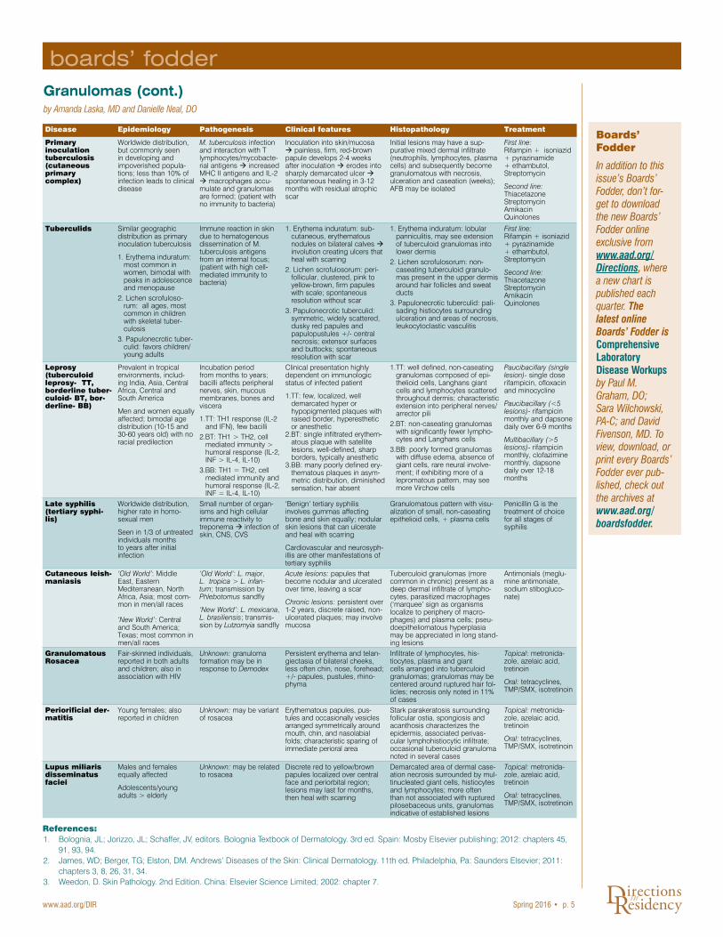
Granulomas (cont.)by Amanda Laska, MD and Danielle Neal, DO
Disease Epidemiology Pathogenesis Clinical features Histopathology Treatment
Primary inoculation tuberculosis (cutaneous primary complex)
Worldwide distribution, but commonly seen in developing and impoverished popula-tions; less than 10% of infection leads to clinical disease
M. tuberculosis infection and interaction with T lymphocytes/mycobacte-rial antigens increased MHC II antigens and IL-2 macrophages accu-mulate and granulomas are formed; (patient with no immunity to bacteria)
Inoculation into skin/mucosa painless, firm, red-brown papule develops 2-4 weeks after inoculation erodes into sharply demarcated ulcer spontaneous healing in 3-12 months with residual atrophic scar
Initial lesions may have a sup-purative mixed dermal infiltrate (neutrophils, lymphocytes, plasma cells) and subsequently become granulomatous with necrosis, ulceration and caseation (weeks); AFB may be isolated
First line: Rifampin + isoniazid + pyrazinamide + ethambutol, Streptomycin
Second line: Thiacetazone StreptomycinAmikacinQuinolones
Tuberculids Similar geographic distribution as primary inoculation tuberculosis
1. Erythema induratum: most common in women, bimodal with peaks in adolescence and menopause
2. Lichen scrofuloso-rum: all ages, most common in children with skeletal tuber-culosis
3. Papulonecrotic tuber-culid: favors children/young adults
Immune reaction in skin due to hematogenous dissemination of M. tuberculosis antigens from an internal focus; (patient with high cell-mediated immunity to bacteria)
1. Erythema induratum: sub-cutaneous, erythematous nodules on bilateral calves involution creating ulcers that heal with scarring
2. Lichen scrofulosorum: peri-follicular, clustered, pink to yellow-brown, firm papules with scale; spontaneous resolution without scar
3. Papulonecrotic tuberculid: symmetric, widely scattered, dusky red papules and papulopustules +/- central necrosis; extensor surfaces and buttocks; spontaneous resolution with scar
1. Erythema induratum: lobular panniculitis, may see extension of tuberculoid granulomas into lower dermis
2. Lichen scrofulosorum: non-caseating tuberculoid granulo-mas present in the upper dermis around hair follicles and sweat ducts
3. Papulonecrotic tuberculid: pali-sading histiocytes surrounding ulceration and areas of necrosis, leukocytoclastic vasculitis
First line: Rifampin + isoniazid + pyrazinamide + ethambutol, Streptomycin
Second line: Thiacetazone StreptomycinAmikacinQuinolones
Leprosy (tuberculoid leprosy- TT, borderline tuber-culoid- BT, bor-derline- BB)
Prevalent in tropical environments, includ-ing India, Asia, Central Africa, Central and South America
Men and women equally affected; bimodal age distribution (10-15 and 30-60 years old) with no racial predilection
Incubation period from months to years; bacilli affects peripheral nerves, skin, mucous membranes, bones and viscera
1.TT: TH1 response (IL-2 and IFN), few bacilli
2.BT: TH1 > TH2, cell mediated immunity > humoral response (IL-2, INF > IL-4, IL-10)
3.BB: TH1 = TH2, cell mediated immunity and humoral response (IL-2, INF = IL-4, IL-10)
Clinical presentation highly dependent on immunologic status of infected patient
1.TT: few, localized, well demarcated hyper or hypopigmented plaques with raised border, hyperesthetic or anesthetic
2.BT: single infiltrated erythem-atous plaque with satellite lesions, well-defined, sharp borders, typically anesthetic
3.BB: many poorly defined ery-thematous plaques in asym-metric distribution, diminished sensation, hair absent
1.TT: well defined, non-caseating granulomas composed of epi-thelioid cells, Langhans giant cells and lymphocytes scattered throughout dermis; characteristic extension into peripheral nerves/arrector pili
2.BT: non-caseating granulomas with significantly fewer lympho-cytes and Langhans cells
3.BB: poorly formed granulomas with diffuse edema, absence of giant cells, rare neural involve-ment; if exhibiting more of a lepromatous pattern, may see more Virchow cells
Paucibacillary (single lesion)- single dose rifampicin, ofloxacin and minocycline
Paucibacillary (<5 lesions)- rifampicin monthly and dapsone daily over 6-9 months
Multibacillary (>5 lesions)- rifampicin monthly, clofazimine monthly, dapsone daily over 12-18 months
Late syphilis (tertiary syphi-lis)
Worldwide distribution, higher rate in homo-sexual men
Seen in 1/3 of untreated individuals months to years after initial infection
Small number of organ-isms and high cellular immune reactivity to treponema infection of skin, CNS, CVS
‘Benign’ tertiary syphilis involves gummas affecting bone and skin equally; nodular skin lesions that can ulcerate and heal with scarring
Cardiovascular and neurosyph-illis are other manifestations of tertiary syphilis
Granulomatous pattern with visu-alization of small, non-caseating epithelioid cells, + plasma cells
Penicillin G is the treatment of choice for all stages of syphilis
Cutaneous leish-maniasis
‘Old World’: Middle East, Eastern Mediterranean, North Africa, Asia; most com-mon in men/all races
‘New World’: Central and South America; Texas; most common in men/all races
‘Old World’: L. major, L. tropica > L. infan-tum; transmission by Phlebotomus sandfly
‘New World’: L. mexicana, L. brasiliensis; transmis-sion by Lutzomyia sandfly
Acute lesions: papules that become nodular and ulcerated over time, leaving a scar
Chronic lesions: persistent over 1-2 years, discrete raised, non-ulcerated plaques; may involve mucosa
Tuberculoid granulomas (more common in chronic) present as a deep dermal infiltrate of lympho-cytes, parasitized macrophages (‘marquee’ sign as organisms localize to periphery of macro-phages) and plasma cells; pseu-doepitheliomatous hyperplasia may be appreciated in long stand-ing lesions
Antimonials (meglu-mine antimoniate, sodium stibogluco-nate)
Granulomatous Rosacea
Fair-skinned individuals, reported in both adults and children; also in association with HIV
Unknown: granuloma formation may be in response to Demodex
Persistent erythema and telan-giectasia of bilateral cheeks, less often chin, nose, forehead; +/- papules, pustules, rhino-phyma
Infiltrate of lymphocytes, his-tiocytes, plasma and giant cells arranged into tuberculoid granulomas; granulomas may be centered around ruptured hair fol-licles; necrosis only noted in 11% of cases
Topical: metronida-zole, azelaic acid, tretinoin
Oral: tetracyclines, TMP/SMX, isotretinoin
Periorificial der-matitis
Young females; also reported in children
Unknown: may be variant of rosacea
Erythematous papules, pus-tules and occasionally vesicles arranged symmetrically around mouth, chin, and nasolabial folds; characteristic sparing of immediate perioral area
Stark parakeratosis surrounding follicular ostia, spongiosis and acanthosis characterizes the epidermis, associated perivas-cular lymphohistiocytic infiltrate; occasional tuberculoid granuloma noted in several cases
Topical: metronida-zole, azelaic acid, tretinoin
Oral: tetracyclines, TMP/SMX, isotretinoin
Lupus miliaris disseminatus faciei
Males and females equally affected
Adolescents/young adults > elderly
Unknown: may be related to rosacea
Discrete red to yellow/brown papules localized over central face and periorbital region; lesions may last for months, then heal with scarring
Demarcated area of dermal case-ation necrosis surrounded by mul-tinucleated giant cells, histiocytes and lymphocytes; more often than not associated with ruptured pilosebaceous units, granulomas indicative of established lesions
Topical: metronida-zole, azelaic acid, tretinoin
Oral: tetracyclines, TMP/SMX, isotretinoin
References:1. Bolognia, JL; Jorizzo, JL; Schaffer, JV, editors. Bolognia Textbook of Dermatology. 3rd ed. Spain: Mosby Elsevier publishing; 2012: chapters 45,
91, 93, 94. 2. James, WD; Berger, TG; Elston, DM. Andrews’ Diseases of the Skin: Clinical Dermatology. 11th ed. Philadelphia, Pa: Saunders Elsevier; 2011:
chapters 3, 8, 26, 31, 34.3. Weedon, D. Skin Pathology. 2nd Edition. China: Elsevier Science Limited; 2002: chapter 7.
p. 6 • Spring 2016Directionsesidencyin R
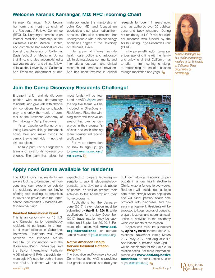
Winter 2015 RFTC was submitted by Travis Morrell, MD, MPH — a resident physician at Loma Linda University Dermatology.
A 55-year-old female was admit-ted for pneumonia complicated by sepsis. On hospital day three, she was noted to have six asymptomat-ic, non-tender, edematous papules and plaques scattered on her neck, upper arms, and left hand. Her med-ical history including a hematologic malignancy, for which she received her first course of chemotherapy two weeks prior. On biopsy, the inflam-matory infiltrate was centered on sweat ducts.1. What is the name of the dis-
order? Neutrophilic Eccrine Hidradenitis
2. This was first characterized in association with what malignan-cy? Acute myelogenous leukemia
3. What is the most common chemotherapy association? Cytarabine
4. Classic finding on pathology? Peri-eccrine neutrophilic inflammation
5. What is another name for this
disorder? Toxic Erythema of
Chemotherapy
6. What is the name for the disor-
der that shares similar pathol-
ogy findings, but is found on
children’s soles? Idiopathic
palmoplantar hidradenitis / pal-
moplantar eccrine hidradenitis
Race for the Case: Spring 2016By Emily de Golian, MD
A 69-year-old Caucasian male pre-sented for treatment evaluation for a 4.3 x 3.7 cm left hip plaque, which was present for 10 years prior to recent biopsy by an outside physi-cian. Firm palpable nodules were present within this asymptomatic, growing lesion. His medical his-tory is otherwise non-contributory.
1. What translocation is most like-ly present within this lesion?
2. What are the histopathologic findings?
3. Identify the immunohisto-chemical pattern classic to this diagnosis.
4. What is the recommended stan-dard treatment option with the highest cure rate without recur-rence?
5. What treatment is recommend-ed for patients with recurrent or metastatic lesions?
Respond online with the cor-rect answers at www.aad.org/RaceForTheCase for the opportunity to win a Starbucks gift card! If you win, we will also publish your mug (face), and if you have an interest-ing story to tell residents, we might share it (see our current winner pro-file to the right). Good luck! D R
Answers to Winter 2015 Race for the Case
D R
Emily de Golian, MD, is a PGY-3 resident physician, department of dermatology at Loma Linda University Medical Center
Race for the Case Winner Profile: Winter 2015
Congratulations to Nicole Harter, MD — a second-year resident at the University of Southern California in Los Angeles, California. She is originally from Hilo, Hawaii, but grew up in the charm-ing, small-town of Prescott, Arizona. Nicole is pursuing a fellowship in pediatric der-matology and plans to con-tinue a career in academic medicine, with particular interest in pediatric derma-tologic surgery. When she is not with her USC resi-dency-family, she loves to be active by running, hik-ing, biking, and enjoying the year-round beauty of Pasadena, California with her adventurous husband and adorable pup. Together they love to explore and live to travel! Nicole loves to cook, and her specialty is fun-flavored cupcakes — best when shared among family and co-residents!
Nicole Harter, MD
D R
Spring 2016 • p. 7www.aad.org/DIR Directionsesidencyin R
D R
D R
D R
Faranak Kamangar, MD is a senior dermatology resident at the University of California, Davis department of dermatology.
Welcome Faranak Kamangar, MD: RFC incoming Chair!
Join the Camp Discovery Residents Challenge!
Apply now! Grants available for residents
Faranak Kamangar, MD, begins her term this month as chair of the Residents / Fellows Committee (RFC). Dr. Kamangar completed an Internal Medicine internship at the California Pacific Medical Center, and completed her medical educa-tion at the University of California, Davis School of Medicine. During that time, she also accomplished a two-year research and clinical fellow-ship at the University of California, San Francisco department of der-
matology under the mentorship of John Koo, MD, and focused on psoriasis and complex medical ther-apeutics. She also completed her undergraduate with a biotechnology bachelor’s degree at the University of California, Davis.
Her areas of interest include health care policy and advocacy within dermatology; community and international outreach; and clinical research and therapeutic innovation. She has been involved in clinical
research for over 11 years now, and has authored over 20 publica-tions and book chapters. During her residency at UC Davis, her clini-cal research was funded by the ASDS Cutting Edge Research Grant (CERG).
In her personal time, Dr. Kamangar enjoys spending time with her family and enjoying all that California has to offer — from surfing to hiking, to maintaining a peaceful balance through meditation and yoga.
Engage in a fun and friendly com-petition with fellow dermatology residents, and give kids with chronic skin conditions the chance to laugh, play, and enjoy the magic of sum-mer at the American Academy of Dermatology’s Camp Discovery.
It’s an experience like no other, letting kids swim, fish, go horseback riding, hike and make friends. At camp, they’re just kids — not their skin conditions.
To take part, just put together a team and raise funds however you choose. The team that raises the
most funds will be fea-tured in AAD’s Aspire, and the top five teams will be included in Directions in Residency. Plus, the win-ning team will receive an award that can be dis-played in their program’s offices, and each winning team member will receive a gift card!
For more information on how to sign up, go to: www.events.aad.org/ residents.
The AAD knows that residents are always looking to broaden their hori-zons and gain experience outside the residency program, so they’re offering two exciting opportunities to travel and provide care for under-served communities. Deadlines are fast approaching!
Resident International GrantThis is an opportunity for 15 U.S. and Canadian senior dermatology residents to participate in a four- to six-week elective in Gaborone, Botswana. Residents will rotate between the Princess Marina Hospital (in conjunction with the Botswana-UPenn Partership) and the Baylor International Pediatric AIDS Initiative (BIPAI) to provide der-matologic HIV care for both children and adults. Residents will also be
expected to prepare lectures/pre-sentations, submit teledermatology consults, and develop a database of photos, as well as present their activities to the Academy and their home programs.
Applications for the January–June (2017) travel rotation must be submitted by April 1, 2016, while applications for the July–December (2017) travel rotation may be sub-mitted until September 30, 2016. For more information, visit www.aad.org/international, or contact Janine Mueller at [email protected].
Native American Health Service Resident Rotation ProgramThe Education and Volunteers Abroad Committee at the AAD is providing four grants to second- and third-year
U.S. dermatology residents to par-ticipate in a rural health elective in Chinle, Arizona for one to two weeks. Residents will provide dermatologic care to the Navajo Nation population and will assist primary health care providers with diagnoses and dis-ease management. Residents will be expected to keep records of consults, prepare lectures, and submit an eval-uation of activities to the Academy within one month of the rotation.
Applications must be submitted by April 1, 2016 for the 2016-2017 rotations: November 2016; March 2017; May 2017; and August 2017. Applications submitted after April 1 will be considered for the 2017-2018 rotation series. For more information, please visit www.aad.org/native american, or email Janine Mueller at [email protected].
Amer
ican
Aca
dem
y of
Der
mat
olog
y As
soci
atio
nP.
O. B
ox 4
014
Scha
umbu
rg, I
llino
is 6
0168
-401
4
ZO Skin Health Inc. by Zein Obagi, MD, proudly supports the American Academy of Dermatology
and the Directions in Residency newsletter.
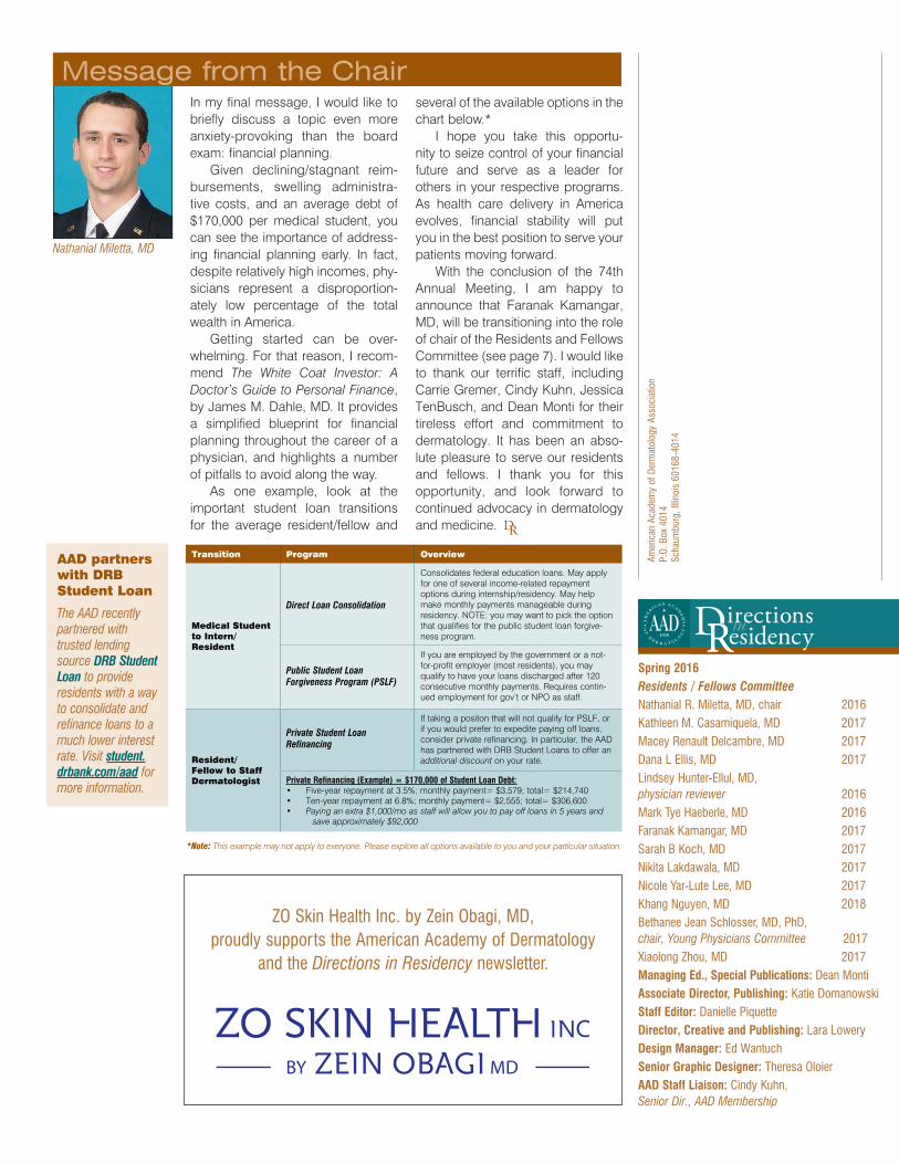
Nathanial Miletta, MD
Message from the Chair In my final message, I would like to briefly discuss a topic even more anxiety-provoking than the board exam: financial planning.
Given declining/stagnant reim-bursements, swelling administra-tive costs, and an average debt of $170,000 per medical student, you can see the importance of address-ing financial planning early. In fact, despite relatively high incomes, phy-sicians represent a disproportion-ately low percentage of the total wealth in America.
Getting started can be over-whelming. For that reason, I recom-mend The White Coat Investor: A Doctor’s Guide to Personal Finance, by James M. Dahle, MD. It provides a simplified blueprint for financial planning throughout the career of a physician, and highlights a number of pitfalls to avoid along the way.
As one example, look at the important student loan transitions for the average resident/fellow and
several of the available options in the chart below.*
I hope you take this opportu-nity to seize control of your financial future and serve as a leader for others in your respective programs. As health care delivery in America evolves, financial stability will put you in the best position to serve your patients moving forward.
With the conclusion of the 74th Annual Meeting, I am happy to announce that Faranak Kamangar, MD, will be transitioning into the role of chair of the Residents and Fellows Committee (see page 7). I would like to thank our terrific staff, including Carrie Gremer, Cindy Kuhn, Jessica TenBusch, and Dean Monti for their tireless effort and commitment to dermatology. It has been an abso-lute pleasure to serve our residents and fellows. I thank you for this opportunity, and look forward to continued advocacy in dermatology and medicine.
Spring 2016Residents / Fellows CommitteeNathanial R. Miletta, MD, chair 2016Kathleen M. Casamiquela, MD 2017Macey Renault Delcambre, MD 2017Dana L Ellis, MD 2017Lindsey Hunter-Ellul, MD, physician reviewer 2016Mark Tye Haeberle, MD 2016 Faranak Kamangar, MD 2017Sarah B Koch, MD 2017Nikita Lakdawala, MD 2017Nicole Yar-Lute Lee, MD 2017Khang Nguyen, MD 2018Bethanee Jean Schlosser, MD, PhD, chair, Young Physicians Committee 2017Xiaolong Zhou, MD 2017Managing Ed., Special Publications: Dean Monti Associate Director, Publishing: Katie Domanowski Staff Editor: Danielle PiquetteDirector, Creative and Publishing: Lara LoweryDesign Manager: Ed WantuchSenior Graphic Designer: Theresa OloierAAD Staff Liaison: Cindy Kuhn, Senior Dir., AAD Membership
ininDirectionsesidency Rin R
D R
*Note: This example may not apply to everyone. Please explore all options available to you and your particular situation.
Transition Program Overview
Medical Student to Intern/Resident
Direct Loan Consolidation
Consolidates federal education loans. May apply for one of several income-related repayment options during internship/residency. May help make monthly payments manageable during residency. NOTE: you may want to pick the option that qualifies for the public student loan forgive-ness program.
Public Student Loan Forgiveness Program (PSLF)
If you are employed by the government or a not-for-profit employer (most residents), you may qualify to have your loans discharged after 120 consecutive monthly payments. Requires contin-ued employment for gov’t or NPO as staff.
Resident/Fellow to Staff Dermatologist
Private Student Loan Refinancing
If taking a positon that will not qualify for PSLF, or if you would prefer to expedite paying off loans, consider private refinancing. In particular, the AAD has partnered with DRB Student Loans to offer an additional discount on your rate.
Private Refinancing (Example) = $170,000 of Student Loan Debt:• Five-year repayment at 3.5%; monthly payment= $3,579; total= $214,740• Ten-year repayment at 6.8%; monthly payment= $2,555; total= $306,600• Paying an extra $1,000/mo as staff will allow you to pay off loans in 5 years and
save approximately $92,000
AAD partners with DRB Student Loan
The AAD recently partnered with trusted lending source DRB Student Loan to provide residents with a way to consolidate and refinance loans to a much lower interest rate. Visit student.drbank.com/aad for more information.