Embed Size (px)
Citation preview
Master
Every patientdeserves theGOLD STANDARD ...
Anatomic PathologyChecklist
CAP Accreditation Program
College of American Pathologists325 Waukegan RoadNorthfield, IL 60093-2750www.cap.org 07.29.2013
Disclaimer and Copyright NoticeIf you are enrolled in the CAP's Laboratory Accreditation Program and are preparing for an inspection,you must use the Checklists that were mailed in your application or reapplication packet, not those postedon the Web site. The Checklists undergo regular revision and Checklists may be revised after you receiveyour packet.
If a Checklist has been updated since receiving your packet, you will be inspected based upon the Checkliststhat were mailed. If you have any questions about the use of Checklists in the inspection process, pleasee-mail the CAP ([email protected]), or call (800) 323-4040, ext. 6065.
The checklists used in connection with the inspection of laboratories by the Laboratory AccreditationProgram of the College of American Pathologists have been created by the College and are copyrightedworks of the College. The College has authorized copying and use of the checklists by College inspectorsin conducting laboratory inspections for the CLA and by laboratories that are preparing for such inspections.Except as permitted by section 107 of the Copyright Act, 17 U.S.C. sec. 107, any other use of the checklistsconstitutes infringement of the College’s copyrights in the checklists. The College will take appropriatelegal action to protect these copyrights.
All Checklists are ©2013. College of American Pathologists. All rights reserved.
2 of 72
07.29.2013Anatomic Pathology Checklist
Anatomic PathologyChecklist
TABLE OF CONTENTS
SUMMARY OF CHANGES.....................................................................................................................5UNDERSTANDING THE CAP ACCREDITATION CHECKLIST COMPONENTS..................................8HOW TO INSPECT USING R.O.A.D INSPECTION TECHNIQUES.......................................................9INTRODUCTION..................................................................................................................................10DEFINITION OF TERMS......................................................................................................................10GENERAL ANATOMIC PATHOLOGY.................................................................................................12
INTERLABORATORY COMPARISONS.............................................................................................................................12SAFETY..............................................................................................................................................................................13
SURGICAL PATHOLOGY....................................................................................................................14QUALITY MANAGEMENT..................................................................................................................................................14QUALITY CONTROL..........................................................................................................................................................17
SURGICAL SPECIMEN EXAMINATION......................................................................................................................17INTRA-OPERATIVE CONSULTATION (RAPID DIAGNOSIS)......................................................................................21FINE NEEDLE ASPIRATE (FNA) SPECIMENS...........................................................................................................24SURGICAL PATHOLOGY REPORTS..........................................................................................................................25
HISTOLOGY LABORATORY...............................................................................................................30GENERAL QUALITY CONTROL.........................................................................................................31SPECIAL STAINS (HISTOCHEMISTRY).............................................................................................32IMMUNOLOGIC AND MOLECULAR METHODS................................................................................33
IMMUNOFLUORESCENCE MICROSCOPY......................................................................................................................33IMMUNOHISTOCHEMISTRY.............................................................................................................................................34FLUORESCENCE AND NON-FLUORESCENCE IN SITU HYBRIDIZATION (FISH, ISH)................................................38PREDICTIVE MARKERS...................................................................................................................................................40DIGITAL IMAGE ANALYSIS................................................................................................................................................45
Validation and Calibration.............................................................................................................................................45Quality Control.............................................................................................................................................................46Specimen Analysis.......................................................................................................................................................48DNA Staining................................................................................................................................................................49Reports.........................................................................................................................................................................50Personnel.....................................................................................................................................................................50
INSTRUMENTS AND EQUIPMENT...................................................................................................................................51Instruments and Equipment Maintenance....................................................................................................................51Pipettes and Thermometers.........................................................................................................................................52Tissue Processor.........................................................................................................................................................53Flotation Baths.............................................................................................................................................................54Microtomes...................................................................................................................................................................54Cryostat........................................................................................................................................................................55
PHYSICAL FACILITIES........................................................................................................................55STORAGE AND SUPPLY...................................................................................................................................................55
HISTOLOGY LABORATORY SAFETY................................................................................................55AUTOPSY PATHOLOGY......................................................................................................................58
QUALITY MANAGEMENT..................................................................................................................................................58AUTOPSY CONSENT PROCEDURES..............................................................................................................................60
3 of 72
07.29.2013Anatomic Pathology Checklist
AUTOPSY ROOM...............................................................................................................................................................61AUTOPSY PERFORMANCE AND DOCUMENTATION.....................................................................................................62AUTOPSY SAFETY............................................................................................................................................................66
ELECTRON MICROSCOPY.................................................................................................................68QUALITY CONTROL..........................................................................................................................................................68
ELECTRON MICROSCOPY SAMPLE PREPARATION...............................................................................................69INSTRUMENTS AND EQUIPMENT............................................................................................................................70REPORTS....................................................................................................................................................................70RECORDS, FILES AND PHOTOGRAPHS..................................................................................................................71
LABORATORY SAFETY.....................................................................................................................................................71
4 of 72
07.29.2013Anatomic Pathology Checklist
SUMMARY OF CHECKLIST EDITION CHANGESAnatomic Pathology Checklist
07/29/2013 Edition
The following lists of requirements provide information on what has changed in this edition of the checklist, or inthe previous edition. This information is provided in three categories:
1. New — requirements that have been added2. Revised — requirements listed in this section fall into two categories:
● A major change to a requirement or a note that would necessitate a change in procedure for thelaboratory
● A change to the Phase3. Deleted/Moved/Merged — requirements listed in this section fall into three categories:
● Deleted — requirements that have been removed● Moved — requirements that have been relocated from this checklist into another checklist, or have
been moved within this checklist and given a new checklist requirement number (resequenced)● Merged — requirements that have been combined with a similar requirement in the checklist
If this checklist was created for an on-site inspection or self-evaluation, it has been customized based on thelaboratory's activity menu. The listing below is comprehensive; therefore, some of the requirements included maynot appear in the customized checklist. Such requirements are not applicable to the testing performed by thelaboratory.
Note: For the detail of the changes, refer to the "Changes Only" document which may be found on the CAP websitethrough e-LAB Solutions (Laboratory Accreditation Program Master and Custom Checklists). To access thisdocument select "Changes Only" from the Checklist Type drop-down menu.
The "Changes Only" document contains the text of new and deleted checklist requirements, major and minorrequirement revisions, and changes to explanatory text. These changes are presented, in order, as they appearin the checklist. Major requirement revisions will display a "Revised" flag. Minor revisions will not display a "Revised"flag and are defined as those editorial changes that are not likely to affect your laboratory operations, but areworded to better convey the intent of the requirement. Changes appear in redline/strikeout format that comparesthe previous checklist edition to this edition. Requirements that have been moved or merged will appear at theend of that file.
NEW Checklist Requirements
Effective DateRequirement07/29/2013ANP.1165007/31/2012ANP.1171607/31/2012ANP.1217007/29/2013ANP.1217307/29/2013ANP.2136007/29/2013ANP.2146007/31/2012ANP.2298507/29/2013ANP.2304507/29/2013ANP.2313007/29/2013ANP.33025
REVISED Checklist Requirements
Effective DateRequirement07/29/2013ANP.0821607/29/2013ANP.1015007/29/2013ANP.10260
5 of 72
07.29.2013Anatomic Pathology Checklist
07/29/2013ANP.1160507/29/2013ANP.1164007/31/2012ANP.1167007/29/2013ANP.1171307/31/2012ANP.1173407/29/2013ANP.1175607/31/2012ANP.1180007/31/2012ANP.1190007/29/2013ANP.1209207/29/2013ANP.1209407/31/2012ANP.1238507/31/2012ANP.1250007/31/2012ANP.2138207/29/2013ANP.2145007/29/2013ANP.2257007/31/2012ANP.2261507/31/2012ANP.2296407/29/2013ANP.2297007/29/2013ANP.2297607/29/2013ANP.2297807/29/2013ANP.2299909/25/2012ANP.2300207/31/2012ANP.2300307/31/2012ANP.2301807/31/2012ANP.2303707/31/2012ANP.2303807/31/2012ANP.2304807/29/2013ANP.2309507/29/2013ANP.2310007/29/2013ANP.2312007/29/2013ANP.2315007/29/2013ANP.2430007/29/2013ANP.3245007/31/2012ANP.3310007/29/2013ANP.3312007/31/2012ANP.3320007/29/2013ANP.55100
DELETED/MOVED/MERGED Checklist Requirements
Effective DateRequirement07/30/2012ANP.1000007/30/2012ANP.1145007/30/2012ANP.1245007/30/2012ANP.2136607/30/2012ANP.2139007/30/2012ANP.2300607/28/2013ANP.2301407/30/2012ANP.2307507/28/2013ANP.3005007/30/2012ANP.3215007/30/2012ANP.3260007/30/2012ANP.3265007/30/2012ANP.3325007/30/2012ANP.5110007/30/2012ANP.5410007/30/2012ANP.55050
6 of 72
07.29.2013Anatomic Pathology Checklist

07/30/2012ANP.5515007/30/2012ANP.57050
7 of 72
07.29.2013Anatomic Pathology Checklist
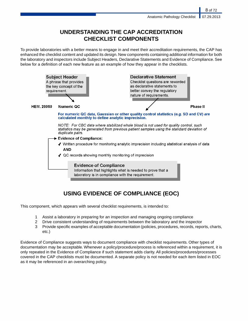
UNDERSTANDING THE CAP ACCREDITATIONCHECKLIST COMPONENTS
To provide laboratories with a better means to engage in and meet their accreditation requirements, the CAP hasenhanced the checklist content and updated its design. New components containing additional information for boththe laboratory and inspectors include Subject Headers, Declarative Statements and Evidence of Compliance. Seebelow for a definition of each new feature as an example of how they appear in the checklists.
USING EVIDENCE OF COMPLIANCE (EOC)
This component, which appears with several checklist requirements, is intended to:
1 Assist a laboratory in preparing for an inspection and managing ongoing compliance2 Drive consistent understanding of requirements between the laboratory and the inspector3 Provide specific examples of acceptable documentation (policies, procedures, records, reports, charts,
etc.)
Evidence of Compliance suggests ways to document compliance with checklist requirements. Other types ofdocumentation may be acceptable. Whenever a policy/procedure/process is referenced within a requirement, it isonly repeated in the Evidence of Compliance if such statement adds clarity. All policies/procedures/processescovered in the CAP checklists must be documented. A separate policy is not needed for each item listed in EOCas it may be referenced in an overarching policy.
8 of 72
07.29.2013Anatomic Pathology Checklist
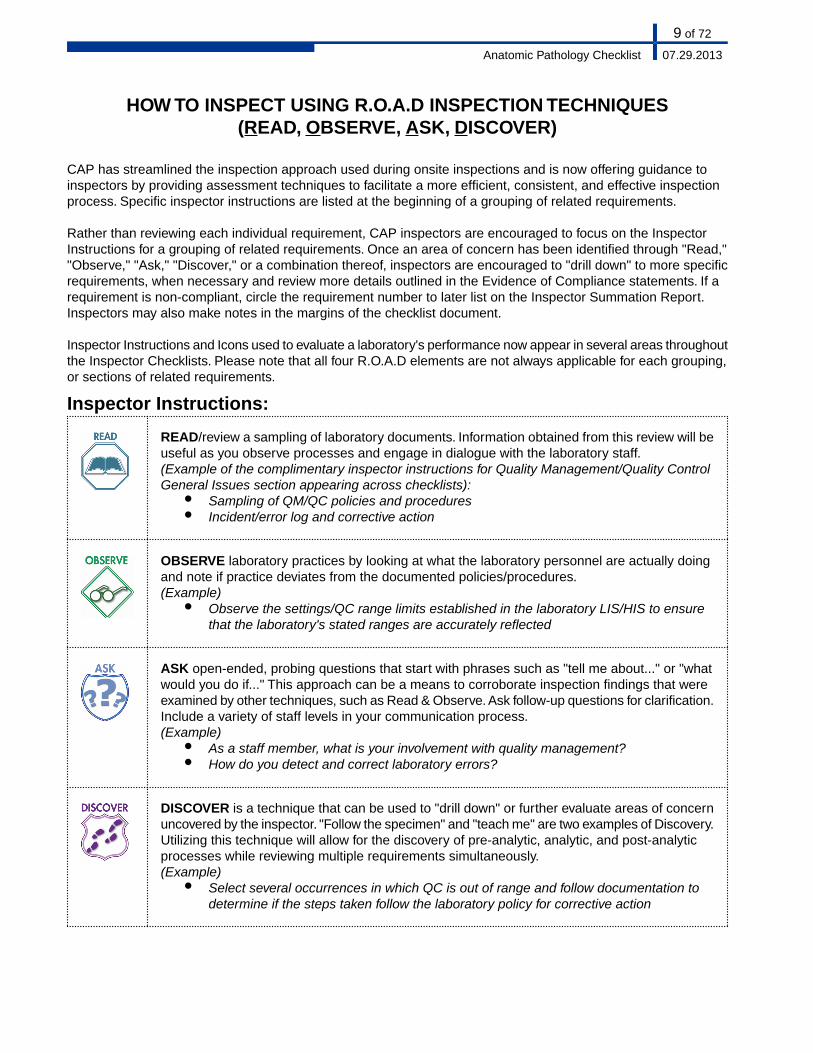
HOW TO INSPECT USING R.O.A.D INSPECTION TECHNIQUES(READ, OBSERVE, ASK, DISCOVER)
CAP has streamlined the inspection approach used during onsite inspections and is now offering guidance toinspectors by providing assessment techniques to facilitate a more efficient, consistent, and effective inspectionprocess. Specific inspector instructions are listed at the beginning of a grouping of related requirements.
Rather than reviewing each individual requirement, CAP inspectors are encouraged to focus on the InspectorInstructions for a grouping of related requirements. Once an area of concern has been identified through "Read,""Observe," "Ask," "Discover," or a combination thereof, inspectors are encouraged to "drill down" to more specificrequirements, when necessary and review more details outlined in the Evidence of Compliance statements. If arequirement is non-compliant, circle the requirement number to later list on the Inspector Summation Report.Inspectors may also make notes in the margins of the checklist document.
Inspector Instructions and Icons used to evaluate a laboratory's performance now appear in several areas throughoutthe Inspector Checklists. Please note that all four R.O.A.D elements are not always applicable for each grouping,or sections of related requirements.
Inspector Instructions:
READ/review a sampling of laboratory documents. Information obtained from this review will beuseful as you observe processes and engage in dialogue with the laboratory staff.(Example of the complimentary inspector instructions for Quality Management/Quality ControlGeneral Issues section appearing across checklists):
● Sampling of QM/QC policies and procedures● Incident/error log and corrective action
OBSERVE laboratory practices by looking at what the laboratory personnel are actually doingand note if practice deviates from the documented policies/procedures.(Example)
● Observe the settings/QC range limits established in the laboratory LIS/HIS to ensurethat the laboratory's stated ranges are accurately reflected
ASK open-ended, probing questions that start with phrases such as "tell me about..." or "whatwould you do if..." This approach can be a means to corroborate inspection findings that wereexamined by other techniques, such as Read & Observe. Ask follow-up questions for clarification.Include a variety of staff levels in your communication process.(Example)
● As a staff member, what is your involvement with quality management?● How do you detect and correct laboratory errors?
DISCOVER is a technique that can be used to "drill down" or further evaluate areas of concernuncovered by the inspector. "Follow the specimen" and "teach me" are two examples of Discovery.Utilizing this technique will allow for the discovery of pre-analytic, analytic, and post-analyticprocesses while reviewing multiple requirements simultaneously.(Example)
● Select several occurrences in which QC is out of range and follow documentation todetermine if the steps taken follow the laboratory policy for corrective action
9 of 72
07.29.2013Anatomic Pathology Checklist
INTRODUCTION
An inspection of a laboratory section, or department will include the discipline-specific checklist(s), the LaboratoryGeneral Checklist, and the All Common Checklist (COM).
In response to the ongoing request to reduce the redundancy within the Accreditation Checklists, the CAPaccreditation program is introducing the All Common Checklist.
The purpose of the All Common Checklist is to group together those requirements that were redundant in LaboratoryGeneral and the discipline-specific checklists.Therefore, the CAP centralized all requirements regarding: proficiencytesting, procedure manuals, test method validation, and critical results into one checklist, the COM checklist.
Laboratories that do not file slides on-site (for example, some "read-only" laboratories) must retain a sample ofslides on-site for review by the inspector on all days when the laboratory is subject to its regular on-site inspection.The sample must, at a minimum, include all slides accessioned over a continuous 2-week period within the previous2 years.
DEFINITION OF TERMS
Annual - Every 12 calendar months
Biennial - Every 24 calendar months
Calibrator, historical - The set of archived results of a single-point calibrator that demonstrates stability of the assayover time
Check - Examination to determine the accuracy, quality or presence of any attribute of a test system
Confirmation - Establishment of the correctness of a value or process
Correlation - Establishment of agreement between two or more measured values
Credentialing - The process of obtaining, verifying, and assessing the qualifications of a practitioner to providecare in a health care organization
Digital image analysis - The computer-assisted detection or quantification of specific features in an image followingenhancement and processing of that image, including immunohistochemistry, DNA analysis, morphometric analysis,and in situ hybridization
Equipment - Single apparatus or set of devices or apparatuses needed to perform a specific task
Examination - In the context of checklist requirements, examination refers to the process of inspection of tissuesand samples prior to analysis. An examination is not an analytical test.
FDA - In the context of checklist requirements, FDA should be taken to mean the national, state, or provincialauthority having jurisdiction over in vitro diagnostic test systems.
Function Check - Operational check performed to confirm that instruments and equipment (electrical, mechanicalsystems) are working according to manufacturer's specifications on a daily basis or before use. Examples mayinclude base line calibration, balancing/zero adjustment, component operational checks (electronics, lamps, tubing),operational readiness (thermometer calibration, reagent delivery), and electronic function and peak performance.
10 of 72
07.29.2013Anatomic Pathology Checklist
High complexity - Rating given by the FDA to commercially marketed in vitro diagnostic tests based on their risksto public health. Tests in this category are seen to have the highest risks to public health.
Instrument - An analytical unit that uses samples to perform chemical or physical assays (e.g. chemistry analyzer,hematology analyzer)
Laboratory Director - The individual who is responsible for the overall operation and administration of the laboratory,including provision of timely, reliable and clinically relevant test results and compliance with applicable regulationsand accreditation requirements.This individual is listed on the laboratory's CAP and CLIA certificate (as applicable).
Maintenance - Those activities that prolong the life of an instrument or minimize breakdowns or mechanicalmalfunctions. Examples include cleaning or changing parts, fluids, tubing, lubrication, electronic checks, etc.
Moderate complexity - Rating given by the FDA to commercially marketed in vitro diagnostic tests based on theirrisks to public health
Modification of manufacturer's instructions - Any change to the manufacturer's supplied ingredients or modificationsto the assay as set forth in the manufacturer's labeling and instructions, including specimen type, instrumentationor procedure that could affect its performance specifications for sensitivity, specificity, accuracy, or precision orany change to the stated purpose of the test, its approved test population, or any claims related to interpretationof the results
Nonwaived - Tests categorized as either moderately complex (including provider-performed microscopy) or highlycomplex by the US Food and Drug Administration (FDA), according to a scoring system used by the FDA
Reagent - Any substance in a test system other than a solvent or support material that is required for the targetanalyte to be detected and its value measured in a sample.
Report errors - A report element (see GEN.41096) that is either incorrect or incomplete
Section Director - The individual who is responsible for the medical, technical and/or scientific oversight of aspecialty or section of the laboratory.
Semiannual - Every 6 calendar months
Subject to U.S. Regulations - Laboratories located within the United States and laboratories located outside of theUS that have obtained or applied for a CLIA certificate to perform laboratory testing on specimens collected in theUS for the assessment of the health of human beings.
Telepathology - The practice in which the pathologist views digitized or analog video or still image(s), and rendersan interpretation that is included in a formal diagnostic report or document in the patient record.
Test system - The process that includes pre-analytic, analytic, and post-analytic steps used to produce a test resultor set of results. A test system may be manual, automated, multi-channel or single-use and can include reagentscomponents, equipment or instruments required to produce results. A test system may encompass multiple identicalanalyzers or devices. Different test systems may be used for the same analyte.
Validation - A defined process by which a laboratory confirms that a laboratory developed or modifiedFDA-cleared/approved test performs as intended or claimed.
Verification - The process by which a laboratory determines that an FDA-cleared/approved test performs accordingto the specifications set forth by the manufacturer.
Waived - A category of tests defined as "simple laboratory examinations and procedures which have an insignificantrisk of an erroneous result." Laboratories performing waived tests are subject to minimal regulatory requirements.
11 of 72
07.29.2013Anatomic Pathology Checklist
GENERAL ANATOMIC PATHOLOGY
Do NOT use this Checklist if the laboratory does NOT perform any on-site preparation or examination of anatomicpathology specimens, but refers all submitted material to an outside laboratory, or if the laboratory's involvementin anatomic pathology is limited to filing of reports and/or slides
This Checklist covers several areas of anatomic pathology services, and is divided into the following sections:Surgical Pathology, Histology Laboratory, Autopsy Pathology, and Electron Microscopy. Cytopathology (bothgynecologic and non-gynecologic) is covered in a separate Checklist.The sequence for inspection of the anatomicpathology service is at the discretion of the inspection team. The sequence herein is consistent with that used forall other sections of the laboratory, but is not restrictive.
INTERLABORATORY COMPARISONS
Inspector Instructions:
● Sampling of records of peer educational program participation
● How does your laboratory participate in peer educational performance comparisons?
Phase IEducation ParticipationANP.02000
As applicable, the laboratory participates in a peer educational program in anatomic pathology(e.g. CAP Educational Anatomic Pathology Programs).
NOTE:The laboratory should consider participation in programs appropriate to its scope of service.Such programs provide valuable educational opportunities for peer performance comparisons inboth technical and diagnostic arenas. While none of these completely emulates the precise clinicalsetting involving anatomic pathology preparations and rendering of anatomic or clinical diagnoses,they can be a useful benchmark of peer-based performance in a national database.
Evidence of Compliance:✓ Records such as CAP order form, purchase order AND✓ Completed/submitted results indicating that the laboratory is participating in a CAP educational
AP program OR records of enrollment/participation in another AP peer educational programOR records for participation in a laboratory-developed program by circulating case materialwith other laboratories or within the laboratory's own practice with documentation of peer review
12 of 72
07.29.2013Anatomic Pathology Checklist
SAFETY
Inspector Instructions:
● Sampling of formaldehyde/xylene vapor monitoring records
● Space, storage, cleanliness, ventilation, outlets, sinks, lighting are all sufficient
● Is the work area sufficient for you to perform your duties safely and accurately?
**REVISED** 07/29/2013Phase IIFormaldehyde/Xylene SafetyANP.08216
Formaldehyde and xylene vapor concentrations are maintained below the following maxima,expressed as parts per million, in all areas of the Anatomic Pathology Department whereformaldehyde or xylene are used.
NOTE: Formaldehyde and xylene vapor concentrations must be monitored in all areas where thesereagents are used: e.g. surgical pathology gross dissection room, frozen section area, histologylaboratory, autopsy room, etc. Xylene vapor concentration monitoring in histology laboratoriesshould include manual and automated coverslipping areas, as these locations are often not ventilated.Initial monitoring involves identifying all employees who may be exposed at or above the actionlevel or at or above the STEL and accurately determining the exposure of each employee identified.Further formaldehyde monitoring is mandated at least every 6 months if results of the initial monitoringequal or exceed 0.5 ppm (8 hr time-weighted exposure, the “action level”) or at least once per yearif the results exceed the short term exposure limit (STEL) 2.0 ppm.The laboratory may discontinueperiodic formaldehyde monitoring if results from 2 consecutive sampling periods taken at least 7days apart show that employee exposure is below the action level and the short-term exposurelimit, and 1) no change has occurred in production, equipment, process or personnel or controlmeasures that may result in new or additional exposure to formaldehyde, and 2) there have beenno reports of conditions that may be associated with formaldehyde exposure.
Formaldehyde monitoring must be repeated any time there is a change in production, equipment,process, personnel, or control measures which may result in new or additional exposure toformaldehyde for any employee involved in the activity. If any personnel report signs or symptomsof respiratory or dermal conditions associated with formaldehyde exposure, the laboratory mustpromptly monitor the affected person's exposure.
Xylene must be monitored initially, but there is no requirement for periodic monitoring of xylene.
13 of 72
07.29.2013Anatomic Pathology Checklist
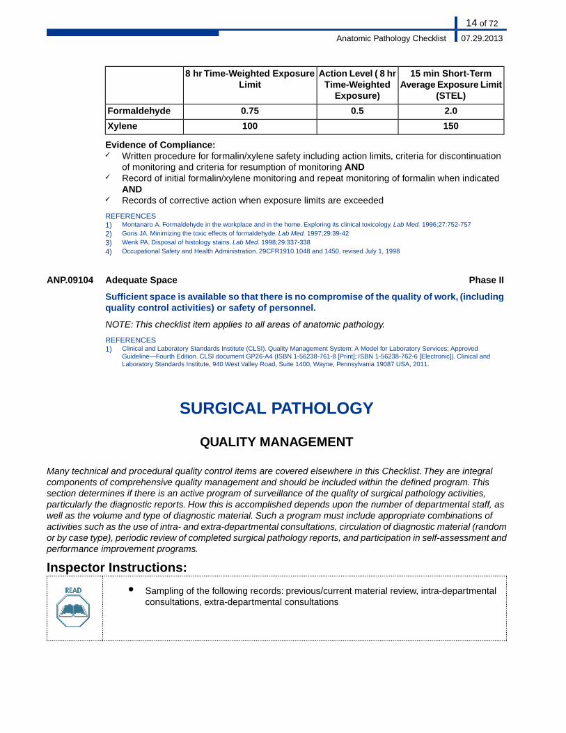
15 min Short-TermAverage Exposure Limit
(STEL)
Action Level ( 8 hrTime-Weighted
Exposure)
8 hr Time-Weighted ExposureLimit
2.00.50.75Formaldehyde
150100Xylene
Evidence of Compliance:✓ Written procedure for formalin/xylene safety including action limits, criteria for discontinuation
of monitoring and criteria for resumption of monitoring AND✓ Record of initial formalin/xylene monitoring and repeat monitoring of formalin when indicated
AND✓ Records of corrective action when exposure limits are exceeded
REFERENCES1) Montanaro A. Formaldehyde in the workplace and in the home. Exploring its clinical toxicology. Lab Med. 1996;27:752-757
2) Goris JA. Minimizing the toxic effects of formaldehyde. Lab Med. 1997;29:39-42
3) Wenk PA. Disposal of histology stains. Lab Med. 1998;29:337-338
4) Occupational Safety and Health Administration. 29CFR1910.1048 and 1450, revised July 1, 1998
Phase IIAdequate SpaceANP.09104
Sufficient space is available so that there is no compromise of the quality of work, (includingquality control activities) or safety of personnel.
NOTE: This checklist item applies to all areas of anatomic pathology.
REFERENCES1) Clinical and Laboratory Standards Institute (CLSI). Quality Management System: A Model for Laboratory Services; Approved
Guideline—Fourth Edition. CLSI document GP26-A4 (ISBN 1-56238-761-8 [Print]; ISBN 1-56238-762-6 [Electronic]). Clinical andLaboratory Standards Institute, 940 West Valley Road, Suite 1400, Wayne, Pennsylvania 19087 USA, 2011.
SURGICAL PATHOLOGY
QUALITY MANAGEMENT
Many technical and procedural quality control items are covered elsewhere in this Checklist. They are integralcomponents of comprehensive quality management and should be included within the defined program. Thissection determines if there is an active program of surveillance of the quality of surgical pathology activities,particularly the diagnostic reports. How this is accomplished depends upon the number of departmental staff, aswell as the volume and type of diagnostic material. Such a program must include appropriate combinations ofactivities such as the use of intra- and extra-departmental consultations, circulation of diagnostic material (randomor by case type), periodic review of completed surgical pathology reports, and participation in self-assessment andperformance improvement programs.
Inspector Instructions:
● Sampling of the following records: previous/current material review, intra-departmentalconsultations, extra-departmental consultations
14 of 72
07.29.2013Anatomic Pathology Checklist
● Does your laboratory exclude any specimen types from routine submission to thepathology department?
● What is your laboratory's course of action when a significant disparity exists betweenthe initial intra-operative consultation and final pathology diagnosis?
Phase ISurgical Pathology ExclusionANP.10016
There is a policy that lists specimens that an institution may choose to exclude from routinesubmission to the pathology department for examination.
NOTE: This policy should be made in conjunction with the hospital administration and appropriatemedical staff departments. The laboratory director should have participated in or been consultedby the medical staff in deciding which surgical specimens are to be sent to the pathology departmentfor examination.
This checklist item is not applicable if 1) all specimens are submitted to pathology, or 2) the laboratoryis not part of an institution that provides surgical services.
REFERENCES1) Kassan MA, et al. Value of routine pathology in herniorrhaphy performed upon adults. Surg Gynecol Obstet. 1986;163:518-522
2) Netser JC, et al. Value-based pathology: a cost-benefit analysis of the examination of routine and non-routine tonsil and adenoidspecimens. Am J Clin Pathol. 1997;108:158-165
3) Zarbo RJ, Nakleh RE. Surgical pathology specimens for gross examination only and exempt from submission. A College of AmericanPathologists Q-Probes study of current policies in 413 institutions. Arch Pathol Lab Med. 1999;123:133-139
4) College of American Pathologists. Policies and guidelines manual. Surgical specimens to be submitted to pathology for examination.Northfield, IL: CAP, 1999:Appendix M
5) Nakhleh RE, Fitzgibbons PL, eds. College of American Pathologists. Quality improvement manual in anatomic pathology, second edition.Northfield, IL: CAP, 2002
Phase ISurgical Pathology Microscopic ExemptionsANP.10032
There is a policy regarding what types of surgical specimens (if any) may be exempt frommicroscopic examination.
NOTE: Irrespective of any exemptions, microscopic examination should be performed wheneverthere is a request by the attending physician, or at the discretion of the pathologist when indicatedby the clinical history or gross findings. If there is such a policy, it should be approved by the medicalstaff or appropriate committee. Typical exempt specimens include foreskins in children, prostheticcardiac valves without attached tissue, torn meniscus, varicose veins, tonsils in children below acertain age, etc.
REFERENCES1) Weibel E. Pathological findings of clinical value in tonsils and adenoids. Acta Otolaryngol. 1965;60:331-338
2) Wolkomir AF, et al. Selective microscopic examination of gallbladders, hernia sacs and appendices. Am Surg. 1991:57:289-292
3) Boutin P, Hogshead H. Surgical pathology of the intervertebral disc: is routine examination necessary? Spine. 1992;17:1236-1238
4) Cornell WB, Levin HS. The inguinal hernia sac: trash or treasure? Anatomic pathology II check sample, APII-9. Chicago, IL: AmericanSociety of Clinical Pathology, 1993:17(4)
5) Delong WH, Grignon DJ. Pathologic findings in ribs removed at the time of radical nephrectomy for renal cell carcinoma. Int J SurgPathol. 1994;1:177-180
6) Raab SS. The cost-effectiveness of routine histologic examination. Am J Clin Pathol. 1998;110:391-396
7) Zarbo RJ, Nakleh RE. Surgical pathology specimens for gross examination only and exempt from submission. A College of AmericanPathologists Q-Probes study of current policies in 413 institutions. Arch Pathol Lab Med. 1999;123:133-139
8) College of American Pathologists. Policies and guidelines manual. Surgical specimens to be submitted to pathology for examination.Northfield, IL: CAP, 1999:Appendix M
Phase IIPrevious/Current Material ReviewANP.10050
Whenever appropriate, pertinent previous cytologic and/or histologic material from thepatient is reviewed with current material being examined.
NOTE: Because sequential analysis of cytologic and histologic specimens may be critical in patientmanagement and follow-up, efforts must be made to routinely review pertinent previous material.Documentation of the retrospective review should be included in the current patient report.
15 of 72
07.29.2013Anatomic Pathology Checklist
REFERENCES1) Bozzo P. Implementing quality assurance. Chicago, IL: American Society of Clinical Pathology, 1991:72-74
Phase IIIntra-operative/Final Diagnosis DisparityANP.10100
When significant disparity exists between initial intra-operative consultation (e.g. frozensection, cytology, gross evaluation) and final pathology diagnosis, it is reconciled anddocumented in the surgical pathology report and in the departmental quality managementfile.
REFERENCES1) Gephardt GN, Zarbo RJ. Interinstitutional comparison of frozen section consultations. A College of American Pathologists Q-Probes
study of 90 538 cases in 461 institutions. Arch Pathol Lab Med. 1996;120:804-8092) Nakhleh RE, Zarbo RJ. Amended reports in surgical pathology and implications for diagnostic error detection and avoidance. A College
of American Pathologists Q-Probes study of 1 667 547 accessioned cases in 359 laboratories. Arch Pathol Lab Med. 1998;122:303-3093) Firlik KS, et al. Use of cytological preparations for the intraoperative diagnosis of stereotactically obtained brain biopsies: a 19year
experience and survey of neuropathologists. J Neurosurg. 1999;91:454-458
**REVISED** 07/29/2013Phase IIntra and Extra-Departmental ConsultationsANP.10150
The laboratory has a policy for inclusion of intra- and extra-departmental consultations inthe patient's final report.
NOTE: Intra-departmental consultations may be included in the patient's final report, or filedseparately.The pathologist in charge of the surgical pathology case must decide whether the resultsof intra-departmental consultations provide relevant information for inclusion in some manner in thepatient's report.
Documentation of extra-departmental consultations must be readily accessible within the pathologydepartment.The method used to satisfy this requirement is at the discretion of the laboratory director,and can be expected to vary according to the organization of the department. These consultationscan be maintained with the official surgical pathology reports or kept separately, so long as theycan be readily linked.
REFERENCES1) Leslie KO, et al. Second opinions in surgical pathology. Am J Clin Pathol. 1996;106(suppl 1):S58-S64
2) Tomaszewski JE, et al. Consensus conference on second opinions in diagnostic anatomic pathology. Who, what, and when. Am J ClinPathol. 2000;114:329-335
3) Hahm GK, et al. Quality assurance of second opinion in gastrointestinal and liver pathology. Am J Clin Pathol. 2000;114:631
4) Renshaw AA, et al. Blinded review as a method of quality improvement in surgical pathology. Arch Pathol Lab Med. 2002;126:961-963
5) Azam M, Nakhleh RE. Surgical pathology extradepartmental consultation practices. A College of American Pathologists Q-probes studyof 2746 consultations from 180 laboratories. Arch Pathol Lab Med. 2002;126:405-412
6) Cooper K, et al. Institutional consultations in surgical pathology. How should diagnostic disagreements be handled? Arch Pathol LabMed. 2002;126:650-651
Phase IExtra-Departmental ConsultationANP.10250
When extra-departmental cases are submitted to the laboratory for consultation, they areaccessioned according to the standard practices of the laboratory, and a documented reportis prepared, with a copy sent to the originating laboratory.
NOTE: In most cases, original materials including slides and blocks should be promptly returnedto the original institution. However, in some situations (for example, when the patient is receivingongoing care at the referral institution pending tumor resection, etc.) it may be appropriate for thereferral laboratory to retain slides/blocks for a period of time. In such situations, a letter should besent to the originating laboratory along with the consultation report, requesting permission to retainthe slides/blocks and accepting transfer of stewardship of the patient materials from the originallaboratory to the referral institution.
Evidence of Compliance:✓ Written procedure for handling and reporting of extra-departmental cases
REFERENCES
16 of 72
07.29.2013Anatomic Pathology Checklist
1) Custodianship of Human Biospecimens and Their Derived Products. Policy Statement. College of American Pathologists, Northfield,IL. Adopted February 1997. Revised May 2009
**REVISED** 07/29/2013Phase ISlide/Block HandlingANP.10260
There is a policy defining the handling of original slides/blocks for consultation and legalproceedings.
NOTE: This must include appropriate handling and documentation of the use, circulation, referral,transfer, and receipt of original slides and blocks. The laboratory must have a record of the locationof original slides and blocks that have been referred for consultation or legal proceedings.
Phase IOff-Site AutopsiesANP.10270
As applicable, there is a policy for performance of autopsies off-site.
NOTE: If feasible, the autopsy room should be located within the institution. Requirements in theAutopsy Pathology section that relate to the physical facility, dissection and handling of organs andtissues apply only to those cases that are performed at the site under CAP accreditation. Thepathologist should encourage off-site locations where autopsies are performed (e.g. Funeral homes)to provide facilities that meet the standards expected for accredited autopsy rooms.
REFERENCES1) Hanzlick RL, et al. Autopsy facility design. In: Collins KA, et al, eds. Autopsy Performance and Reporting. 2nd ed. Northfield, IL: College
of American Pathologists: 2003; chap 14
QUALITY CONTROL
SURGICAL SPECIMEN EXAMINATION
Inspectors and laboratories are reminded that requirements relating to collection and accessioning of specimensare covered in the Laboratory General Checklist. During the on-site inspection, the handling of surgical specimensmust be evaluated.
Laboratories that do not file slides on-site (for example, some "read-only" laboratories) must retain a sample ofslides on-site on all days when the laboratory is subject to its regular on-site inspection. The sample must, at aminimum, include all slides accessioned over a continuous 2-week period within the previous 2 years.
Inspector Instructions:
● Sampling of surgical specimen handling and retention policies and procedures● Sampling of sub-optimal specimen records/log● Sampling of records of daily review of histologic slide quality● Sampling of non-pathologist performance evaluations● Documentation of non-pathologist personnel education and experience
● Sampling of slides (quality, labeling)
17 of 72
07.29.2013Anatomic Pathology Checklist
● What is your course of action when you receive sub-optimal specimens?● How does your laboratory ensure specimen identity throughout processing and
examination?● How does your laboratory ensure quality testing when non-pathologists assist in gross
examinations?
Phase IAdequate StorageANP.11250
Refrigerated storage is available for large or unfixed specimens.
Phase IIRadioactive Material HandlingANP.11275
There are specific policies and procedures for the safe handling of tissues that may containradioactive material (e.g. sentinel lymph nodes, breast biopsies, prostate "seeds," etc.).
NOTE: These procedures should be developed in conjunction with the institutional radiation safetyofficer, and must comply with any state regulations for the safe handling of tissues containingradionuclides. The policy should distinguish between low radioactivity specimens such as sentinellymphadenectomy and implant devices with higher radiation levels.
The pathology department may wish to monitor these specimens for radioactivity, with safe storageof specimens until sufficient decaying has occurred, before proceeding with processing in thehistology laboratory.
REFERENCES1) Glass EC, et al. Editorial: radiation safety considerations for sentinel node techniques. Ann Surg Oncol. 1999:6:10
2) Miner TJ, et al. Guideline for the safe use of radioactive materials during localization and resection of sentinel lymph nodes. Ann SurgOncol. 1999;6:75-82
3) Cibull ML. Handling sentinel lymph node biopsy specimens. A work in progress. Arch Pathol Lab Med. 1999;123:620-621
4) Pfeifer JD. Sentinel lymph node biopsy. Am J Clin Pathol. 1999;112:599-602
5) Barnes CA. False-negative frozen section results. Am J Clin Pathol. 2000;113:900; 6) Fitzgibbons PL, et al. Recommendations forhandling radioactive specimens obtained by sentinel lymphadenectomy. Am J Surg Pathol. 2000;24:1549-1551
Phase ISub-Optimal SpecimensANP.11475
There are documented procedures for handling sub-optimal specimens (e.g. specimens thatare unlabeled, not labeled with two patient identifiers on the container, unaccompanied byadequate requisition information, left unfixed or unrefrigerated for an extended period,received in a container/bag with a contaminated outside surface).
Phase IISpecimen IdentityANP.11500
The identity of every specimen is maintained at all times during the processing andexamination steps.
Evidence of Compliance:✓ Written procedure describing system for maintaining specimen identity
Phase ISpecimen RetentionANP.11550
Gross specimens are retained until at least 2 weeks after the final reports are signed andresults reported to the referring physician.
Evidence of Compliance:✓ Written policy for specimen retention
REFERENCES1) Travers H. Q&A Section. Northfield, IL: College of American Pathologists CAP Today, March 1992:63
2) Travers H. Q&A Section. Savage RA, editor. CAP Today, November 1993:86-87
18 of 72
07.29.2013Anatomic Pathology Checklist
3) Tracey ME. Hospital takes closer look at specimen returns. CAP Today, July 1992:81
4) Lester SC. Manual of surgical pathology. New York, NY: Churchill Livingstone, 2001:18-20
5) Nakhleh RE, Fitzgibbons PL. Quality improvement manual in anatomic pathology, second edition. Northfield, IL: CAP; 2002:14
Phase IIGross Examination - PathologistANP.11600
All macroscopic tissue gross examinations are performed by a pathologist or pathologyresident, or under the supervision of a qualified pathologist.
NOTE: Specific requirements for supervision of non-pathologists who assist in grossing specimens,are given below.
Evidence of Compliance:✓ Written procedure with defined criteria for macroscopic examination
**REVISED** 07/29/2013Phase IIGross Examination - Non-PathologistANP.11605
When individuals other than a pathologist or pathology resident assist in gross examinations,the extent of their activities and the nature of supervision (direct vs. indirect) is defined ina documented protocol.
NOTE: This protocol must list the specific types of specimens for which non-pathologists arepermitted to assist in the gross examination. The nature of the supervision must be establishedindividually, for each non-pathologist. The laboratory director is responsible for this protocol. ForMohs surgery a dermatologist is also qualified to perform the gross examination and to supervisenon-pathologists.
REFERENCES1) Department of Health and Human Services, Centers for Medicare and Medicaid Services. Clinical laboratory improvement amendments
of 1988; final rule. Fed Register. 1992(Feb 28):7183 [42CFR493.1489(b)(6)]2) Cibull ML. Q&A. Northfield, IL: College of American Pathologists CAP Today. 1997;11(7):112
3) Grzybicki DM, et al. National practice characteristics and utilization of pathologists' assistants. Arch Pathol Lab Med. 2001;125:905-912
Phase IIGross Examination QualificationsANP.11610
If individuals other than a pathologist or pathology resident assist in gross examinations,such individuals qualify as high complexity testing personnel under CLIA regulations.
NOTE: The laboratory director may delegate the dissection of specimens to non-pathologistindividuals; these individuals must be qualified as high complexity testing personnel under CLIAregulations. The minimum training/experience required of such personnel is:
1. An earned associate degree in a laboratory science or medical laboratory technology,obtained from an accredited institution, OR
2. Education/training equivalent to the above that includes at least 60 semester hours orequivalent from an accredited institution. This education must include 24 semesterhours of medical laboratory technology courses, OR 24 semester hours of sciencecourses that includes 6 semester hours of chemistry, 6 semester hours of biology, and12 semester hours of chemistry, biology or medical laboratory technology in anycombination. In addition, the individual must have laboratory training including eithercompletion of a clinical laboratory training program approved or accredited by theABHES, NAACLA, or other organization approved by HHS (note that this training maybe included in the 60 semester hours listed above), OR at least 3 months documentedlaboratory training in each specialty in which the individual performs high complexitytesting.
It is the responsibility of the laboratory director to determine whether an individual's education,training and experience satisfies the requirements of this checklist requirement.
This checklist requirement applies only to laboratories subject to US regulations.
19 of 72
07.29.2013Anatomic Pathology Checklist
Evidence of Compliance:✓ Records of qualifications including degree or transcript and work history in related field OR
documentation of grandfathered exception
REFERENCES1) Department of Health and Human Services, Centers for Medicare and Medicaid Services. Clinical laboratory improvement amendments
of 1988; final rule. Fed Register. 2003(Oct 1):1070-1071 [42CFR493.1489], 1071-1072 [42CFR493.1491]
**REVISED** 07/29/2013Phase IICompetency Assessment of Non-PathologistsANP.11640
The competency of non-pathologist(s) who assist in the performance of gross tissueexaminations is assessed by the pathologist at least annually.
NOTE: Please refer to GEN.55500, Competency Assessment, in the Laboratory General checklistfor a list of criteria and frequency for competency assessment. Not all six elements may apply inall cases.
For Mohs surgery a dermatologist is also qualified to perform the gross examination and evaluatenon-pathologists.
Evidence of Compliance:✓ Written procedure and schedule for assessing competency of non-pathologists AND✓ Records of competency assessment documented at defined frequency
REFERENCES1) Cibull ML. Q&A. Northfield, IL: College of American Pathologists CAP Today. 1997;11(7):112
2) Grzybicki DM, et al. The usefulness of pathologists' assistants. Am J Clin Pathol. 1999;112:619-626
3) Galvis CO, et al. Pathologists' assistants practice. A measurement of performance. Am J Clin Pathol. 2001;116:816-822
**NEW** 07/29/2013Phase IMohs DiagnosisANP.11650
Mohs surgically excised tissue diagnoses are made by a dermatologist, dermatopathologist,or pathologist.
NOTE: The diagnosis includes whether or not tumor is present, assessment of the margins, etc.
Phase IIPathologist DiagnosisANP.11660
All surgical tissue diagnoses are made by a pathologist.
Evidence of Compliance:✓ Pathology reports signed by diagnosing pathologist
REFERENCES1) Cibull ML. Q&A. Northfield, IL: College of American Pathologists CAP Today. 1997;11(7):112
**REVISED** 07/31/2012Phase ISpecimen - Gross ExaminationANP.11670
Documented instructions or guidelines are readily available in the laboratory for the properdissection, description, and histologic sampling of various specimen types (e.g. mastectomy,colectomy, hysterectomy, renal biopsy, etc.).
NOTE:The guidelines should address large or complicated specimen types and smaller specimensrequiring special handling, such as muscle biopsies, renal biopsies, and rectal suction biopsies forHirschsprung's disease. Guidelines serve an important educational function in departments withpostgraduate (residency) programs. However, they also are useful in providing consistency in thehandling of similar specimen types in departments without such training programs.
**REVISED** 07/29/2013
20 of 72
07.29.2013Anatomic Pathology Checklist
Phase IHistologic Prep QualityANP.11713
There is documented evidence of daily review of the technical quality of histologicpreparations by the pathologist.
NOTE: If specimens are referred to an outside laboratory for histologic processing, there must bea procedure for providing feedback on slide quality to the outside laboratory.
This checklist requirement is intended to apply to routine histology slides. Specific quality controlrequirements for special stains, immunohistochemistry, and other special studies are found elsewherein this checklist.
Evidence of Compliance:✓ Documentation of corrective action when problems are identified
**NEW** 07/31/2012Phase IIParaffin MicrotomyANP.11716
There is a written procedure that indicates the sectioning thickness of paraffin embeddedtissue for various tissue types and procedures.
NOTE: Paraffin embedded sections are routinely sectioned at 4-5 microns. Some tissues (e.g. renalbiopsy) may require thinner sections, while some special stain techniques (e.g. congo red stain)may require thicker sections. Use of the recommendations in the table below is at the discretion ofthe laboratory director.
ThicknessTissue4 to 5 micronsRoutine Paraffin1 to 3 micronsRenal Sections2 to 3 micronsBone Marrow6 to 15 micronsNerve histochemical staining6 to 12 micronsAmyloid demonstration
**REVISED** 07/31/2012Phase IISlide QualityANP.11734
Slides are of sufficient quality for diagnosis.
NOTE: Histopathology slides must be of adequate technical quality to be diagnostically useful.Criteria to evaluate include adequate tissue fixation, processing, thickness of sections, absence ofinterfering tissue folds and tears, and good staining technique and cover slipping. For hematoxylinand eosin and other routine stains, the patient slide serves as the internal control to ensure adequatestaining technique. The sections must be cut from sufficient depth in the block to include the entiretissue plane.
INTRA-OPERATIVE CONSULTATION (RAPID DIAGNOSIS)
NOTE:This checklist subsection applies to intra-operative consultations including gross examination of specimens,frozen sections, touch preparations, scrape preparations, etc.
21 of 72
07.29.2013Anatomic Pathology Checklist
Inspector Instructions:
● Sampling of policies and procedures (gross examinations, frozen sections, touch preps,scrape preps)
● Sampling of verbal report records● Sampling of final intra-operative consultation reports● Sampling of cryostat decontamination records
● Sampling of reagents and slides (labeling)● Sampling of frozen section cases (quality of sectioning and staining)
● What is your laboratory's course of action regarding residual frozen tissue?
**REVISED** 07/29/2013Phase IIReagentsANP.11756
All solutions and stains are properly labeled and changed on a defined schedule.
NOTE: All solutions and stains must be properly labeled with the contents, and, if applicable, datethey are changed/filtered and expiration date. All solutions and stains should be changed or filteredfollowing a defined process, determined by the usage of the reagents.
Evidence of Compliance:✓ Written policy defining reagent labeling requirements AND✓ Written documentation of reagent change process OR documentation of reagent change on a
QC log
**REVISED** 07/31/2012Phase IIIntra-Operative Slide/Container LabelingANP.11800
Each slide and container used to submit residual tissue for routine processing is labeledwith two identifiers.
NOTE: Acceptable patient identifiers include name, date of birth, medical record, and accessionnumber.
Evidence of Compliance:✓ Written procedure for slide/container labeling
Phase IIFrozen Section Preparation QualityANP.11810
Frozen section, touch and scrape preparations are adequate for intra-operative diagnosis.
Phase IIIntra-Operative ResultsANP.11850
The results of intra-operative surgical consultations are documented and signed by theindividual who made the diagnosis.
22 of 72
07.29.2013Anatomic Pathology Checklist
NOTE: The intent of this requirement is for the laboratory to maintain a contemporaneous report ofthe consultation. This may be a handwritten, signed report or a computer-generated report withelectronic signature.
**REVISED** 07/31/2012Phase IIVerbal ReportsANP.11900
If verbal reports are given, the pathologist is able to speak directly with intra-operativemedical/surgical personnel.
Evidence of Compliance:✓ Records documenting verbal reports
Phase IIVerbal Report/Patient IDANP.11950
The patient's identification is checked and confirmed before delivery of any verbal report.
Evidence of Compliance:✓ Written policy for reporting frozen section results
Phase IIFinal ReportANP.12000
All intra-operative consultation reports are made a part of the final surgical pathology report.
Phase IIFrozen Section SlidesANP.12050
All frozen section, touch and scrape preparation slides are permanently stained, mounted,properly labeled, and retained with the rest of the slides from the case.
Evidence of Compliance:✓ Written procedure for handling and retention of frozen section preparations
REFERENCES1) Nakhleh RE, Fitzgibbons PL, eds. College of American Pathologists. Quality improvement manual in anatomic pathology, second edition.
Northfield, IL: CAP, 2002
Phase IResidual Frozen TissueANP.12075
Following frozen section examination, the residual frozen tissue is routinely processed intoparaffin, and a histologic section prepared and examined for comparison with the frozensection interpretation.
NOTE: The laboratory must prepare a paraffin block and stained slide(s) from each frozen sectionblock, and such paraffin blocks must be retained in accordance with CAP guideline for retention ofsurgical pathology blocks (ANP.12500).
Correlation of frozen section findings with a permanent section prepared from routinely fixed andprocessed residual frozen tissue is an important quality improvement mechanism. Evaluation ofsuch permanent sections provides important feedback on the accuracy of frozen section diagnosesand improves recognition of specific frozen section morphologic alterations.
The only exceptions to this requirement are as follows: 1) Frozen tissue that must be submitted forspecialized studies; 2) Mohs frozen sections. However, the CAP strongly recommends preparationof paraffin sections from frozen tissue used for Mohs frozen sections, for quality managementpurposes. CAP also recommends retention of the tissue used for Mohs frozen sections in accordancewith CAP retention guidelines.
Evidence of Compliance:✓ Written procedure for the processing and examination of residual frozen tissue including
correlation of the findings
23 of 72
07.29.2013Anatomic Pathology Checklist
REFERENCES1) Rickert RR. Quality assurance goals in surgical pathology. Arch Pathol Lab Med. 1990;114:1157-1162
2) Association of Directors of Anatomic and Surgical Pathology. Recommendations on quality control and quality assurance in anatomicpathology. Am J Surg Pathol. 1991;15:1007-1009
3) Gephardt GN, et al. Interinstitutional comparison of frozen section consultations. A College of American Pathologists Q-probes studyof 90 538 cases in 461 institutions. Arch Pathol Lab Med. 1996;120:804-809
4) Novis DA, et al. Interinstitutional comparison of frozen section consultation in small hospitals. Arch Pathol Lab Med. 1996;120:1087-1093
5) Nakhleh RE, Fitzgibbons PL, eds. College of American Pathologists. Quality improvement manual in anatomic pathology, second edition.Northfield, IL: CAP, 2002
FINE NEEDLE ASPIRATE (FNA) SPECIMENS
NOTE: This checklist section applies if FNA specimens are evaluated and reported in the Surgical Pathologysection, with two exceptions:
1. This section does not apply if FNA specimens are used only to evaluate specimen adequacy (and notused alone to establish a diagnosis)
2. This section does not apply if FNA slides are screened by cytotechnologists; if such is the case, theCytopathology checklist must be used.
Inspector Instructions:
● Sampling of FNA policies and procedures
● Sampling of slides (approximately 5 cases for labeling, quality)● Sampling of primary specimen containers (labeling)
● How do you ensure there is no cross contamination of FNA specimens?
**REVISED** 07/29/2013Phase IIFNA Specimen LabelingANP.12092
If the pathologist performs FNA procedures or if laboratory personnel participate in FNAprocedures, two patient identifiers are placed on the prepared slides and any specimencontainer at the time of the procedure.
NOTE: All specimens must be labeled at the time of collection to provide unique identification. Eachprepared slide must be labeled separately and any specimen container with collected material (e.g.fluid from aspiration) must also be labeled.
Evidence of Compliance:✓ Written procedure defining FNA specimen labeling requirements at the time of collection
**REVISED** 07/29/2013Phase IIError Prevention Patient IDANP.12094
24 of 72
07.29.2013Anatomic Pathology Checklist
If the pathologist performs FNA procedures, there is a documented procedure to assureproper identification of the patient, the site and the procedure.
REFERENCES1) http://www.jointcommission.org/PatientSafety/UniversalProtocol/
Phase IIFNA Cross ContaminationANP.12096
There is a procedure to prevent cross contamination of FNA specimens during processingand staining.
NOTE: Methods to minimize this problem may include cytocentrifuge, filter and monolayerpreparations. Smears made from highly cellular cases should be stained after the other cases, andthe staining fluids must be changed or filtered between each of the highly cellular cases. Oneprocedure to detect contamination is to insert a clean blank slide in each staining run and examineit for contaminating cells.
Evidence of Compliance:✓ Written procedure for staining FNA specimens, including methods to prevent cross contamination
SURGICAL PATHOLOGY REPORTS
Inspector Instructions:
● Sampling of documentation of communication of significant/unexpected findings● Sampling of surgical pathology reports for completeness, including required CAP cancer
protocol data elements, pathologist review and ASR disclaimer, when appropriate
● How does your laboratory correlate the results of specialized studies with the morphologicdiagnosis?
**NEW** 07/31/2012Phase IIReport ReviewANP.12170
All reports are reviewed and signed by the pathologist.
NOTE: The inspector must review a broad sampling of surgical pathology reports issued since theprevious on-site inspection, representing at least the most common types of specimens seen in thelaboratory. When diagnostic reports are generated by computer or telecommunications equipment,the actual signature or initials of the pathologist may not appear on the report. It is neverthelessessential that the laboratory have a procedure that ensures and documents that the responsiblepathologist has reviewed and approved the completed report before its release. In the occasionalsituation when the diagnosing pathologist is not available for timely review and approval of thecompleted report, the laboratory may have a policy and procedure for review and approval of thatreport by another pathologist. In that circumstance, the names and responsibilities of both thepathologist who made the diagnosis and the pathologist who performs final verification must appearon the report.
**NEW** 07/29/2013Phase IMohs ReportANP.12173
There is a written report generated for each Mohs surgical procedure.
25 of 72
07.29.2013Anatomic Pathology Checklist
NOTE: A written note, report, or diagram must be included in the patient's medical record or operativereport. The report should include required elements such as gross description, accession number,designation of relationship of blocks to the slides, and clear diagnosis on each specimen.
Phase IISignificant/Unexpected FindingsANP.12175
There is a policy regarding the communication, and documentation thereof, of significantand unexpected surgical pathology findings.
NOTE: Certain surgical pathology diagnoses may be considered significant and unexpected. Suchdiagnoses may include: malignancy in an uncommon location or specimen type (e.g. hernia sac,intervertebral disk material, tonsil, etc.), or change of a frozen section diagnosis after review ofpermanent sections.There should be a reasonable effort to ensure that such diagnoses are receivedby the clinician, by means of telephone, pager or other system of notification. There must bedocumentation of the date of communication of these diagnoses.
The pathology department may designate certain surgical pathology diagnoses for promptcommunication to the clinician. Such diagnoses may include, for example, neoplasms causingparalysis, or fat in an endometrial curettage. There must be documentation of the date ofcommunication of such results.
Diagnoses to be defined as “significant and unexpected,” and those for prompt communicationshould be determined by the pathology department, in cooperation with local clinical medical staff.
Documentation of communication of these diagnoses may be included in the pathology report, orin other laboratory records.
Evidence of Compliance:✓ Records of date of communication of significant/unexpected findings
REFERENCES1) Rosai J, Bonfiglio TA, Corson JM, et al. Standardization of the surgical pathology report. Mod Pathol.1992 Mar;5(2):197-9
2) Zarbo RJ, Nakhleh RE, Walsh M; Quality Practices Committee, College of American Pathologists. Customer satisfaction in anatomicpathology. A College of American Pathologists Q-Probes study of 3065 physician surveys from 94 laboratories. Arch Pathol Lab Med.2003 Jan;127(1):23-9
3) Silverman JF, Pereira TC. Critical values in anatomic pathology. Arch Pathol Lab Med. 2006;130:638-640
4) LiVolsi VA. Critical values in anatomic pathology; how do we communicate? Am J Clin Pathol 204;122:171-172
5) Allen TC. Critical Values in anatomic pathology? Arch Pathol Lab Med 2007;131:684-68
6) Pereira TC, Liu Y, Silverman JF. Critical Values in surgical pathology. Am J Clin Pathol 2004;122:201-205
7) Association of Directors of Anatomic and Surgical Pathology. Critical diagnosis (critical values) in anatomic pathology. Am J Surg Pathol2006;30:897-899
8) Nakhleh RE, Souers R, Brown RW. Significant and Unexpected Diagnoses in Surgical Pathology: A College of American PathologistsSurvey of 1130 Laboratories. Arch Pathol Lab Med. In press
9) Sarewitz SJ, Williams RB. Significant and Unexpected versus Critical Results in Surgical Pathology. Editorial. Arch Pathol Lab Med.In press
Phase IIGross Description ReportingANP.12200
All surgical pathology reports include gross descriptions, information essential for diagnosisand patient care, and record-essential processing information.
NOTES:1. Descriptions should include information regarding type, number, dimensions and/or
weight of specimens, measurements and extent of gross lesions2. Processing information should include a summary of block/slide designations for special
sections as appropriate (e.g. margins of resection, breast quadrants, lymph node levels,etc.)
3. Annotated drawings and photographs are valuable tools for documenting gross findings,but are not adequate replacements for a text description
Evidence of Compliance:✓ Written procedure for the reporting of the gross examination findings on the surgical pathology
report
REFERENCES
26 of 72
07.29.2013Anatomic Pathology Checklist
1) Association of Directors of Anatomic and Surgical Pathology. Recommendations for the reporting of resected large intestinal carcinomas.Am J Clin Pathol. 1996;106:12-15
2) Imperato PJ, et al. Radical prostatectomy specimens among Medicare patients in New York State. A review of pathologists' reports.Arch Pathol Lab Med. 1998;122:966-971
3) Cochran AJ, et al. Recommendations for the reporting of tissues removed as part of the surgical treatment of cutaneous melanoma.Am J Clin Pathol. 1998;110:719-722
4) Ruby SG. Clinician interpretation of pathology reports. Confusion or comprehension? Arch Pathol Lab Med. 2000;124:943-944
5) Powsner SM, et al. Clinicians are from Mars and pathologists are from Venus. Clinician interpretation of pathology reports. Arch PatholLab Med. 2000;124:104-1046
Phase IICancer ProtocolsANP.12350
All data elements required in applicable CAP Cancer Protocols are included in the surgicalpathology report.
NOTE:1. The use of these protocols is encouraged, but not required, providing that the data
elements required by the protocols are present in the report.2. Data elements not applicable to the specimen need not be included in the report. (For
example, if a mastectomy specimen does not include lymph nodes, no reference tolymph nodes is required.)
3. This checklist requirement is not applicable to cancer reports for which no CAP CancerProtocol applies (for example, incisional biopsy of the breast) nor to reports onspecimens that do not contain cancer.
4. Reports must include the required data elements from the current edition of the CAPCancer Protocols. Laboratories may use the previous edition of the Protocols for up to8 months after publication of the current edition.
This checklist requirement should be cited by the inspector only if there is a pattern of repeatedfailure to include all requirements in multiple reports.
REFERENCES1) College of American Pathologists. Practicing Pathology: Cancer Protocols. http://www.cap.org/cancerprotocols/protocols/intro.html
NOTE: The following Phase 0 checklist item is used for information-gathering purposes, but is not a requirementof the Laboratory Accreditation Program. The laboratory is not required to respond or provide documentation ofcorrective action if cited.
**REVISED** 07/31/2012Phase 0Synoptic ReportingANP.12385
Data elements required by applicable CAP Cancer Protocols are reported using a synopticformat.
NOTE:1 All required cancer data from an applicable cancer protocol that are included in the
report must be displayed using a format consisting of the required checklist item(required data element), followed by its answer (response), e.g. "Tumor size 5.5 cm."Outline format without the paired required data element (RDE): response format is notconsidered synoptic.
2 Each diagnostic parameter pair (checklist RDE: response) is listed on a separate lineor in a tabular format, to achieve visual separation. For example:
● Headers may be used to separate or group data elements● Any line may be indented to visually group related data elements or indicate a
subordinate relationship● Text attributes (e.g. color, bold, font, size, capitalization/case, or animations) are
optional● Blank lines may be used to separate data elements and group related elements
Note: the following are allowed to be combined on the same line:
27 of 72
07.29.2013Anatomic Pathology Checklist
● Anatomic site or specimen, laterality and procedure● Pathologic Staging Tumor Node Metastasis (pTNM) staging elements● Negative margins, as long as all negative margins are specifically enumerated
3. If multiple responses are permitted for the same data element, the responses may belisted on a single line.
4. The synopsis can appear in the diagnosis section of the pathology report, at the endof the report or in a separate section, but all RDE and responses must be listed togetherin one location.
5. Additional items (not required for the CAP checklist) may be included in the synopsisbut all required RDE must be present.
6. Narrative style comments are permitted in addition to, but are not as a substitute for,the synoptic reporting. It is not uncommon for narrative style comments to be used forclinical history, gross descriptions and microscopic descriptions.
REFERENCES1) College of American Pathologists. Practicing Pathology: Cancer Protocols. http://www.cap.org/cancerprotocols/protocols/intro.html
Phase IICorrelation of ResultsANP.12400
There is a mechanism to correlate the results of specialized studies (e.g.immunohistochemistry, nucleic acid probes, cytogenetics, flow cytometry, electronmicroscopy) with the morphologic diagnosis.
NOTE: It is not in the best interests of the patient to have potentially conflicting diagnoses orinterpretations rendered by different sections of the laboratory.The pathologist should issue a reportreconciling potentially conflicting data, when appropriate.
Evidence of Compliance:✓ Written procedure for correlation of specialized studies with morphologic diagnoses
REFERENCES1) Editorial. Incorporation of immunostaining data in anatomic pathology reports. Am J Clin Pathol. 1993;99:1
2) Putti T, et al. Cost-effectiveness of immunohistochemistry in surgical pathology. Am J Clin Pathol. 1998;110:51
3) Raab SS. The cost-effectiveness of immunohistochemistry. Arch Pathol Lab Med. 2000;124:1185-1191
Phase IIASR DisclaimerANP.12425
If patient testing is performed using Class I analyte-specific reagents (ASRs) obtained orpurchased from an outside vendor, the patient report includes the disclaimer required byfederal regulations.
NOTE: ASRs are antibodies, both polyclonal and monoclonal, specific receptor proteins, ligands,nucleic acid sequences, and similar reagents which, through specific binding or chemical reactionwith substances in a specimen, are intended for use in a diagnostic application for identificationand quantification of an individual chemical substance or ligand in biological specimens.
By definition, an ASR is the active ingredient of a laboratory-developed test system. ASRs may beobtained from outside vendors or synthesized in-house. ASRs from outside vendors are suppliedindividually. They are not bundled with other materials in kit form, and the accompanying productliterature does not include any claims with respect to use or performance of the reagent.
Class I ASRs in use in the anatomic pathology laboratory include some antibodies forimmunohistochemistry and nucleic acid probes for FISH and ISH.
Class I ASRs are not subject to preclearance by the US Food and Drug Administration or to specialcontrols by FDA. Thus, if the laboratory performs patient testing using Class I ASRs obtained orpurchased from an outside vendor, federal regulations require that the following disclaimeraccompany the test result on the patient report:
28 of 72
07.29.2013Anatomic Pathology Checklist
"This test was developed and its performance characteristics determined by (laboratory name). Ithas not been cleared or approved by the US Food and Drug Administration."
The CAP recommends additional language, such as "FDA does not require this test to go throughpremarket FDA review. This test is used for clinical purposes. It should not be regarded asinvestigational or for research.This laboratory is certified under the Clinical Laboratory ImprovementAmendments (CLIA) as qualified to perform high complexity clinical laboratory testing."
The above disclaimer is not required when using reagents that are sold in kit form with other materialsand/or an instrument, and/or with instructions for use, and/or when labeled by the manufacturer asClass I for in vitro diagnostic use (IVD), Class II IVD, or Class III IVD.
Most antibodies used in immunohistochemistry are labeled “for in vitro diagnostic use” and thus doNOT require the disclaimer.
The laboratory must establish or verify the performance characteristics of tests using Class I ASRsin accordance with the Method Performance Specifications section of the All Common Checklist.
The laboratory may put an ASR disclaimer on the pathology report for all immunostains, FISH andISH studies collectively used in a particular case. Separately tracking each reagent used for a caseand selectively applying the disclaimer to only the class I ASRs is unnecessary.
REFERENCES1) Department of Health and Human Services, Food and Drug Administration. Medical devices; classification/reclassification; restricted
devices; analyte specific reagents. Final rule. Fed Register. 1997(Nov 21);62243-45 [21CFR809, 21CFR864]2) Caldwell CW. Analyte-specific reagents in the flow cytometry laboratory. Arch Pathol Lab Med. 1998;122:861-864
3) Graziano. Disclaimer now needed for analyte-specific reagents. Northfield, IL: College of American Pathologists CAP Today.1998;12(11):5-11
4) Analyte Specific Reagents; Small Entity Compliance Guidance. http://www.fda.gov/cdrh/oivd/guidance/1205.html, February 26, 2003
5) Shapiro JD and Prebula RJ. FDA's Regulation of Analyte-Specific Reagents. Medical Devicelink, February 2003.http://www.devicelink.com/mddi/archive/03/02/018.html
6) U.S. Department of Health and Human Services, Food and Drug Administration. www.fda.gov/cdrh/ode/immuno.html
7) U.S. Department of Health and Human Services, Food and Drug Administration. http://www.fda.gov/cdrh/oivd/index.html
**REVISED** 07/31/2012Phase IIRecord RetentionANP.12500
Surgical pathology records and materials are retained for an appropriate period.
NOTE 1: Minimum requirements for surgical pathology, providing these are not less stringent thanlocal, state and national regulations, are:
1. Accession log records - 2 years2. Wet tissue (stock bottle) - 2 weeks after final report3. Paraffin blocks - 10 years (subject to Note 2, below)4. Glass slides (including control slides) and reports - 10 years (slides must remain
readable for this period)5. Surgical pathology reports - 10 years (see Notes 3 and 4, below)6. Fluorochrome-stained slides - at the discretion of the laboratory director7. Fine needle aspiration slides - 10 years8. Images of FISH studies - 10 years (see Note 5, below)
There must be a documented policy for protecting and preserving the integrity and retrieval ofsurgical pathology materials and records.The retention period should be extended, when appropriate,to provide documentation for adequate quality control and medical care.
NOTE 2: Regarding extra-institutional release of blocks for research purposes: Federal regulationsrequire that a laboratory retain paraffin blocks for two years unless the tissue is blocked specificallyfor research and not used for patient diagnostic purposes.* The CLA requires, however, that paraffinblocks used for patient diagnostic purposes must be kept for at least 10 years. Nevertheless, suchblocks may be released for research purposes after the two-year regulatory requirement if all of thefollowing criteria are met:
29 of 72
07.29.2013Anatomic Pathology Checklist
1. For laboratories subject to US regulations, formal written authorization is obtained inaccordance with the requirements of HIPAA if identifiable patient information is released.
2. The laboratory retains sufficient blocks to support the diagnosis for the full 10-yearperiod.
3. Provision is made for retrieval by the laboratory of any blocks or material that remainafter use in research, if the blocks or material are needed for diagnostic, legal, or otherlegitimate purposes.
4. The laboratory meets other relevant requirements including but not limited to therequirements of the institution, the directives of any applicable institutional review board(IRB) or similar entity; and state and local laws and regulations.
NOTE 3: Pathology reports may be retained in either paper or electronic format. If retained inelectronic format alone, however, the electronic reports must include a secure pathologist electronicsignature. Images of paper reports--such as microfiche or PDF files--are acceptable.
NOTE 4: Reports of outside consultations performed on cases from the laboratory (whether or notsuch consultation was requested by the laboratory) must be retained for 10 years after the date onwhich the original report was issued.
NOTE 5: There is no retention requirement for images when the source slides remain readable forthe required 10-year retention period. The 10-year retention requirement applies to images of slidepreparations that are not readable for the 10-year period (e.g. FISH studies).
*The restriction on release of blocks does not prohibit release of blocks for purposes of treatment,diagnosis, prognosis, etc., for patients on research protocols as long as release is consistent withpatient privacy regulations (e.g. HIPAA) and applicable state and local regulations; and there is IRBapproval, as applicable.
Evidence of Compliance:✓ Written record and specimen retention policy(ies)
REFERENCES1) College of American Pathologists. http://www.cap.org/apps/docs/laboratory_accreditation/RetentionGuidelines010405.pdf
HISTOLOGY LABORATORY
The requirements on procedure manuals in the Quality Management section of the All Common Checklist applyto the histology laboratory.
Inspector Instructions:
● Sampling of specimen preparation records● Sampling of histology QC policies and procedures● Sampling of QC records (immunologic, FISH/ISH methods, histochemical)
● Sampling of tissue blocks (identification)● Sampling of slides (labeling, quality)● Sampling of reagents (expiration date)
30 of 72
07.29.2013Anatomic Pathology Checklist
● How does your laboratory ensure specimen identity throughout processing?
● If problems are identified during the review of histology procedures, further evaluate thelaboratory's responses, corrective actions and resolutions
● Select a representative specimen and follow from receipt in the department throughaccessioning, grossing, processing, time reported and availability in the LIS
GENERAL QUALITY CONTROL
Phase IISpecimen IdentityANP.21050
The identity of every specimen is maintained through each step of tissue processing andblock and slide preparation.
NOTE: An unambiguous system of specimen identification coupled with a legible, sequential cassetteand slide labeling system that withstands reagents and stains are essential to fulfill this requirement.
Each block of tissue must be identified by the entire accession number assigned to the case andby any descriptive letter(s)/number(s) added by the prosector during the dissection. If additionalblocks are prepared later, all lists and logs must reflect these additions. Identification number andletter(s)/numbers(s) must be affixed to all blocks in a manner that remains legible.
Each slide must be identified by the entire accession number and descriptive letters unique to theblock from which it is cut. Other appropriate identifiers should be included as applicable (e.g. levelsof sectioning). Automated prelabeling systems are acceptable. Regardless of whether the identifyinginformation is on the slide or on a label, the information must be indelible, legible and able towithstand all stages of processing and conditions of storage.
Evidence of Compliance:✓ Written procedure describing system for maintaining specimen identity
Phase IISpecimen Preparation RecordsANP.21350
The histology laboratory maintains records of the number of blocks, slides, and stainsprepared.
NOTE: Laboratories must be capable of demonstrating volumes for any given period of time.
**NEW** 07/29/2013Phase IIAutomated StainerANP.21360
There is a schedule to change the solutions in automated stainers.
NOTE: Solutions must be changed at intervals appropriate for the laboratory's workload. Changing,filtering, or addition to solutions should be documented when performed.
Evidence of Compliance:✓ Written procedure defining frequency of changing staining solutions AND✓ QC records that document compliance with the procedure
31 of 72
07.29.2013Anatomic Pathology Checklist
**REVISED** 07/31/2012Phase IIReagent Expiration DateANP.21382
All reagents are used within their indicated expiration dates.
NOTE: The laboratory must assign an expiration date to any reagents that do not have amanufacturer-provided expiration date. The assigned expiration date should be based on knownstability, frequency of use, storage conditions, and risk of contamination.
This checklist requirement applies to all reagents used in the laboratory (histochemical,immunohistochemical, and immunofluorescent reagents, and reagents used for molecular tests).
The acceptable performance of histochemical stains is determined by technical assessment onactual case material, use of suitable control sections, and as part of the pathologist's diagnosticevaluation of a surgical pathology or autopsy pathology case.
Some histochemical stains used in the histology laboratory are not subject to outdating, so thatassignment of expiration dates may have no meaning. The acceptable performance of such stainsshould be confirmed at least annually by technical assessment, as described above. (If themanufacturer assigns an expiration date, it must be observed.)
For laboratories not subject to US regulations, expired reagents may be used only under the followingcircumstances: 1.The reagents are unique, rare or difficult to obtain; or 2. Delivery of new shipmentsof reagents is delayed through causes not under control of the laboratory. The laboratory mustdocument verification of the performance of expired reagents in accordance with written laboratorypolicy. Laboratories subject to US regulations must not use expired reagents.
Evidence of Compliance:✓ Written policy for evaluating reagents lacking manufacturer's expiration date
REFERENCES1) Department of Health and Human Services, Centers for Medicare and Medicaid Services. Clinical laboratory improvement amendments
of 1988; final rule. Fed Register. 2003(Jan 24):7164 [42CFR493.1252(c)]
Phase IISpecial Stains/StudiesANP.21395
For special stains and studies using immunologic and FISH/ISH methods, results of controlsare documented to be acceptable before reporting patient results.
REFERENCES1) Department of Health and Human Services, Centers for Medicare and Medicaid Services. Clinical laboratory improvement amendments
of 1988; final rule. Fed Register. 2003(Jan 24):7166 [42CFR493.1256(f)]2) Department of Health and Human Services, Centers for Medicare and Medicaid Services. Clinical laboratory improvement amendments
of 1988; final rule. Fed Register. 2003(Jan 24):3708 [42CFR493.1256(d)(6)]3) ASCO/CAP ER/PgR guidelines
SPECIAL STAINS (HISTOCHEMISTRY)
The current histochemical test menu should be made available to the inspector. The inspector should select avariety of stained slides from the menu and evaluate for quality.
Inspector Instructions:
● Sampling of slides (quality)
**REVISED** 07/29/2013
32 of 72
07.29.2013Anatomic Pathology Checklist
Phase IISpecial Stain QualityANP.21450
All histochemical stains must be of high quality, and daily controls must demonstrate oneach day of use the tissue components or organisms for which they were designed.
Evidence of Compliance:✓ Written procedure for special stain QC AND✓ Records of special stain QC
REFERENCES1) Department of Health and Human Services, Centers for Medicare and Medicaid Services. Clinical laboratory improvement amendments
of 1988; final rule. Fed Register. 2003(Jan 24):7166 [42CFR493.1256(e)(2)]2) Thaxton PH. Recipes for rapid special stains. Advance/Lab. 1999(Oct);8(10):57-58
**NEW** 07/29/2013Phase ISpecial Stain ControlsANP.21460
Validated tissue controls are required for each special stain.
NOTE: Positive tissue controls assess the performance of the special stain. Special stains areperformed on sections of control tissue known to contain components specific to each special stain.Validation of tissue used as a positive control must be performed and documented before beingused with clinical specimens.
Evidence of Compliance:✓ Written results of special stain control tissue validation
IMMUNOLOGIC AND MOLECULAR METHODS
IMMUNOFLUORESCENCE MICROSCOPY
Inspector Instructions:
● IF QC policy or procedure● Sampling of IF QC records
Phase IIQC - ImmunofluorescenceANP.21850
For immunofluorescence microscopy, appropriate positive and negative controls areperformed.
NOTE: Internal antigens serve as positive controls (e.g. IgA in tubular casts, IgG in protein dropletsand C3 in blood vessels).When internal positive controls are absent, daily external positive controlsare required. Non-reactive elements in the patient specimen may serve as a negative tissue control.A negative reagent control in which the patient tissue is processed in an identical manner to thetest specimen but with the primary antibody omitted must be performed for each patient testspecimen.
Evidence of Compliance:✓ Written procedure for immunofluorescence QC AND✓ Records of immunofluorescence QC
REFERENCES1) Walker PD, et al. Practice guidelines for the renal biopsy. Mod. Pathol. 2004;17:1555-1563
33 of 72
07.29.2013Anatomic Pathology Checklist
IMMUNOHISTOCHEMISTRY
The requirements on procedure manuals in the Quality Management section of the All Common Checklist applyto the immunohistochemistry laboratory.
Inspector Instructions:
● Sampling of IHC policies and procedures● Sampling of antibody/new reagent/new shipment validation records● Sampling of antibody QC records● Sampling of buffer pH records● Sampling of batch control records● Sampling of maintenance records
● Sampling of slides (quality)
● How does your laboratory validate new antibodies?● How does your laboratory validate new reagent lots?● How does your laboratory distinguish non-specific false-positive staining from
endogenous biotin?
Phase IISpecimen ModificationANP.22300
If the laboratory performs immunohistochemical staining on specimens other thanformalin-fixed, paraffin-embedded tissue, the procedure manual includes appropriatemodifications to address such specimens.
NOTE: Such specimens include frozen sections, air-dried imprints, cytocentrifuge or otherliquid-based preparations, decalcified tissue, and tissues fixed in alcohol blends or other fixatives.
REFERENCES1) Perkins SL, Kjeldsberg CR. Immunophenotyping of lymphomas and leukemias in paraffin-embedded tissues. Am J Clin Pathol
1993:99(4):362-3732) Clinical and Laboratory Standards Institute (CLSI). Quality Assurance for Design Control and Implementation of Immunohistochemistry
Assays; Approved Guideline—Second Edition. CLSI document I/LA28-A2 (ISBN 1-56238-745-6). Clinical and Laboratory StandardsInstitute, 940 West Valley Road, Suite 1400, Wayne, Pennsylvania 19087 USA, 2011.
Phase IIBuffer pHANP.22500
The pH of the buffers used in immunohistochemistry is routinely monitored.
NOTE: pH must be tested when a new batch is prepared or received.
Evidence of Compliance:✓ Written procedure defining pH range for each buffer in use AND✓ Records of initial and subsequent QC on each buffer
Phase IIQC - AntibodiesANP.22550
Positive tissue controls are used for each antibody.
34 of 72
07.29.2013Anatomic Pathology Checklist
NOTE: Positive controls assess the performance of the primary antibody. They are performed onsections of tissue known to contain the target antigen, using the same epitope retrieval andimmunostaining protocols as the patient tissue. Results of controls must be documented, either ininternal laboratory records, or in the patient report. A statement in the report such as, “All controlsshow appropriate reactivity” is sufficient.
Ideally, the positive control tissue would be the same specimen type as the patient test specimen(e.g. small biopsy, large tissue section, cell block), and would be processed and fixed in the samemanner (e.g. formalin-fixed, alcohol-fixed, decalcified) as the patient specimen. However, for mostlaboratories, it is not practical to maintain separate positive control samples to cover every possiblecombination of fixation, processing and specimen type. Thus, it is reasonable for a laboratory tomaintain a bank of formalin-fixed tissue samples as its positive controls; these controls can be usedfor patient specimens that are of different type, or fixed/processed differently, providing that thelaboratory can show that these patient specimens exhibit equivalent immunoreactivity. This can beaccomplished by parallel testing a small panel of common markers to show that specimens ofdifferent type, or processed in a different way (e.g. alcohol-fixed cytology specimens, decalcifiedtissue) have equivalent immunoreactivity to routinely processed, formalin-fixed tissue.
A separate tissue section may be used as a positive control, but test sections often contain normalelements that express the antigen of interest (internal controls). Internal positive controls areacceptable for these antigens, but the laboratory manual must clearly state the manner in whichinternal positive controls are used.
A positive control section included on the same slide as the patient tissue is optimal practice becauseit helps identify failure to apply primary antibody or other critical reagent to the patient test slide;however, one separate positive control per staining run for each antibody in the run (batch control)may be sufficient provided that the control slide is closely scrutinized by a qualified reviewer.
Ideally, positive control tissues possess low levels of antigen expression, as is often seen inneoplasms. Exclusive use of normal tissues that have high levels of antigen expression may resultin antibody titers of insufficient sensitivity, leading to false-negative results.
Evidence of Compliance:✓ Written procedure for the selection and use of positive tissue controls for each antibody AND✓ Patient reports or worksheet with control results
REFERENCES1) O'Leary TJ. Standardization in immunohistochemistry. Appl Immunohistochem Molecul Morphol 2001;9:3-8
**REVISED** 07/29/2013Phase IIQC - AntibodiesANP.22570
Appropriate negative controls are used.
NOTE: Negative controls must assess the presence of nonspecific staining in patient tissue as wellas the specificity of each antibody with the exception listed below. Results of controls must bedocumented, either in internal laboratory records, or in the patient report. A statement in the reportsuch as, "All controls show appropriate reactivity" is sufficient.
For laboratories using older biotin-based detection systems, it is important to use a negative reagentcontrol to assess nonspecific or aberrant staining in patient tissue related to the antigen retrievalconditions and/or detection system used. A separate section of patient tissue is processed usingthe same reagent and epitope retrieval protocol as the patient test slide, except that the primaryantibody is omitted, and replaced by any one of the following:
■ An unrelated antibody of the same isotype as the primary antibody (for monoclonalprimary antibodies)
■ An unrelated antibody from the same animal species as the primary antibody (forpolyclonal primary antibodies)
■ The negative control reagent included in the staining kit
35 of 72
07.29.2013Anatomic Pathology Checklist
■ The diluent/buffer solution in which the primary antibody is diluted
In general, a separate negative reagent control should be run for each block of patient tissue beingimmunostained; however, for cases in which there is simultaneous staining of multiple blocks fromthe same specimen with the same antibody (e.g. cytokeratin staining of multiple axillary sentinellymph nodes), performing a single negative control on one of the blocks may be sufficient providedthat all such blocks are fixed and processed identically. This exception does not apply to stains ondifferent types of tissues or those using different antigen retrieval protocols or antibody detectionsystems. The laboratory director must determine which cases will have only one negative reagentcontrol, and this must be specified in the department's procedure manual.
The negative reagent control would ideally control for each reagent protocol and antibody retrievalcondition; however, large antibody panels often employ multiple antigen retrieval procedures. Insuch cases, a reasonable minimum control would be to perform the negative reagent control usingthe most aggressive retrieval procedure in the particular antibody panel. Aggressiveness of antigenretrieval (in decreasing order) is as follows: pressure cooker; enzyme digestion; boiling; microwave;steamer; water bath. High pH retrieval should be considered more aggressive than comparableretrieval in citrate buffer at pH 6.0.
Immunohistochemical tests using polymer-based detection systems (biotin-free) are sufficientlyfree of background reactivity to obviate the need for a negative reagent control and such controlsmay be omitted at the discretion of the laboratory director following appropriate validation.
It is also important to assess the specificity of each antibody by a negative tissue control, whichmust show no staining of tissues known to lack the antigen.The negative tissue control is processedusing the same fixation, epitope retrieval and immunostaining protocols as the patient tissue.Unexpected positive staining of such tissues indicates that the test has lost specificity, perhapsbecause of improper antibody concentration or excessive antigen retrieval. Intrinsic properties ofthe test tissue may also be the cause of "non-specific" staining. For example, tissues with highendogenous biotin activity such as liver or renal tubules may simulate positive staining when usinga detection method based on biotin labeling.
A negative tissue control must be processed for each antibody in a given run. Any of the followingcan serve as a negative tissue control:
1. Multitissue blocks.These can provide simultaneous positive and negative tissue controls,and are considered “best practice” (see below).
2. The positive control slide or patient test slides, if these slides contain tissue elementsthat should not react with the antibody.
3. A separate negative tissue control slide.
The type of negative tissue control used (i.e. separate sections, internal controls or multitissueblocks) must be specified in the laboratory manual.
Multitissue blocks may be considered best practice and can have a major role in maintaining quality.When used as a combined positive and negative tissue control as mentioned above, they can serveas a permanent record documenting the sensitivity and specificity of every stain, particularly whenmounted on the same slide as the patient tissue. When the components are chosen appropriately,multitissue blocks may be used for many different primary antibodies, decreasing the number ofdifferent control blocks needed by the laboratory. Multitissue blocks are also ideal for determiningoptimal titers of primary antibodies since they allow simultaneous evaluation of many different piecesof tissue. Finally, they are a useful and efficient means to screen new antibodies for sensitivity andspecificity or new lots of antibody for consistency, which should be done before putting any antibodyinto diagnostic use.
Evidence of Compliance:✓ Written procedure for the selection and use of negative reagent (as appropriate) and tissue
controls for IHC AND✓ Patient reports or worksheet with control results
REFERENCES
36 of 72
07.29.2013Anatomic Pathology Checklist
1) Leong AS-Y, Cooper K, Leong FJW-M. Manual of Diagnostic Antibodies for Immunohistology. 2nd ed. London: Greenwich MedicalMedia; 2003
2) Dabbs DJ, ed. Diagnostic Immunohistochemistry: Theranostic and Genomic Applications. Philadelphia: Saunders/Elsevier; 2010
3) Burry RW. Specificity controls for immunocytochemical methods. J Histochem Cytochem 2000;48:163-166
4) Weirauch M. Multitissue control block for immunohistochemistry. Lab Med. 1999;30:448-449
5) Miller RT. Multitumor “sandwich” blocks in immunohistochemistry. Simplified method and preparation and practical uses. ApplImmunohistochem 1993;1: 156-159
6) Chan JKC, Wong CSC, Ku WT, Kwan MY. Reflections on the use of controls in immunohistochemistry and proposal for application ofa multitissue spring-roll control block. Ann Diagn Pathol 2000;4: 329-336
**REVISED** 07/31/2012Phase IEndogenous BiotinANP.22615
If the laboratory uses an avidin-biotin complex (ABC) detection system (or a related systemsuch as streptavidin-biotin or neutravidin-biotin), there is a policy that addresses nonspecificfalse-positive staining from endogenous biotin.
NOTE: Biotin is a coenzyme present in mitochondria, and cells that have abundant mitochondriasuch as hepatocytes, kidney tubules and many tumors (particularly carcinomas) are rich inendogenous biotin. Biotin-rich intranuclear inclusions are also seen in gestational endometrium andin some tumors that form morules. If steps are not included in the immunostaining method to blockendogenous biotin before applying the ABC detection complex, nonspecific false-positive stainingmay occur, particularly when using heat-induced epitope retrieval (which markedly increases thedetectability of endogenous biotin).This artifact is often exquisitely localized to tumor cells and maybe easily misinterpreted as true immunoreactivity.
Blocking endogenous biotin involves incubating the slides with a solution of free avidin (which bindsto endogenous biotin), followed by incubation with a biotin solution (which saturates any emptybiotin-binding sites remaining on the avidin). Biotin-blocking steps should be performed immediatelyafter epitope retrieval and before incubation with primary antibody.
REFERENCES1) Miller RT, Kubier P. Blocking of endogenous avidin-binding activity in immunohistochemistry: the use of egg whites. Appl Immunohistochem
1997; 5: 63-662) Miller RT, Kubier P, Reynolds B, Henry T. Blocking of endogenous avidin-binding activity in immunohistochemistry: the use of skim milk
as an economical and effective substitute for commercial biotin solutions. Appl Immunohistochem & Molec Morphol 1999;7:63-65
Phase IIControl Slide ReviewANP.22660
When batch controls are run, the laboratory director or designee reviews all control slideseach day of patient testing.
NOTE: Records of this daily review must be maintained and should clearly document that positiveand negative controls for all antibodies stain appropriately. Batch control records must be retainedfor 2 years.
The batch control slides must be readily available to pathologists who are signing out cases. Thelocation of the slides should be stated in the procedure manual.
REFERENCES1) Shellhorn N. IHC troubleshooting tips. Advance/Lab. 2000;9(1):33-37
Phase IIAntibody ValidationANP.22750
The laboratory has documented validation of new antibodies, prior to use in patient diagnosis.
NOTE: The performance characteristics of each assay in the immunohistochemistry laboratorymust be appropriately validated before being placed into clinical use. The initial goal is to establishthe optimal antibody titration, detection system, and antigen retrieval protocol. Once optimized, apanel of tissues must be tested to determine the assay's sensitivity and specificity. The scope ofthe validation is at the discretion of the laboratory director and will vary with the antibody. For awell-characterized antibody with a limited spectrum of antigenic targets, like chromogranin or prostatespecific antigen, the validation can be limited. A panel of 10 positive and 10 negative neoplasmswould be sufficient in this setting. For an antibody that is not well characterized and/or has a wide
37 of 72
07.29.2013Anatomic Pathology Checklist
range of reported reactivity, a more extensive validation is necessary.The number of tissues testedshould, in this circumstance, be large enough to determine whether the staining profile matchesthat previously described.
An exception to the above requirements is that studies may not be feasible for antigens such asALK that are only seen in rare tumors.
This checklist has additional validation requirements for HER2 and estrogen/progesterone receptortesting in breast carcinoma. Please refer to the subsection "Predictive Markers," below.
Evidence of Compliance:✓ Written procedure for the evaluation/validation of new antibodies
REFERENCES1) Hsi ED. A practical approach for evaluating new antibodies in the clinical immunohistochemistry laboratory. Arch Pathol Lab Med.
2001;125:289-2942) Clinical and Laboratory Standards Institute (CLSI). Quality Assurance for Design Control and Implementation of Immunohistochemistry
Assays; Approved Guideline—Second Edition. CLSI document I/LA28-A2 (ISBN 1-56238-745-6). Clinical and Laboratory StandardsInstitute, 940 West Valley Road, Suite 1400, Wayne, Pennsylvania 19087 USA, 2011.
Phase IINew Reagent Lot Confirmation of AcceptabilityANP.22760
The performance of new lots of antibody and detection system reagents is compared withold lots before or concurrently with being placed into service.
NOTE: Parallel staining is required to control for variables such as disparity in the lots of detectionreagents or instrument function. New lots of primary antibody and detection system reagents mustbe compared to the previous lot using an appropriate panel of control tissues. This comparisonmust be made on slides cut from the same control block.
Evidence of Compliance:✓ Written procedure for the confirmation of acceptability of new reagent lots prior to use AND✓ Records of confirmation of new reagent lots
Additional requirements are in the REAGENTS section of the All Common Checklist.
Phase IISlide QualityANP.22900
The immunohistochemical stains produced are of acceptable technical quality.
NOTE: The inspector must examine examples of the immunohistochemical preparations offeredby the laboratory. A reasonable sample might include 5-10 diagnostic antibody panels.
REFERENCES1) Shellhorn N. IHC troubleshooting tips. Advance/Lab. 2000;9(1):33-37
FLUORESCENCE AND NON-FLUORESCENCE IN SITU HYBRIDIZATION(FISH, ISH)
This section is intended for the application of FISH (e.g. HER2) and ISH (e.g. HPV, HBV) techniques in histologicand cytologic sections.
Inspector Instructions:
● Sampling of FISH/ISH policies and procedures● Sampling of probe validation records● Sampling of QC records● Sampling of patient test reports
38 of 72
07.29.2013Anatomic Pathology Checklist
● What is your course of action if a probe does not produce an internal control signal?● How does your laboratory validate assay performance prior to test implementation?
Phase IIFISH/ISH Probe ValidationANP.22956
There is documentation of validation of all probes, including commercially available ISH andFISH probes, and probes developed by the laboratory (including those using analyte-specificreagents).
Evidence of Compliance:✓ Written procedure for validation of ISH and FISH probes
REFERENCES1) American College of Medical Genetics Laboratory. Standards and guidelines for clinical genetics laboratories, 2nd ed. Bethesda, MD:
ACMG, 19992) NCCLS. Fluorescence In Situ Hybridization (FISH) Methods for Medical Genetics; Approved Guideline. NCCLS document MM7-A (ISBN
1-56238-524-0). NCCLS, 940 West Valley Road, Suite 1400, Wayne, Pennsylvania 1908-1898 USA, 2004.3) Lawrence Jennings, Vivianna M. Van Deerlin, Margaret L. Gulley (2009) Recommended Principles and Practices for Validating Clinical
Molecular Pathology Tests. Archives of Pathology & Laboratory Medicine: Vol. 133, No. 5, pp. 743-755
Phase IFISH/ISH ScoringANP.22963
There are documented procedures for scoring results, including the number of cells scored;quantitative and semi-quantitative analyses are scored according to these procedures asapplicable.
REFERENCES1) American College of Medical Genetics Laboratory. Standards and guidelines for clinical genetics laboratories, 2nd ed. Bethesda, MD:
ACMG, 19992) NCCLS. Fluorescence In Situ Hybridization (FISH) Methods for Medical Genetics; Approved Guideline. NCCLS document MM7-A (ISBN
1-56238-524-0). NCCLS, 940 West Valley Road, Suite 1400, Wayne, Pennsylvania 1908-1898 USA, 2004.
**REVISED** 07/31/2012Phase IIFISH/ISH ControlsANP.22964
Control loci (internal or external) are used with and documented for each FISH analysis.
NOTE: When normal chromosome targets are expected to be present within a sample, an internalcontrol for that target should be used during each hybridization (i.e. a locus specific probe at adifferent site on the same chromosome and/or a normal locus on the abnormal homolog). If a probeis used that does not produce an internal control signal (e.g. a Y chromosome probe in a female),or in the case of translocation FISH another sample that is known to have the probe target shouldbe run in parallel with the patient sample.
REFERENCES1) American College of Medical Genetics Laboratory. Standards and Guidelines for Clinical Genetics Laboratories, 3rd ed. Bethesda, MD:
ACMG, 2003. Available at: Accessed 20062) NCCLS. Fluorescence In Situ Hybridization (FISH) Methods for Medical Genetics; Approved Guideline. NCCLS document MM7-A (ISBN
1-56238-524-0). NCCLS, 940 West Valley Road, Suite 1400, Wayne, Pennsylvania 1908-1898 USA, 2004.
Phase IRetention - ImagesANP.22965
Photographic or digitized images are retained for documentation of all FISH assays (at leastone cell for assays with normal results, and at least two cells for assays with abnormalresults).
NOTE: For assays where multiple chromosomal loci (>2) are targets in part of a single test, animage of at least one cell must be retained for documentation of each target. Images of at least twocells are required to document all abnormalities. Images of FISH assays must be retained for 10years.
39 of 72
07.29.2013Anatomic Pathology Checklist
Evidence of Compliance:✓ Written policy for retention of photographic/digitized images✓ Verification of viewable images
Phase IIMorphologic InterpretationANP.22966
For in situ hybridization studies, the morphologic interpretation and correlation of resultsare performed by a qualified pathologist as appropriate.
Phase IReport - InterpretationANP.22967
Appropriate interpretation of in situ hybridization results are provided in the report.
REFERENCES1) NCCLS. Fluorescence In Situ Hybridization (FISH) Methods for Medical Genetics; Approved Guideline. NCCLS document MM7-A (ISBN
1-56238-524-0). NCCLS, 940 West Valley Road, Suite 1400, Wayne, Pennsylvania 1908-1898 USA, 2004.
PREDICTIVE MARKERS
This checklist section applies only to immunohistochemical and FISH/ISH tests used to predict responsiveness toa specific treatment independent of other histopathologic findings. This section does not apply to tests performedto determine tumor or cell lineage. For example, this section applies to estrogen receptor testing used to determineeligibility for hormonal treatment of breast carcinoma, but does not apply to estrogen receptor testing used solelyto assist in determining the primary site of origin of a metastatic neoplasm.
Inspector Instructions:
● Sampling of predictive markers policies and procedures● Sampling of patient reports for completeness, including ASCO/CAP scoring when
applicable● Records of annual benchmark comparison● HER2, ER, PgR proficiency testing policy● HER2, ER, PgR proficiency testing records● Review of HER2, ER and PgR assay validation studies
● What is your laboratory's course of action when negative HER2 and/or negative ER/PgRby IHC results are obtained and the fixation was not appropriate?
Phase IReport ElementsANP.22969
For immunohistochemical and FISH/ISH tests that provide independent predictive information,the patient report includes information on specimen processing, the antibody clone/probe,and the scoring method used.
NOTE: For immunohistochemical and FISH/ISH studies used to provide diagnostic predictiveinformation independent of other histopathologic findings (e.g. hormone receptors in breastcarcinoma, HER2, EGFR), the laboratory should include the following information in the patientreport:
1. The type of specimen fixation and processing (e.g. formalin-fixed paraffin-embeddedsections, air-dried imprints, etc.).
40 of 72
07.29.2013Anatomic Pathology Checklist
2. The antibody clone or probe and general form of detection system used (e.g. LSAB,polymer, proprietary kit, etc.; information on the vendor name or type of equipmentused is not necessary)
3. Criteria used to determine a positive vs. negative result, and/or scoring system (e.g.percent of stained cells, staining pattern, etc.)
Evidence of Compliance:✓ Written procedure for reporting IHC results for tests involving predictive markers OR report
template containing all required elements AND✓ Copies of patient reports confirming inclusion of the required elements
REFERENCES1) Fisher ER, et al. Solving the dilemma of the immunohistochemical and other methods used for scoring ER and PR receptors in patients
with invasive breast cancer. Cancer. 2005;103:164-732) Collins LC, et al. Bimodal frequency distribution of estrogen receptor immunohistochemical staining results in breast cancer: an analysis
of 825 cases. Am J Clin Pathol. 2005;123:16-203) Allred DC, et al. ER expression is not bimodal in breast cancer. Am J Clin Pathol. 2005;124:474-5
**REVISED** 07/29/2013Phase IIAnnual Result ComparisonANP.22970
For immunohistochemical and FISH/ISH tests that provide independent predictive information,the laboratory at least annually compares its patient results with published benchmarks,and evaluates interobserver variability among the pathologists in the laboratory.
NOTE: Individuals interpreting the assay must also have their concordance compared with eachother and this concordance should also be at least 95%.
With specific reference to estrogen and progesterone receptor studies: in general, the overallproportion of ER-negative breast cancers (invasive and DCIS) should not exceed 30%.The averageis somewhat lower in postmenopausal than premenopausal women (approximately 20% vs. 35%).The average is considerably lower in well-differentiated carcinomas (<10%) and certain specialtypes of invasive carcinomas (<10% in lobular, tubular, and mucinous types). The proportion ofPgR-negative cases is 10-15% higher than for ER in each of these settings. Investigation is warrantedif the proportion of negative cases is significantly higher in any of these settings.
REFERENCES1) Wolff AC, Hammond ME, Schwartz JN, et al. American Society of Clinical Oncology/College of American Pathologists guideline
recommendations for human epidermal growth factor receptor 2 testing in breast cancer. Arch Pathol Lab Med 2007;131:18-432) Hammond ME, Hayes DF, Dowsett M, et al. American Society of Clinical Oncology/College of American Pathologists guideline
recommendations for immunohistochemical testing of estrogen and progesterone receptors in breast cancer. Arch Pathol Lab Med2010;134(6): 907-922
3) Fitzgibbons PL, Murphy DA, Hammond ME, et al. Recommendations for validating estrogen and progesterone receptorimmunohistochemistry assays. Arch Pathol Lab Med 2010;134:930-935
4) Dunnwald LK, Rossing MA, Li CI. Hormone receptor status, tumor characteristics, and prognosis: a prospective cohort of breast cancerpatients. Breast Cancer Research 2007;9:R6
Phase IIPT for HER2, ER, and PgRANP.22973
The laboratory is enrolled in the appropriate CAP Surveys, or other CAP-accepted proficiencytesting (PT) program, for HER2, ER, and PgR testing in breast carcinoma.
NOTE: HER2 PT is method specific, and laboratories performing HER2 testing by multiple methodsmust participate in PT for each method. Details are available on the CAP website www.cap.org.Satisfactory performance requires correct responses on at least 90% of graded challenges in eachtesting event (mailing).
If the laboratory interprets HER2 test results from immunohistochemical stains prepared at anotherfacility, the laboratory must (1) enroll in an appropriate PT survey, (2) send PT materials to thestaining facility for preparation, and (3) interpret the resulting stains using the same procedures thatare used for patient specimens. If the laboratory interprets FISH (or ISH) stains for HER2 preparedat another facility, the laboratory must not participate in PT, but must perform an alternativeassessment of the test twice annually.
41 of 72
07.29.2013Anatomic Pathology Checklist
Evidence of Compliance:✓ Records such as CAP order form or purchase order indicating that the laboratory is enrolled in
CAP Surveys for HER2 PT OR record of completed/submitted result forms
REFERENCES1) Wolff AC, Hammond ME, Schwartz JN, et al. American Society of Clinical Oncology/College of American Pathologists guideline
recommendations for human epidermal growth factor receptor 2 testing in breast cancer. Arch Pathol Lab Med 2007;131:18-432) Nakhleh RE, Grimm EE, Idowu MO, et al. Laboratory compliance with the American Society of Clinical Oncology/College of American
Pathologists (ASC/CAP) guidelines for human epidermal growth factor 2 (HER2) testing: a College of American Pathologists survey of757 laboratories. Arch Pathol Lab Med 134:728034, 2010
NOTE: THE REMAINING ITEMS ON PREDICTIVE MARKERS APPLY ONLY TO ASSAYS PERFORMED ONBREAST CARCINOMA
**REVISED** 07/29/2013Phase IIER/PgR ValidationANP.22976
If the laboratory performs immunohistochemistry for estrogen receptor (ER) and/orprogesterone receptor (PgR) as a prognostic/predictive marker on breast carcinoma, thelaboratory has documented appropriate validation for the assay(s).
NOTE: Initial test validation should include a minimum of 40 cases (20 positive and 20 negativecases) for FDA-cleared/approved tests; laboratories should consider using higher numbers of testcases if a Laboratory Developed or Laboratory Modified Test is to be validated. Validation shouldbe performed by comparing the laboratory's results with another assay that has been appropriatelyvalidated. Acceptable concordance levels are 90% for positive results and 95% for negative results.
If significant changes are made to the testing methods (e.g. antibody clones, antigen retrievalprotocol or detection system), revalidation is required.
This requirement is applicable to both new and existing assays. If review of the initial validationdoes not meet the current standard, it must be supplemented and brought into compliance. It ispossible to do this retroactively by review and documentation of past proficiency testing challengesor by sending unstained slides from recent cases to a reference laboratory for correlation. If nodocumentation exists from the initial validation, the assay must be fully revalidated and documented.
REFERENCES1) Fitzgibbons PL, Murphy DA, Hammond ME, Allred DC, Valenstein P. Recommendations for validating estrogen and progesterone
receptor immunohistochemistry assays. Arch Pathol Lab Med 2010 (in press)2) Hammond ME, Hayes DF, Dowsett M, et al. American Society of Clinical Oncology/College of American Pathologists Guideline
recommendations for immunohistochemical testing of estrogen and progesterone receptors in breast cancer. Arch Pathol Lab Med2010;134:E1-E25
**REVISED** 07/29/2013Phase IIHER2 Assay ValidationANP.22978
If the laboratory performs HER2 testing (HER2 protein over-expression byimmunohistochemistry or HER2 gene amplification by in situ hybridization [e.g. FISH, CISH*,SISH*, etc.]), the laboratory has documented appropriate validation for the assay(s).
NOTE: Initial test validation must be performed on a minimum of 25 cases (recommended 25-100).Validation may be performed by comparing the results of testing with a validated alternative method(i.e. IHC vs. FISH) either in the same laboratory or another laboratory, or with the same validatedmethod performed in another laboratory; validation testing must be done using the same set ofcases in both labs. If the same validated method is used as the comparison method for validation,the laboratory must also compare its method with the alternative method (whether IHC or (F)ISHto define which cases should be reflexed to the alternative method).
The validation data should clearly show the degree of concordance between methods for eachpossible result (i.e. for IHC: 0, 1+, 2+, 3+; for FISH, CISH, SISH: positive, negative, and indeterminate(if applicable), as defined by the cut-offs listed in the latest version of the CAP/ASCO guideline).
The characteristics of the cases used for validation should be similar to those seen in the laboratory'spatient population (i.e. core biopsies vs. open biopsy material, primary vs. metastatic tumor, etc.).
42 of 72
07.29.2013Anatomic Pathology Checklist
The validation documentation should identify the comparative test method(s) used.
Samples used for validation must be handled in conformance with the guidelines in this checklist.If specimens are fixed in a medium other than 10% neutral buffered formalin, the validation studymust show that results are concordant with results from formalin-fixed tissues.
If significant changes are made in testing methods (e.g. antibody clone, antigen retrieval protocolor detection system, FISH probe or pretreatment protocol), revalidation is required.
This checklist item applies to laboratories that perform the technical testing of specimens for HER2amplification. Patient specimens should be fixed in the same manner as the specimens used forthe validation study(ies).
This requirement is applicable to both new and existing assays. If review of the initial validationdoes not meet the current standard, it must be supplemented and brought into compliance. It ispossible to do this retroactively by review and documentation of past proficiency testing challengesor by sending unstained slides from recent cases to a reference laboratory for correlation. If nodocumentation exists from the initial validation, the assay must by fully revalidated and documented.
*CISH = chromogenic in-situ hybridization; SISH = silver-enhanced in-situ hybridization
Evidence of Compliance:✓ Documentation of validation data including criteria for concordance
The following checklist item applies to laboratories that perform the technical testing of specimens for HER2,estrogen receptor and/or progesterone receptor tests.
Phase IHER2; ER/PgR by IHC - FixationANP.22983
If the laboratory assesses HER2 protein over-expression by immunohistochemistry, HER2gene amplification by in situ hybridization, or estrogen/progesterone receptor expressionby immunohistochemistry, there is a documented procedure to ensure appropriate specimenfixation time.
NOTE: Specimens subject to these tests should be fixed in 10% neutral buffered formalin for atleast 6 hours, up to a maximum of 48 hours for HER2 testing and 72 hours for estrogen receptorand progesterone receptor testing. The volume of formalin should be at least 10 times the volumeof the specimen. Decalcification solutions with strong acids should not be used. For cases withnegative HER2 results by IHC that were fixed outside these limits, consideration should be givento performing confirmatory analysis by in-situ hybridization.
Laboratories should communicate the following fixation guidelines to clinical services:
1. Specimens should be immersed in fixative within 1 hour of the biopsy or resectionprocedure
2. If delivery of a resection specimen to the pathology department is delayed (e.g.specimens from remote sites), the tumor should be bisected prior to immersion infixative. In such cases, it is important that the surgeon ensure that the identity of theresection margins is retained in the bisected specimen; alternatively, the margins maybe separately submitted.
3. The time of removal of the tissue and the time of immersion of the tissue in fixativeshould be recorded and submitted to the laboratory
Communication may be through memoranda, website, phone, face-to-face meetings, or othermeans. The laboratory should consider monitoring compliance and contacting clients when theseguidelines are not met.
If specimens are fixed in a medium other than 10% neutral buffered formalin, the laboratory mustperform a validation study showing that results are concordant with results from formalin-fixedtissues.
43 of 72
07.29.2013Anatomic Pathology Checklist
Laboratories testing specimens obtained from another institution should have a policy that addressestime of fixation. Information on time of fixation may be obtained by appropriate questions on thelaboratory’s requisition form.
Laboratories should qualify any negative results for specimens not meeting the above guidelines.
**NEW** 07/31/2012Phase IPredictive Marker Testing - Decalcified TissueANP.22985
If the laboratory performs FISH and/or immunohistochemistry for ER, PgR, and/or HER2 ondecalcified tissues, the results include a disclaimer noting that these assays have not beenvalidated on decalcified specimens.
NOTE: Separate validation for ER, PgR, and/or HER2 testing on decalcified specimens is notfeasible for most laboratories. As such, a disclaimer should be included in the surgical pathologyreport which may read, "This assay has not been validated on decalcified tissues. Results shouldbe interpreted with caution given the likelihood of false negativity on decalcified specimens.
**REVISED** 07/29/2013Phase IIHER2 by IHC - ScoringANP.22999
If the laboratory interprets HER2 protein over-expression by immunohistochemistry (IHC),results are reported using either the manufacturer's instructions or the ASCO/CAP scoringcriteria.
NOTE: If the ASCO/CAP scoring criteria are used, the report includes the ASCO/CAP referencewith the version number (e.g. year of publication).
REFERENCES1) Wolff AC, Hammond ME, Schwartz JN, et al. American Society of Clinical Oncology/College of American Pathologists guideline
recommendations for human epidermal growth factor receptor 2 testing in breast cancer. Arch Pathol Lab Med 2007;131:18-43
**REVISED** 09/25/2012Phase IIHER2 by ISH/FISH - ScoringANP.23002
If the laboratory interprets HER2 gene amplification by in situ hybridization (e.g. FISH, CISH,SISH), results are reported using the ASCO/CAP scoring criteria.
NOTE: For HER2 gene status determined by in-situ hybridization, positive (amplified) cases arethose with ratios of HER2 to CEP17 of > 2.2. Negative cases are defined as those with ratios of <1.8. Equivocal cases are those with a ratio of 1.8 to 2.2.* For test systems without an internal controlprobe, positive (amplified) cases are those with an average HER2 gene copy number > sixsignals/nucleus, negative cases are those with < four signals/nucleus, and equivocal cases arethose with an average HER2 gene copy number of four to six signals/nucleus. Careful attentionshould be paid to the recommended exclusion criteria for performing or interpreting in situhybridization for HER2 (e.g. signal obscured by background; for FISH, difficulty in defining areasof invasive carcinoma under UV light; see table 6 in reference 1, below).
*For FDA-cleared/approved test systems that do not provide an equivocal category, themanufacturer's instructions should be followed.
REFERENCES1) Wolff AC, Hammond ME, Schwartz JN, et al. American Society of Clinical Oncology/College of American Pathologists guideline
recommendations for human epidermal growth factor receptor 2 testing in breast cancer. Arch Pathol Lab Med 2007;131:18-43
**REVISED** 07/31/2012Phase IIReceptor ReportingANP.23003
Immunohistochemical estrogen receptor and/or progesterone receptor test results arereported using the ASCO/CAP scoring criteria.
44 of 72
07.29.2013Anatomic Pathology Checklist
NOTE: The ASCO/CAP scoring guidelines are as follows:
1. A positive test is defined as positive staining of greater than or equal to 1% of tumorcell nuclei.
2. A negative test is defined as staining of less than 1% of tumor cell nuclei.3. The report includes the percent of positive-staining tumor cells. The percent of
positive-staining tumor cell nuclei may be determined by estimation or quantification.4. In addition to the above, for positive tests the report includes an estimate of the intensity
of staining over the entire tumor present on the tissue section. Staining intensity isreported as weak moderate or strong.*
5. A test result is defined as "indeterminate" if there is a problem in specimen handling,processing or staining that compromises test reliability, in the judgment of the pathologist.
The laboratory should consider adding a qualifying note to the test report, when, 1) there are negativeresults for tumor types that are usually positive--for example, invasive tubular, lobular and mucinouscarcinomas, and low grade invasive carcinomas; 2) the estrogen receptor test is negative and theprogesterone receptor test is positive (which raises the possibility of a false negative estrogenreceptor test, or a false positive progesterone receptor test); 3) results are negative in a breastspecimen, in the absence of internal controls; 4) there is weak staining, or staining of few cells, ina small sample (e.g. less than 100 tumor cells). In the above four circumstances, considerationshould be given to repeating the test on a different tissue block, if available, or resection specimen.
For specimens containing both duct carcinoma in situ (DCIS) and invasive carcinoma, estrogenreceptor, and progesterone receptor tests should be scored only for the invasive component. Thestaining status of the DCIS component may be reported in a comment, at the option of the pathologist.
*Calculation of an intensity score (e.g. Allred, H or Quick score) is optional.
REFERENCES1) Hammond ME, Hayes DF, Dowsett M, et al. American Society of Clinical Oncology/College of American Pathologists guideline
recommendations for immunohistochemical testing of estrogen and progesterone receptors in breast cancer. Arch Pathol Lab Med2010;134(6): 907-922
DIGITAL IMAGE ANALYSIS
This section applies to laboratories using digital image analysis to quantify specific features in a tissue sectionimage following enhancement and processing of that image, including but not limited to, IHC, morphometric analysis,FISH/ISH and DNA ploidy. This checklist section does not apply to laboratories that are imaging slides for manualscoring or review by an individual.
VALIDATION AND CALIBRATION
Inspector Instructions:
● Sampling of validation and calibration policies and procedures● Sampling of validation/calibration records
45 of 72
07.29.2013Anatomic Pathology Checklist
● Sampling of calibration materials (labeling)● Sampling of calibration slides (labeling)
● What is your course of action if calibration is unacceptable?
Phase IIPreanalytic DocumentationANP.23004
There is documentation that the preanalytic phase of the test system has been validated foreach assay, including definition of acceptable specimen preservation, fixation and processing,and definition of how microscopic fields are selected for analysis.
NOTE: Test results may be affected by fixation parameters including time of fixation and the typeof fixative used, and by hemorrhage, necrosis, and autolysis of tissue.
Phase IICalibrationANP.23009
Appropriate slides are used for calibration.
NOTE: There are two types of image analysis systems in current use in anatomic pathology. Thefirst evaluates pixels without regard for pixel location (e.g. nucleus, cytoplasm, extracellular areas,etc.).The second initially identifies objects (the nucleus, for example) in an image and then classifiesthe object as positive or negative.
For pixel-based systems, calibration is accomplished by use of a slide that includes all of the possibleintensity values that a pixel can occupy; this slide is then run for verification of calibration. For somesystems, the vendor provides the calibration slide and calibration must be verified before a patientsample can be analyzed.
For object-based systems, calibration must address two processes: 1) capturing or ignoring objects,as appropriate; and 2) correct classification of captured objects, with establishment of the minimumthreshold for scoring an object as positive. Typically calibration slides are used to adjust for objectdetection based on minimum or maximum size, shape, etc. Then the minimal staining threshold isestablished for classifying an object as positive, by comparing to background staining and counterstaining for each run of patient specimens. A weakly expressed antigen should be used to establishthe threshold (e.g. secretory endometrium for estrogen receptor). The calibration of the imagingsystem may be confined to the type of analysis e.g. nuclear, membrane or cytoplasm, etc.
Calibration should include adjustment of light output, if applicable, to ensure that output is matchedto the sensor's dynamic range.
(This requirement does not apply to systems that feature "internal calibration." The Quality Controlrequirements below, however, do apply.)
QUALITY CONTROL
Controls are samples that act as surrogates for patient/client specimens. They are periodically processed like apatient/client sample to monitor the ongoing performance of the analytic process.
46 of 72
07.29.2013Anatomic Pathology Checklist
Inspector Instructions:
● Sampling of QC policies and procedures● Sampling of QC records
● How do you determine when QC is unacceptable and corrective actions are needed?
● Select several occurrences in which QC is out of range and follow documentation todetermine if the steps taken follow the laboratory policy for corrective action
**REVISED** 07/31/2012Phase IIDaily QCANP.23018
Control materials at more than one expression (level) are run concurrently with patientspecimens.
NOTE: Controls should check test performance at relevant decision points. For many tests, a positiveand a negative control are sufficient. Controls need be run only on days when patient specimensare tested. For immunohistochemistry, the laboratory must follow the control requirements in theImmunohistochemistry section of this checklist.
Evidence of Compliance:✓ Written procedure documenting QC requirements and✓ QC results
REFERENCES1) Department of Health and Human Services, Centers for Medicare and Medicaid Services. Medicare, Medicaid and CLIA programs;
CLIA fee collection; correction and final rule. Fed Register. 2003(Jan 24):5232 [42CFR493.1256(d)(3)(i)]2) Clinical and Laboratory Standards Institute (CLSI). Statistical Quality Control for Quantitative Measurement Procedures: Principles and
Definitions; Approved Guideline—Third Edition. CLSI document C24-A3 (ISBN 1-56238-613-1). Clinical and Laboratory StandardsInstitute, 940 West Valley Road, Suite 1400, Wayne, Pennsylvania 19087-1898 USA, 2006
Phase IIQC HandlingANP.23020
Control specimens are tested in the same manner and by the same personnel as patient/clientsamples.
NOTE: QC specimens must be analyzed by personnel who routinely perform patient/client testing- this does not imply that each operator must perform QC daily, so long as each instrument and/ortest system has QC performed at required frequencies, and all analysts participate in QC on aregular basis.To the extent possible, all steps of the testing process must be controlled, recognizingthat pre-analytic and post-analytic variables may differ from those encountered with patient/clients.
Evidence of Compliance:✓ Records reflecting that QC is run by the same personnel performing patient testing
REFERENCES1) Department of Health and Human Services, Centers for Medicare and Medicaid Services. Clinical laboratory improvement amendments
of 1988; final rule. Fed Register. 2003(Jan 24):7166 [42CFR493.1256(d)(8)]; 2) ibid, 2003(Jan 24):3708[42CFR493.1256(d)(7-8)
Phase IIPositive Threshold LevelANP.23021
47 of 72
07.29.2013Anatomic Pathology Checklist
A negative control is used to ensure that non-staining areas are scored as negative.
NOTE: The negative control may be a separate slide or an area on the patient test slide that isknown to be negative.
Evidence of Compliance:✓ Written procedure for defining threshold for positive-staining cells
Phase IIQC Confirmation of AcceptabilityANP.23022
The results of controls are reviewed for acceptability before reporting results.
NOTE: Control results must be reviewed before reporting patient/client results. It is implicit in qualitycontrol that patient/client test results will not be reported when controls do not yield acceptableresults.
Evidence of Compliance:✓ Written policy/procedure stating that controls are reviewed and acceptable prior to reporting
patient results AND✓ Evidence of corrective action taken when QC results are not acceptable
REFERENCES1) Department of Health and Human Services, Centers for Medicare and Medicaid Services. Clinical laboratory improvement amendments
of 1988; final rule. Fed Register. 2003(Jan 24):7166 [42CFR493.1256(f)]2) Department of Health and Human Services, Centers for Medicare and Medicaid Services. Clinical laboratory improvement amendments
of 1988; final rule. Fed Register. 2003(Jan 24):3708 [42CFR493.1256(d)(6)]
Phase IIMonthly QC ReviewANP.23025
Quality control data are reviewed and assessed at least monthly by the laboratory directoror designee.
NOTE: The QC data for tests performed less frequently than once per month should be reviewedwhen the tests are performed.
Evidence of Compliance:✓ Records of QC review with documented follow-up for outliers, trends or omissions
SPECIMEN ANALYSIS
Inspector Instructions:
● Sampling of specimen analysis policies and procedures
Phase IIArea of AnalysisANP.23027
A qualified pathologist selects the appropriate areas for analysis.
Phase IIAnalysis GuidelinesANP.23028
There are documented guidelines for identification of appropriate areas and cells for analysis.
NOTE: Evaluation of heterogeneous cell populations requires use of specific guidelines andprocedures, particularly if there is background or nonspecific staining, or if there is cell debris,endogenous pigment, and/or artifacts of aging, sectioning or preparation.
48 of 72
07.29.2013Anatomic Pathology Checklist
DNA ANALYSIS
Inspector Instructions:
● Sampling of DNA analysis policies and procedures● Sampling of QC records
● How does your laboratory ensure detection of DNA aneuploidy
Phase IIHistogram Acceptability CriteriaANP.23031
There are documented criteria for acceptability of histograms for interpretation.
NOTE: The histogram should represent a statistically relevant and representative sample of cells.The characteristic contour of a cell cycle should be evident. There should be a sufficient numberof stem-line events to permit accurate S-phase determination and a limit on background debris.
Phase IIG0/G1 PeakANP.23033
Appropriate internal or external control cells of known DNA content are evaluated with eachspecimen or batch of specimens to establish an acceptable coefficient of variation for theG0/G1 peak.
Evidence of Compliance:✓ Written procedure defining controls used for DNA analysis AND✓ Records of QC results
REFERENCES1) Hiddemann W, Et Al. Convention on Nomenclature for DNA Cytometry. Cytometry. 1984;5:445-446
Phase IIAneuploid Cell Population IDANP.23034
Criteria are established for identification of an aneuploid cell population in the test specimen.
NOTE: Detection of DNA aneuploidy depends largely on the ability of the test system to resolveone or more peaks in the histogram that are separate from the non-neoplastic cells that are present.Resolution of separate peaks is dependent upon the coefficient of variation (CV) of the analysis,which is in turn highly dependent upon the tissue type, the manner in which the specimen is preparedand the relative frequency of non-neoplastic cells. Periodic evaluation of the CV’s for control cellsis necessary to ensure adequate resolution of the test system.
In paraffin-embedded tissue, the position of the diploid peak is variable, depending on the processingtechniques. In the event that only one G0/G1 peak is found, it is assumed to be diploid unlessmorphologic assessment indicates otherwise. In the event of more than one peak, if the relativeDNA content cannot be determined to be DNA hypodiploid or DNA hyperdiploid, the paraffin sectionmust be re-processed, accompanied by the processing of multiple blocks each containing variableproportions of normal and neoplastic tissue from the same patient, as defined morphologically bythe pathologist.
REFERENCES
49 of 72
07.29.2013Anatomic Pathology Checklist
1) Hiddemann W, Et Al. Convention On Nomenclature for DNA Cytometry. Cytometry. 1984;5:445-446
2) Coon JS, Landay AL, Weinstein RS. Advances In Flow Cytometry For Diagnostic Pathology. Lab New Invest. 1987;57:453-479
REPORTS
Inspector Instructions:
● Sampling of patient reports for completeness
Phase IIFinal Report InterpretationANP.23036
The final report includes an interpretation by the responsible pathologist.
NOTE: Interpretation requires correlation with the light microscopic features such as routine histology,immunohistochemistry, cytologic material, cytogenetic and molecular studies, and/or clinicalinformation.
**REVISED** 07/31/2012Phase IIFinal Report ElementsANP.23037
The final report includes the criteria for positive and negative results including referencerange.
NOTE: The reference range may be determined by the laboratory’s validation of the test system,or through evaluation of manufacturer’s or other published information.
REFERENCES1) Henry, Cannon, Winkleman, Eds., Clinical Chemistry-Principles and Technique, 2nd Ed., 1974:343-371
2) Clinical and Laboratory Standards Institute (CLSI). Defining, Establishing, and Verifying Reference Intervals in the Clinical Laboratory- Approved Guideline- Third Edition - CLSI Document C28-A3c. (ISBN 1-56238-682-4) Clinical and Laboratory Standards Institute, 940West Valley Road, Suite 1400, Wayne, PA, 19087-1898, USA, 2010.
**REVISED** 07/31/2012Phase IIFinal Report ElementsANP.23038
The final report includes the specimen source, name of the vendor and imaging systemused, the antibody clone or probe, and the detection method, as well as any limitations ofthe test result, if applicable.
NOTE: For DNA staining, the CV (coefficient of variation) should be included in the patient report.
PERSONNEL
Inspector Instructions:
● Documentation of operator and section director/technical supervisor education andexperience
50 of 72
07.29.2013Anatomic Pathology Checklist
Phase IIImaging Operator QualificationsANP.23041
Personnel who operate the imaging system are qualified as high-complexity testing personnel.
Evidence of Compliance:✓ Records of qualifications including degree or transcript and work history in related field
Phase IIPersonnel - Technical OperationsANP.23042
The person in charge of technical operations is qualified to perform high-complexity testing,with at least one year's experience in image analysis under a qualified laboratory director.
Evidence of Compliance:✓ Records of qualifications including degree or transcript, certification/registration, current license
(if required) and work history in related field
INSTRUMENTS AND EQUIPMENT
Inspector Instructions:
● Sampling of instrument/equipment policies and procedures● Sampling of instrument/equipment maintenance logs and repair records● Sample of function check records for instruments/equipment● Sampling of pipette/dilutor checks● Records of traceability to NIST standards● Sampling of temperature logs
● Instrument/equipment records (promptly retrievable)● Instruments/equipment (clean and well-maintained)
● What is your laboratory's course of action prior to using non-certified thermometers?● How does your laboratory prevent cross-contamination of paraffin sections in the flotation
bath?
● If problems are identified during the review of instruments and equipment, or whenasking questions, further evaluate the laboratory's responses, corrective actions andresolutions
● Select a representative assay and follow the entire process from specimen receipt tofinal result reporting
INSTRUMENTS AND EQUIPMENT MAINTENANCE
A variety of instruments and equipment are used to support the performance of analytical procedures. All instrumentsand equipment should be properly operated, maintained, serviced, and monitored to ensure that malfunctions ofthese instruments and equipment do not adversely affect the analytical results. The procedures and schedules forinstrument and equipment maintenance and function checks must be as thorough and as frequent as specified bythe manufacturer. Examples of equipment include, but are not limited to centrifuges, microscopes, incubators, heatblocks, biological safety cabinets, fume hoods, etc. Off-site storage of records, such as with centralized medical
51 of 72
07.29.2013Anatomic Pathology Checklist
maintenance or computer files, is not precluded if the inspector is satisfied that the records can be promptlyretrieved.
**NEW** 07/29/2013Phase IIInstrument/Equipment PerformanceANP.23045
The performance of all instruments and equipment is verified before use.
NOTE: The function of all instruments and equipment is verified upon installation and before useto ensure that it will function as intended. Instrument and equipment function should be re-verifiedafter scheduled preventive maintenance, after major instrument repairs, or if it is relocated.Instruments and equipment include tissue processors, microtomes, cryostats, automated stainers(H&E, histochemical, and IHC), coverslippers, cassette and slide label printers, and digital imagescanners.
Evidence of Compliance:✓ Written procedure for function verification AND✓ Records of function checks
**REVISED** 07/31/2012Phase IIInstrument/Equipment Service RecordsANP.23048
There is a schedule for checking, servicing and maintenance of all instruments and equipment(e.g. microscopes), and records are readily available to technical staff.
NOTE: Effective utilization of instruments and equipment by the technical staff depends upon theprompt availability of maintenance, repair, and service documentation (copies are acceptable).Laboratory personnel are responsible for the reliability and proper function of their instruments andmust have access to this information.
Evidence of Compliance:✓ Documentation of corrective action when problems are identified
PIPETTES AND THERMOMETERS
NOTE TO THE INSPECTOR:This section applies if the laboratory uses volumetric pipettes or procedures requiringprecise temperatures.
Phase IIPipette Accuracy - Non Class AANP.23085
Pipettes that are used for quantitative dispensing of material are checked for accuracy andreproducibility at least annually, and results documented.
NOTE: Such checks are most simply done gravimetrically. This consists of transferring a numberof measured samples of water from the pipette to a balance. Each weight is recorded, the weightsare converted to volumes, and then means (for accuracy), and SD/CV (for imprecision) are calculated.Alternative approaches include spectrophotometry or (less frequently) the use of radioactive isotopes,and commercial kits are available from a number of vendors. Computer software is useful wherethere are many pipettes, and provides convenient documentation. This checklist requirement doesnot apply to Class A volumetric pipettes that meet the American Society for Testing and Materialscalibration (accuracy) specifications.
REFERENCES1) Curtis RH. Performance verification of manual action pipets. Part I. Am Clin Lab. 1994;12(7):8-9
2) Curtis RH. Performance verification of manual action pipets. Part II. Am Clin Lab. 1994;12(9):16-17
3) Perrier S, et al. Micro-pipette calibration using a ratiometric photometer-reagent system as compared to the gravimetric method. ClinChem. 1995;41:S183
52 of 72
07.29.2013Anatomic Pathology Checklist
4) Bray W. Software for the gravimetric calibration testing of pipets. Am Clin Lab. Oct 1995 (available on the internet athttp://www.labtronics.com/di/resources/art122_pt.asp
5) Clinical and Laboratory Standards Institute (CLSI). Laboratory Instrument Implementation, Verification, and Maintenance; ApprovedGuideline. CLSI Document GP31-A. (ISBN 1-56238-697-2). Clinical and Laboratory Standards Institute, 940 West Valley Road, Suite1400, Wayne, PA 19087-1898, USA, 2009.
6) Johnson B. Calibration to dye for: Artel's new pipette calibration system. Scientist. 1999;13(12):14
7) Connors M, Curtis R. Pipetting error: a real problem with a simple solution. Parts I and II. Am Lab News. 1999;31(13):20-22
8) Skeen GA, Ashwood ER. Using spectrophotometry to evaluate volumetric devices. Lab Med. 2000;31:478-479
9) American Society for Testing and Materials. Standard specification for glass volumetric (transfer) pipets, designation E 969-95.Philadelphia, PA: ASTM, 1995
Phase IINIST ThermometerANP.23090
An appropriate thermometric standard device of known accuracy (guaranteed by manufacturerto meet NIST Standards) is available.
NOTE:Thermometers should be present on all temperature-controlled instruments and environmentsand checked daily. Thermometric standard devices should be recalibrated or recertified prior to thedate of expiration of the guarantee of calibration.
Evidence of Compliance:✓ Thermometer certificate of accuracy
**REVISED** 07/29/2013Phase IINon-certified ThermometersANP.23095
All non-certified thermometers in use are checked against an appropriate thermometricstandard device before initial use.
NOTE: Thermometers should be present on all temperature-controlled instruments and equipmentand be checked each day of use.
Evidence of Compliance:✓ Written procedure defining criteria for validation of non-certified thermometers AND✓ Records of validation prior to being placed in service
TISSUE PROCESSOR
**REVISED** 07/29/2013Phase ITissue Processor SolutionsANP.23100
Solutions are changed as needed.
NOTE: Tissue processor solutions must be changed at intervals appropriate for workload.
Evidence of Compliance:✓ Written procedure defining frequency for changing tissue processor solutions based on workload
AND✓ QC records documented at defined frequency
REFERENCES1) Baunoch DA, et al. Troubleshooting problems in processing, staining. Advance/Lab. 1999(Oct));8(10):59-64
**NEW/REVISED** 07/29/2013Phase IITissue Processing ProgramsANP.23120
Tissue processing programs are validated.
NOTE: To validate new processing programs, laboratories should run tissue samples of the samesize, thickness and fixation in duplicate. Reagents on the processor(s) should be comparable, e.g.all fresh reagents. Process, embed, cut, and stain slides at the same time and evaluate the quality
53 of 72
07.29.2013Anatomic Pathology Checklist
of the blocks, e.g. firmness, ease of cutting. The slides should be evaluated by the pathologistwithout knowledge of which processing program was used and graded on quality of section andstaining. The new processing program must be of equal or better quality before being put into use.
This method may also be used to verify a routine processing program before putting a new processorinto production.
Evidence of Compliance:✓ Written procedure for validation of new tissue processing programs AND✓ QC records documenting validation of recent processing program changes
**NEW** 07/29/2013Phase ITissue Processing ProgramsANP.23130
Specific tissue processing programs are available for different types and sizes of specimens.
NOTE:To achieve acceptable results for diagnostic purposes, processing programs may be neededfor different sizes and types of specimens. Biopsy specimens may be processed on a shorterschedule than larger specimens; large, dense or fatty specimens and brain specimens will notprocess adequately on a shorter schedule. A variety of processing programs should be used toachieve good processing results.
Evidence of Compliance:✓ Written procedure defining processing programs for various types and sizes of specimen tissues
**REVISED** 07/29/2013Phase IParaffin Bath and Dispenser TemperatureANP.23150
Paraffin baths and dispensers are controlled and maintained.
NOTES:1. Instruments must be clean and well-maintained2. The temperature of the dispenser must be correct for the type of paraffin used3. Temperatures are checked each day of use and recorded
FLOTATION BATHS
Phase IIFlotation BathsANP.23350
Flotation baths are clean and well-maintained, and there is a procedure for preventingcross-contamination of paraffin sections in the bath.
NOTE: Of particular importance are periodic water changes or blotting of the water surface so thatsections from one patient block are not inadvertently carried over to another case (so-called "floaters"or "extraneous tissue").
REFERENCES1) Gephardt GN, Zarbo RJ. Extraneous tissue in surgical pathology. A College of American Pathologists Q-Probes study of 275 laboratories.
Arch Pathol Lab Med. 1996;120:1009-1014
MICROTOMES
Phase IMicrotome MaintenanceANP.23400
Microtomes and microtome knives are clean and well-maintained.
54 of 72
07.29.2013Anatomic Pathology Checklist
NOTES:1. Microtomes must be clean, properly lubricated, and without excessive play in the
advance mechanism2. Knives must be sharp and free of nicks
CRYOSTAT
Phase IICryostat DecontaminationANP.23410
There is a documented procedure for the routine decontamination of the cryostat at definedintervals, and decontamination records are evident.
NOTE: The cryostat must be defrosted and decontaminated by wiping all exposed surfaces withtuberculocidal disinfectant. The cryostat should be at room temperature during decontaminationunless otherwise specified by the manufacturer. This should be done at an interval appropriate forthe institution; this must be weekly for instruments used daily. Trimmings and sections of tissuethat accumulate inside the cryostat must be removed during decontamination. Although not arequirement, steel mesh gloves should be worn when changing knife blades.
REFERENCES1) Clinical and Laboratory Standards Institute (CLSI). Protection of Laboratory Workers From Occupationally Acquired Infections; Approved
Guideline—Third Edition. CLSI document M29-A3 (ISBN 1-56238-567-4). Clinical and Laboratory Standards Institute, 940 West ValleyRoad, Suite 1400, Wayne, Pennsylvania 19087-1898 USA, 2005.
2) http://www.well.ox.ac.uk/_asset/file/leica-disinifection-2.pdf
3) http://www.epa.gov/oppad001/list_b_tuberculocide.pdf
PHYSICAL FACILITIES
STORAGE AND SUPPLY
Phase IStorage OrganizationANP.23700
Slides and paraffin blocks are properly stored in an organized manner (i.e. accessible forretrieval, and properly identified).
NOTE: Slides and blocks should be stored in a manner to prevent contamination from blood orother fluids or tissues. The storage area for blocks should be cool to prevent blocks from meltingtogether.
HISTOLOGY LABORATORY SAFETY
NOTE TO THE INSPECTOR: The inspector should review relevant requirements from the Safety section of theLaboratory General checklist, to assure that the histology laboratory is in compliance.
The following requirements pertain specifically to the histology laboratory.
55 of 72
07.29.2013Anatomic Pathology Checklist
Inspector Instructions:
● Sampling of histology safety policies and procedures● Sampling of microwave reproducibility and ventilation checks
● How does your laboratory ensure the safe handling of suspected CJD tissues?
Phase IIAutomated Tissue ProcessorANP.24050
Each open (i.e. generative of flammable vapors into the ambient workspace) automatedtissue processor is operated at least 5 feet from the storage of combustible materials andfrom the paraffin dispenser.
NOTE: Tissue processors that operate as a closed system confine ignitable vapor hazards withinthe processor and thus do not pose a hazard requiring a 1.52 m (5 ft.) separation.
Each open (i.e. generative of flammable vapors into the ambient workspace) automated tissueprocessor must be located at least 5 feet from the storage of combustible materials unless separatedby one-hour fire-resistive construction. Flammable and combustible liquids must not be positionednear sources of heat or ignition. At least 5 feet must separate each open system tissue processorfrom the paraffin dispenser.
REFERENCES1) A.11.4.2.1 (NFPA 99-2002)
Phase IMicrotome Knife StorageANP.24100
Microtome knives are stored in original containers or by some other means to avoid personnelinjury or equipment damage.
Phase IIWaste DisposalANP.24200
Infectious tissues and other contaminated materials are disposed of with minimum dangerto professional, technical, and custodial personnel.
NOTE: Waste disposal must be in accord with all regulations and disposed of with minimum dangerto professional, technical, and custodial personnel.
Evidence of Compliance:✓ Written procedure for waste disposal in accordance with local regulations
REFERENCES1) Clinical and Laboratory Standards Institute (CLSI). Protection of Laboratory Workers From Occupationally Acquired Infections; Approved
Guideline—Third Edition. CLSI document M29-A3 (ISBN 1-56238-567-4). Clinical and Laboratory Standards Institute, 940 West ValleyRoad, Suite 1400, Wayne, Pennsylvania 19087-1898 USA, 2005
2) Clinical and Laboratory Standards Institute (CLSI). Clinical Laboratory Waste Management; Approved Guideline—Third Edition. CLSIdocument GP05-A3 (ISBN 1-56238-744-8). CLSI, 940 West Valley Road, Suite 1400, Wayne, Pennsylvania 19087-1898, USA 2011.
**REVISED** 07/29/2013Phase IICreutzfeldt-Jakob Disease (CJD) Special HandlingANP.24300
There are documented procedures for the special handling of tissues in the histologylaboratory from cases in which Creutzfeldt-Jakob disease is suspected.
56 of 72
07.29.2013Anatomic Pathology Checklist
NOTE: In addition to specimen handling, the policy should include guidelines for appropriateintralaboratory communication.
Neuropathology tissues from cases of Creutzfeldt-Jakob disease should be treated with formic acid.Paraffin blocks and slides prepared from formic-acid-treated tissue may be handled routinely.
If tissue has not been treated with formic acid, it must be hand-processed and treated as containingpotentially transmissible prions. Double gloves must be worn at all times when handling such tissue.All solutions, including water washes, must be collected and treated with equal volumes of freshundiluted household bleach for 60 minutes before disposal. Disposables, glassware, tools, etc.must be handled according to the procedures employed in the autopsy room described elsewherein this checklist. All scraps of paraffin and unused sections should be collected on a disposablesheet. The microtome may be wiped with bleach or NaOH solution. No special precautions areneeded in handling intact glass slides once they have been coverslipped. Broken slides should bedecontaminated and discarded. Paraffin blocks should be stored in a bag or box and labeled asinfectious. Alternatively, the laboratory may reseal the cut surface of the blocks with paraffin.Additional information may be found in the Autopsy section of this checklist.
REFERENCES1) Brown W, et al. A simple and effective method for inactivating virus activity in formalin-fixed tissue samples from patients with
Creutzfeldt-Jakob disease. Neurology. 1990;40:887-8902) Brown P. Guidelines for high risk autopsy cases: special precautions for Creutzfeldt-Jakob disease. In: Hutchins G, ed. Autopsy
performance and reporting. Northfield, IL: College of American Pathologists, 1990:68-743) Greenblatt, M. Q&A. Northfield, IL: College of American Pathologists, CAP Today 1993(March);7(3):69-70
4) Crain BJ. Safety tips for anatomic studies of possible CJD. Northfield, IL: College of American Pathologists, CAP Today. 1996(Jan);10(1):56
5) Rank JP. How can histotechnologists protect themselves from Creutzfeldt-Jakob disease. Lab Med. 1999;30:305
6) Nixon RR. Prions and prion diseases. Lab Med. 1999;30:335-338
Phase IGlass Slide/Block DisposalANP.27150
There are documented procedures for safe disposal of used glass slides and paraffin blocks.
NOTE:The laboratory must follow CAP retention requirements for slides and blocks (refer to checklistrequirement in the “Surgical Pathology Reports” section of this checklist).
REFERENCES1) Clinical and Laboratory Standards Institute (CLSI). Clinical Laboratory Waste Management; Approved Guideline—Third Edition. CLSI
document GP05-A3 (ISBN 1-56238-744-8). CLSI, 940 West Valley Road, Suite 1400, Wayne, Pennsylvania 19087-1898, USA 2011.2) Title 45, CFR; parts 160, 162, And 164, Health Insurance Reform: Security Standards; Final Rule, Federal Register, Published Feb.
20, 2003. http://www.cms.hhs.gov/HIPAAGenInfo
NOTE: The following four requirements apply to microwave devices used in the histology laboratory.
Phase IMicrowave UsageANP.27170
Microwave devices are used in accordance with manufacturer's instructions.
NOTE: Microwave devices should be used in accordance with manufacturer's instructions, unlessCAP requirements are more stringent.
Evidence of Compliance:✓ Written procedure for microwave usage
Phase IMicrowave MonitoringANP.28290
Microwave devices are at least annually monitored for reproducibility.
NOTE: “Reproducibility” is defined as consistency in diagnostic quality obtained from microwaveequipment and procedures. For some devices, reproducibility may be evaluated by monitoring thetemperatures of identical samples after microwave processing. For those microwave devices(particularly those incorporated into histology processing equipment) that usetemperature-independent methods to evaluate reproducibility, the laboratory should have a writtenprocedure for monitoring reproducibility that follows instrument manufacturer's instructions.Information on such procedures is given in the reference to this checklist requirement (see below).
57 of 72
07.29.2013Anatomic Pathology Checklist
The microwave device should be tested for radiation leakage if there is visible damage to the device.
Evidence of Compliance:✓ Written procedure for monitoring the diagnostic quality of specimens processed using microwaves
REFERENCES1) Clinical and Laboratory Standards Institute. Microwave Device Use in the Histology Laboratory; Approved Guideline. CLSI document
GP28-A [ISBN 1-56238-563-1]. Clinical and Laboratory Standards Institute, 940 West Valley Road, Suite 1400, Wayne, Pennsylvania19087-1898 USA, 2005
Phase IMicrowave Container VentingANP.28860
All containers used in microwave devices are vented.
NOTE: Venting of containers is necessary so that processing occurs at atmospheric pressure, toprevent explosion. For procedures using pressure above that of the atmosphere, specializedcontainers must be used, with strict adherence to manufacturer instructions.
Evidence of Compliance:✓ Written procedure for the use of appropriately vented containers
REFERENCES1) Clinical and Laboratory Standards Institute. Microwave Device Use in the Histology Laboratory; Approved Guideline. CLSI document
GP28-A [ISBN 1-56238-563-1]. Clinical and Laboratory Standards Institute, 940 West Valley Road, Suite 1400, Wayne, Pennsylvania19087-1898 USA, 2005
Phase IMicrowave VentingANP.29430
Microwave devices are properly vented.
NOTE: This checklist item does not apply to microwave devices that are designed by themanufacturer to operate without venting.
Microwave devices should be placed in an appropriate ventilation hood to contain airborne chemicalcontaminants and potentially infectious agents. Before operation of the microwave device, flammableand corrosive reagents should be removed from the hood, to prevent fire or chemical damage tothe electronic components of the device. Microwave devices used outside a fume hood should havean integral fume extractor that is certified by the manufacturer for use in a clinical laboratory.
The effectiveness of ventilation should be monitored at least annually.
This checklist requirement does not apply if only non-hazardous reagents (and non-infectiousspecimens) are used in the device (e.g. water, certain biological stains, paraffin sections). Thelaboratory should consult the MSDS sheets received with reagents and stains to assist in determiningproper handling requirements and safe use.
Evidence of Compliance:✓ Records of annual evaluation of ventilation effectiveness
REFERENCES1) Clinical and Laboratory Standards Institute. Microwave Device Use in the Histology Laboratory; Approved Guideline. CLSI document
GP28-A [ISBN 1-56238-563-1]. Clinical and Laboratory Standards Institute, 940 West Valley Road, Suite 1400, Wayne, Pennsylvania19087-1898 USA, 2005
AUTOPSY PATHOLOGY
QUALITY MANAGEMENT
The purpose of this section is to determine if there is an active program of surveillance of the quality of autopsydiagnostic reports and utilization of the information obtained to enhance the quality of patient care.
58 of 72
07.29.2013Anatomic Pathology Checklist
Inspector Instructions:
● Sampling of the following records: intra- and extra-departmental consultations,documented autopsy teaching activities
● How does your laboratory communicate important autopsy findings that were undetectedclinically?
● How does your laboratory incorporate autopsy findings into the institution's QM plan?
● Select a representative case and follow the entire process from receipt to final reporting
Phase IIPostmortem Teaching ActivityANP.30100
The findings of the postmortem examination are used for correlative clinicopathologicalteaching purposes that are designed to enhance the quality of patient care.
NOTE:The autopsy has an important role in medical education and quality improvement.The valueof the final autopsy report is enhanced when the findings are used for teaching that emphasizesclinicopathological correlations. This teaching activity should be documented and may take any ofseveral forms, including a correlative note in the autopsy report, interdepartmental note or summary,or a clinical teaching conference.
Autopsy findings that were clinically unapparent but important should be specifically documentedin the report. Inter-departmental communication of such findings may, in addition, also beaccomplished via presentation at an inter-departmental conference.
Evidence of Compliance:✓ Representative report containing clinical pathological correlation OR✓ Evidence of presentation at interdepartmental conference
REFERENCES1) McPhee SJ. Maximizing the benefits of autopsy for clinicians and families.What needs to be done. Arch Pathol Lab Med. 1996;120:743-748
2) Feinstein AR. Epidemiologic and clinical challenges in reviving the necropsy. Arch Pathol Lab Med. 1996;120:749-752
3) Baker PB, et al. Quality assurance of autopsy face sheet reporting, final autopsy report turnaround time, and autopsy rates. A Collegeof American Pathologists Q-Probes study of 10 003 autopsies from 418 institutions. Arch Pathol Lab Med. 1996;120:1003-1008
4) Nakhleh RE, et al. Autopsy result utilization. A College of American Pathologists Q-Probes study of 256 laboratories. Arch Pathol LabMed. 1999;123:290-295
5) Hutchins GM, et al. Practice guidelines for autopsy pathology. Autopsy reporting. Arch Pathol Lab Med. 1999;123:1085-1092
6) Hanzlick RL. The autopsy lexicon. Suggested headings for the autopsy report. Arch Pathol Lab Med. 2000;124:594-603
7) Bayer-Garner IB, et al. Pathologists in a teaching institution assess the value of the autopsy. Arch Pathol Lab Med. 2002;126:442-447
8) Baker PB. Communication of autopsy results. In: Collins KA, et al, eds. Autopsy Performance and Reporting. 2nd ed. Northfield, IL:College of American Pathologists: 2003; chap 33
9) Davis GJ, et al. The autopsy in medical education. In: Collins KA, et al, eds. Autopsy Performance and Reporting. 2nd ed. Northfield,IL: College of American Pathologists: 2003; chap 34
10) Hutchins GM, et al. Autopsy reporting. In: Collins KA, et al, eds. Autopsy Performance and Reporting. 2nd ed. Northfield, IL: Collegeof American Pathologists: 2003; chap 28
11) Anderson RE, et al. A model for autopsy based quality assessment of medical diagnostics. Hum Pathol.1990;21:174-181
12) Hill RB, et al. Autopsy-based quality assessment program for improvement of diagnostic accuracy. Qual Assur HealthCare. 1993;5:351-359
13) Sinard J, Blood D. Quality Improvement on an academic autopsy service. Arch Pathol Lab Med. 2001;125:237-245
Phase IAutopsy QMANP.30150
59 of 72
07.29.2013Anatomic Pathology Checklist
The findings from autopsies are incorporated into the institutional quality managementprogram.
NOTE: Some examples of this could include: 1) reporting newly diagnosed infectious diseases tothe hospital infection prevention committee, 2) presentation and/or review by institutional qualityassurance committees, 3) reporting issues related to quality of care to risk management or sentinelevent review committees.
REFERENCES1) Suchii-Bernal L, et al. Application of an autopsy report specifically designed for cardiovascular diseases. Arch Pathol Lab Med.
1994;118:71-782) O'Leary DS. Relating autopsy requirements to the contemporary accreditation process. Arch Pathol Lab Med. 1996;120:763-766
3) Nakhleh RE, et al. Autopsy result utilization. A College of American Pathologists Q-Probes study of 256 laboratories. Arch Pathol LabMed. 1999;123:290-295
4) Sens MA, et al. Quality assurance in autopsy pathology. In: Collins KA, et al, eds. Autopsy Performance and Reporting. 2nd ed. Northfield,IL: College of American Pathologists: 2003; chap 37
AUTOPSY CONSENT PROCEDURES
Inspector Instructions:
● Sampling of autopsy consent policies and procedures
● How does your laboratory identify cases that are subject to medical examiner and/orcoroner jurisdiction?
Phase IIAutopsy ConsentANP.31070
There is a documented procedure for obtaining autopsy consent, including who may giveconsent.
Evidence of Compliance:✓ Written policy defining procedure for obtaining autopsy consent
REFERENCES1) Warren JW, et al. Organ-limited autopsies. Obtaining permission for postmortem examination of the urinary tract. Arch Pathol Lab Med.
1995;119:440-4432) Saqi A, Hoda S. Limitations on autopsy by next-of-kin: a significant hindrance to pathologic evaluation? Am J Clin Pathol. 1998;110:512-513
3) Chariot P, et al. Declining autopsy rate in a French hospital. Physicians' attitudes to the autopsy and use of autopsy material in researchpublications. Arch Pathol Lab Med. 2000;124:739-745
4) Bierig JR. Informed consent in the practice of pathology. Arch Pathol Lab Med. 2001;125:1425-1429
5) College of American Pathologists documents pertaining to the autopsy. In: Collins KA, et al, eds. Autopsy Performance and Reporting.2nd ed. Northfield, IL: College of American Pathologists: 2003; chap 5
Phase IIMedical Examiner JurisdictionANP.31100
There are instructions covering possible medical examiner or coroner jurisdiction overhospital deaths to assess the appropriateness of performing a hospital autopsy.
NOTE: To assess the appropriateness of performing a hospital autopsy, the department must befamiliar with applicable statutes and/or regulations that identify hospital deaths subject to medicalexaminer or coroner jurisdiction. The department should maintain a copy of applicable statute(s)and/or regulation(s) that identify those deaths that are in the jurisdiction of the medical examinerand/or coroner.
60 of 72
07.29.2013Anatomic Pathology Checklist
Evidence of Compliance:✓ Written policy defining jurisdiction of medical examiner or coroner
REFERENCES1) Randall BB, et al. Forensic pathology. In: Collins KA, et al, eds. Autopsy Performance and Reporting. 2nd ed. Northfield, IL: College of
American Pathologists: 2003; chap 7
AUTOPSY ROOM
Inspector Instructions:
● Sampling of temperature checks/logs● Sampling of scale/balance calibration records
● Autopsy room (clean, sufficient lighting and space)● Photographic facilities
Phase IAdequate Space and LightingANP.32200
There is sufficient space and the autopsy room is clean and well-maintained, with adequatelighting.
NOTE: The space should be sufficient for the workload requirements of the service. The autopsyroom should be dedicated to the performance of autopsies. Other functions (e.g. storage teaching,tissue procurement) should not interfere with the safe performance of the autopsy and the cleaningof the facility.
REFERENCES1) Hazlett SO. Perspectives in pathology. The newly designed morgue. Advance/Lab. 2000;9(1):10-11
2) Hanzlick RL, et al. Autopsy facility design. In: Collins KA, et al, eds. Autopsy Performance and Reporting. 2nd ed. Northfield, IL: Collegeof American Pathologists: 2003; chap 14
Phase IIAdequate StorageANP.32400
Provisions are available for satisfactory storage of bodies (refrigeration or embalming).
NOTE: For refrigeration, the temperature should be in the range of 34-40º F (1.1-4.4º C).
Evidence of Compliance:✓ Records of temperature checks
REFERENCES1) Hanzlick RL, et al. Autopsy facility design. In: Collins KA, et al, eds. Autopsy Performance and Reporting. 2nd ed. Northfield, IL: College
of American Pathologists: 2003; chap 142) Cooperation between pathologists and funeral directors. In: Collins KA, et al, eds. Autopsy Performance and Reporting. 2nd ed. Northfield,
IL: College of American Pathologists: 2003; chap 16
**REVISED** 07/29/2013Phase IScale/BalanceANP.32450
A scale and/or balance are provided for reliable weighing of organs.
61 of 72
07.29.2013Anatomic Pathology Checklist
NOTE: If infants or fetuses are autopsied at the institution, accuracy of balances to 1.0 gm for infantsand 0.1 gm for fetuses must be documented by periodic calibration.
Evidence of Compliance:✓ Record of scale calibration checks and scale in use is appropriate for the types of cases
performed
REFERENCES1) Hutchins GM. Autopsy performance. In: Collins KA, et al, eds. Autopsy Performance and Reporting. 2nd ed. Northfield, IL: College of
American Pathologists: 2003; chap 152) Hanzlick RL, et al. Autopsy facility design. In: Collins KA, et al, eds. Autopsy Performance and Reporting. 2nd ed. Northfield, IL: College
of American Pathologists: 2003; chap 143) Bove KE, et al. The Perinatal and Pediatric Autopsy. In: Collins KA, et al, eds. Autopsy Performance and Reporting. 2nd ed. Northfield,
IL: College of American Pathologists: 2003; chap 18
Phase ITemperature and VentilationANP.32500
Ambient temperature and ventilation control are adequate.
NOTE: Airborne infectious agent control requires appropriate ventilation.
REFERENCES1) Montanaro A. Formaldehyde in the workplace and in the home. Exploring its clinical toxicology. Lab Med. 1996;27:752-757
2) Hazlett SO. Perspectives in pathology. The newly designed morgue. Advance/Lab. 2000;9(1):10-11
3) Hanzlick RL, et al. Autopsy facility design. In: Collins KA, et al, eds. Autopsy Performance and Reporting. 2nd ed. Northfield, IL: Collegeof American Pathologists: 2003; chap 14
Phase IPhotographic EquipmentANP.32550
Photographic equipment is available, convenient, and functional.
REFERENCES1) Belanger AJ, et al. Implementation of a practical digital imaging system for routine gross photography in an autopsy environment. Arch
Pathol Lab Med. 2000;124:160-1652) Lantz PE, et al. Photography in autopsy pathology. In: Collins KA, et al, eds. Autopsy Performance and Reporting. 2nd ed. Northfield,
IL: College of American Pathologists: 2003; chap 263) Hanzlick RL, et al. Autopsy facility design. In: Collins KA, et al, eds. Autopsy Performance and Reporting. 2nd ed. Northfield, IL: College
of American Pathologists: 2003; chap 14
AUTOPSY PERFORMANCE AND DOCUMENTATION
Inspector Instructions:
● Sampling of records of case review/pre-autopsy discussion● Sampling of final autopsy reports for completeness● Record retention policy
● Autopsy records (organized, readily available)
● How does your laboratory ensure prompt retrieval of cases according to diagnosis?
62 of 72
07.29.2013Anatomic Pathology Checklist
● If problems are identified during the review of autopsy records, or when asking questions,further evaluate the laboratory's responses, corrective actions and resolutions
Phase IIClinical Record ReviewANP.33000
Available clinical records are reviewed and/or clinical information discussed with the attendingphysician or clinical housestaff/fellows before conducting the autopsy.
Evidence of Compliance:✓ Autopsy records documenting patient clinical history
REFERENCES1) Hutchins GM et al. Autopsy reporting. In: Collins KA, et al, eds. Autopsy Performance and Reporting. 2nd ed. Northfield, IL: College of
American Pathologists: 2003; chap 282) Hanzlick RL. The autopsy: its impact on clinical medicine, health care, and research. In: Collins KA, et al, eds. Autopsy Performance
and Reporting. 2nd ed. Northfield, IL: College of American Pathologists: 2003; chap 313) Baker PB. Communication of autopsy results. In: Collins KA, et al, eds. Autopsy Performance and Reporting. 2nd ed. Northfield, IL:
College of American Pathologists: 2003; chap 33
**NEW** 07/29/2013Phase IPatient Identity ConfirmationANP.33025
The identity of deceased patients is confirmed prior to beginning the autopsy.
Evidence of Compliance:✓ Written policy defining procedure for verifying patient identity during preparation for the autopsy
Phase IIAutopsy PerformanceANP.33050
All autopsies are performed, or directly supervised, by a pathologist who is board certifiedin anatomic pathology, or possesses qualifications equivalent to those required forcertification in anatomic pathology.
**REVISED** 07/31/2012Phase IIPreliminary ReportsANP.33100
A documented preliminary report of the gross pathologic diagnoses is submitted to theattending physician and the institutional record in 90% of the cases within a reasonabletime.
NOTE: For preliminary reports based on gross examination only, two working days is therecommended TAT. For cases with complicated dissections or rush histology, up to 4 working daysis recommended. For some cases such as single organ only examination or slide consults, aProvisional Report may not be appropriate or required.
Evidence of Compliance:✓ Review of TAT
REFERENCES1) Zarbo RJ, et al. Quality assurance of autopsy permit form information, timeliness of performance, and issuance of preliminary report.
Arch Pathol Lab Med. 1996;120:346-3522) Adickes ED, Sims KL. Enhancing autopsy performance and reporting. A system for a 5-day completion time. Arch Pathol Lab Med.
1996;120:249-2533) Smith MT, Garvin AJ. Anatomic pathology turnaround times. Use and abuse. Am J Clin Pathol 1996;106(Suppl 1):S70-S73
4) Baker PB, et al. Quality assurance of autopsy face sheet reporting, final autopsy report turnaround time, and autopsy rates. A Collegeof American Pathologists Q-Probes study of 10 003 autopsies from 418 institutions. Arch Pathol Lab Med. 1996;120:1003-1008
5) Hanzlick RL. The autopsy lexicon. Suggested headings for the autopsy report. Arch Pathol Lab Med. 2000;124:594-603
6) Hutchins GM, et al. Autopsy reporting. In: Collins KA, et al, eds. Autopsy Performance and Reporting. 2nd ed. Northfield, IL: Collegeof American Pathologists: 2003; chap 28
63 of 72
07.29.2013Anatomic Pathology Checklist
7) Baker PB. Communication of autopsy results. In: Collins KA, et al, eds. Autopsy Performance and Reporting. 2nd ed. Northfield, IL:College of American Pathologists: 2003; chap 33
**NEW/REVISED** 07/29/2013Phase IFinal Report TATANP.33120
The final autopsy report is produced within 60 working days in 90% of the cases.
NOTE: The 90% threshold is used in recognition of the fact that occasional unusual cases mayrequire more than 60 days for completion, particularly when external consultation is required. Ifcases exceed 60 days, there should be documentation of the reason for the delay and of ongoingreview of this information by the director of the service.
Evidence of Compliance:✓ Review of turnaround time data for the final autopsy report
REFERENCES1) Adickes ED, Sims KL. Enhancing autopsy performance and reporting. A system for a 5-day completion time. Arch Pathol Lab Med.
1996;120:249-2532) Smith MT, Garvin AJ. Anatomic pathology turnaround times. Use and abuse. Am J Clin Pathol 1996;106(Suppl 1):S70-S73
3) Baker PB, et al. Quality assurance of autopsy face sheet reporting, final autopsy report turnaround time, and autopsy rates. A Collegeof American Pathologists Q-Probes study of 10 003 autopsies from 418 institutions. Arch Pathol Lab Med. 1996;120:1003-1008
4) Hanzlick RL. The autopsy lexicon. Suggested headings for the autopsy report. Arch Pathol Lab Med. 2000;124:594-603
5) Bove KE, et al. The role of the autopsy in medical malpractice cases, II. Controversy related to autopsy performance and reporting.Arch Pathol Lab Med. 2002;126:1032-1035
6) Hutchins GM, et al. Autopsy reporting. In: Collins KA, et al, eds. Autopsy Performance and Reporting. 2nd ed. Northfield, IL: Collegeof American Pathologists: 2003; chap 28
7) Baker PB. Communication of autopsy results. In: Collins KA, et al, eds. Autopsy Performance and Reporting. 2nd ed. Northfield, IL:College of American Pathologists: 2003; chap 33
**REVISED** 07/31/2012Phase IIGross and Microscopic DescriptionsANP.33200
Gross descriptions are clear and pertinent findings are adequately described. If microscopyis performed, microscopic descriptions are included in the report and a key of block and/orslide designations is included to identify the source of specific microscopic sections.
NOTE: At a minimum, the key should include information on laterality and on specific lesionssampled. Annotated drawings and photographs are valuable tools for documenting the autopsyfindings, but are not adequate replacements for a text description.
REFERENCES1) Hutchins GM, et al. Autopsy reporting. In: Collins KA, et al, eds. Autopsy Performance and Reporting. 2nd ed. Northfield, IL: College
of American Pathologists: 2003; chap 282) Hanzlick RL, et al. The autopsy lexicon: suggested headings for the autopsy report. In: Collins KA, et al, eds. Autopsy Performance
and Reporting. 2nd ed. Northfield, IL: College of American Pathologists: 2003; chap 29
Phase IIFinal Report ContentANP.33350
The final autopsy report contains sufficient information in an appropriate format so that aphysician may ascertain the patient's major disease processes and probable cause of death.
Evidence of Compliance:✓ Review of representative autopsy report(s)
REFERENCES1) Hutchins GM, et al. Autopsy reporting. In: Collins KA, et al, eds. Autopsy Performance and Reporting. 2nd ed. Northfield, IL: College
of American Pathologists: 2003; chap 282) Hanzlick RL, et al. The autopsy lexicon: suggested headings for the autopsy report. In: Collins KA, et al, eds. Autopsy Performance
and Reporting. 2nd ed. Northfield, IL: College of American Pathologists: 2003; chap 293) Hanzlick RL. Medical certification of death and cause-of-death statements. In: Collins KA, et al, eds. Autopsy Performance and Reporting.
2nd ed. Northfield, IL: College of American Pathologists: 2003; chap 30
Phase IAutopsy RecordsANP.33400
Autopsy records are organized and readily available for review and are entered into a databaseto allow for retrieval of cases by diagnosis.
64 of 72
07.29.2013Anatomic Pathology Checklist
NOTE: At the facility's discretion, the database may be a card file, log book, or an electronic record,depending on the size of the database.
REFERENCES1) Moore GW. Computer-based indexing. In: Collins KA, et al, eds. Autopsy Performance and Reporting. 2nd ed. Northfield, IL: College
of American Pathologists: 2003; chap 322) Hutchins GM, et al. Autopsy reporting. In: Collins KA, et al, eds. Autopsy Performance and Reporting. 2nd ed. Northfield, IL: College
of American Pathologists: 2003; chap 28
Phase IIRecord RetentionANP.33500
Autopsy pathology records and materials are retained for an appropriate period.
NOTE 1:There must be a documented policy for preserving the integrity of retained autopsy servicematerials. The laboratory must define the period of time that such materials are retained. Theretention period shall be sufficient for use of the materials in the institution's quality improvementactivities (e.g. morbidity and mortality conferences). In establishing retention requirements, careshould be taken to comply with state and federal regulations. Minimum requirements for autopsypathology, providing these are not less stringent than state and federal regulations, are:
1. Accession log records - 2 years2. Wet tissue (stock bottle) - 3 months after final report3. Paraffin blocks - 10 years4. Glass slides and reports - 10 years
NOTE 2: Regarding release of blocks for research purposes: Federal regulations require that alaboratory retain paraffin blocks for two years unless the tissue is blocked specifically for researchand not used for patient diagnostic purposes.The CLA requires, however, that paraffin blocks usedfor patient diagnostic purposes must be kept for at least 10 years. Nevertheless, such blocks maybe released for research purposes after the two-year regulatory requirement if all of the followingcriteria are met:
1. For a laboratory subject to U.S. law, formal written authorization is obtained inaccordance with the requirements of HIPAA if identifiable patient information is releasedunless, in accordance with 45CFR164.512(i), the laboratory obtains from the researchera representation that use of the blocks protects the health information of decedents
2. The laboratory retains sufficient blocks to support the diagnosis for the full 10-yearperiod.
3. Provision is made for retrieval by the laboratory of any blocks or material that remainafter use in research, if the blocks or material are needed for diagnostic, legal, or otherlegitimate purposes.
4. The laboratory meets other relevant requirements including but not limited to therequirements of the institution, the directives of any applicable institutional review board(IRB) or similar entity; and state and local laws and regulations.
Evidence of Compliance:✓ Written record retention policy
REFERENCES1) College of American Pathologists. Guidelines for the retention of laboratory records and materials. Northfield, IL: CAP, current edition
2) College of American Pathologists documents pertaining to the autopsy. In: Collins KA, et al, eds. Autopsy Performance and Reporting.2nd ed. Northfield, IL: College of American Pathologists: 2003; chap 5
65 of 72
07.29.2013Anatomic Pathology Checklist
AUTOPSY SAFETY
NOTE TO THE INSPECTOR: This section applies to the on-site autopsy laboratory. The inspector should reviewrelevant requirements from the safety section of the Laboratory General checklist, to assure that the autopsylaboratory is in compliance.
The following requirements pertain specifically to the autopsy laboratory.
Inspector Instructions:
● Sampling of autopsy safety policies and procedures
● Posting of autopsy safety policies
● How does your laboratory ensure inactivation of hepatitis B virus when disinfectingtables, reusable instruments and aprons?
Phase IIAutopsy FacilitiesANP.33650
Appropriate facilities, equipment and instruments are available to meet safety policies andprocedures.
NOTE: Containers must be available for contaminated waste and hazardous chemicals and policiesmust be in place for their disposal. Equipment and apparel must be available to provide protectionto eyes, hands, and skin surfaces from direct and aerosolized exposures during autopsy performance.Procedures must be in place for the disposition or cleaning of these items for re-use upon completionof the autopsy.
REFERENCES1) Baker PB. Autopsy safety. In: Collins KA, et al, eds. Autopsy Performance and Reporting. 2nd ed. Northfield, IL: College of American
Pathologists: 2003; chap 112) Hanzlick RL, et al. Autopsy facility design. In: Collins KA, et al, eds. Autopsy Performance and Reporting. 2nd ed. Northfield, IL: College
of American Pathologists: 2003; chap 143) Bull AD, et al. Should eye protection be worn when performing necropsies? J Clin Pathol. 1991;44:782-
4) Towfighi J, et al. A protective device for performing cranial autopsies. Hum Pathol. 1989;20:288-289
5) Kembach-Wighton G, et al. Bone-dust in autopsies: reduction of spreading. Forensic Sci Intl. 1996;83:95-103
6) Wetli CV. Autopsy safety. Lab Med. 2001;32:451-453
7) http://www.cdc.gov/h1n1flu/tissuesubmission.htm
Phase IISafetyANP.34000
There is appropriate signage at entries to the autopsy laboratory warning of the potentialpresence of hazardous chemicals and biologic materials, and the need for universalprecautions. Policies and procedures for contaminated cases/specimens, hazardouschemicals, etc. are written and posted in the autopsy suite.
66 of 72
07.29.2013Anatomic Pathology Checklist
NOTE: It is important that persons entering the autopsy laboratory be aware of potential hazardsand take appropriate protective measures. Postings may include information such as details ofpersonal protective equipment and emergency contact information.
REFERENCES1) Johnson MD, et al. Autopsy risk and acquisition of human immunodeficiency virus infection. A case report and reappraisal. Arch Pathol
Lab Med. 1997;121:64-662) The implantable cardioverter-defibrillator. A potential hazard for autopsy pathologists. Arch Pathol Lab Med. 1997;121:1076-1080
3) Wetli CV. Autopsy safety. Lab Med. 2001;32:451-453
4) Baker PB. Autopsy safety. In: Collins KA, et al, eds. Autopsy Performance and Reporting. 2nd ed. Northfield, IL: College of AmericanPathologists: 2003; chap 11
Phase IIDecontaminationANP.34050
The safety policies and procedures provide instructions for daily cleaning, cleaning after anautopsy, proper handling of highly infectious cases, and disposal of tissues.
NOTE: Tables and reusable instruments and aprons must be adequately disinfected after use.Either autoclaving or chemical disinfection of instruments is acceptable, but the method chosenmust be adequate to inactivate the hepatitis B virus.
REFERENCES1) Baker PB. Autopsy safety. In: Collins KA, et al, eds. Autopsy Performance and Reporting. 2nd ed. Northfield, IL: College of American
Pathologists: 2003; chap 112) Nichols WS, et al. High risk autopsy cases. In: Collins KA, et al, eds. Autopsy Performance and Reporting. 2nd ed. Northfield, IL: College
of American Pathologists: 2003; chap 123) Clinical and Laboratory Standards Institute (CLSI). Protection of Laboratory Workers From Occupationally Acquired Infections; Approved
Guideline—Third Edition. CLSI document M29-A3 (ISBN 1-56238-567-4). Clinical and Laboratory Standards Institute, 940 West ValleyRoad, Suite 1400, Wayne, Pennsylvania 19087-1898 USA, 2005.
Phase IICreutzfeldt-Jakob Disease (CJD) Special HandlingANP.34150
There are documented procedures for the special handling of cases in whichCreutzfeldt-Jakob disease is suspected.
NOTE: In addition to practicing universal precautions during the autopsy, procedures must be writtenfor the special precautions to be taken for autopsies on patients in whom the diagnosis ofCreutzfeldt-Jakob disease is suspected. Pathologists should consider taking these special precautionsas well in cases of (a) rapidly progressive dementia, (b) dementia with seizures, especially myoclonicseizures, and (c) dementia associated with cerebellar or lower motor neuron signs.The recommendedmethod for handling these brains to reduce infectivity is immersion of tissue blocks in 95% formicacid. Aerosol formation must be avoided during removal of the brain.
If there is any suspicion of Creutzfeldt-Jakob disease, the autopsy should be limited to the brain,and the tissue treated as outlined below. There should be very few exceptions to this rule.
Autopsy brain tissues should be handled as follows:
The intact brain is fixed in formalin for 1-2 weeks before cutting. Tissue blocks (representativeregions of neocortex, basal ganglia, and cerebellum) are taken, agitated in at least 50-100 mL of95-100% formic acid for 1 hour, and then returned to formalin for 2 days before embedding.Alternatively, one may take the necessary diagnostic sections from the fresh brain, fix them informalin for 2-7 days, treat with formic acid for 1 hour, fix again in formalin for 2 days, and thenembed in paraffin. This method significantly reduces infectivity.
At the conclusion of the autopsy, the area of incision and other contaminated skin surfaces arewashed with freshly opened undiluted commercial household bleach (sodium hypochlorite). Assodium hypochlorite deteriorates after several months, a newly opened container should be usedfor each autopsy. After 10 minutes, the skin may be washed with water. All gowns, gloves, plasticsheets, and other disposable supplies are placed in a red or orange biohazard bag and incinerated.Alternatively, they may be autoclaved (132º C steam) and discarded. Hard surfaces aredecontaminated with freshly opened undiluted bleach or NaOH. 1N NaOH is adequate unless therewill be dilution by surface liquid, in which case 2N NaOH should be used. Bleach and NaOH areequally effective, but NaOH is preferred for steel instruments and surfaces because it is less corrosive
67 of 72
07.29.2013Anatomic Pathology Checklist
than bleach.The disinfectant should remain in contact with the surface for at least 15 and preferably60 minutes. Autopsy instruments should have any visible blood removed, then decontaminatedwith undiluted bleach or 1-2N NaOH as above. Alternatively, they may be autoclaved for 1 hour at132º C and 20 psi.
For information on handling slides and blocks, refer to the checklist requirement in the HistologyLaboratory Safety section of this checklist.
Evidence of Compliance:✓ Written policy for handling CJD cases
REFERENCES1) Brown PW, et al. A simple and effective method for inactivating virus activity in formalin-fixed tissue samples from patients with
Creutzfeldt-Jakob disease. Neurology. 1990;40:887-8902) Greenblatt, M. Q&A. In: CAP Today. 1993(March);7(3):6970. Northfield, IL: College of American Pathologists
3) Brown P. Special precautions for autopsies of patients with Creutzfeldt-Jakob disease. In: Collins KA, et al. . Autopsy Performance andReporting. 2nd ed. Northfield, IL: College of American Pathologists;2003:chap 13
4) Johnson MD, et al. Autopsy risk and acquisition of human immunodeficiency virus infection: a case report and reappraisal. Arch PatholLab Med. 1997;121:64-66
ELECTRON MICROSCOPY
If the electron microscopy service is a separate and distinct laboratory in the Anatomic Pathology section, theinspector may find it more convenient to use an additional copy of the Anatomic Pathology Checklist for theinspection, answering all applicable requirements.
Inspector Instructions:
● Sampling of EM policies and procedures
● Select a representative EM sample and follow the entire process from specimen receiptto final result reporting
QUALITY CONTROL
The requirements on procedure manuals in the Quality Management section of the All Common Checklist applyto the electron microscopy service.
68 of 72
07.29.2013Anatomic Pathology Checklist
ELECTRON MICROSCOPY SAMPLE PREPARATION
Inspector Instructions:
● Sampling of blocks (adequately identified)● Sampling of slides and electron micrographs (quality, adequately identified)
● How does your laboratory ensure specimen identity throughout testing?● How does your laboratory ensure appropriate tissue areas are selected for EM
examination?
Phase IISpecimen IDANP.52000
The identity of every specimen and image, including blocks, slides, and electron micrographs,is maintained through each step in processing.
NOTE: Each block of tissue must be individually labeled with unique patient identifier(s), e.g.accession number etched onto the block or embedded into it. Storage of unlabeled blocks in separatecontainers that are labeled with patient number or name does not meet this requirement.
Evidence of Compliance:✓ Written procedure describing system for maintaining specimen identity
Phase IITissue Section ReviewANP.52100
Sections of embedded tissue (face sections) are reviewed by the pathologist to ensure thatappropriate areas are selected for electron microscopic examination.
Phase ITissue Section ReviewANP.52150
Where appropriate, one micron sections (prepared after trimming or ultra thin sectioning)are also reviewed by the pathologist to ensure that appropriate areas have been selected.
NOTE: An example might be a mesenchymal neoplasm where confusion between tumor cells andadmixed stromal elements could occur.
Phase IISlide/Electron Micrograph QualityANP.52300
Examine several slides and electron photomicrographs.They are of sufficient quality forproper interpretation of ultrastructural changes.
69 of 72
07.29.2013Anatomic Pathology Checklist
INSTRUMENTS AND EQUIPMENT
Inspector Instructions:
● Sampling of EM maintenance and repair records● Sampling of EM calibration records
● Sampling of ultramicrotomes (condition)● Instrument/equipment records (promptly retrievable)
Phase IIAdequate UltramicrotomeANP.53000
Ultramicrotomes are adequate and in good repair.
Phase IIEM MaintenanceANP.53100
The electron microscope is under a regular, documented maintenance and repair system.
NOTES:1. Service and repair records must be readily available to the laboratory staff2. Off-site storage of maintenance records is acceptable if the records can be promptly
retrieved
Phase IMagnification CalibrationANP.53150
The magnification is calibrated after major maintenance, as appropriate.
Evidence of Compliance:✓ Written procedure for calibration of magnification AND✓ Records of calibration
REPORTS
Inspector Instructions:
● Sampling of EM reports (signed, appropriate correlations)
Phase IIReport FormatANP.54000
The report format provides for correlation with routine light microscope and other (e.g.immunohistochemical and immunofluorescent) studies.
70 of 72
07.29.2013Anatomic Pathology Checklist
Phase IIReport SignatureANP.54050
All reports are signed by the pathologist.
NOTE: Where diagnostic reports are generated by computer or telecommunications equipment,the actual signature or initials of the pathologist may not appear. It is nevertheless essential thatthe laboratory have a procedure that ensures and documents that the responsible pathologist hasreviewed and approved the completed report before its release.
RECORDS, FILES AND PHOTOGRAPHS
Inspector Instructions:
● Specimen retention policies and procedures
● Tissue storage (readily retrievable)
**REVISED** 07/29/2013Phase IRecord RetentionANP.55100
Electron microscopy records and materials are retained for an appropriate period of time.
NOTE:1. Accession log records - 2 years2. Wet tissue - 2 weeks after the final report3. Resin blocks and grids - 10 years4. Pictures and reports - 10 years
Evidence of Compliance:✓ Written specimen retention policy
LABORATORY SAFETY
NOTE TO THE INSPECTOR: The inspector should review relevant requirements from the Safety section of theLaboratory General checklist, to assure that the electron microscopy laboratory is in compliance.
The following requirements pertain specifically to the electron microscopy laboratory.
Inspector Instructions:
● Sampling of EM safety policies and procedures● Sampling of radiation leakage check records
71 of 72
07.29.2013Anatomic Pathology Checklist
Phase IIEM SafetyANP.57000
Safety policies and procedures are established for electron microscopy sample preparationsand instrument operation.
Phase IIHazardous ChemicalsANP.57070
Procedures are adequate for the safe handling and disposal of osmium tetroxide, epoxyresins, and other hazardous chemicals.
NOTE: Osmium tetroxide is volatile and toxic. Exposure to its vapor can lead to blindness andserious respiratory complications. There must be a clearly stated and posted policy as to whatshould be done if there is accidental spillage. Material for dealing with such a spill should be readilyavailable, e.g. corn oil and an absorbent such as saw dust. For US laboratories, disposal of osmiumtetroxide should be according to OSHA regulations for toxic compounds. Epoxy resins are highlyallergenic, and direct contact should be avoided. The laboratory should have documentation thatpersonnel have been trained in the handling of these materials.
REFERENCES1) Cooper K. Neutralization of osmium tetroxide in case of accidental spillage and for disposal. Micros Soc Canada Bull. 1988;8(3):24-28
2) Wenk PA. Disposal of histology stains. Lab Med. 1998;29:337-338
Phase IIX-Ray LeakageANP.57100
The electron microscope is checked for x-ray leakage at the time of installation and aftermajor repair.
NOTE: Periodic monitoring is also required for devices operating at 70,000 volts or above. Recordsof radiation leakage checks must be maintained.
72 of 72
07.29.2013Anatomic Pathology Checklist