Embed Size (px)
Citation preview

Dr. Andrea D. Székely
BLASTULATION, IMPLANTATION,FORMATION OF THE ECTODERM
AND ENDODERM

4 days after the ovulation
early blastocyst formation0.1 - 0.2 mm
BLASTOCYST
- continuous mitotic divisions- cavity formation (blastocoel)- the cells start to compact and flatten - BUT the zona pellucida does not enlarge
-2 cell types will differentiate: - embryoblast (inner)- trophoblast (outer)
CAVITATION OF THE MORULA
EARLY BLASTOCYSTS

Within the BLASTOCYST an inner cell mass (ICM) is separated, this contains the EMBRYOBLASTS
The surrounding cells will be the TROPHOBLASTS, responsible forthe formation of the chorion.
BLASTULATION

- enters the uterus 0.1 - 0.2 mm - „hatches" from the zona pellucida
The implantation starts 5 - 6 days after the ovulation, the blastula descends in the uterine cavity, then adheres to the endometrium (the trophoblasts produce enzymes which pierce the endometrium)
APPOSITION (the embryo turns towards the endometrium with the ICM being deep)- The superficial proteoglycans bind to the cells- hCG, progesterone release grows Pregnancy test!!
The glands enlargeThe endometrium thickensA richer vascular network grows
HATCHING OF THE BLASTOCYST
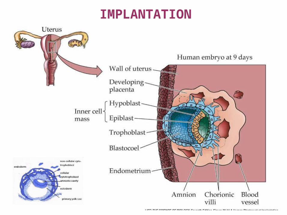
IMPLANTATION

STEPS OF IMPLANTATIONAPPOSITION – the ICM faces the endometrium
ADPLANTATION – the endometrial cells grow processes(they swell and catch the blastocyst - reversible binding)
ADHESION – the microvilli of the trophoblasta interact with the cells of the endometrium (proteoglycans) irreversible binding
DIFFERENTIATION - trophoblast derivatives - syncytiotrophoblast - cytotrophoblast
IMPLANTATION – the syncytiotrophoblasts form a syncytiumot and penetrate the membrana basalis as well as the endometrium
DECIDUAL REACTION – apoptosis within the endometrial cells, then the stroma cells undergo an epitheloid transformation.
The extracellular vacuoles will be filled with blood and merge to form the LACUNAE
the invasive growth will be stopped by the zona compacta
By week 2. the pregnency can be tested from urine too.

MOLECULAR FACTORS OF IMPLANTATION

BILAMINAR EMBRYO

EMBRYOGENESE
Cindy C Lu*, Jane Brennan† and Elizabeth J Robertson‡Current Opinion in Genetics & Development 2001, 11:384–392
= Hypoblast
Extraembryonicstructures
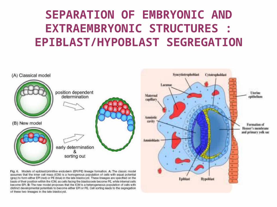
SEPARATION OF EMBRYONIC AND EXTRAEMBRYONIC STRUCTURES :
EPIBLAST/HYPOBLAST SEGREGATION

TRENNUNG DER EMBRYONALEN UND EXTRAEMBRYONALEN STRUKTUREN

EMBRYONIC DISK, HYPOBLAST – EPIBLAST2. weekthe blastocyst partially is implanted, the primitive yolk sac is formed from the blastocoel
EMBRYOBLASTS: FORM THE EMBRYONIC DISK hypoblast - cuboidal cells, line the primitive yolk sac epiblast - tall cylindrical cells lining the amnion cavity D 11-12. a minimal enlargement of the implanted embryo – bulges into the uterine cavity
extraembryonic mesoderm – formed between the trophoblasts and amnion, or yolk sac – the cavities here merge to form the extraemberyonic coelom which surrounds the embryonic disk (except for the site of the body stalk „allantois” – later umbilical cord
D 13. the endometrium is completely sealed
Hypoblasts form the secondary or definitive yolk sacSo the extraembryonic coelom will turn to the cavity of the chorion
CRANIAL changes a thickening forms - praechordal plate (BPM)
CAUDAL changes a similar fusion - cloaca membrane, Similar to the praechordal plate, it is formed by the adjacent layers of hypo- and epiblast cells

IN VITRO FERTILIZATIONHSG
Ovarian stimulation (FSH)
Ultrasound assistedharvesting of the oocyte
„assisted hatching”
ICSI – „intracytoplasmic sperm injection”
Embryo biopsy

FURTHER MOVIES
http://www.visembryo.com/baby/index.htmlhttp://embryo.soad.umich.edu/
www.advancedfertility.comhttp://www.med.uc.edu/embryology/contents.htm