Embed Size (px)
Citation preview
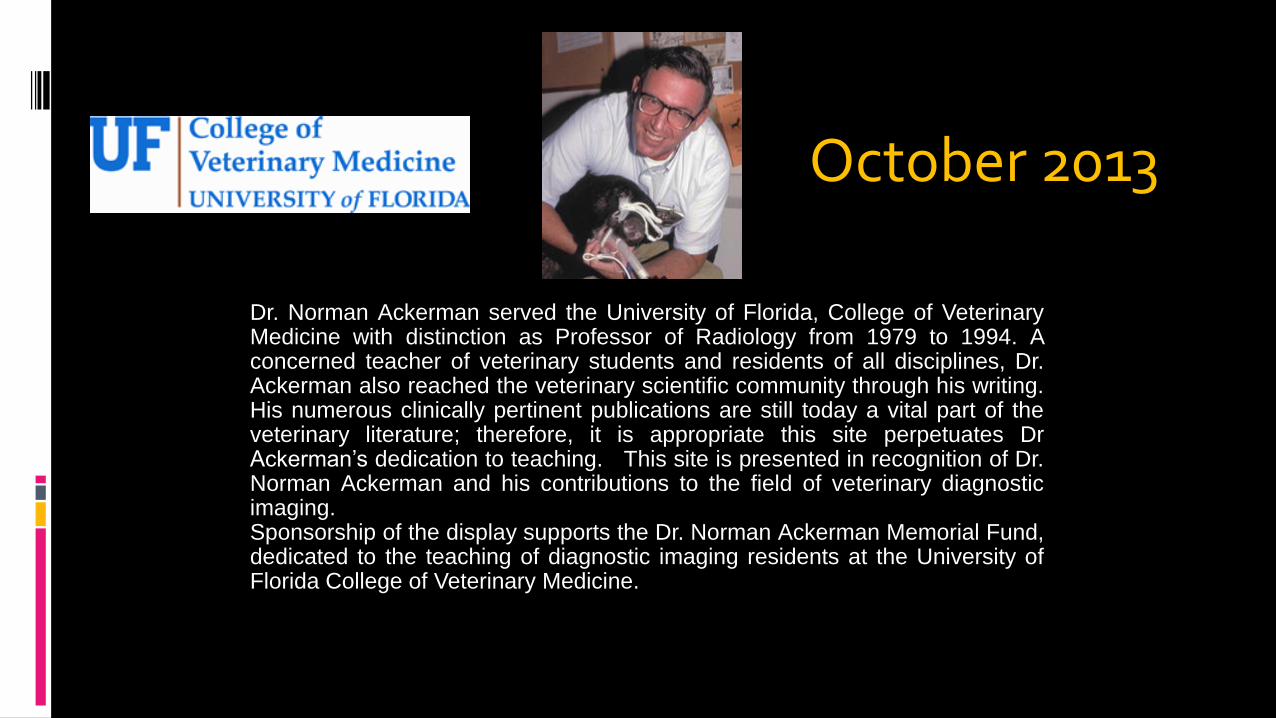
Dr. Norman Ackerman served the University of Florida, College of VeterinaryMedicine with distinction as Professor of Radiology from 1979 to 1994. Aconcerned teacher of veterinary students and residents of all disciplines, Dr.Ackerman also reached the veterinary scientific community through his writing.His numerous clinically pertinent publications are still today a vital part of theveterinary literature; therefore, it is appropriate this site perpetuates DrAckerman’s dedication to teaching. This site is presented in recognition of Dr.Norman Ackerman and his contributions to the field of veterinary diagnosticimaging.Sponsorship of the display supports the Dr. Norman Ackerman Memorial Fund,dedicated to the teaching of diagnostic imaging residents at the University ofFlorida College of Veterinary Medicine.
October 2013
Norman Ackerman Memorial Radiography Case Challenge
Daisy
13 year old FS Mixed Breed Dog
History and case presentation
Daisy presents to your clinic with a 1 month history of vomiting and decreased appetite
On physical examination, Daisy is quiet but alert and responsive, and you hear decreased lung sounds on the left side
You order thoracic radiographs
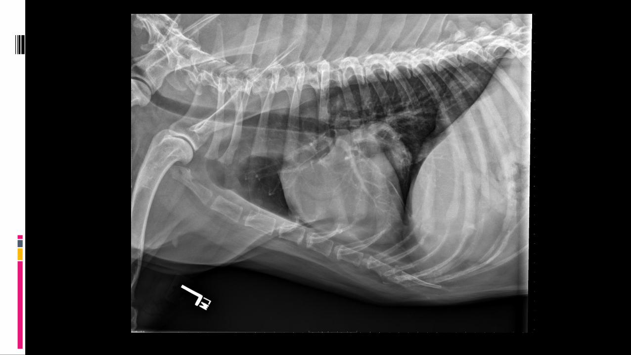
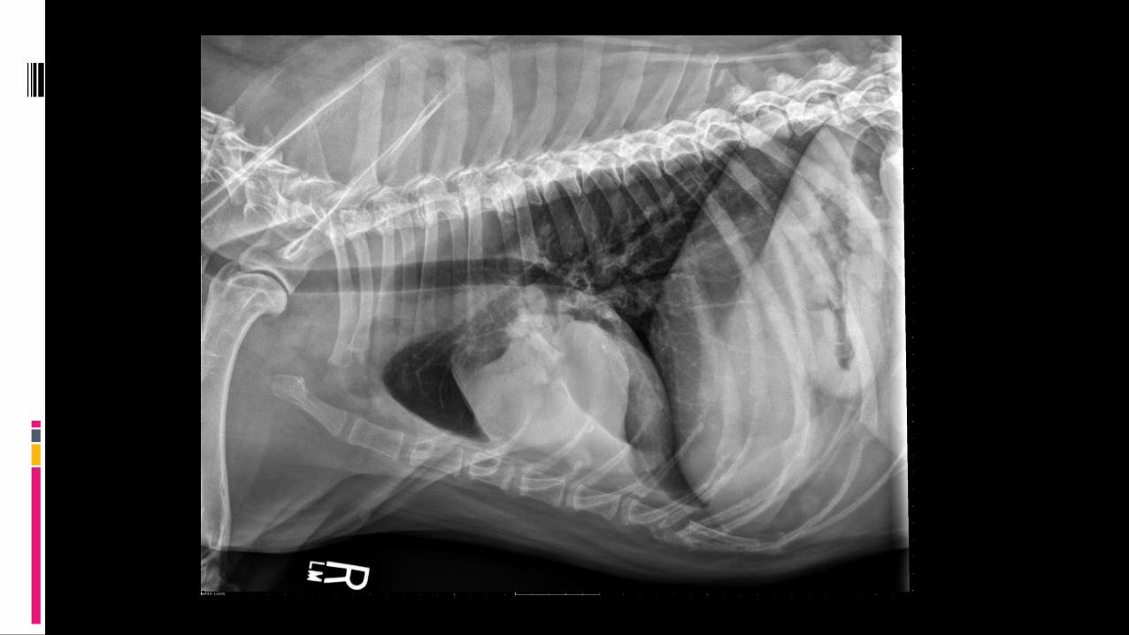
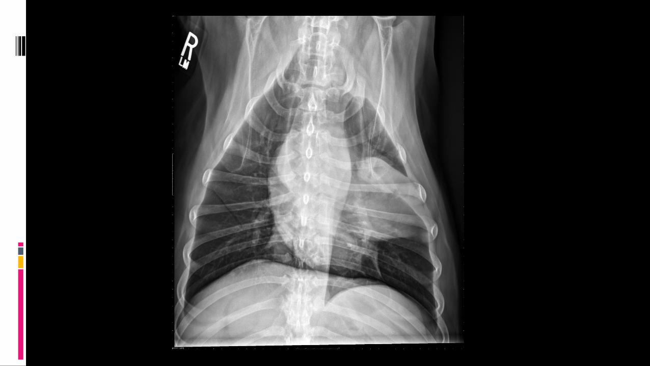
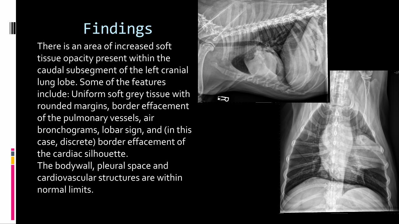
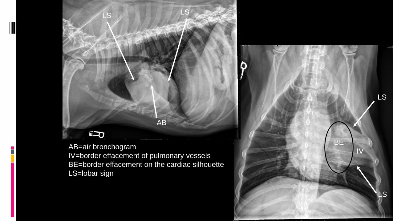
FindingsThere is an area of increased soft tissue opacity present within the caudal subsegment of the left cranial lung lobe. Some of the features include: Uniform soft grey tissue with rounded margins, border effacement of the pulmonary vessels, air bronchograms, lobar sign, and (in this case, discrete) border effacement of the cardiac silhouette.The bodywall, pleural space and cardiovascular structures are within normal limits.
LS
LS
8
IV
AB
BEAB=air bronchogram
IV=border effacement of pulmonary vessels
BE=border effacement on the cardiac silhouette
LS=lobar sign
LSLS
9
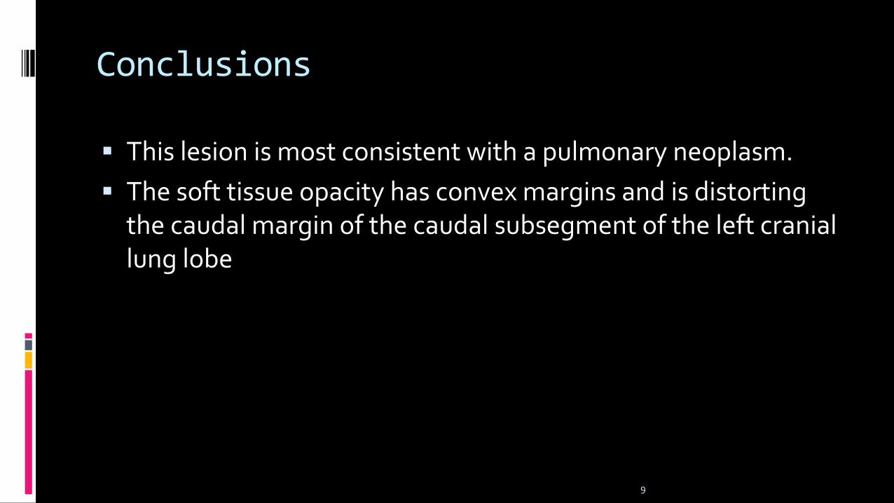
Conclusions
This lesion is most consistent with a pulmonary neoplasm.
The soft tissue opacity has convex margins and is distorting the caudal margin of the caudal subsegment of the left cranial lung lobe
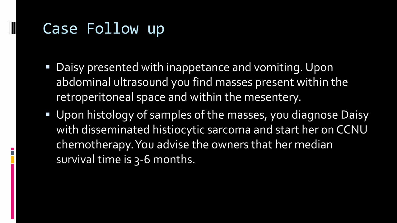
Case Follow up
Daisy presented with inappetance and vomiting. Upon abdominal ultrasound you find masses present within the retroperitoneal space and within the mesentery.
Upon histology of samples of the masses, you diagnose Daisy with disseminated histiocytic sarcoma and start her on CCNU chemotherapy. You advise the owners that her median survival time is 3-6 months.
11
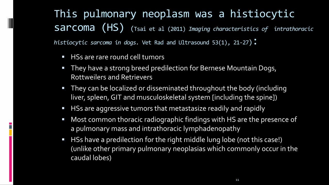
This pulmonary neoplasm was a histiocyticsarcoma (HS) (Tsai et al (2011) Imaging characteristics of intrathoracichistiocytic sarcoma in dogs. Vet Rad and Ultrasound 53(1), 21-27):
HSs are rare round cell tumors
They have a strong breed predilection for Bernese Mountain Dogs, Rottweilers and Retrievers
They can be localized or disseminated throughout the body (including liver, spleen, GIT and musculoskeletal system [including the spine])
HSs are aggressive tumors that metastasize readily and rapidly
Most common thoracic radiographic findings with HS are the presence of a pulmonary mass and intrathoracic lymphadenopathy
HSs have a predilection for the right middle lung lobe (not this case!) (unlike other primary pulmonary neoplasias which commonly occur in the caudal lobes)