Embed Size (px)
Citation preview
DOSE LIMITS AND DOSIMETRY
Voss Associates
1
2
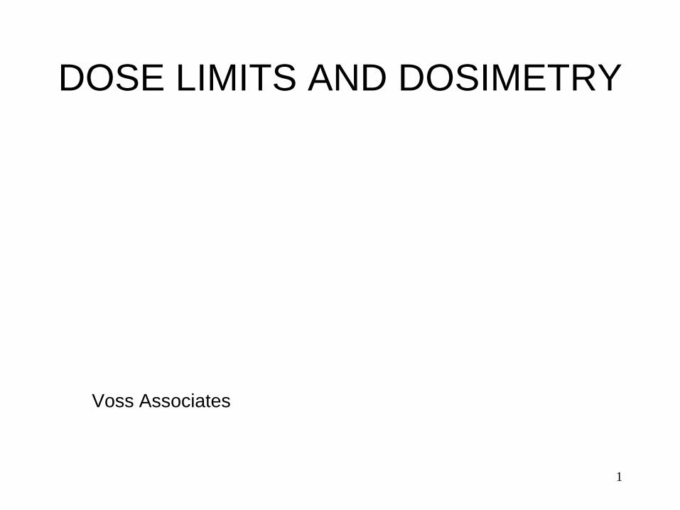
10CFR835
DOSE LIMITS
Radiological Workers
whole body (internal + external) (TEDE) 5 rem/year
lens of the eye (external) 15 rem/year
skin and extremities (external shallow dose) 50 rem/year
any organ or tissue (other than
lens of eye) (internal + external) 50 rem/year
embryo/fetus (internal + external) 0.5 rem/
gestation period
3
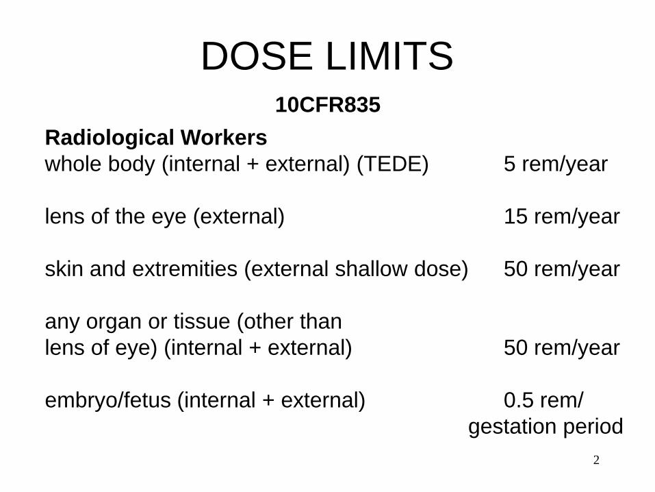
10CFR20
DOSE LIMITS
Radiological Workers
whole body total effective dose equivalent 5 rem/year
lens of the eye 15 rem/year
skin and extremities (external shallow dose) 50 rem/year
any organ or tissue (other than
lens of eye) (sume of DDE + CDE) 50 rem/year
4
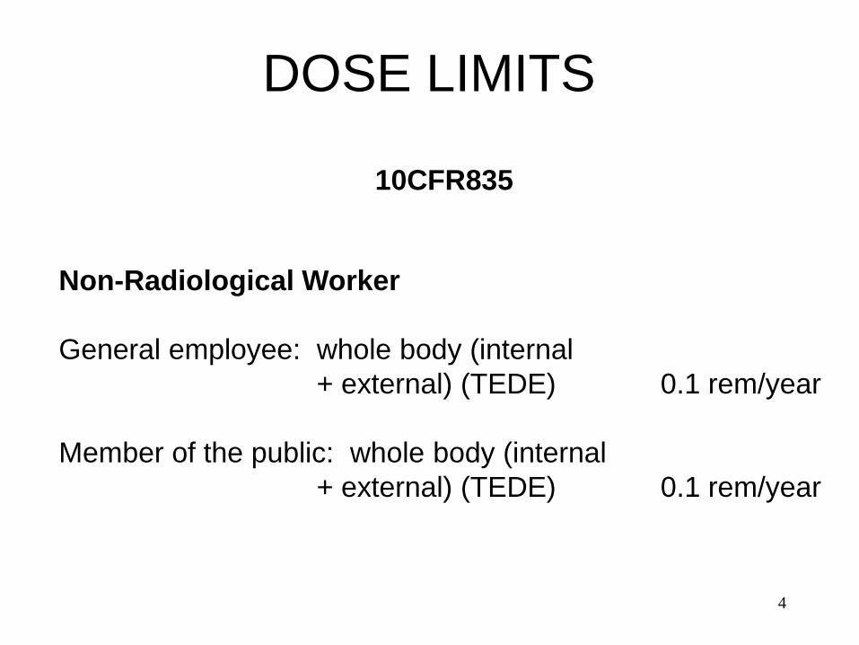
10CFR835
DOSE LIMITS
Non-Radiological Worker
General employee: whole body (internal
+ external) (TEDE) 0.1 rem/year
Member of the public: whole body (internal
+ external) (TEDE) 0.1 rem/year
5
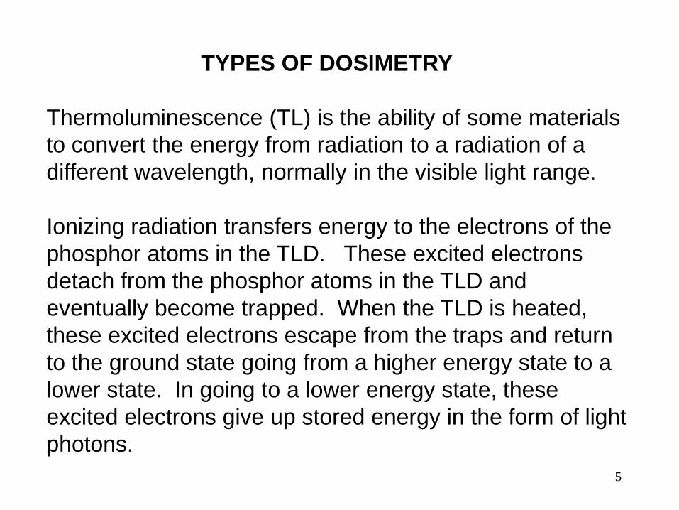
TYPES OF DOSIMETRY
Thermoluminescence (TL) is the ability of some materials
to convert the energy from radiation to a radiation of a
different wavelength, normally in the visible light range.
Ionizing radiation transfers energy to the electrons of the
phosphor atoms in the TLD. These excited electrons
detach from the phosphor atoms in the TLD and
eventually become trapped. When the TLD is heated,
these excited electrons escape from the traps and return
to the ground state going from a higher energy state to a
lower state. In going to a lower energy state, these
excited electrons give up stored energy in the form of light
photons.
6
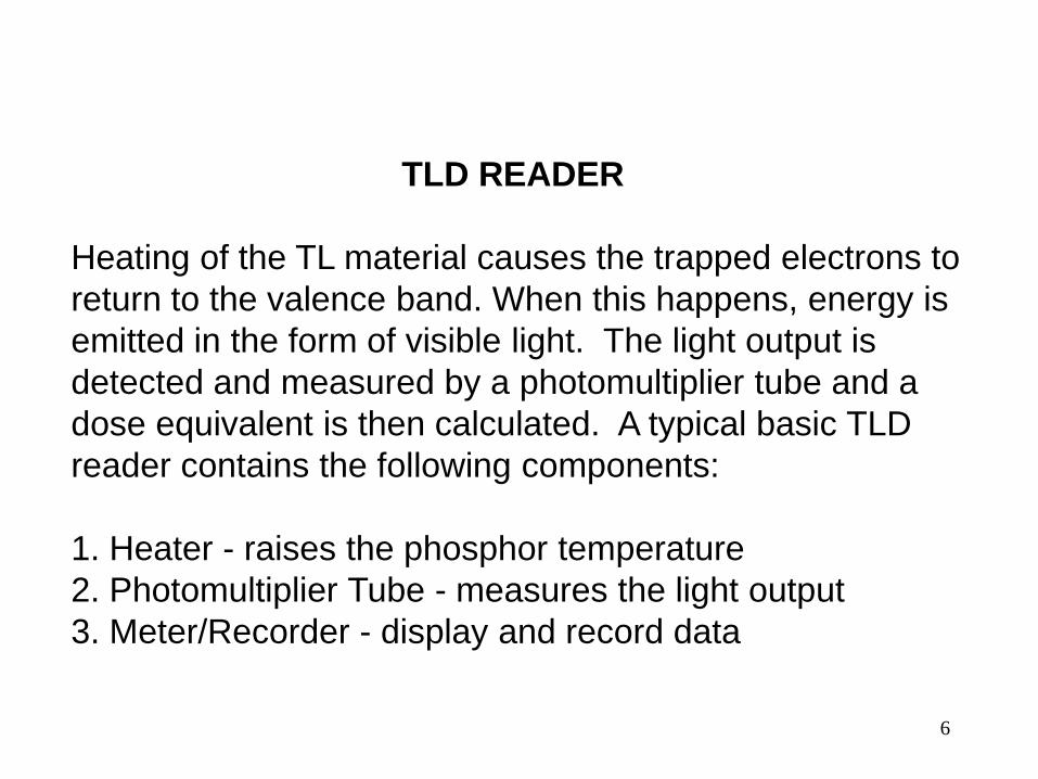
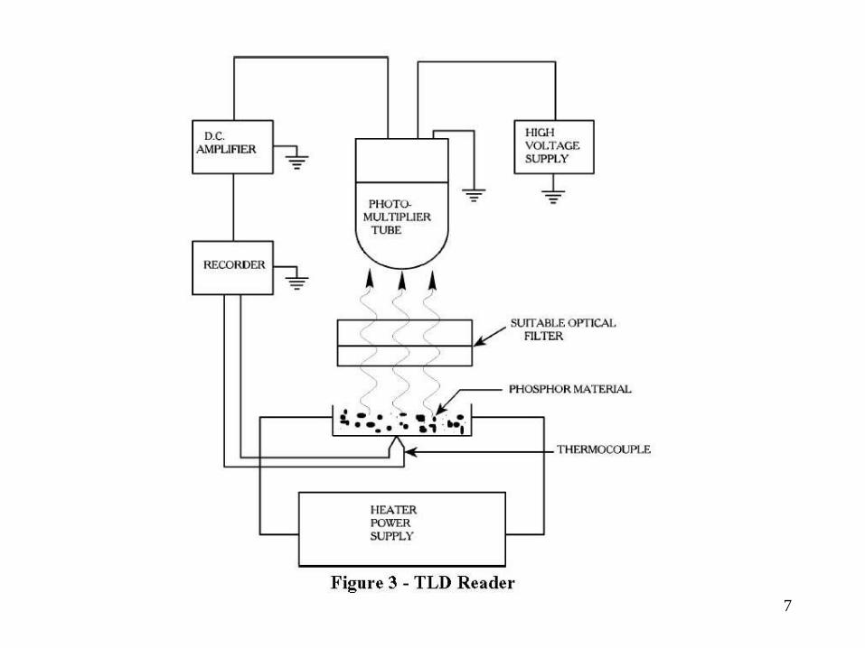
TLD READER
Heating of the TL material causes the trapped electrons to
return to the valence band. When this happens, energy is
emitted in the form of visible light. The light output is
detected and measured by a photomultiplier tube and a
dose equivalent is then calculated. A typical basic TLD
reader contains the following components:
1. Heater - raises the phosphor temperature
2. Photomultiplier Tube - measures the light output
3. Meter/Recorder - display and record data
7
8
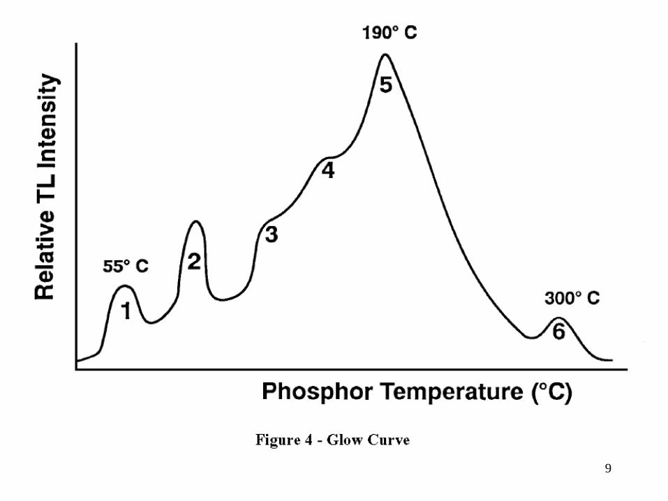
A glow curve can be obtained from the heating process.
The light output from TL material is not easily interpreted.
Multiple peaks result as the material is heated and electrons
trapped in "shallow" traps are released.
This results in a peak as these traps are emptied.
As heating continues, the electrons in deeper traps are
released.
This results in additional peaks.
The area under the curve represents the radiation energy
deposited in the TLD.
9
10
ADVANTAGES AND DISADVANTAGES OF TLDs
1. Advantages (as compared to film dosimeter badges)
include:
a. Able to measure a greater range of doses
b. Doses may be easily obtained
c. They can be read on site instead of being sent
away for developing
d. Quicker turnaround time for readout
e. Reusable
2. Disadvantages
a. Each dose cannot be read out more than once
b. The readout process effectively "zeroes" the
TLD
11
The TLD materials used in many dosimetry badges are
lithium fluoride (LiF) and calcium fluoride (CaF).
Multiple chips are used to provide a separate
measurements of neutron dose and photon deep dose
and shallow dose.
7LiF materials are sensitive to beta and photon radiation
and 6LiF is sensitive to neutron radiation.
CaF materials are sensitive to beta and photon
radiation.
12
To measure the “shallow dose” a relatively thicker mylar
window is placed on top of one of the CaF chips.
This reduces that chip’s response to shallow dose.
By comparing the two CaF chip responses a shallow dose
can be determined.
Calcium Fluoride Dysprosium, Aluminum Oxide, Calcium
Sulfate Dysprosium, and Calcium Fluoride Manganese are
also used in TLD materials. Each has areas of usefulness.
13
Track-etch Dosimeter
Track-etch dosimeters are used to measure high energy
neutrons (> 5 MeV).
High-energy neutrons knock protons out of hydrogen
atoms or other light nuclei in the plastic material.
The recoil protons cause secondary ionization in the
plastic material leaving a track of weakened material.
A chemical bath etches the track left by the recoil proton.
These etched tracks scatter light in the automatic reader.
The amount of scattered light is proportional to the
number of tracks, which is proportional to the neutron
dose.
14
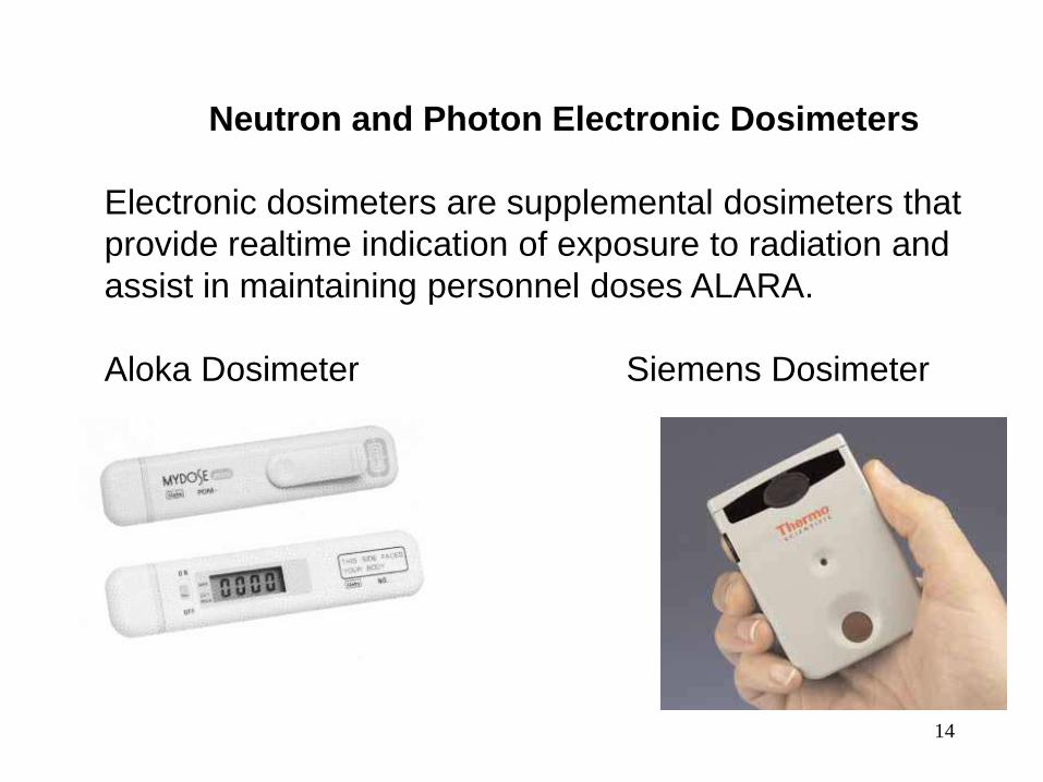
Neutron and Photon Electronic Dosimeters
Electronic dosimeters are supplemental dosimeters that
provide realtime indication of exposure to radiation and
assist in maintaining personnel doses ALARA.
Aloka Dosimeter Siemens Dosimeter
15
Internal Dosimetry
Internal dose evaluation programs (including routine
bioassay programs) should be conducted for those who,
under typical conditions, are likely to receive 0.1 rem
(0.001 sievert) or more committed effective dose
equivalent from all occupational radionuclide intakes in a
year.
BIOASSAY ASSESSMENT METHODS
Bioassay assessment used to determine internal dose
contributions relies on calculation of dose to affected
portions of the body based on the quantities of
radioactive materials in the body.
16
There are two types of bioassay measurements
employed in nuclear industries: in vivo and in vitro.
In vivo bioassay involves counting the living tissue using
a radiation detector.
This method is possible only for those radionuclides
emitting penetrating radiation, e.g., Co-60 and Cs-137 or
bremsstrahlung, e.g., P-32 and Sr-90.
Many radionuclides, Na-22, Fe-59, Co-60, Zn-65, Rb-86,
Sr-85, Te-132, I-131, Cs-137, Ba-140, Ce-144, Au-198,
U-235, Np-239, and Am-241 emit electromagnetic
radiation of sufficient energy to be measured by external
counting.
17
In vitro involves counting an excreted sample, such as
urine.
The amount of material present in the body is estimated
using the amount of materials present in excretions or
secretions from the body.
Samples could include urine, feces, blood, sputum,
saliva, hair, and nasal discharges.
Calculation of dose requires knowledge and use of
metabolic models which allow sample activity to be
related to activity present in the body.

18
Dosimetry Terms
Absorbed Dose (D): Energy absorbed by matter from
ionizing radiation per unit mass of irradiated material at
the place of interest in that material. The absorbed dose is
expressed in units of rad (or gray) (1 rad = 0.01 gray).
Dose Equivalent (H): The product of the absorbed dose
(D)(in rad or gray) in tissue, a quality factor (Q), and all
other modifying factors (N). Dose equivalent is expressed
in units of rem (or sievert) (1 rem = 0.01 sievert).
Deep Dose Equivalent (DDE): The dose equivalent
derived from external radiation at a tissue depth of 1 cm in
tissue (1000 mg/cm2).

19
Shallow Dose Equivalent (SDE): The dose equivalent
derived from external radiation at a depth of 0.007 cm in
tissue (7 mg/cm2).
Whole Body: For the purposes of external exposure,
head, trunk (including male gonads), arms above and
including the elbow, or legs above and including the
knee.
Extremity: Hands and arms below the elbow or feet and
legs below the knee.
20
Committed Dose Equivalent (CDE): The dose equivalent
calculated to be received by a tissue or organ over a 50-
year period after the intake of a radionuclide into the body.
It does not include contributions from radiation sources
external to the body. Committed Dose Equivalent is
expressed in units of rem (or sievert).
Committed effective dose equivalent (H E,50)— The sum
of the committed dose equivalents to various tissues in the
body (HT,50), each multiplied by the appropriate weighting
factor (WT) - that is HE,50=ΣWTHT,50.
Committed effective dose equivalent is expressed in units
of rem (or sievert).

21
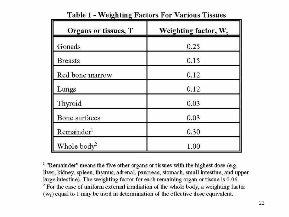
Weighting factor (WT)—The fraction of the overall health
risk, resulting from uniform, whole-body irradiation,
attributable to specific tissue (T).
The dose equivalent to tissue (HT) is multiplied by the
appropriate weighting factor to obtain the effective dose
equivalent contribution from that tissue.
22

23
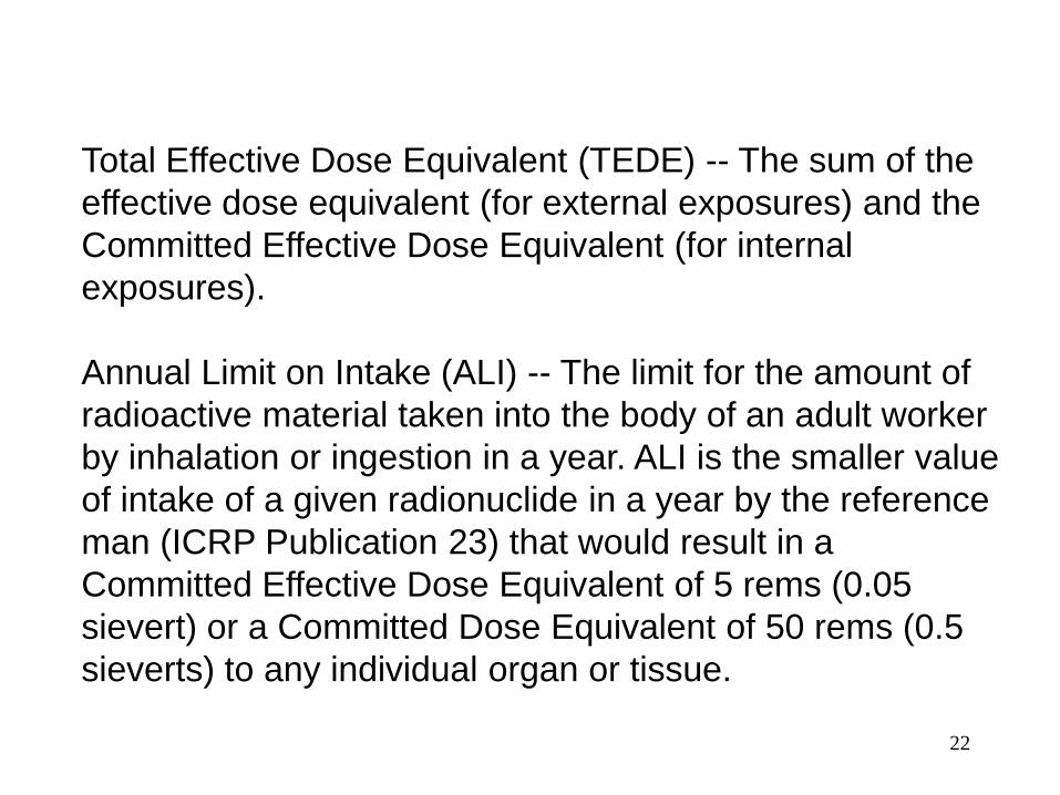
Total Effective Dose Equivalent (TEDE) -- The sum of the
effective dose equivalent (for external exposures) and the
Committed Effective Dose Equivalent (for internal
exposures).
Annual Limit on Intake (ALI) -- The limit for the amount of
radioactive material taken into the body of an adult worker
by inhalation or ingestion in a year. ALI is the smaller value
of intake of a given radionuclide in a year by the reference
man (ICRP Publication 23) that would result in a
Committed Effective Dose Equivalent of 5 rems (0.05
sievert) or a Committed Dose Equivalent of 50 rems (0.5
sieverts) to any individual organ or tissue.
24
Derived Air Concentration (DAC) -- The airborne
concentration that equals the ALI divided by the volume
breathed by an average worker for a working year of 2000
hours (assuming a breathing volume of 2400m3).
Bioassay -- The determination of kinds, quantities, or
concentrations, and, in some cases, locations of radioactive
material in the human body, whether by direct measurement
or by analysis, and evaluation of radioactive materials
excreted or removed from the human body.
Declared pregnant worker -- A woman who has voluntarily
declared to her employer, in writing, her pregnancy for the
purpose of being subject to the occupational dose limits to
the embryo/fetus in accordance with 10CFR835.
1
Radiological Protection
Standards
Voss Associates
2
History Of Standards
Because there are still unknowns, the setting of exposure
limits involves judgments that cannot be wholly based upon
the present body of scientific knowledge.
For this reason, the concept of an "acceptable risk" is used.
The benefits are weighed against the potential damage and
then limits are set at some level at which the most benefit to
mankind will accrue.
Since all exposure is assumed to involve risk to the individual,
exposures should always be kept as low as practicable.
3
Erythema Dose
Early efforts at control were hampered by a lack of
quantitative methods.
There were no units by which one could assess the
amount of radiation.
As a result of the use of radiation by doctors in treating
patients, a unit called the erythema dose came into use.
This was a highly qualitative unit; defined in terms of the
amount of radiation that would produce a well defined
reddening of the skin.
4
Erythema Dose
It varied not only with the type of radiation and the dose rate,
but also with the response of different parts of the body.
This lack of a certain value for this unit made protection work
more or less of a trial-and-error process.
Around 1914, radiation began to be used in industry -- radium
dial-painting and x-rays used for showing flaws in materials.

5
ICRU, ICRP, AND NCRP
In 1925, at the First International Congress of Radiology, the
International Commission on Radiological Units and
Measurements (ICRU) was formed.
In 1928, this group adopted the definition of an international
unit, the Roentgen.
For the first time measurements throughout the world could
be made in terms of the same unit.
Over the years the ICRU has been the main force in defining
and adopting units for use on an international basis.
6
ICRU, ICRP, AND NCRP
At the Second International Congress of Radiology in 1928,
the first international body concerned with protection
standards was formed.
At first known as the International X-ray and Radium
Protection Commission, this group is now called the
International Commission on Radiological Protection
(ICRP).
This group discusses and reviews basic protection
principles, and these recommendations then serve as a
guide from which regulations can be drawn up by each
country to suit its needs.
7
ICRU, ICRP, AND NCRP
In 1934, the ICRP made its first recommendation of a
tolerance level of exposure: 0.2 R/day.
This limit remained in force until 1950.
Because of World War II, the ICRP did not meet
between 1937 and 1950.
This left much of the study of protection standards
during this time to the national committees.

8
ICRU, ICRP, AND NCRP
The National Committee on Radiation Protection and
Measurements (NCRP) was formed in the United States in
1929.
The work of this body was coordinated by the National
Bureau of Standards.
The early recommendations of the Committee appeared in
the National Bureau of Standards Handbooks.
The NCRP recommendations as outlined in Handbooks 20
and 23, served as the basis for protection practices during
the days of the Manhattan project.
9
ICRU, ICRP, AND NCRP
The development of the atomic bomb had a dramatic impact
on radiation protection problems.
Before the war, most of the problems concerned rather low
energy x-rays.
During WWII, not only were there these to treat, but also
other types of radiation with a wide range of energies.
There was a large increase of workers in the radiation field.
10
ICRU, ICRP, AND NCRP
New units were needed to define the dose contributed by
radiation other than x-rays.
Large amounts of waste were produced and methods of
disposal were being developed.
With reactors in use, not only the workers, but also others
not connected with the work, had to be considered.
11
ICRU, ICRP, AND NCRP
The NCRP met in 1946 to reorganize.
At this time a number of subcommittees were formed to
deal with the new problems more effectively.
This resulted in the publication of a number of handbooks
after the war that represented changes and additions to the
old recommendations.
The Committee was replaced by a non-profit corporation
chartered by Congress in 1964 and is now known as the
National Council on Radiation Protection and
Measurements.
12
ICRU, ICRP, AND NCRP
These various NCRP committees develop proposed recommendations on various aspects of radiation protection and radiation measurements, which when approved by the Council, are published as NCRP Reports.
The three organizations, ICRU, ICRP and NCRP, have figured prominently in the development of present day radiation protection practices.
13
Radiation Exposure Concerns
Initial concerns resulted from medical exposure to external
radiation from the use of x-rays for diagnosis and therapy.
World War II introduced considerations about internal
exposure and the dose to the general public.
Potential genetic effects of radiation and the impact of long-
term exposure at low dose rates emerged.
Data from biological studies seemed to indicate that one could
not assume that all effects had a threshold dose.
14
Radiation Exposure Concerns
Efforts have been directed toward quantifying the risk
associated with a certain level of exposure.
The Non-Threshold relationship assumes that any dose
carries some risk of producing damage therefore all
exposure should be kept at the lowest practical levels.
15
Factors to be considered:
Information available for the quantification of risks is
imperfect.
The assumptions of a risk by an individual presumes a
willingness to chance the risk in exchange for some
resultant benefit which justifies the risk.
The balancing of risk versus benefit in order to obtain a net
benefit is not easily accomplished.
The approach adopted by both the ICRP and the NCRP is to
keep exposures ALARA.
16
Radiation Exposure Concerns
The continuing reviews of biological data have revealed
two types of radiation effects.
–Practical threshold dose -- nonstochastic effects.
–No threshold -- stochastic effects.
Nonstochastic effects -- prevented by limiting the dose
to the individual to a value below the threshold dose for
occurrence of the effects.
Stochastic effects -- limit the probability of occurrence to
some level (deemed acceptable) by limiting the
radiation exposure.
17
Federal Policy on Radiation Matters
In 1959, the Federal Radiation Council (FRC) was
formed (Public Law 86-373).
– Advised the President concerning radiation matters
– Provided guidance for all Federal agencies in setting
standards and in working with the States.
– The recommendations of the FRC were approved in
1960 and formed the basis of the Federal radiation
protection guidance.
– The FRC was abolished by Reorganization Plan
No. 3 in 1970.
18
Office of Radiation Programs (ORP) of the EPA took over
the activities of the FRC.
In 1981 the EPA drafted proposed revised
recommendations in the Federal Register regarding
occupational exposure, and solicited comments.
The EPA believes that it is appropriate to adopt the general
features of the ICRP approach in radiation protection
guidance for use by Federal agencies for occupational
exposure.
The revised EPA guidance was approved and issued in
January 1987.
19
Regulating Agencies
In 1954 under the Atomic Energy Act the United States
Atomic Energy commission (AEC) was given the
responsibility of regulating the atomic energy industry.
– The Act authorized the AEC to set up a licensing
program to be augmented by whatever rules or regulations
were deemed appropriate.
– The bases for these rules are: to protect the public health
and safety, and provide for national defense and security.
In 1974 the Energy Reorganization Act abolished the AEC
and established two agencies to perform the functions of the
AEC, Nuclear Regulatory Commission (NRC) and the Energy
Research and Development Administration (ERDA).
20
Regulating Agencies
NRC
The NRC is charged with inspection and review in order to
assure compliance.
– This function is carried out by NRC personnel (inspectors)
at regular intervals.
– Their job is to make the inspections and report their
findings.
– In the event that a failure to comply is noted, the licensee
is required to correct this.
States have set up their own safety standards with the
assistance of the NRC to assure that the state and
Commission programs are compatible. These states are
referred to as Agreement States.

21
Regulating Agencies
NRC
The NRC has established radiation protection standards that
pertain to many areas of activities.
10CFR20
10CFR30
10CFR34
10CFR35
10CFR70
10CFR71
are the regulations of most interest for NRC-regulated
activities.
22
Regulating Agencies
DOE
In 1977 the U.S. Department of Energy (DOE) replaced
ERDA.
The DOE activities relate to energy research and
development.
The DOE has issued occupational radiation protection
standards that pertain to its own activities as well as to those
of its contractors.
– These standards appear in 10 CFR Part 835.
– These standards are based upon the recommendations of
the ICRP, NCRP and the guidance of the EPA.
23
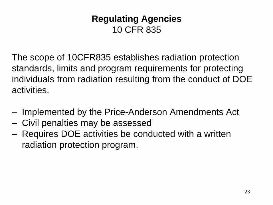
Regulating Agencies
10 CFR 835
The scope of 10CFR835 establishes radiation protection
standards, limits and program requirements for protecting
individuals from radiation resulting from the conduct of DOE
activities.
– Implemented by the Price-Anderson Amendments Act
– Civil penalties may be assessed
– Requires DOE activities be conducted with a written
radiation protection program.
24
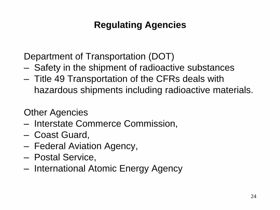
Regulating Agencies
Department of Transportation (DOT)
– Safety in the shipment of radioactive substances
– Title 49 Transportation of the CFRs deals with
hazardous shipments including radioactive materials.
Other Agencies
– Interstate Commerce Commission,
– Coast Guard,
– Federal Aviation Agency,
– Postal Service,
– International Atomic Energy Agency
CONTAMINATION CONTROL
Voss Associates
1
2
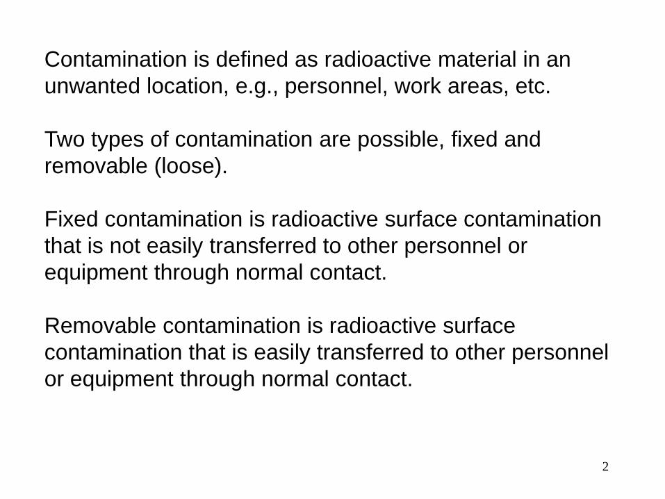
Contamination is defined as radioactive material in an
unwanted location, e.g., personnel, work areas, etc.
Two types of contamination are possible, fixed and
removable (loose).
Fixed contamination is radioactive surface contamination
that is not easily transferred to other personnel or
equipment through normal contact.
Removable contamination is radioactive surface
contamination that is easily transferred to other personnel
or equipment through normal contact.
3
Fixed contamination is measured by use of a direct survey
technique using a portable radiation survey instrument.
This technique, commonly referred to as "frisking" or
“scanning”, indicates the total contamination on a surface
apparent to the detector from both fixed and removable
contamination.
When non-removable levels are to be recorded, the
removable level must be subtracted from the total.
4
Removable contamination is measured by a transfer test
using a suitable sampling material. Common materials used
for the monitoring are the standard paper disk smear or cloth
smear. The standard technique involves wiping approximately
100 cm2 of the surface of interest using moderate pressure. A
common sampling practice used to ensure a 100 cm2 sample
is to wipe a 16 square inch "S" shape on the surface (i.e., 4
inches by 4 inches).
Qualitative, large area wipe surveys may be taken using other
materials, such as Masslinn cloth or Kimwipe, to indicate the
presence of removable contamination. Large area swipes
are commonly used when exact levels of contamination are
not required.
5
Constant (Continuous) Monitoring
There are various types of constant monitoring
instruments throughout the facilities to warn personnel of
radiation and contamination hazards.
Some instruments are permanently installed, and some
instruments are portable to allow movement from place to
place as deemed appropriate by the radiological control
staff.
6
Continuous Air Monitor (CAM)
These instruments continuously sample the air for
radioactive contamination in specific locations.
The air being sampled is typically drawn through a moving
particulate filter which is then monitored by a detector
system or through an internal detector to directly identify
radioactive materials present.
A CAM can give both a visual and audible alarm to warn
personnel of the presence of airborne contamination.
7
Process Monitoring Systems
Process monitoring systems monitor certain operations in
various facilities to alert operators of abnormal conditions
which might lead to the release of excessive amounts of
radioactivity to the facility or environment.
Process monitors include temperature, pressure, and flow
sensors.
8
Area and Equipment Surveys
Area and equipment surveys are conducted routinely
throughout the facilities to locate sources of radiation and
contamination and to detect potential changes in
radiological conditions.
Pre-job surveys are performed prior to work in radiological
areas in order to evaluate the hazards and determine work
limitations and physical safeguards.
Direct surveys with portable radiation detection
instruments and removable contamination surveys with
smears or swipes are used.
9
External Personnel Surveys
Personnel surveys are either performed by the individual
(self-monitoring) using handheld or automated
instruments or by a radiological control technician.
Self-monitoring is typically performed upon exiting a
contaminated area at established boundary points.
Personnel monitoring by a RCT is usually conducted
whenever contamination of the body or clothing is
suspected, or as required by exit monitoring when self-
monitoring is not feasible (remote location) or not
allowed.
10
Personnel Internal Monitoring
A routine program of internal contamination monitoring is
conducted as a final check on contamination control
procedures.
The program consists of external whole/partial body
counting and/or urinalysis.
11
Once the presence of radioactive material has been
located, the basic goal underlying any effective
contamination control program is to minimize
contaminated areas and maintain contamination levels as
low as reasonably achievable.
In some situations, this is not always possible due to:
• Economical conditions: Cost of time and labor to
decontaminate a location(s) outweighs the hazards of
the contamination present.
• Radiological conditions: Radiation dose rates or other
radiological conditions present hazards which far
exceed the benefits of decontamination.
• Operating conditions: Some areas, e.g., hot cells, will
be contaminated due to normal operations.
12
Other means of control must be initiated when
decontamination is not possible.
Engineering control (ventilation and containment),
administrative procedures (RWPs), and personnel protective
equipment are alternatives for the control of contamination.
In Fixed Contamination Areas the contamination may be
covered by paint, floor tiles, etc. when decontamination is not
possible.
13
CONTAMINATION CONTROL MEASURES
• Access/Administrative Controls
• Engineering Controls
• Personnel Protective Measures
• Decontamination
• Preventive Methods
14
Access/Administrative Controls
Once contamination has been located and quantified
and radiological areas have been determined, access
control to these areas must be adequately established.
Work Authorization, Radiological Posting, and
Radiological Work Permits are the primary administrative
controls.

15
ENGINEERING CONTROLS
Ventilation and Containment (Confinement) are the main
engineering controls.
Ventilation is a process of providing adequate air flow to
keep the radiological area well ventilated and to keep
the potential airborne radioactivity from migrating to
other non-radiological areas. This is done thru negative
pressure, much like some businesses use to keep their
shops temperature and humidity controlled without trying
to air condition the outside environment.
Containment (or confinement) is simply keeping
radioactive materials in enclosures adequate to prevent
the materials from getting outside the container.
16
In vitro involves counting an excreted sample, such as urine.
The amount of material present in the body is estimated
using the amount of materials present in excretions or
secretions from the body.
Samples could include urine, feces, blood, sputum, saliva,
hair, and nasal discharges.
Calculation of dose requires knowledge and use of
metabolic models which allow sample activity to be related
to activity present in the body.
PERSONNEL PROTECTIVE MEASURES
17
Dosimetry Terms
Absorbed Dose (D): Energy absorbed by matter from
ionizing radiation per unit mass of irradiated material at
the place of interest in that material. The absorbed dose is
expressed in units of rad (or gray) (1 rad = 0.01 gray).
Dose Equivalent (H): The product of the absorbed dose
(D)(in rad or gray) in tissue, a quality factor (Q), and all
other modifying factors (N). Dose equivalent is expressed
in units of rem (or sievert) (1 rem = 0.01 sievert).
Deep Dose Equivalent (DDE): The dose equivalent
derived from external radiation at a tissue depth of 1 cm in
tissue (1000 mg/cm2).
18
Shallow Dose Equivalent (SDE): The dose equivalent
derived from external radiation at a depth of 0.007 cm in
tissue (7 mg/cm2).
Whole Body: For the purposes of external exposure,
head, trunk (including male gonads), arms above and
including the elbow, or legs above and including the
knee.
Extremity: Hands and arms below the elbow or feet and
legs below the knee.

19
Committed Dose Equivalent (CDE): The dose equivalent
calculated to be received by a tissue or organ over a 50-
year period after the intake of a radionuclide into the body.
It does not include contributions from radiation sources
external to the body. Committed Dose Equivalent is
expressed in units of rem (or sievert).
Committed effective dose equivalent (H E,50)— The sum
of the committed dose equivalents to various tissues in the
body (HT,50), each multiplied by the appropriate weighting
factor (WT) - that is HE,50=ΣWTHT,50.
Committed effective dose equivalent is expressed in units
of rem (or sievert).
20
Weighting factor (WT)—The fraction of the overall health
risk, resulting from uniform, whole-body irradiation,
attributable to specific tissue (T).
The dose equivalent to tissue (HT) is multiplied by the
appropriate weighting factor to obtain the effective dose
equivalent contribution from that tissue.

21
22
Total Effective Dose Equivalent (TEDE) -- The sum of the
effective dose equivalent (for external exposures) and the
Committed Effective Dose Equivalent (for internal
exposures).
Annual Limit on Intake (ALI) -- The limit for the amount of
radioactive material taken into the body of an adult worker
by inhalation or ingestion in a year. ALI is the smaller value
of intake of a given radionuclide in a year by the reference
man (ICRP Publication 23) that would result in a
Committed Effective Dose Equivalent of 5 rems (0.05
sievert) or a Committed Dose Equivalent of 50 rems (0.5
sieverts) to any individual organ or tissue.

23
Derived Air Concentration (DAC) -- The airborne
concentration that equals the ALI divided by the volume
breathed by an average worker for a working year of 2000
hours (assuming a breathing volume of 2400m3).
Bioassay -- The determination of kinds, quantities, or
concentrations, and, in some cases, locations of radioactive
material in the human body, whether by direct measurement
or by analysis, and evaluation of radioactive materials
excreted or removed from the human body.
Declared pregnant worker -- A woman who has voluntarily
declared to her employer, in writing, her pregnancy for the
purpose of being subject to the occupational dose limits to
the embryo/fetus in accordance with 10CFR835.
1
External Exposure Control
Voss Associates
2
Basic Methods for Exposure Reduction
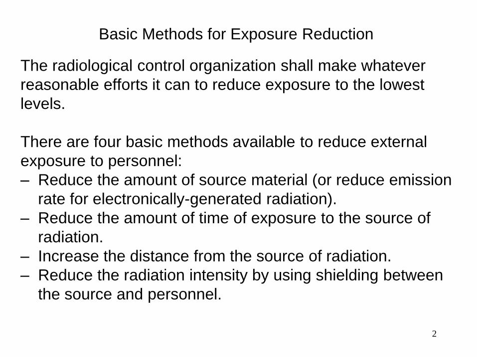
The radiological control organization shall make whatever
reasonable efforts it can to reduce exposure to the lowest
levels.
There are four basic methods available to reduce external
exposure to personnel:
– Reduce the amount of source material (or reduce emission
rate for electronically-generated radiation).
– Reduce the amount of time of exposure to the source of
radiation.
– Increase the distance from the source of radiation.
– Reduce the radiation intensity by using shielding between
the source and personnel.
3
Basic Methods for Exposure Reduction
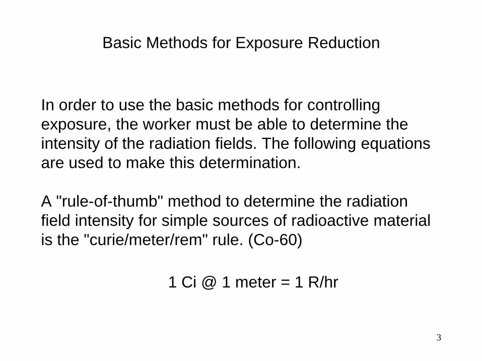
In order to use the basic methods for controlling
exposure, the worker must be able to determine the
intensity of the radiation fields. The following equations
are used to make this determination.
A "rule-of-thumb" method to determine the radiation
field intensity for simple sources of radioactive material
is the "curie/meter/rem" rule. (Co-60)
1 Ci @ 1 meter = 1 R/hr
4
Basic Methods for Exposure Reduction
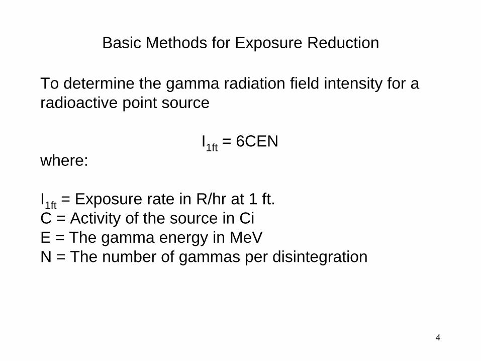
To determine the gamma radiation field intensity for a
radioactive point source
I1ft = 6CEN
where:
I1ft = Exposure rate in R/hr at 1 ft.
C = Activity of the source in Ci
E = The gamma energy in MeV
N = The number of gammas per disintegration
5
Basic Methods for Exposure Reduction
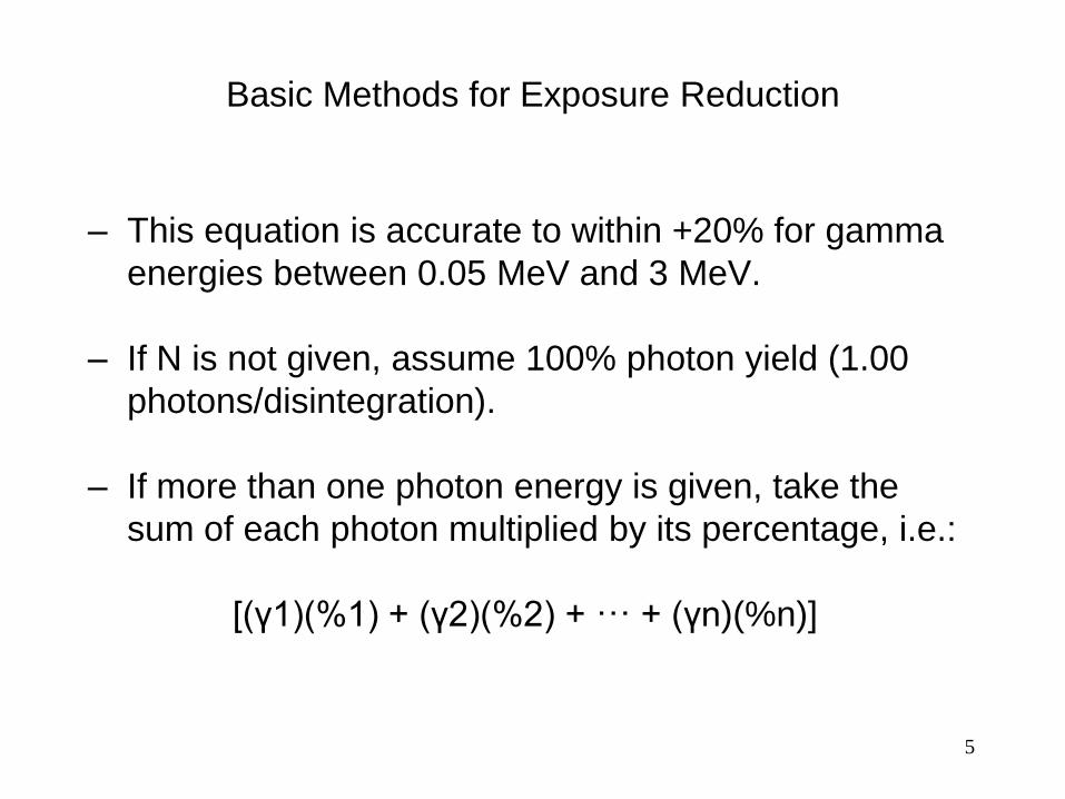
– This equation is accurate to within +20% for gamma
energies between 0.05 MeV and 3 MeV.
– If N is not given, assume 100% photon yield (1.00
photons/disintegration).
– If more than one photon energy is given, take the
sum of each photon multiplied by its percentage, i.e.:
[(γ1)(%1) + (γ2)(%2) + ··· + (γn)(%n)]
6
Basic Methods for Exposure Reduction
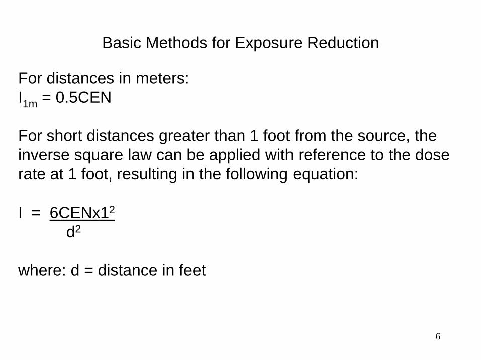
For distances in meters:
I1m = 0.5CEN
For short distances greater than 1 foot from the source, the
inverse square law can be applied with reference to the dose
rate at 1 foot, resulting in the following equation:
I = 6CENx12
d2
where: d = distance in feet
7
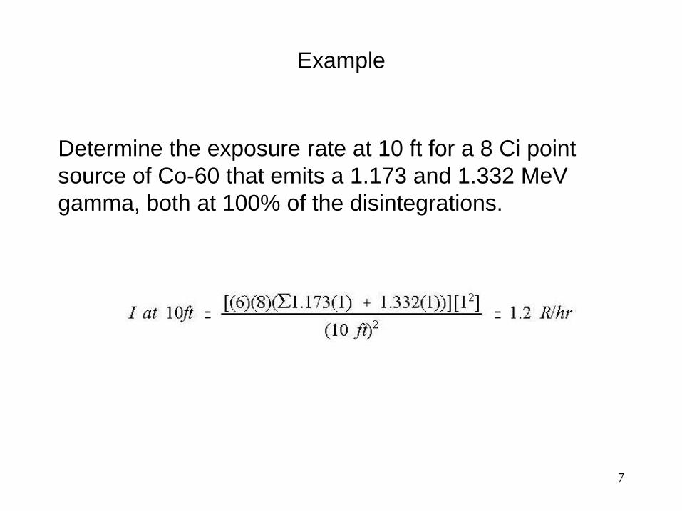
Example
Determine the exposure rate at 10 ft for a 8 Ci point
source of Co-60 that emits a 1.173 and 1.332 MeV
gamma, both at 100% of the disintegrations.
8
Source Reduction
The first method that should be employed to reduce personnel
external exposure is source reduction. If a source can be
eliminated or if its hazard potential can be significantly
reduced, then other engineering means may not be necessary.
Various techniques are employed to accomplish external
exposure reduction using source reduction.
Allow natural decay to reduce source strength
– If the radioisotopes involved are short-lived, then waiting to
perform the task may significantly reduce the hazard.
– By waiting for natural decay to reduce the source strength, a
considerable savings in external exposure can be achieved.
9
Source Reduction
Move the source material to another location
– Decontaminate the equipment or material through mechanical or chemical means to remove the source material prior to working in the area or on the equipment.
– Reduce the source material in the system by flushing equipment with hot water or chemical solutions and collect itin a less frequently occupied area.
– Discharge or remove the resin or filtering media prior toworking in the area or on the system.
– Move the radioactive source (e.g., a drum, barrel or calibration source) to another location prior to starting work.
10
Time Savings
Personnel working in radiation fields must limit their exposure
time so that they do not exceed their established permissible
dose limits and are able to keep exposures ALARA.
The longer the time spent in the radiation field, the greater
the exposure to the individual; therefore, the amount of time
spent in radiation fields should be reduced.
The Radiological Control Technician needs to be aware that
radiation exposures are directly proportional to the time spent
in the field. If the amount of time is doubled, then the amount
of exposure received is doubled.
11
Time Savings
Analyze and train using mock-ups of the work site
– A particular task can be analyzed on a mock-up of the
system to determine the quickest and most efficient
method to perform the task.
– The team of workers assigned to the task can rehearse,
without radioactive materials, so that problems can be
worked out and the efficiency of the team increased prior
to any exposure.
– By determining the most efficient method and rehearsing
the task, the amount of time, and therefore the exposure,
can be reduced.

12
Time Savings
Pre-job briefings are an important part of any good ALARA
program
– Discussions at the pre-job briefing with the individuals
assigned to the task can identify any potential problems not
previously identified.
– Identifying personnel responsibilities and the points at
which various individuals are required to be present can
reduce the overall time required to perform the job.
Review job history files -- Review the files from previously
completed tasks of the same nature to identify previous
problems and spots where time could be saved.

13
Time Savings
Pre-stage all tools and equipment -- All tools should be
staged prior to entry to prevent the worker from waiting in a
radiation field for a tool to be brought.
Pre-assemble equipment and tools outside the area
– Equipment that can be preassembled should be
preassembled prior to any entry into the radiation field.
– Tools that require assembly, pre-testing, and/or calibration
should be performed outside the radiation field.
14
Time Savings
Use time limiting devices -- Time limitations for workers
can be monitored and limited using various devices
such as stopwatches, alarming dosimeters, or radio-
transmitting dosimeters.
Use communication devices such as walkie-talkies
– Poor communication can lead to incorrect or poor
quality work and prolonged waiting in the radiation
field while supervisors or experts are contacted.
– Communication devices such as walkie-talkies or
radio headsets can alleviate these problems and
reduce the amount of time that is spent in the
radiation field.
15
Time Savings
Use a team of workers instead of allowing one individual to
receive all of the exposure
– Even if the task requires a minimum amount of time, if it
causes one individual to receive an exposure greater than
allowable, a team of workers should be used to reduce the
individual exposures.
– If a team of workers is used, good communications are
necessary to ensure the total exposure for the job does
not increase significantly.
16
Use experienced personnel
- The total time required to perform a job is reduced if experts
are used instead of inexperienced personnel.
- Inexperienced personnel should not be trained in significant
radiation fields
Time Savings

17
Exposure Calculation
The exposure received by personnel will increase as
the time spent in the radiation field increases.
18
Stay Time Calculation
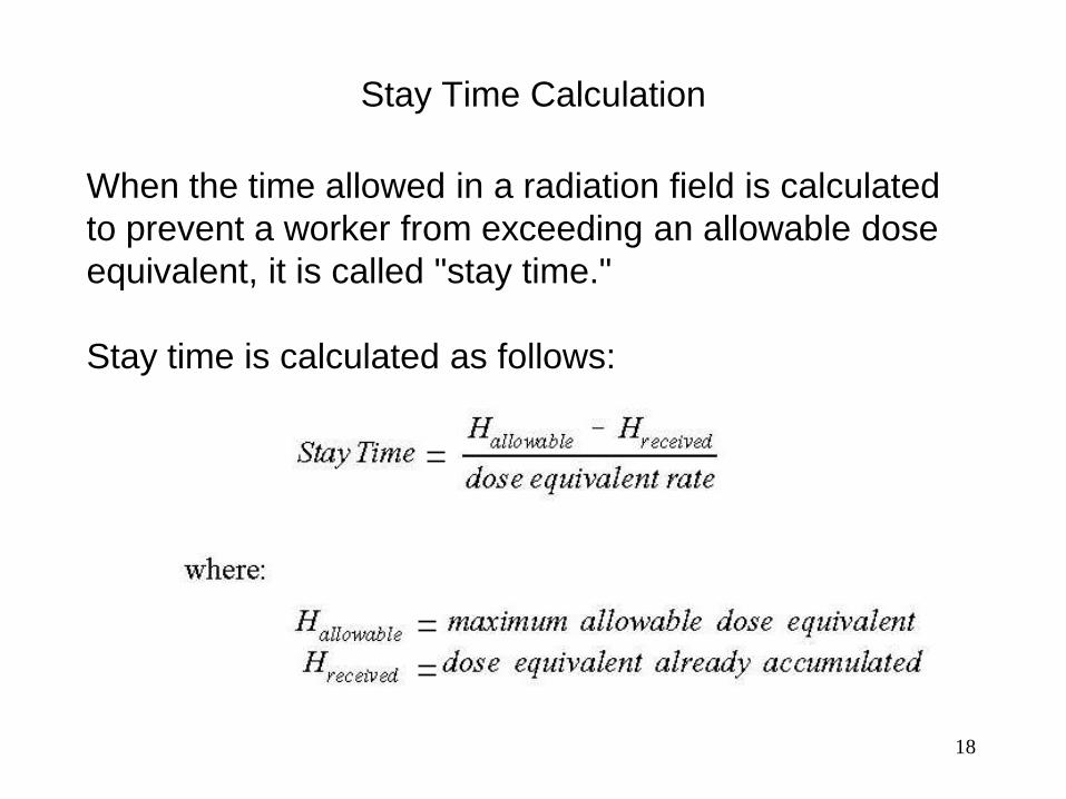
When the time allowed in a radiation field is calculated
to prevent a worker from exceeding an allowable dose
equivalent, it is called "stay time."
Stay time is calculated as follows:
19
Distance
The intensity of the radiation field decreases as the distance
from the source increases.
Therefore, increasing the distance will reduce the amount of
exposure received.
In many cases, increasing the distance from the source is
more effective than decreasing the time spent in the radiation
field.
20
Distance
The radiation intensity for a point source decreases
according to the Inverse Square Law which states that
as the distance from a point source changes the dose
rate decreases or increases by the square of the ratio of
the distances from the source.
The inverse square law becomes inaccurate close to
the source (i.e., about 10 times the diameter of the
source).
For a point source, if the distance is doubled, the
radiation intensity will be reduced by a factor of (2)2 or 4.
21
Distance
Remote handling tools/remote control devices – Tools, such as tongs or long-handled tools, are an
effective means of increasing the distance from a point source to a worker.
– For very high radiation fields, remote control devices may be appropriate, especially if the task is performed frequently.
Remote observation by cameras or indicators– Gauges or meters can be moved to a location remote from
the source of radiation.– Closed-circuit television and video cameras can be used
to allow observation of work activities or system operations from a location remote to the source of radiation.
22
Distance
Move work to another location
– If the source of radiation can not be reduced, then
possibly the work can be moved to a low exposure
area.
– For example, if a pump or valve needs reworking,
then an exposure savings could be achieved by
removing the component from the system and
performing all repair work in a lower exposure area.
23
Distance
Posting of areas -- Posting of radiological areas based
on radiation level is a method for increasing the
distance between the workers and the radiation source.
Extendable Instruments -- Extendable radiation survey
instruments, such as the Eberline Teletector or RO-7,
can reduce the exposure to the surveyor by increasing
the distance.
24
Distance
– For workers or inspectors not actively engaged in the workactivity in the radiation field, moving to a lower exposurerate "waiting" area can be effective.
– Identifying "low dose rate waiting areas" can notify workersof the location of the lowest exposure rate in an area or room.
– Be aware of the location of radiation sources at theworksite and locate the worker at a point farthest from the source.
– Work at arm's length and do not lie on or hug radioactive components.
25
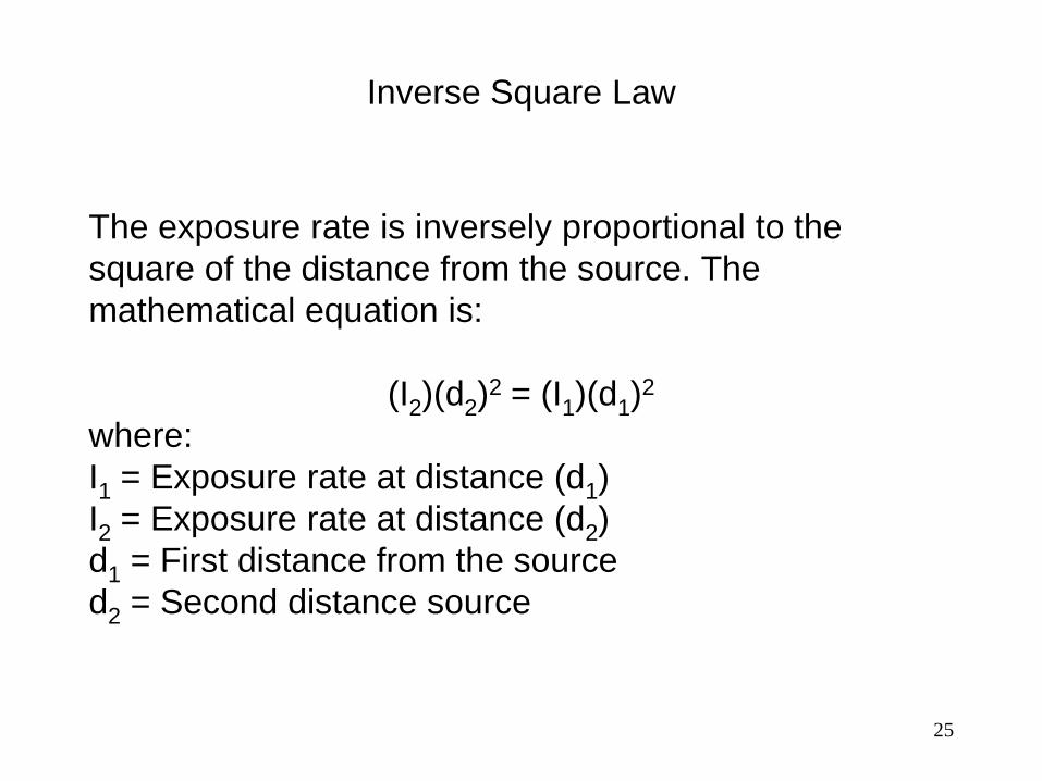
Inverse Square Law
The exposure rate is inversely proportional to the
square of the distance from the source. The
mathematical equation is:
(I2)(d2)2 = (I1)(d1)
2
where:
I1 = Exposure rate at distance (d1)
I2 = Exposure rate at distance (d2)
d1 = First distance from the source
d2 = Second distance source
26
Inverse Square Law
A somewhat useful variation on the inverse square law
is the use of a quadratic equation when the actual
distance to the source is not known.
How does the inverse square law apply to alpha, beta,
and neutron radiation ?
27
Inverse Square Law
The inverse square law holds true only for point
sources; however, it gives a good approximation when
the source dimensions are smaller than the distance
from the source to the exposure point.
Some sources, such as a pipe or tank, cannot be
treated as a point source unless the distance to the
source is greater than the length of the source.
28
Line Source Calculations
The actual calculations for a line source involve
calculus; however, the mathematics can be simplified if
the line source is treated as a series of point sources
placed side by side along the length of the source.
If the line source is treated in this manner, the
relationship between distance and exposure rate can be
written mathematically as:
I1d1 = I2d2
29
Line Source Calculations
The exposure rate is inversely proportional to the distance from the source
Assuming the source material is distributed evenly along the line
Assuming the point at which the exposure rate is calculated is on a line perpendicular to the center of the line source
Assuming the width or diameter of the line is small compared to the length
30
Planar Or Surface Sources
Planar or surface sources of radiation can be any type of
geometry where the width or diameter is not small compared
to the length.
When the distance to the plane source is small compared to
the longest dimension, then the exposure rate falls off a little
slower than 1/d (i.e. not as quickly as a line source).
As the distance from the plane source increases, then the
exposure rate drops off at a rate approaching 1/d2
31
Mass Attenuation Coefficient
The linear attenuation coefficient ( µ ) is dependent on:
–photon energy
–chemical composition of the absorber (Z)
–the physical density of the absorber
The linear attenuation is the probability of a photon
interaction per path length and has units of (length)-1
(typically cm-1).
The linear attenuation coefficient will change depending
on the physical density of the absorber.
32
Mass Attenuation Coefficient
Mathematically:
µm = µl/ρ
where:
µm = mass attenuation coefficient
µl = linear attenuation coefficient
ρ = physical density
When the units of the linear attenuation coefficient are
cm-1 and the units of physical density are mg/cm3 then
the units of mass attenuation coefficient become
cm2/mg.
33
Density-Thickness
The mass attenuation coefficient µm is used in the
attenuation equation
I = I0e-µmx
where:
I = shielded (attenuated) radiation intensity
I0 = unshielded radiation intensity
µm = mass attenuation coefficient
x = density-thickness value

34
Density-Thickness
When µm is used in the attenuation equation, x must
have units of mg/cm2 to cancel out the units of µm. With
these units, x is called density-thickness.
Density-thickness is a value equal to the product of the
density of the absorbing material and its thickness.
This value is given in units of mg/cm2.
The density of any material is a measure of its mass per
unit volume, as compared to the density of water. Water
has a density of 1 g/cm3, or 1000 mg/cm3.
35
Density-Thickness
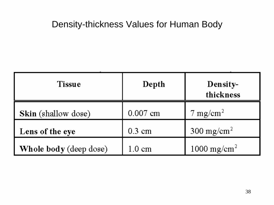
According to ICRP 15, the density of soft human tissue is
equal to 1000 mg/cm3. Using this value we can calculate
density-thickness values for various depths that radiation
may penetrate into the human body and cause damage.
For purposes of reporting radiation dose the tissue depths of
concern are the skin (shallow dose), the lens of the eye, and
the whole body (deep dose).
The concept of "density-thickness" is important to
discussions of radiation attenuation by human tissue, as well
as detector shielding and windows, and dosimetry filters.
36
Density-Thickness
Although materials may have different densities and
thicknesses, if their density-thickness values are the same,
they will attenuate radiation in a similar manner.
For example, a piece of mylar used as a detector window
with a density of 7 mg/cm2 will attenuate radiation similar to
the skin of the human body.
These values can be used to design radiation detection
instrumentation such that detector windows and shields have
the same or similar density-thickness values.
37
Density-Thickness
Any radiation passing the detector window would also
penetrate to the basal layer of the skin on the human
body and deposit energy in living tissue.
External dosimetry can be designed around these
values such that dose equivalent is determined for the
skin of the whole body, lens of the eye, and whole body.
For example, a dosimeter filter may be designed as
1000 mg/cm2. Any radiation passing this filter would
also pass through the skin of the whole body and
deposit energy in vital human organs.
38
Density-thickness Values for Human Body
39
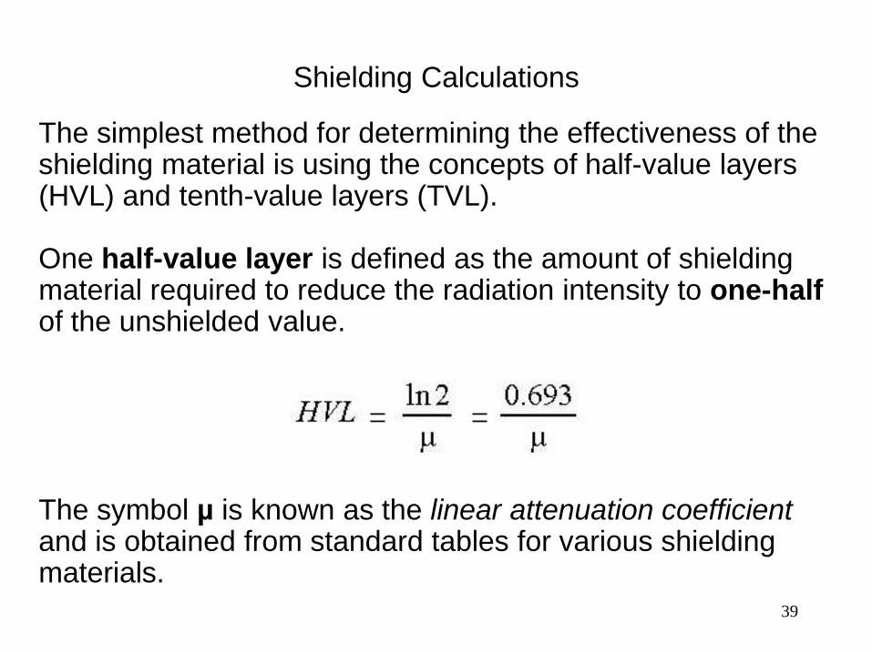
Shielding Calculations
The simplest method for determining the effectiveness of the shielding material is using the concepts of half-value layers (HVL) and tenth-value layers (TVL).
One half-value layer is defined as the amount of shielding material required to reduce the radiation intensity to one-half of the unshielded value.
The symbol µ is known as the linear attenuation coefficient and is obtained from standard tables for various shielding materials.
40
Shielding Calculations
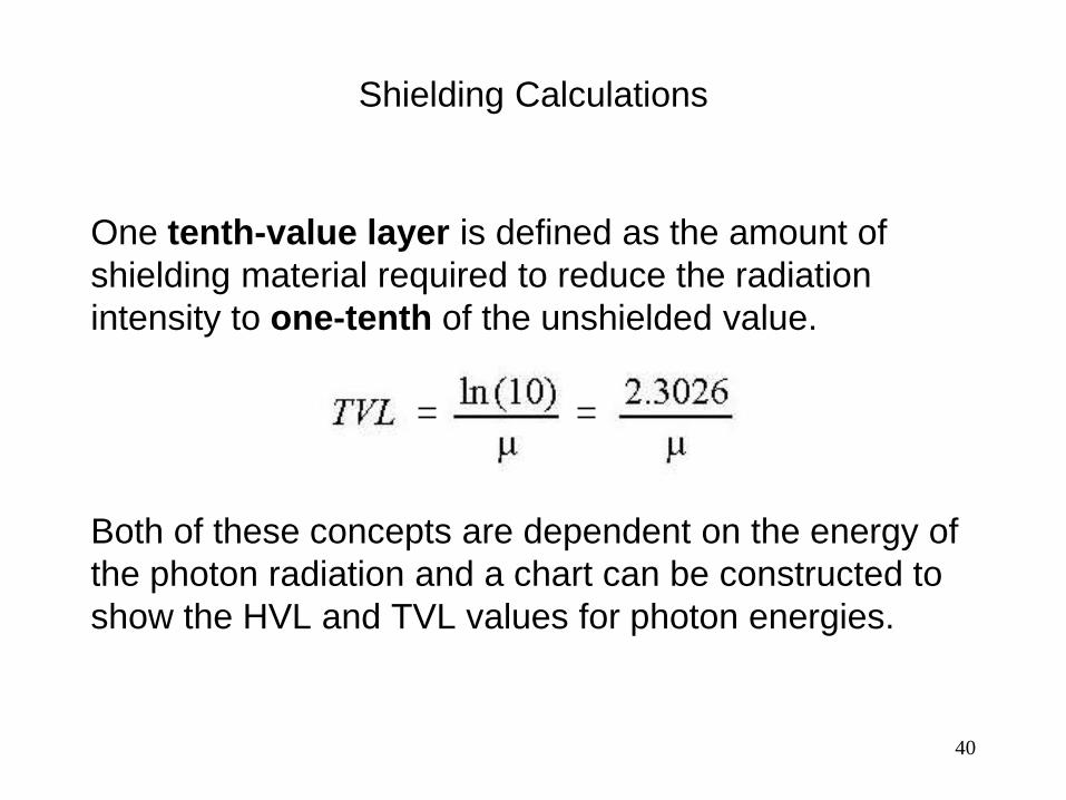
One tenth-value layer is defined as the amount of
shielding material required to reduce the radiation
intensity to one-tenth of the unshielded value.
Both of these concepts are dependent on the energy of
the photon radiation and a chart can be constructed to
show the HVL and TVL values for photon energies.

41
Half-Value Layers
42
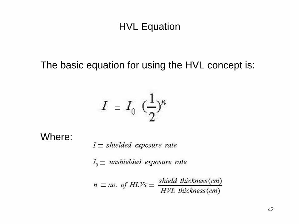
HVL Equation
The basic equation for using the HVL concept is:
Where:
43
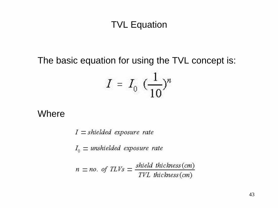
TVL Equation
The basic equation for using the TVL concept is:
Where
1
Internal Exposure Control
Voss Associates
2
Entry Of Radioactive Materials Into The Body
Modes of Entry
Inhalation: Materials enter the body in the air that is
breathed.
Ingestion: Materials enter the body through the mouth.
Absorption: Material enters the body through intact skin.
Entry through wounds:
– Penetration: Materials enter (passively) through
existing wounds which were not adequately covered.
– Injection: Materials enter (forcefully) through wounds
incurred on the job.
3
Preventive Measures
Inhalation -- assessment of conditions, use of
engineering controls, respiratory protection equipment
Ingestion -- proper radiological controls and work
practices
Absorption -- assessment of conditions and protective
clothing
Entry through wounds -- not allowing contamination
near a wound by work restriction or proper radiological
controls if an injury occurs in a contaminated area.
4
Preventive Measures
Note that the preventive measures are designed to do
one of two things:
– Minimize the amount of radioactive materials present
which are available to enter the body, or
– Block the pathway from the source of radioactive
materials into the body.
5
Annual Limit On Intake
Derived Air Concentration
Assimilation of radioactive materials in the workplace occurs
most often as a result of inhalation of airborne radioactive
contaminants. With some nuclides, specifically tritium,
absorption through the skin is also a major concern.
Two limiting values have been calculated and are available
for use in limiting the inhalation of radioactive materials.
These limiting values are:
–Annual Limit on Intake (ALI)
–Derived Air Concentration (DAC)
6
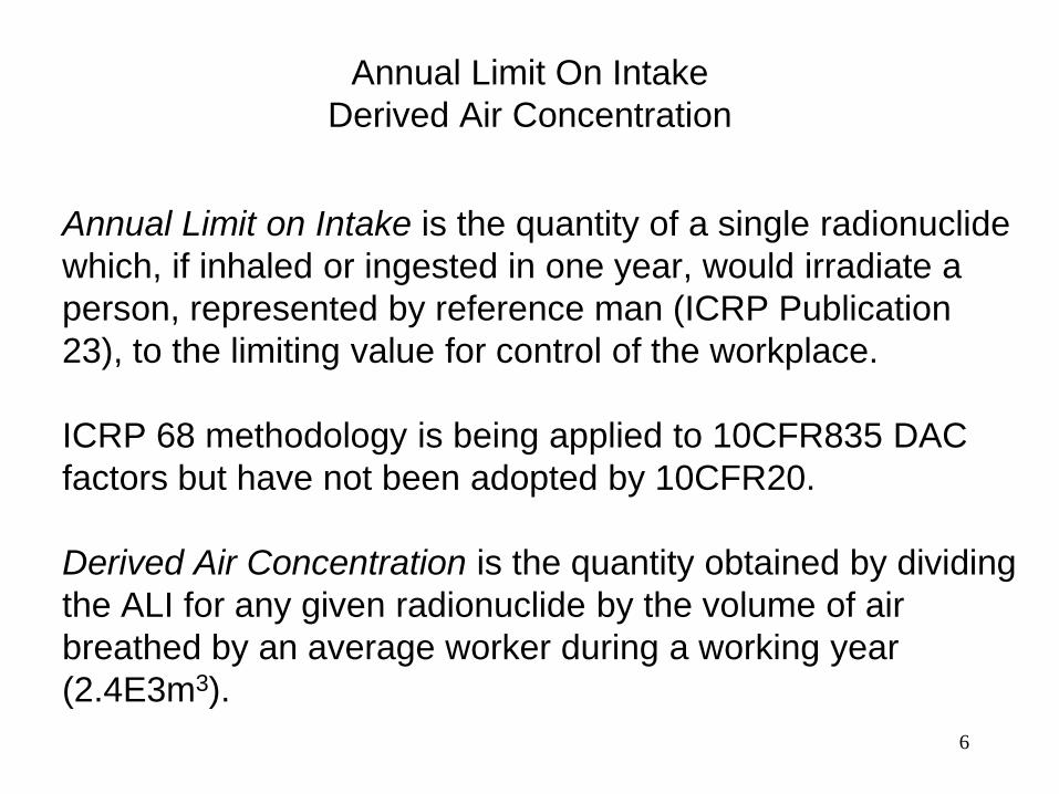
Annual Limit On Intake
Derived Air Concentration
Annual Limit on Intake is the quantity of a single radionuclide
which, if inhaled or ingested in one year, would irradiate a
person, represented by reference man (ICRP Publication
23), to the limiting value for control of the workplace.
ICRP 68 methodology is being applied to 10CFR835 DAC
factors but have not been adopted by 10CFR20.
Derived Air Concentration is the quantity obtained by dividing
the ALI for any given radionuclide by the volume of air
breathed by an average worker during a working year
(2.4E3m3).
7
Annual Limit On Intake
Derived Air Concentration
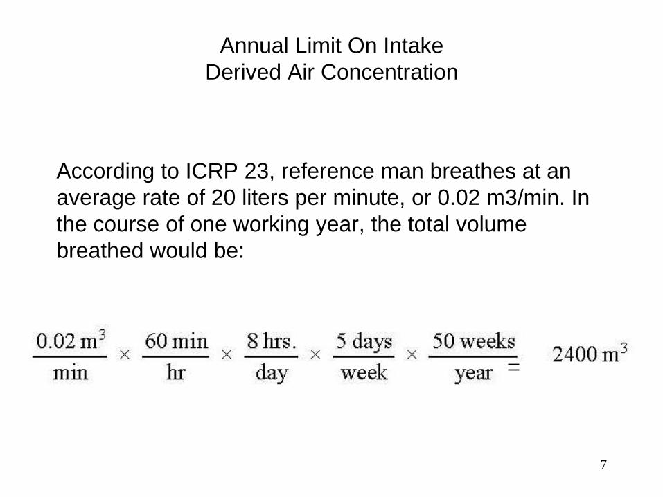
According to ICRP 23, reference man breathes at an
average rate of 20 liters per minute, or 0.02 m3/min. In
the course of one working year, the total volume
breathed would be:
8
Annual Limit On Intake
Derived Air Concentration
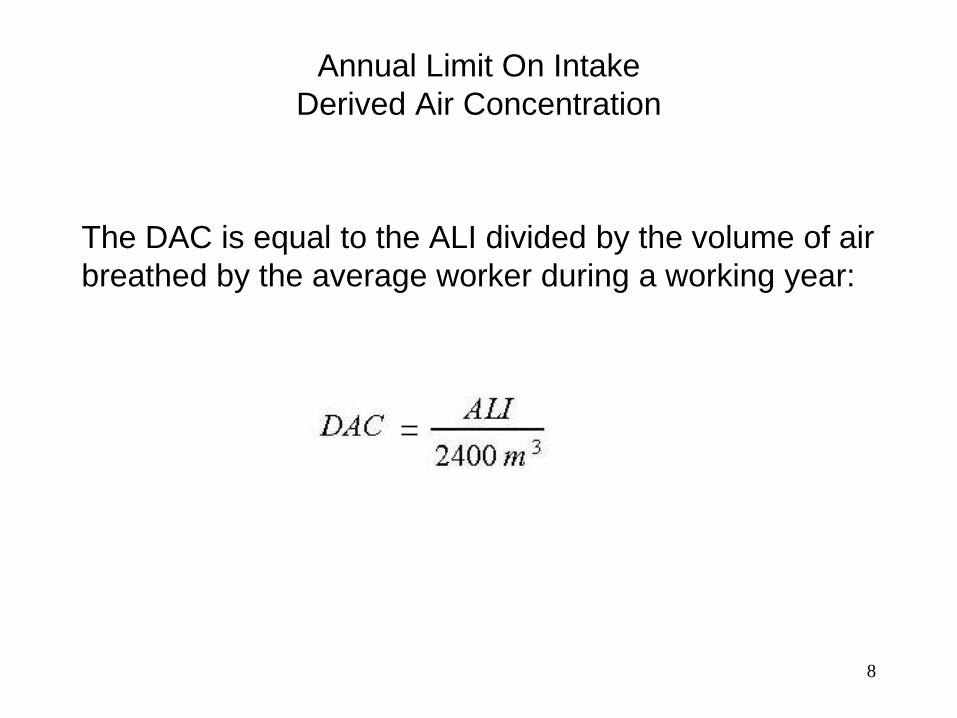
The DAC is equal to the ALI divided by the volume of air
breathed by the average worker during a working year:
9
Annual Limit On Intake
Derived Air Concentration
During routine operations, the combination of physical
design features and administrative controls shall provide
that:
a) The anticipated occupational dose to general employees
shall not exceed the limits.
b) The ALARA process is utilized for personnel exposures
to ionizing radiation.
10
Annual Limit On Intake
Derived Air Concentration
Monitoring of airborne radioactivity shall be performed:
1) Where an individual is likely to receive an exposure of 40
or more DAC-hours in a year; or
2) As necessary to characterize the airborne radioactivity
hazard where respiratory protective devices for protection
against airborne radionuclides have been prescribed.
Real-time air monitoring shall be performed as necessary to
detect and provide warning of airborne radioactivity
concentrations that warrant immediate action to terminate
inhalation of airborne radioactive material.
11
Annual Limit On Intake
Derived Air Concentration
Measures used to minimize the concentration of airborne contaminants remain the primary means of minimizing potential exposure.
Minimizing the concentrations to below DAC values helps insure that workers could not exceed the ALI even if they were in the area continuously for long durations and breathing air at those concentrations.
An Airborne Radioactivity Area is any area where the concentration of airborne radioactivity exceeds or is likely to exceed the DAC value or where an individual present in the area without respiratory protection could receive an intake exceeding 12 DAC-hours in a week.
12
Annual Limit On Intake
Derived Air Concentration
Posting of airborne radioactivity areas controls access to
minimize exposure.
Minimize the stay time of workers in airborne areas to short
periods of time.
Augment installed engineering controls with respiratory
protection equipment to further reduce the concentration of
contaminants in the air the workers are actually breathing.
13
Annual Limit On Intake
Derived Air Concentration
The limitations imposed in terms of dosage to exposed workers are expressed as an annual limit.
Concentrations of contaminants in the air are monitored by continuous monitoring equipment and are supplemented by grab sampling as required.
Engineering controls are augmented with respiratory protection equipment when airborne contaminants exceed or potentially exceed DAC values.
14
Movement of Radioactive Materials
Through the Body
There is no simple device that can be placed on or in the body to determine the quantities of radioactive materials in the body or the dose received by the individual as a result of irradiation of body tissues by these materials.
When radioactive material enters the body, the assessment methods must be based on what happens to the materials, or what the body does with them.
The body does not possess the ability to differentiate between a nonradioactive atom and a radioactive atom of the same element. In terms of metabolic processes, the material is handled the same way.
15
Movement of Radioactive Materials
Through the Body
Once the material is in the body, then its behavior is
governed by the chemical form, its location in the body,
and the body's need for that material.
–Chemical form - solubility
–Location - pathways
–Body's need - intake and incorporation vs. elimination

16
Intake and Uptake
Intake: the amount of radionuclide taken into the body
Uptake: the amount of radionuclide deposited in the body
which makes its way into the body fluids or systemic
system (i.e., blood)
17
Inhaled Radioactive Materials
General Pathways
– Deposition in lungs with eventual transfer to GI tract
or retention
– Transport to body fluids
– Transfer to lymph nodes with eventual movement to
body fluids
– Retention in lymph nodes
18
Inhaled Radioactive Materials
Once in the bodily fluids, possibilities include:
– Transfer to specific organ
– Filtration and elimination by kidneys
– Transport and removal from body fluids through
circulatory systems (perspiration)
19
Inhaled Radioactive Materials
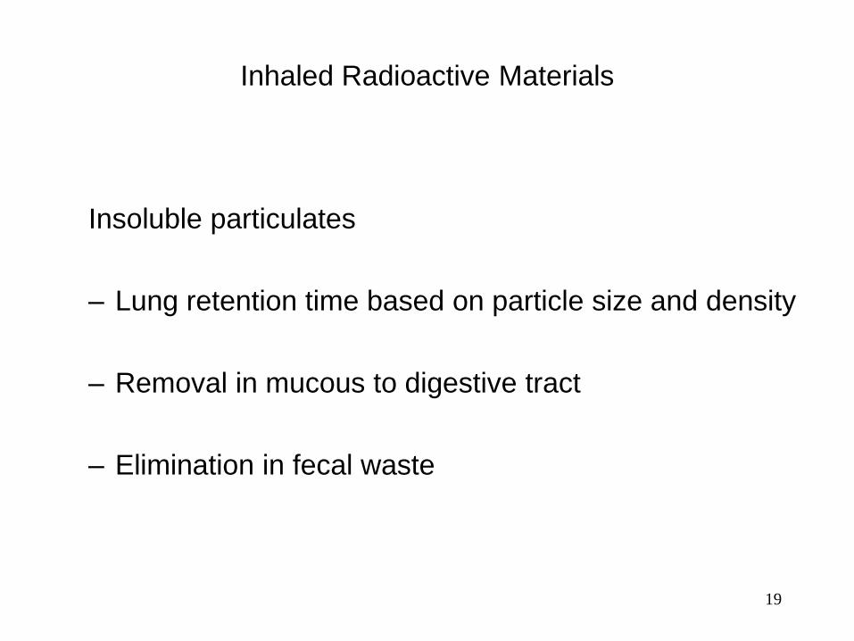
Insoluble particulates
– Lung retention time based on particle size and density
– Removal in mucous to digestive tract
– Elimination in fecal waste
20
Inhaled Radioactive Materials
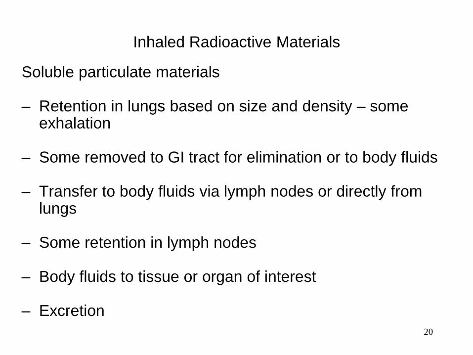
Soluble particulate materials
– Retention in lungs based on size and density – someexhalation
– Some removed to GI tract for elimination or to body fluids
– Transfer to body fluids via lymph nodes or directly from lungs
– Some retention in lymph nodes
– Body fluids to tissue or organ of interest
– Excretion
21
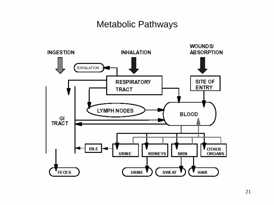
Metabolic Pathways
22
Ingested Radioactive Materials
For elements not used by the body, absorption by
ingestion is poor, and most materials will pass straight
through the body.
Materials pass through stomach to small intestine
where transport of soluble materials to body fluids will
occur.
From body fluids, materials go to the organs and/or are
removed through normal biological elimination
processes.
23
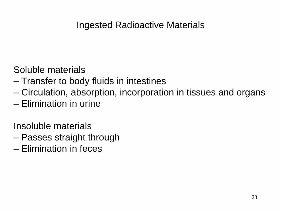
Ingested Radioactive Materials
Soluble materials
– Transfer to body fluids in intestines
– Circulation, absorption, incorporation in tissues and organs
– Elimination in urine
Insoluble materials
– Passes straight through
– Elimination in feces
24
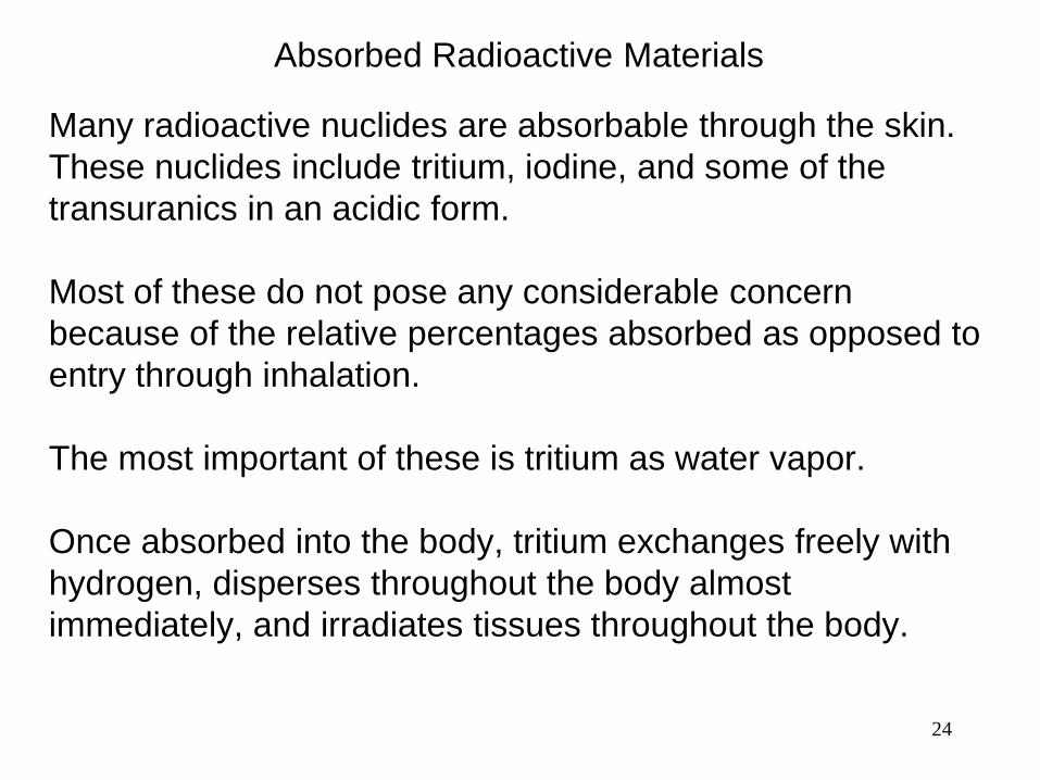
Absorbed Radioactive Materials
Many radioactive nuclides are absorbable through the skin.
These nuclides include tritium, iodine, and some of the
transuranics in an acidic form.
Most of these do not pose any considerable concern
because of the relative percentages absorbed as opposed to
entry through inhalation.
The most important of these is tritium as water vapor.
Once absorbed into the body, tritium exchanges freely with
hydrogen, disperses throughout the body almost
immediately, and irradiates tissues throughout the body.
25
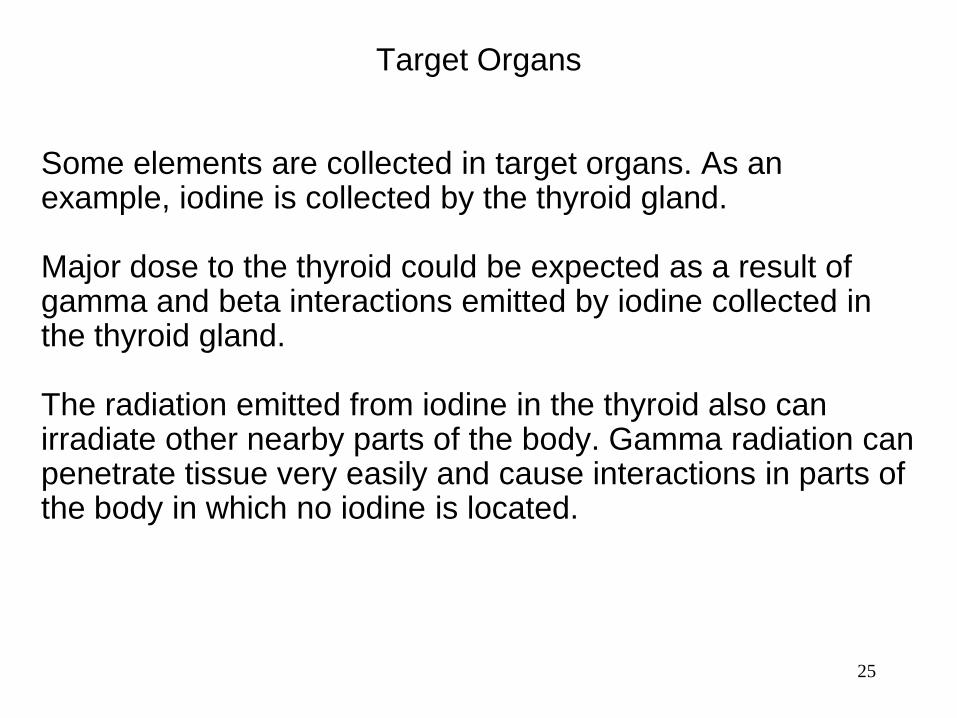
Target Organs
Some elements are collected in target organs. As an example, iodine is collected by the thyroid gland.
Major dose to the thyroid could be expected as a result of gamma and beta interactions emitted by iodine collected in the thyroid gland.
The radiation emitted from iodine in the thyroid also can irradiate other nearby parts of the body. Gamma radiation can penetrate tissue very easily and cause interactions in parts of the body in which no iodine is located.
26
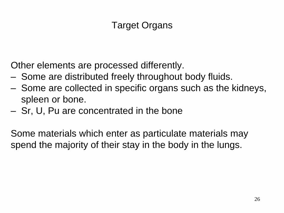
Target Organs
Other elements are processed differently.
– Some are distributed freely throughout body fluids.
– Some are collected in specific organs such as the kidneys,
spleen or bone.
– Sr, U, Pu are concentrated in the bone
Some materials which enter as particulate materials may
spend the majority of their stay in the body in the lungs.
27
Elimination Processes
Normal Biological Elimination
Radioactive materials incorporated into body tissues and organs are eliminated from the body as are their non-radioactive counterparts.
Eliminated through exhalation, perspiration, urination, and defecation.
Each element has a measurable biological half-life - the time required to reduce the amount of material in the body to one-half of its original value.
The biological half-life is independent of the physical or radiological half-life.
28
Elimination Processes
Radioactive Decay
Each radioactive nuclide has a distinctive decay rate that is not influenced by any physical process, including biological functions.
The amount of time required for one half of the material in the body to decay is called the radiological half-life.
Radioactive decay will result in reduction of the quantity of the original nuclides deposited in the body.
It is important to remember that the progeny of these nuclides may also be radioactive.
It is possible that the progeny could introduce completely different concerns for internal dose assessments.
29
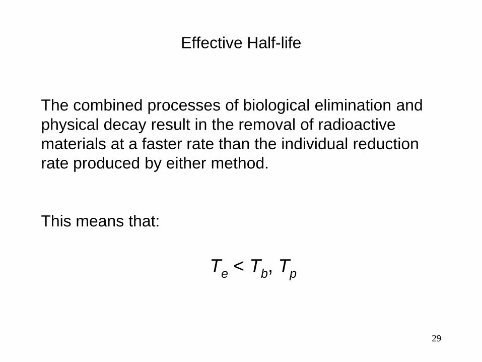
Effective Half-life
The combined processes of biological elimination and
physical decay result in the removal of radioactive
materials at a faster rate than the individual reduction
rate produced by either method.
This means that:
Te < Tb, Tp
30
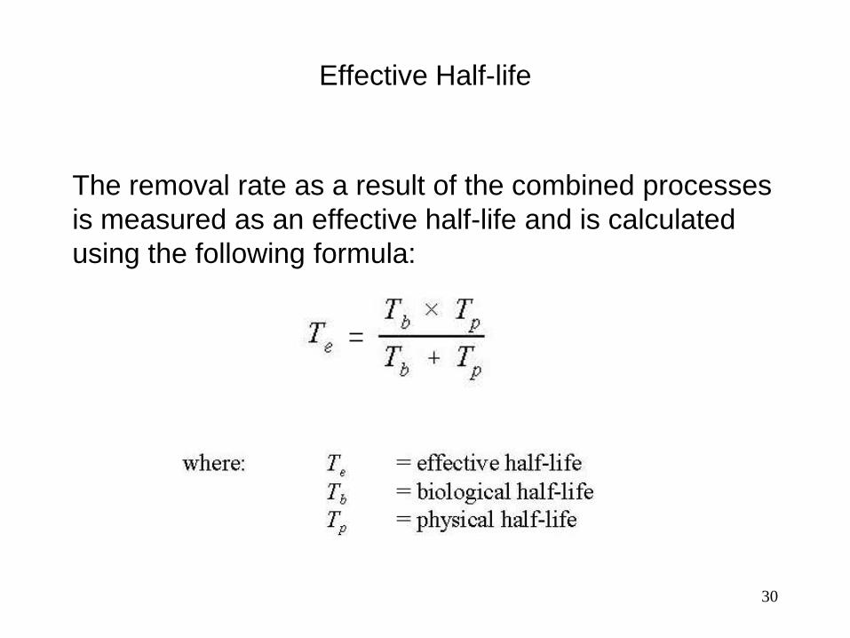
Effective Half-life
The removal rate as a result of the combined processes
is measured as an effective half-life and is calculated
using the following formula:
31
Effective Removal Constant
Another way that this is expressed is the effective
removal constant, λe, which is the composite of the
physical decay constant λp and the biological
elimination constant λb.
λe = λb + λp
32
Example Calculations
Determine the effective half-life of tritium if the biological
half-life is 10 days and the physical half-life is 12.3 years.
Determine the effective half-life of 59-Fe if the biological
half-life is 2000 days and the physical half-life is 44.56 days.
33
Blocking Agents
A blocking agent saturates the metabolic processes in a
specific tissue with the stable element and reduces uptake
of the radioactive forms of the element.
As a rule, these must be administered prior to or almost
immediately after the intake for maximum effectiveness and
must be in a form that is readily absorbed.
The most well known example of this is stable iodine, as
potassium iodide, which is used to saturate the thyroid
gland, thus preventing uptake of radioactive iodine in the
thyroid.
34
Diluting Agents
A diluting agent is a compound which includes a stable
form of the nuclide of concern. By introducing a large
number of stable atoms, the statistical probability of the
body incorporating radioactive atoms is reduced.
Diluting agents can also involve the use of different
elements which the body processes in the same way. This
type of treatment is called displacement therapy.
The compound used must be as readily absorbed and
metabolized as the compound that contains the
radioisotope.
35
Mobilizing Agents
A mobilizing agent is a compound that increases the
natural turnover process, thus releasing some forms of
radioisotopes from body tissues.
Usually most effective within 2 weeks after exposure;
however, use for extended periods may produce less
dramatic reductions.
36
Chelating Agents
A chelating agent is a compound which acts on
insoluble compounds to form a soluble complex ion
which can then be removed through the kidneys.
Commonly used to enhance elimination of transuranics
and other metals.
Therapy is most effective when begun immediately after
exposure if metallic ions are still in circulation and is
less effective once metallic ions are incorporated into
cells or deposited in tissue such as bone.
37
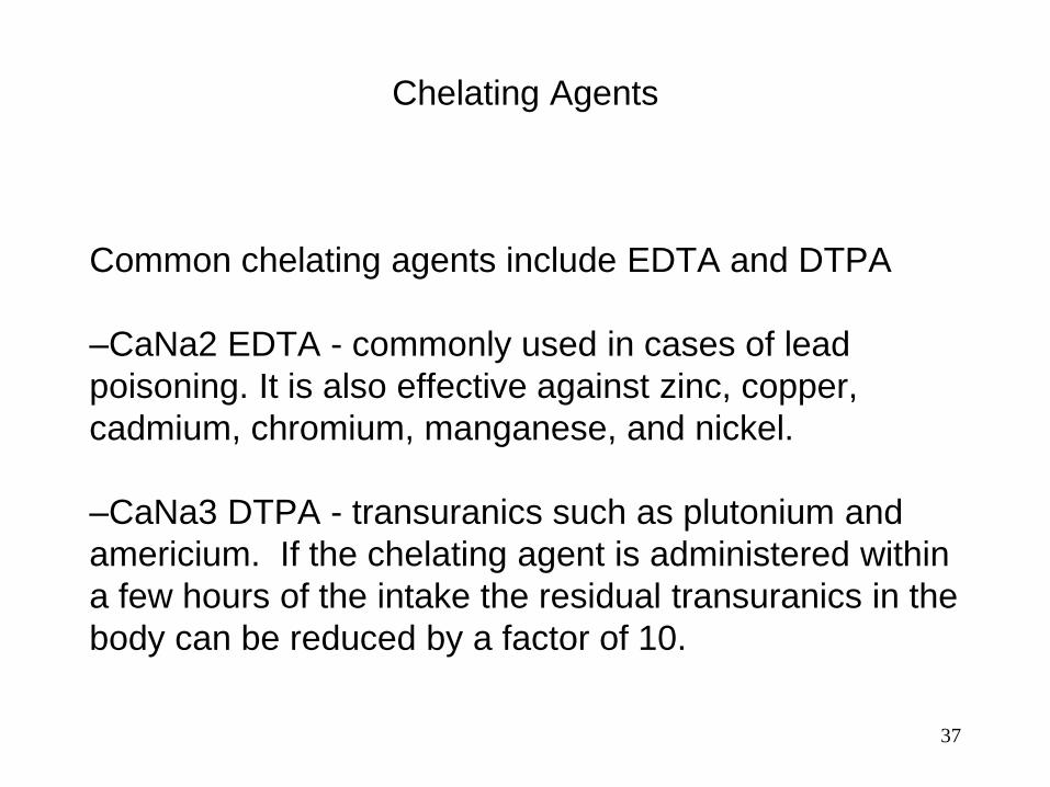
Chelating Agents
Common chelating agents include EDTA and DTPA
–CaNa2 EDTA - commonly used in cases of lead
poisoning. It is also effective against zinc, copper,
cadmium, chromium, manganese, and nickel.
–CaNa3 DTPA - transuranics such as plutonium and
americium. If the chelating agent is administered within
a few hours of the intake the residual transuranics in the
body can be reduced by a factor of 10.
38
Diuretics
Diuretics increase urinary excretion of sodium and water.
Used to reduce internal exposure, however its use has
been limited. Applications include 3-H, 42-K, 38-Cl and
others.
Can lead to dehydration and other complications if not
performed properly.
39
Expectorants and Inhalants
Used to increase flow of respiratory tract excretions.
Inhalants have been used in removing radioactive
particles from all areas of lungs.
40
Lung Lavage
Involves multiple flushing of lungs with appropriate fluid to
remove radioactive materials in the lungs.
Usually limited to applications where resulting exposures
would result in appearance of acute or subacute radiation
effects.
The procedure has been proven effective in cases of “black
lung” disease exhibited by coal miners where the procedure
is used to remove some of the small coal particles that were
deposited at the alveolar region of the lungs.
RADIOACTIVE SOURCE
CONTROL
Voss Associates
1
2
A radioactive source is material used for its emitted
radiation. Sources are constructed as sealed or unsealed
and are classified as accountable or non-accountable .
Radioactive sources are used for response checks in the
field, functional checks, calibration of instruments and
monitors to traceable standards. To ensure the safety and
welfare of all personnel it is important to maintain control of
radioactive sources.
Radioactive sources are controlled to minimize the
potential for:
Spread of contamination
Unnecessary exposure to personnel
Loss or theft
Improper disposal
3
In accordance with 10 CFR 835, Subpart M, the following
provisions apply to sealed sources:
A. Sealed radioactive sources shall be used, handled, and
stored in a manner commensurate with the hazards
associated with operations involving the sources.
B. Each accountable sealed radioactive source shall be
inventoried at intervals not to exceed six months. This
inventory shall:
1. Establish the physical location of each
accountable sealed radioactive source;
2. Verify the presence and adequacy of associated
postings and labels; and
3. Establish the adequacy of storage locations,
containers, and devices.
4
C. Except for sealed sources consisting solely of
gaseous radioactive material or tritium, each accountable
sealed radioactive source shall be subject to a source
leak test upon receipt, when damage is suspected, and
at intervals not to exceed six months. Source leak tests
shall be capable of detecting radioactive material
leakage equal to or exceeding 0.005 μCi.
D. An accountable sealed radioactive source is not
subject to periodic source leak testing if that source has
been removed from service. Such sources shall be
stored in a controlled location, subject to periodic
inventory, and subject to source leak testing prior to
being returned to service.
5
E. An accountable sealed radioactive source is not subject
to periodic inventory and source leak testing if that source
is located in an area that is unsafe for human entry or
otherwise inaccessible.
F. An accountable sealed radioactive source found to be
leaking radioactive material shall be controlled in a manner
that minimizes the spread of radioactive contamination.
6
Sources are controlled using the following precautions:
1. Each source is to be inspected before each use.
2. Remove damaged sources from service.
3. Fingers, whether gloved or not, or other objects should
never be allowed to touch the active surface of unsealed
sources.
4. Protect the source from being contaminated when used
in a surface contamination area.
7
Sealed radioactive sources not in storage containers or
devices and not labeled by the manufacturer must be
clearly marked with a radiation symbol and have a durable
label/ tag containing the following information:
a. Radionuclide
b. Amount of activity
c. Name of manufacturer
d. Date of assay
e. Model and serial numbers (where available)
8
1. Accountable Sealed Radioactive Source means a
sealed radioactive source having a half-life equal to or
greater than 30 days and an isotopic activity equal to or
greater than the corresponding value provided in Appendix
E of 10CFR 835.
2. Radioactive Material Area means any area within a
controlled area, accessible to individuals, in which items or
containers of radioactive material exist and the total activity
of radioactive material exceeds the applicable values
provided in Appendix E to 10 CFR 835.
9
3. Sealed Radioactive Source means a radioactive
source manufactured, obtained, or retained for the
purpose of utilizing the emitted radiation. The sealed
radioactive source consists of a known or estimated quantity
of radioactive material contained within a sealed capsule,
sealed between layer(s) of nonradioactive material, or firmly
fixed to a non-radioactive surface by electroplating or other
means intended to prevent leakage or escape of the
radioactive material.
4. Source Leak Test means a test to determine if a
sealed radioactive source is leaking radioactive material.

10

11

12

13

14

15

16

17

18

19

20

21

22

23

24

25

26

27

28

29

30


![DOSIMETRY PRINCIPLES, DOSE MEASUREMENTS AND … · DOSIMETRY PRINCIPLES, DOSE MEASUREMENTS AND RADIATION ... (ICRU Report 60) [13] ... 60 Applications of ionizing radiation in materials](https://img.pdfslide.us/doc/110x75/5b92fdd109d3f280378c629a/dosimetry-principles-dose-measurements-and-dosimetry-principles-dose-measurements.jpg)