Embed Size (px)
Citation preview
Brief Report
Vol. 28, No. 4, 2016 515
Received February 5, 2015, Revised July 6, 2015, Accepted for publication August 7, 2015
Corresponding author: Sung Eun Chang, Department of Dermatology, Asan Medical Center, University of Ulsan College of Medicine, 88 Olympic-ro 43-gil, Songpa-gu, Seoul 05505, Korea. Tel: 82-2-3010-3460, Fax: 82-2-486-7831, E-mail: [email protected]
This is an Open Access article distributed under the terms of the Creative Commons Attribution Non-Commercial License (http://creativecommons.org/ licenses/by-nc/4.0) which permits unrestricted non-commercial use, distribution, and reproduction in any medium, provided the original work is properly cited.
Copyright © The Korean Dermatological Association and The Korean Society for Investigative Dermatology
http://dx.doi.org/10.5021/ad.2016.28.4.515
Donut Ablation Method as an Alternative to Surgical Excision or Total Ablational Laser Treatment
Joon Min Jung, Seung Seog Han1, Chong Hyun Won, Sung Eun Chang
Department of Dermatology, Asan Medical Center, University of Ulsan College of Medicine, 1I Dermatology Clinic, Seoul, Korea
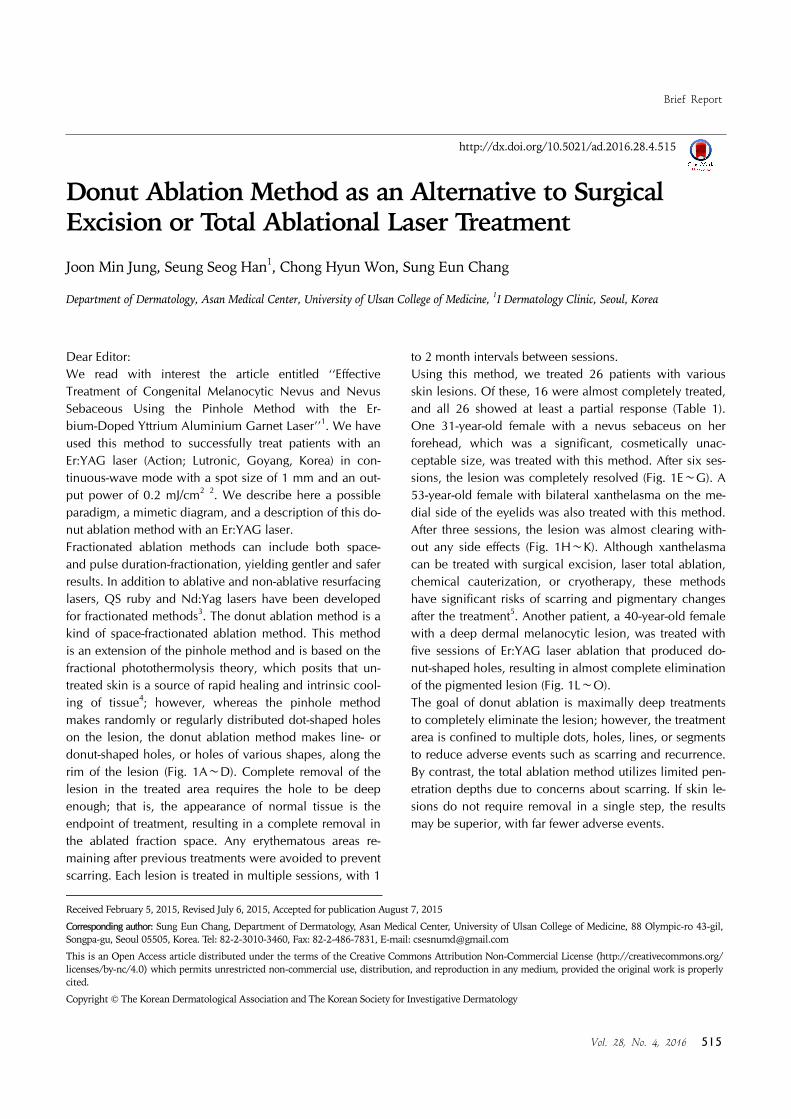
Dear Editor:We read with interest the article entitled ‘‘Effective Treatment of Congenital Melanocytic Nevus and Nevus Sebaceous Using the Pinhole Method with the Er-bium-Doped Yttrium Aluminium Garnet Laser’’1. We have used this method to successfully treat patients with an Er:YAG laser (Action; Lutronic, Goyang, Korea) in con-tinuous-wave mode with a spot size of 1 mm and an out-put power of 0.2 mJ/cm2 2. We describe here a possible paradigm, a mimetic diagram, and a description of this do-nut ablation method with an Er:YAG laser.Fractionated ablation methods can include both space- and pulse duration-fractionation, yielding gentler and safer results. In addition to ablative and non-ablative resurfacing lasers, QS ruby and Nd:Yag lasers have been developed for fractionated methods3. The donut ablation method is a kind of space-fractionated ablation method. This method is an extension of the pinhole method and is based on the fractional photothermolysis theory, which posits that un-treated skin is a source of rapid healing and intrinsic cool-ing of tissue4; however, whereas the pinhole method makes randomly or regularly distributed dot-shaped holes on the lesion, the donut ablation method makes line- or donut-shaped holes, or holes of various shapes, along the rim of the lesion (Fig. 1A∼D). Complete removal of the lesion in the treated area requires the hole to be deep enough; that is, the appearance of normal tissue is the endpoint of treatment, resulting in a complete removal in the ablated fraction space. Any erythematous areas re-maining after previous treatments were avoided to prevent scarring. Each lesion is treated in multiple sessions, with 1
to 2 month intervals between sessions.Using this method, we treated 26 patients with various skin lesions. Of these, 16 were almost completely treated, and all 26 showed at least a partial response (Table 1). One 31-year-old female with a nevus sebaceus on her forehead, which was a significant, cosmetically unac-ceptable size, was treated with this method. After six ses-sions, the lesion was completely resolved (Fig. 1E∼G). A 53-year-old female with bilateral xanthelasma on the me-dial side of the eyelids was also treated with this method. After three sessions, the lesion was almost clearing with-out any side effects (Fig. 1H∼K). Although xanthelasma can be treated with surgical excision, laser total ablation, chemical cauterization, or cryotherapy, these methods have significant risks of scarring and pigmentary changes after the treatment5. Another patient, a 40-year-old female with a deep dermal melanocytic lesion, was treated with five sessions of Er:YAG laser ablation that produced do-nut-shaped holes, resulting in almost complete elimination of the pigmented lesion (Fig. 1L∼O).The goal of donut ablation is maximally deep treatments to completely eliminate the lesion; however, the treatment area is confined to multiple dots, holes, lines, or segments to reduce adverse events such as scarring and recurrence. By contrast, the total ablation method utilizes limited pen-etration depths due to concerns about scarring. If skin le-sions do not require removal in a single step, the results may be superior, with far fewer adverse events.
Brief Report
516 Ann Dermatol
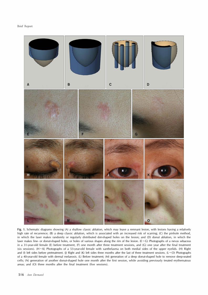
Fig. 1. Schematic diagrams showing (A) a shallow classic ablation, which may leave a remnant lesion, with lesions having a relatively high rate of recurrence; (B) a deep classic ablation, which is associated with an increased risk of scarring; (C) the pinhole method, in which the laser makes randomly or regularly distributed dot-shaped holes on the lesion; and (D) donut ablation, in which the laser makes line- or donut-shaped holes, or holes of various shapes along the rim of the lesion. (E∼G) Photographs of a nevus sebaceus in a 31-year-old female (E) before treatment, (F) one month after three treatment sessions, and (G) one year after the final treatment (six sessions). (H∼K) Photographs of a 53-year-old female with xanthelasma on both medial sides of the upper eyelids. (H) Right and (I) left sides before pretreatment. (J) Right and (K) left sides three months after the last of three treatment sessions. (L∼O) Photographs of a 40-year-old female with dermal melanosis. (L) Before treatment; (M) generation of a deep donut-shaped hole to remove deep-seated cells; (N) generation of another donut-shaped hole one month after the first session, while avoiding previously treated erythematous areas; and (O) three months after the final treatment (five sessions).
Brief Report
Vol. 28, No. 4, 2016 517
Table 1. Summary of the patients treated by fractional resurfacing
DiseaseNumber of
patientsAverage number of treatment sessions
Melanocytic nevus 7 6.7Dermal melanosis 1 5.0Lentigine 5 6.0Café au lait macule 2 3.5Nevus sebaceus 2 5.5Telangiectasia 3 2.7Xanthelasma 1 3.0Scar 5 3.8
Received June 11, 2015, Revised July 21, 2015, Accepted for publication August 10, 2015
Corresponding author: Dong-Youn Lee, Department of Dermatology, Samsung Medical Center, Sungkyunkwan University School of Medicine, 81 Irwon-ro, Gangnam-gu, Seoul 06351, Korea. Tel: 82-2-3410-3543, Fax: 82-2-3410-3869, E-mail: [email protected]
This is an Open Access article distributed under the terms of the Creative Commons Attribution Non-Commercial License (http://creativecommons.org/ licenses/by-nc/4.0) which permits unrestricted non-commercial use, distribution, and reproduction in any medium, provided the original work is properly cited.
Copyright © The Korean Dermatological Association and The Korean Society for Investigative Dermatology
REFERENCES
1. Chung BY, Han SS, Kim BW, Chang SE, Lee MW. Effective
treatment of congenital melanocytic nevus and nevus sebaceous using the pinhole method with the erbium-doped
yttrium aluminium garnet laser. Ann Dermatol 2014;26:651-
653.2. Lee HM, Haw S, Kim JE, Won CH, Lee MW, Choi JH, et al.
A fractional 2940 nm short-pulsed, erbium-doped yttrium
aluminium garnet laser is effective and minimally invasive for the treatment of photodamaged skin in Asians. J Cosmet
Laser Ther 2012;14:253-259.
3. Kim BW, Lee MH, Chang SE, Yun WJ, Won CH, Lee MW, et al. Clinical efficacy of the dual-pulsed Q-switched
neodymium: yttrium-aluminum-garnet laser: comparison
with conservative mode. J Cosmet Laser Ther 2013;15: 340-341.
4. Chung BY, Han SS, Moon HR, Lee MW, Chang SE.
Treatment with the pinhole technique using erbium-doped yttrium aluminium garnet laser for a café au lait macule and
carbon dioxide laser for facial telangiectasia. Ann Dermatol
2014;26:657-659.5. Raulin C, Schoenermark MP, Werner S, Greve B. Xan-
thelasma palpebrarum: treatment with the ultrapulsed CO2
laser. Lasers Surg Med 1999;24:122-127.
http://dx.doi.org/10.5021/ad.2016.28.4.517
Acral Persistent Papular Mucinosis with Partial Response to Tacrolimus Ointment
Ji-Young Jun, Seung Hwan Oh, Joon Ho Shim, Jun-Hwan Kim, Ji-Hye Park, Dong-Youn Lee
Department of Dermatology, Samsung Medical Center, Sungkyunkwan University School of Medicine, Seoul, Korea
Dear Editor:Acral persistent papular mucinosis (APPM) is a rare sub-type of localized lichen myxedematosus (LM)1 with un-known etiology. To our knowledge only six cases tried treatment1-3, and two cases showed family history1. A 53-year-old woman presented with asymptomatic 1∼3 mm flesh-colored papules symmetrically located on both
dorsum of hands and wrists, and on anterior chest. It first appeared 7∼8 years ago and did not disappear. She was previously healthy and no abnormalities were found in an-nual check-up. Her two brothers had same lesions on their dorsum of hands. Biopsy was done on the hand lesion and discrete mucin depositions in upper dermis with spared grenz zone confirmed the diagnosis of APPM (Fig. 1).