Embed Size (px)
Citation preview
Docosahexanoic Acid-Induced Coronary Arterial Dilation:Actions of 17S-Hydroxy Docosahexanoic Acid on K�
Channel Activity
Xiang Li, Song Hong, Pin-Lan Li, and Yang ZhangDepartment of Pharmacology and Toxicology, Medical College of Virginia, Virginia Commonwealth University, Richmond,Virginia (X.L., P.-L.L., Y.Z.); and Ophthalmology and Neuroscience Center, Louisiana State University Health Sciences Center,New Orleans, Louisiana (S.H.)
Received October 29, 2010; accepted December 13, 2010
ABSTRACTDespite extensive studies, the mechanisms mediating the car-diovascular actions of �-3 polyunsaturated fatty acids has notyet been fully understood. The present study determined thepossible actions of an endothelium-derived lipoxygenase prod-uct of docosahexanoic acid (DHA), 17S-hydroxy DHA (17S-HDHA), in bovine coronary arteries. High-performance liquidchromatography (LC) analysis demonstrated that bovine coro-nary arterial endothelial cells can metabolize DHA via lipoxyge-nases, and one of the major products was confirmed to be17S-HDHA by LC-tandem mass spectrometry. In isolated per-fused small bovine coronary arteries, 17S-HDHA (10�9 to 10�5
M) caused a concentration-dependent dilation with a maximumdilator response of 87.8 � 2.5%, which is much more potentthan the dilator response of its precursor, DHA. Moreover,
17S-HDHA-induced vasodilatations were significantly blockedby iberiotoxin, a large conductance Ca2�-activated K� (BKCa)channel blocker, but not altered by an ATP-sensitive K� chan-nel blocker, glibenclamide. In patch-clamp whole-cell record-ing, 17S-HDHA markedly increased K� currents in coronaryarterial smooth muscle cells. In the inside-out mode, but not inthe cell-attached mode, 17S-HDHA dramatically increased theBKCa channel activity, which was substantially blocked by ibe-riotoxin. Collectively, our findings indicate that 17S-HDHA, anendothelium-derived DHA product via lipoxygenase, activatesBKCa channels in coronary arterial smooth muscle cells, leadingto coronary vasodilation, which may represent an importantmechanism mediating the beneficial actions of DHA in coronarycirculation.
IntroductionNumerous epidemiological studies, clinical trials, and an-
imal experiments have demonstrated that fish oils, primarily�-3 polyunsaturated fatty acids (PUFAs), protect againstseveral types of cardiovascular diseases such as myocardialinfarction, arrhythmia, atherosclerosis, stroke, or hyperten-sion (Rapp et al., 1991; McLennan et al., 1996; Nageswari etal., 1999; Kang and Leaf, 2000; Abeywardena and Head,2001; De Caterina and Zampolli, 2001; Jeerakathil and Wolf,2001; Leaf et al., 2003; Holub and Holub, 2004; Harrison andAbhyankar, 2005). Two well known �-3 PUFAs present infish oil are docosahexaenoic acid (DHA) and eicosapentaenoic
acid (Connor et al., 1993). Studies have indicated that DHAmay be a major active component in fish oil conferring car-diovascular protection (Horrocks and Yeo, 1999; Nordøy etal., 2001; Hirafuji et al., 2003). In animal experiments, DHAwas found more effective than eicosapentaenoic acid in re-tarding the development of hypertension in spontaneouslyhypertensive rats and inhibiting thromboxane-like vasocon-strictor responses in the aorta from these rats (McLennan etal., 1996). However, it remains poorly understood how DHAexerts its beneficial action on the cardiovascular system, butseveral possible mechanisms have been suggested, such asreduction of plasma triglycerides, inhibition of platelet func-tion, enhancement of cardiac excitability, and anti-inflamma-tion (McLennan et al., 1996; Salem et al., 2001; Simopoulos,2002).
DHA has been found to be metabolized via cyclooxygenase,lipoxygenase, and P450 metabolic pathways, which generatea series of 17R or 17S monohydroxy, dihydroxy, and trihy-
This study was supported by the National Institute of Health NationalHeart, Lung, and Blood Institute [Grants HL-57244, HL-70726].
Article, publication date, and citation information can be found athttp://jpet.aspetjournals.org.
doi:10.1124/jpet.110.176461.
ABBREVIATIONS: PUFA, polyunsaturated fatty acid; DHA, docosahexanoic acid; 17S-HDHA, 17S-hydroxy DHA; EC, endothelial cell; SMC,smooth muscle cell; BKCa, large conductance Ca2�-activated K�; CDC, cinnamyl-3, 4-dihydroxyl-cyanocinnamate; LC, liquid chromatography;HPLC, high-performance LC; MS, mass spectrometry; ID, internal diameter; U46619, (Z)-7-[(1S,4R,5R,6S)-5-[(E,3S)-3-hydroxyoct-1-enyl]-3-oxabicyclo[2.2.1]heptan-6-yl]hept-5-enoic acid; EET, epoxyeicosa-5,8,11-trienoic acid; DHET, dihydroxyeicosatrienoic acid.
0022-3565/11/3363-891–899$20.00THE JOURNAL OF PHARMACOLOGY AND EXPERIMENTAL THERAPEUTICS Vol. 336, No. 3Copyright © 2011 by The American Society for Pharmacology and Experimental Therapeutics 176461/3669525JPET 336:891–899, 2011 Printed in U.S.A.
891
at Virginia C
omm
onwealth U
niv Tom
pkins McC
aw Lib/A
cq Srv on M
arch 29, 2012jpet.aspetjournals.org
Dow
nloaded from
droxy DHA and various epoxides (Hong et al., 2003). Some ofthese DHA products possess potent bioactivity, in particular,being active as anti-inflammatory and immune-regulatorycompounds (Hong et al., 2003). Inflammation or microinflam-mation plays important roles in the development of athero-sclerosis, ischemic reperfusion injury, and cardiac or vascu-lar remodeling. In this regard, the anti-inflammatory orimmune-regulatory effects of DHA and its products havebeen suggested to contribute to the beneficial actions of �-3PUFAs or fish oil on the cardiovascular system (Simopoulos,2002; Holub and Holub, 2004). However, many classic anti-inflammatory drugs such as commonly used indole and aryl-propionic acid derivatives do not have similar cardiovascularprotective actions to that observed in DHA treatments. Thissuggests that some other mechanisms are involved in theaction of DHA or �-3 PUFAs on the cardiovascular system inaddition to their anti-inflammatory effects. In this regard,previous studies demonstrated that a �-3 PUFA diet en-hanced endothelium-dependent vasodilator response in cor-onary arteries (Shimokawa and Vanhoutte, 1989; Fleis-chhauer et al., 1993). Therefore, DHA may exert its beneficialaction through an endothelium-dependent mechanism in cor-onary circulation.
The present study hypothesized that 17S-HDHA, a lipoxy-genase product, mediates the endothelium-dependent vaso-dilator action of DHA in small coronary arteries. To test thishypothesis, we first separated and analyzed the lipoxygenasemetabolites of DHA produced in coronary arteries and endo-thelial cells (ECs). Then, we tested the ability and potency of17S-HDHA to produce vasodilator response in isolated per-fused coronary arteries. We further determined whether va-sodilator response to 17S-HDHA is associated with the acti-vation of K� channels by using the patch-clamp technique.Our data indicate that 17S-HDHA is a much more potentvasodilator than DHA, and the vasodilator action of 17S-HDHA is associated with the activation of large conductanceCa2�-activated K� (BKCa) channels in coronary arterialsmooth muscle cells (SMCs).
Materials and MethodsVideo Microscopy of Arterial Reactivity. Isolated pressurized
small coronary artery preparation was used to study the vasomotorresponse to DHA and its metabolites as we described previously(Geiger et al., 2000). In brief, the internal diameter (ID) of thesearteries was measured with a microscopic video recording systemcomposed of a stereomicroscope (Leica MZ8; Leica, Wetzlar, Ger-many), a charge-coupled device camera (KP-MI AU; Hitachi, Tokyo,Japan), a video monitor (VM-1220U; Hitachi), a video measuringapparatus (VIA-170; Boeckeler Instruments, Tucson, AZ), and avideo printer (UP890 MD; Sony, Tokyo, Japan). The arterial imageswere also recorded continuously with a videocassette recorder (M-674; Toshiba, Tokyo, Japan). Before testing any compounds, thecannulated artery was equilibrated for 1 to 1.5 h and then precon-tracted to 40 to 60% of their resting diameter with a thromboxane A2
analog, (Z)-7-[(1S,4R,5R,6S)-5-[(E,3S)-3-hydroxyoct-1-enyl]-3-oxabicyclo[2.2.1]heptan-6-yl]hept-5-enoic acid (U46619) (2–20 nM). Once steady-state contraction was obtained, cumulative concentration response to 17S-HDHA (10�9 to 10�5 M; kindly provided by C. S. Serhan, Harvard MedicalSchool, Boston, MA) or DHA (10�9 to 10�5 M; Cayman Chemical, AnnArbor, MI) were determined by measuring changes in the ID. Thevasodilator response was expressed as percentage of relaxation rel-ative to U46619-induced precontraction as calculated in the follow-ing formula: percentage of relaxation � (IDdilator � IDU46619)/(IDbasal
� IDU46619), where IDbasal means the arterial ID under resting con-dition; IDU46619 indicates the ID of U46619-precontracted arteries;and IDdilator means the arterial ID obtained during treatment ofU46619-precontracted arteries with compounds tested. Betweenpharmacological interventions, the arteries were washed three timeswith physiological salt solution and allowed to equilibrate in drug-free physiological salt solution for 20 to 30 min. The arteries weretreated with corresponding inhibitors for 20 min and then concen-tration-response curves to 17S-HDHA were redetermined. To pre-pare endothelium-denuded small coronary arteries, the rings of ar-teries were passed through its lumen by a segment of hair. Theendothelium was removed by gently rotating the hair (Ge et al.,2003).
Extraction and Analysis of DHA Metabolites. DHA metabo-lites were analyzed from the reaction mixtures of arterial or cellhomogenates with DHA. The homogenates of small coronary arterieswere prepared as described previously (Geiger et al., 2000). Thereactions were performed by incubating 200 �g of protein of homog-enates prepared from the arteries, ECs, or SMCs, with DHA (100�M, 15 min at 37°C) with a final volume of 200 �l. The incubationwas stopped upon addition of 2 ml of methanol, which was prechilledto �40°C and placed at �40°C for 30 min. Then, the reaction mix-tures were mixed with an internal standard of 15(S)-hydroxy-5Z,8Z,11Z,13E-eicosatetraenoic acid (1 ng/�l), centrifuged at 3000rpm for 15 min at 4°C, and then acidified to pH 3.5 with HCl. Theextraction manifold was set up using a vacuum apparatus and Ex-tract-Clean/RC500 mg C18 Cartridges (Alltech Associates, Deerfield,IL) (one cartridge per sample) and washed with methanol and dis-tilled H2O. The samples were run through the cartridges followed bywashing of distilled H2O and hexane. The fraction was then collectedafter the addition of 8 ml of methyl formate through the respectivecartridges. The samples containing lipid extracts were dried, redis-solved in methanol, and prepared for analysis. The identity of DHAmetabolite in the extracts was confirmed by using HPLC coupledwith a photodiode-array detector and a tandem mass spectrometer(LC-PDA-MS-MS; Thermo Fisher Scientific, Waltham, MA.
In HPLC analysis, C14-labeled DHA (1 �Ci) was used for eachreaction. The extracts obtained as described above were dissolved inacetonitrile and run on a reversed-phase HPLC system on a nucleosilC18 column (5 �m, 4.6 � 250 mm; Phenomenex, Torrance, CA). Thesolvent program consisted of solvent A that was distilled water andsolvent B that consisted of acetonitrile containing 1% glacial aceticacid. A linear gradient from 50% solvent B to 100% solvent B insolvent A over 40 min was used at a flow rate of 1 ml/min. Thedischarge was collected in 0.2-ml fractions and mixed with scintilla-tion fluid, and then the radioactivity was measured by liquid scin-tillation spectrometry to obtain a profile of radioactive metabolites.
Western Blot Analysis. Forty micrograms of proteins (homoge-nates) prepared from coronary arteries, ECs, and SMCs were boiled,separated by 12% SDS- polyacrylamide gel electrophoresis, andtransferred to nitrocellulose membranes. The blots were blocked by5% nonfat dry milk solution and incubated with polyclonal antibodyagainst 15-lipoxygenase (kindly provided by W. B. Campbell, Medi-cal College of Wisconsin, Milwaukee, WI) overnight at 4°C. Afterwashing three times with TBS-T, the blots were incubated for 1 hwith 1:4000 horseradish peroxidase-labeled goat anti-rabbit IgG anddeveloped using the enhanced chemiluminescence detection system(ECL; GE Healthcare Life Sciences, Little Chalfont, Buckingham-shire, UK).
Patch-Clamp Recording. K� currents were recorded using thepatch-clamp technique according to the procedures described previ-ously (Hamill et al., 1981; Gebremedhin et al., 1992; Li and Camp-bell, 1997). Freshly isolated coronary arterial SMCs were preparedfor patch-clamp studies as described previously (Li and Campbell,1997). The bath solutions used for whole-cell recording mode con-tained 140 mM NaCl, 4 mM KCl, 2 mM CaCl2, 1 mM MgCl2, and 10mM HEPES, 10 mM glucose, pH 7.4, and the pipette solution con-tained 145 mM L-glutamic acid, 1 mM MgCl2,10 mM HEPES, 1 mM
892 Li et al.
at Virginia C
omm
onwealth U
niv Tom
pkins McC
aw Lib/A
cq Srv on M
arch 29, 2012jpet.aspetjournals.org
Dow
nloaded from
EGTA (K�), 1 mM Na2ATP, 0.5 mM NaGTP, and 100 nM ionizedCa2�, pH 7.2. The bath solutions used for single-channel recordingsin the cell-attached mode contained 145 mM) KCl, 1 mM MgCl2, 1mM EGTA (K�), 10 mM HEPES, and 100 nM ionized Ca2�, pH 7.4,and the pipette solution contained 145 mM KCl, 1.8 mM CaCl2, 1mM MgCl2, and 5 mM HEPES, pH 7.4. The bath solutions used inthe inside-out recording mode contained 145 mM KCl, 1 mM MgCl2,5 mM HEPES, and ionized 100 nM Ca2�, pH 7.2, and the pipettesolution contained 5.4 mM) KCl, 1.8 mM CaCl2, 1 mM MgCl2, and 5mM HEPES, pH 7.4. All the solutions were stored at 4°C until use.
Patch pipettes were made from borosilicate glass capillary tubingthat were pulled with a two-stage micropipette puller (PC-87; SutterInstrument Company, Novato, CA) and heat-polished with a micro-forge (MF-90; Narishige, Tokyo, Japan). The pipettes had tip resis-tances of 2 to 5 M� for whole-cell recordings when filled with pipettesolution. SMCs were placed in a 1-ml perfusion chamber mounted onthe stage of a Nikon inverted microscope. A high-resistance seal(5–10 G�) was formed between the pipette tip and the cell mem-brane by applying light suction. The activity of K� channels in themembrane spanning the pipette tip was recorded. These measure-ments represented the cell-attached mode. To establish a whole-cellrecording configuration, a large pulse of suction was used to rupturethe membrane within the pipette, creating a direct continuity be-tween the pipette solution and cytoplasmic contents of the cell.Inside-out membrane patches were excised by lifting the pipettemembrane complex to the air/solution interface. Channel currentswere recorded using voltage-clamping mode of a List EPC-7 patch-clamp amplifier (List Biological Laboratories, Inc., Campbell, CA).For a whole-cell pulse protocol, the patched cell was stimulated byvoltage pulses from �60 to �60 mV in steps of 10 mV. Cell capaci-tance (Cm) was estimated in each cell by integrating capacitativecurrents generated by 10-mV hyperpolarizing pulses after electroniccancellation of the pipette-patch capacitance (pF). Peak K� currentamplitudes were expressed in picoamperes per picofarad (pA/pF) tonormalize for differences in cell membrane area between isolatedbovine coronary arterial SMCs. Here we kept cells with capacitanceas 17.2 � 0.7 pF. For the cell-attached or inside-out recording con-figurations, 3-min recordings were obtained at a membrane potentialof �40 mV before or after treatments. Single-channel conductance (pS)was calculated as amplitude (pA) divided by holding potential (mV).Data were digitized, acquired, and stored using a Micron (Boise, ID)
Pentium III computer and Axon pClamp software (8.1) (Molecular De-vices, Sunnyvale, CA).
The effects of 17S-HDHA on the BKCa channel activity were firstdetermined with the whole-cell recording mode. After a whole-cellpatch was established, control recordings were obtained, and thenthe bath solution was changed to contain 17S-HDHA and a secondpulse recording at each concentration was obtained. To further de-termine the effects of these 17S-HDHA-induced activation of the K�
channels, patch-clamp recordings were performed in the cell-at-tached and inside-out patch mode. A 3-min control recording wasobtained at a membrane potential of �40 mV. The bath solution wasthen exchanged as described above in recording channel activity bywhole-cell patch mode, and then a second successive 3-min recordingwas obtained at each concentration. In another group of cells, ibe-riotoxin (100 nM) was added into the pipette solution and the samerecording protocol was repeated.
Average channel activity (NPo) in patches was determined fromthe following:
NPo � j�1N tj j�/T (1)
where N is the maximal number of channels observed in conditionsproducing high levels of Po, T is the duration of the recording, and tj
is the time with j � 1,2 . . . N channels opening.Statistics. Data are presented as mean � S.E.M.; n represents
the number of independent experiments. The significant differencesin mean values between and within multiple groups were examinedusing an analysis of variance for repeated measures followed by aDuncan’s multiple range test. P � 0.05 was considered statisticallysignificant.
ResultsMetabolism of DHA in Coronary Arteries, ECs, and
SMCs. We first determined whether small coronary arteriesproduce hydroxy metabolites of DHA via the lipoxygenasepathway. In this regard, bovine coronary arterial homoge-nates were incubated with 14C-DHA in the presence andabsence of the lipoxygenase inhibitor, cinnamyl-3,4-dihy-droxyl-cyanocinnamate (CDC) (10 �M, both 15- and 5-lipoxy-genase are inhibited), and the DHA metabolites in the reac-
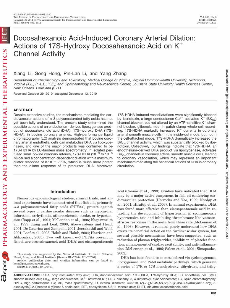
Fig. 1. Metabolism of DHA in coronary arteries. 14C-DHAwas incubated with homogenates of bovine coronary arter-ies for 15 min at 37°C in the presence or absence of indi-cated inhibitors. Then DHA and its metabolites in thereaction mixtures were extracted and applied for HPLCanalysis. A, typical HPLC chromatograms depicting themetabolism of 14C-DHA with (left) or without addition ofthe lipoxygenase inhibitor CDC (10 �M; right) in coronaryarterial tissue homogenates. Metabolites of 14C-DHA wereeluted at a peak of 2.5 min, and 14C-DHA was eluted at apeak of 33 min. B, summarized data showing radioactivityof 14C-DHA metabolites in the reaction mixtures in theabsence or presence of the cyclooxygenase inhibitor indo-methacin (Indo, 100 �M) or CDC (10 �M) (n � 4). C,summarized data showing the conversion rates of DHA toits metabolites in homogenates of coronary arterial tissue(Control), ECs, and SMCs (n � 5). D, representative West-ern blot gel document showing 15-lipoxygenase (15-LO)expression in homogenates of coronary arterial tissue (Con-trol) and their cells (SMC and EC). �, P � 0.05, significantdifference compared with the values from control samples.
17S-Hydroxy DHA on K� Channel Activity in Coronary Arteries 893
at Virginia C
omm
onwealth U
niv Tom
pkins McC
aw Lib/A
cq Srv on M
arch 29, 2012jpet.aspetjournals.org
Dow
nloaded from
tion mixture were extracted followed by separation andanalysis by HPLC. Figure 1A presents typical HPLC chro-matograms depicting the hydroxy metabolites of 14C-DHAwith a radioactive peak at 2.5 min. Inhibition of lipoxygen-ase by CDC markedly reduced the peak of DHA metabo-lites. As summarized in Fig. 1B, the conversion of DHAinto hydroxy metabolites was significantly blocked by CDCbut not by the cyclooxygenase inhibitor, indomethacin (100�M). Similar to indomethacin, P450 monooxygnase inhib-itors, miconazole (10 �M) or 17-octadecynoic acid (1 �M),had no effect on such metabolism of DHA (data not shown).In addition, the hydroxy metabolites of DHA were detectedin homogenates from coronary arterial ECs and SMCs(Fig. 1C). The conversion rate of DHA into hydroxy metab-olites was 3-fold higher in ECs than in SMCs (Fig. 1C).15-Lipoxygenase has been shown to hydrolyze the 17Sposition of DHA to form 17S-HDHA (Hong et al., 2003). Asindicated by Western blot analysis, 15-lipoxygenase hashigher abundance in ECs than in SMCs (Fig. 1D), suggest-ing that the high conversion rate of DHA into hydroxymetabolites in ECs is associated with the higher abun-dance of 15-lipoxygenase in these cells.
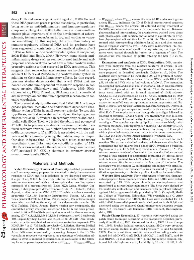
LC/MS Spectrometric Analysis of DHA Metabolites.Figure 2A shows a typical LC chromatogram of 17S-HDHAobtained from LC/MS analysis, which demonstrates that17S-HDHA had the largest peak at a retention time of 21min. Figure 2B presents a MS spectrum and structure of
17S-HDHA. The identity of 17S-HDHA was confirmed byions at m/z of 201 (245-CO2), 229 (273-CO2), 245, 255 (273-H2O), 273, 281 (M-H-H2O-CO2), 299 (M-H-CO2), 325 (M-H-H2O), and 343, which was then further confirmed by MS/MSanalysis. Our HPLC and LC/MS analyses further identifiedthat 17S-HDHA is the primary form of hydroxy metabolites;however, other 17S series hydroxy metabolites of DHA, in-cluding di- and tri-HDHA, were also detected (data notshown). 5-Lipoxygenase metabolizes 17S-HDHA into di- andtri-HDHA (Hong et al., 2003). Thus, our data indicate thatendogenous 15-lipoxygenase and 5-lipoxygenase in bovinecoronary arteries are able to metabolize DHA into 17S seriesHDHA.
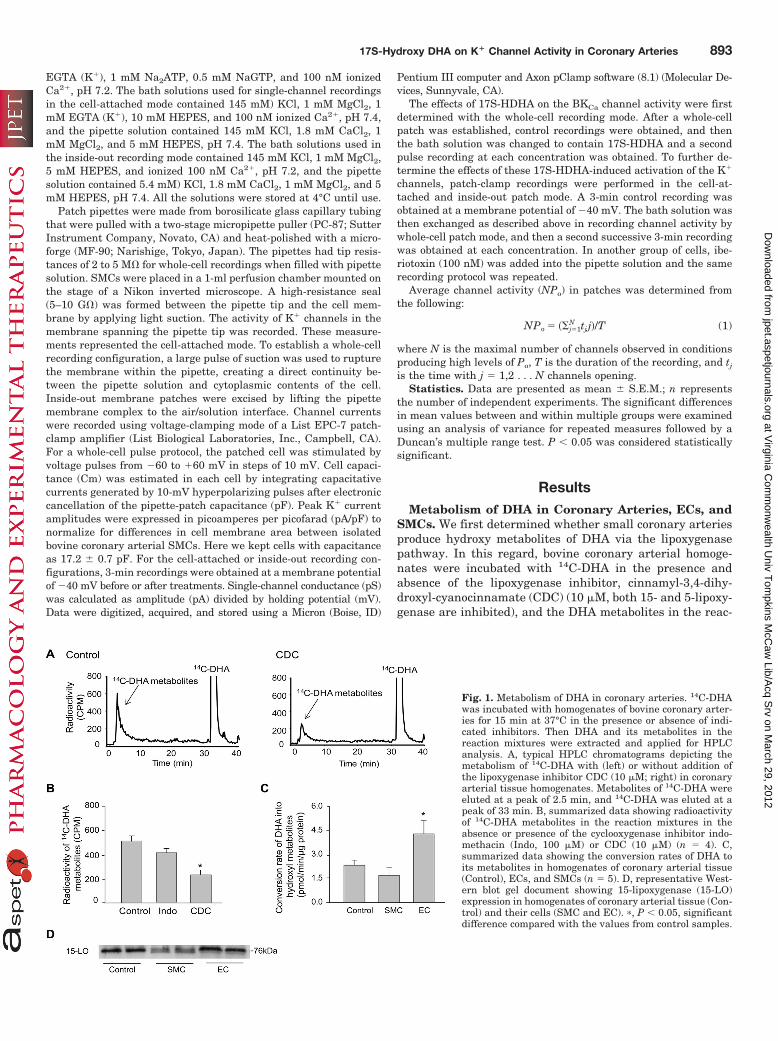
Vasodilator Responses to 17S-HDHA and DHA inSmall Coronary Arteries. We next tested whether DHA or17S-HDHA cause coronary vasodilation. As shown in Fig. 3A,in isolated precontracted small bovine coronary arteries,DHA induced vasodilator response at a relatively high con-centration (10�5 M) with a maximal dilation of 56.9 � 6.2%.Compared with its precursor DHA, 17S-HDHA induced amuch stronger vasodilator response with a maximal dilationof 87.8 � 2.5% at a concentration of 10�5 M. Moreover,17S-HDHA at 10�7 M induced an obvious dilator response(42.9 � 4.9%), whereas DHA had only a trivial dilator re-sponse (11.9 � 2.5%) at the same dose. To quantitate thepotency of dilator responses for 17S-HDHA and DHA, wecalculated the concentration required to induce 50% dilation
Fig. 2. LC/MS analysis of DHA metabolites. A, typical LC chromatogramof 17S-HDHA from LC/MS analysis showing that 17S-HDHA had a peakat a retention time of 21 min. B, typical MS spectrum of lipidomic MS/MSanalysis of DHA metabolites isolated from bovine coronary arteries. In-set, chemical structure of 17S-HDHA as reported previously (Hong et al.,2003). 17S-HDHA was identified on the basis of the diagnostic MS-MSions at m/z of 201 (245-CO2), 229 (273-CO2), 245, 255 (273-H2O), 273, 281(M-H-H2O-CO2), 299 (M-H-CO2), 325 (M-H-H2O), and 343 (M-H).
Fig. 3. A, DHA and 17S-HDHA induce vasodilation of bovine smallcoronary arteries. �, P � 0.05, significant difference compared with thevalues from the arteries treated with DHA (n � 6). B, effects of CDC (10�M) and endothelium removal (EC denuded) on DHA-induced vasodila-tion in bovine small coronary arteries. #, P � 0.05, significant differencecompared with the values from the endothelium intact arteries treatedwith DHA (n � 6).
894 Li et al.
at Virginia C
omm
onwealth U
niv Tom
pkins McC
aw Lib/A
cq Srv on M
arch 29, 2012jpet.aspetjournals.org
Dow
nloaded from
(D50) and found that the 17S-HDHA (D50 � 2.0 � 10�7 M)was 31-fold more potent than DHA (D50 � 6.3 � 10�6 M). Asshown in Fig. 3B, inhibition of lipoxygenase by CDC signifi-cantly reduced DHA-induced vasodilation. Furthermore, re-moval of the endothelium abolished DHA-induced vasodila-tion in these coronary arteries.
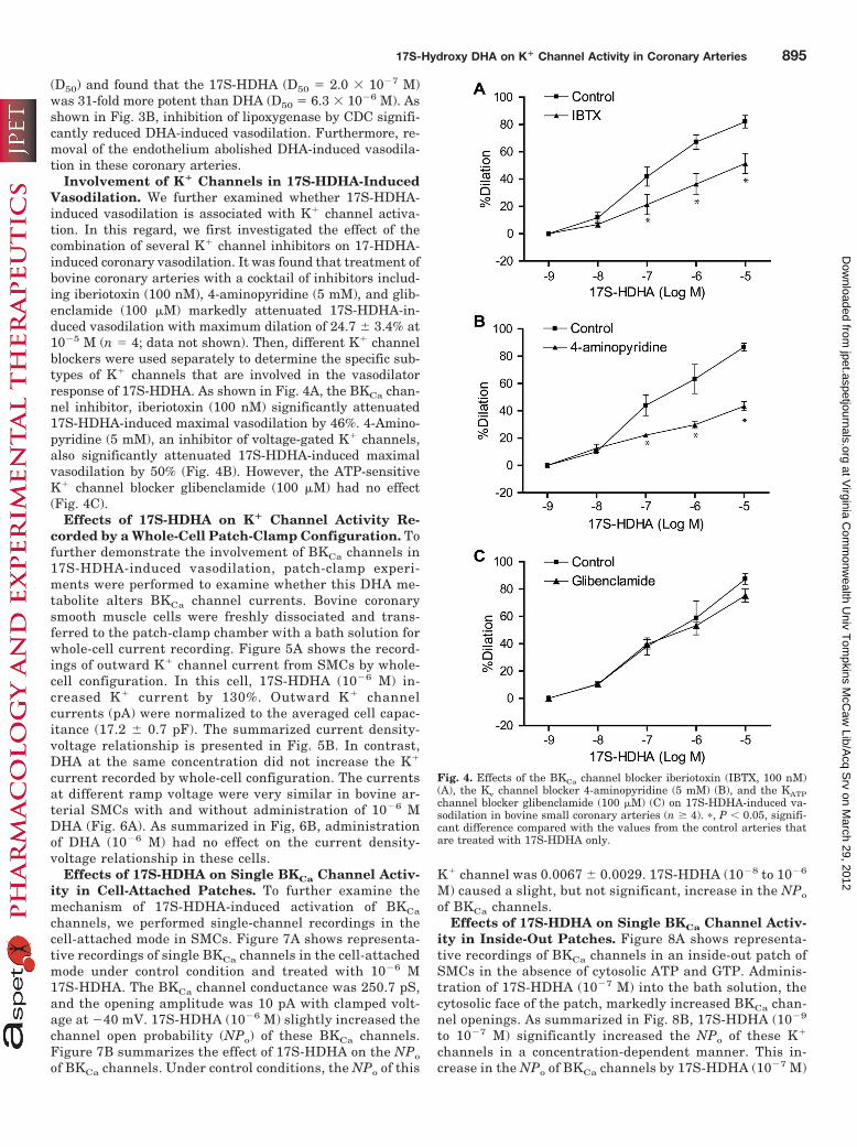
Involvement of K� Channels in 17S-HDHA-InducedVasodilation. We further examined whether 17S-HDHA-induced vasodilation is associated with K� channel activa-tion. In this regard, we first investigated the effect of thecombination of several K� channel inhibitors on 17-HDHA-induced coronary vasodilation. It was found that treatment ofbovine coronary arteries with a cocktail of inhibitors includ-ing iberiotoxin (100 nM), 4-aminopyridine (5 mM), and glib-enclamide (100 �M) markedly attenuated 17S-HDHA-in-duced vasodilation with maximum dilation of 24.7 � 3.4% at10�5 M (n � 4; data not shown). Then, different K� channelblockers were used separately to determine the specific sub-types of K� channels that are involved in the vasodilatorresponse of 17S-HDHA. As shown in Fig. 4A, the BKCa chan-nel inhibitor, iberiotoxin (100 nM) significantly attenuated17S-HDHA-induced maximal vasodilation by 46%. 4-Amino-pyridine (5 mM), an inhibitor of voltage-gated K� channels,also significantly attenuated 17S-HDHA-induced maximalvasodilation by 50% (Fig. 4B). However, the ATP-sensitiveK� channel blocker glibenclamide (100 �M) had no effect(Fig. 4C).
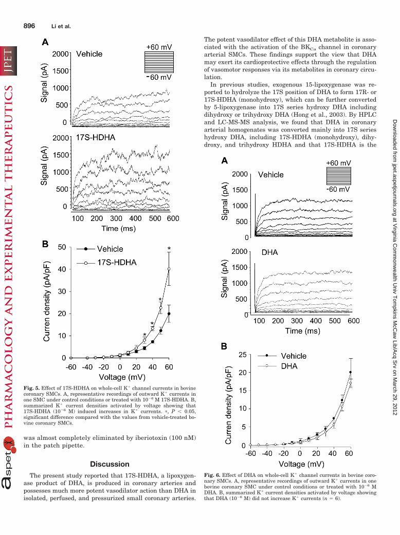
Effects of 17S-HDHA on K� Channel Activity Re-corded by a Whole-Cell Patch-Clamp Configuration. Tofurther demonstrate the involvement of BKCa channels in17S-HDHA-induced vasodilation, patch-clamp experi-ments were performed to examine whether this DHA me-tabolite alters BKCa channel currents. Bovine coronarysmooth muscle cells were freshly dissociated and trans-ferred to the patch-clamp chamber with a bath solution forwhole-cell current recording. Figure 5A shows the record-ings of outward K� channel current from SMCs by whole-cell configuration. In this cell, 17S-HDHA (10�6 M) in-creased K� current by 130%. Outward K� channelcurrents (pA) were normalized to the averaged cell capac-itance (17.2 � 0.7 pF). The summarized current density-voltage relationship is presented in Fig. 5B. In contrast,DHA at the same concentration did not increase the K�
current recorded by whole-cell configuration. The currentsat different ramp voltage were very similar in bovine ar-terial SMCs with and without administration of 10�6 MDHA (Fig. 6A). As summarized in Fig, 6B, administrationof DHA (10�6 M) had no effect on the current density-voltage relationship in these cells.
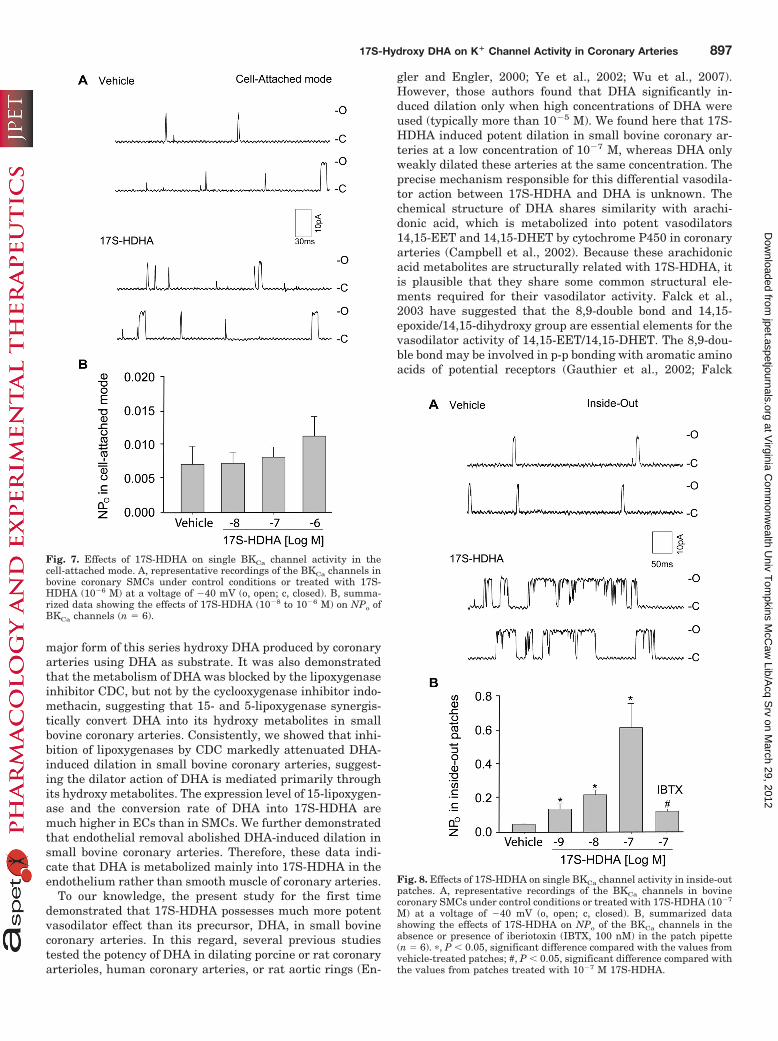
Effects of 17S-HDHA on Single BKCa Channel Activ-ity in Cell-Attached Patches. To further examine themechanism of 17S-HDHA-induced activation of BKCa
channels, we performed single-channel recordings in thecell-attached mode in SMCs. Figure 7A shows representa-tive recordings of single BKCa channels in the cell-attachedmode under control condition and treated with 10�6 M17S-HDHA. The BKCa channel conductance was 250.7 pS,and the opening amplitude was 10 pA with clamped volt-age at �40 mV. 17S-HDHA (10�6 M) slightly increased thechannel open probability (NPo) of these BKCa channels.Figure 7B summarizes the effect of 17S-HDHA on the NPo
of BKCa channels. Under control conditions, the NPo of this
K� channel was 0.0067 � 0.0029. 17S-HDHA (10�8 to 10�6
M) caused a slight, but not significant, increase in the NPo
of BKCa channels.Effects of 17S-HDHA on Single BKCa Channel Activ-
ity in Inside-Out Patches. Figure 8A shows representa-tive recordings of BKCa channels in an inside-out patch ofSMCs in the absence of cytosolic ATP and GTP. Adminis-tration of 17S-HDHA (10�7 M) into the bath solution, thecytosolic face of the patch, markedly increased BKCa chan-nel openings. As summarized in Fig. 8B, 17S-HDHA (10�9
to 10�7 M) significantly increased the NPo of these K�
channels in a concentration-dependent manner. This in-crease in the NPo of BKCa channels by 17S-HDHA (10�7 M)
Fig. 4. Effects of the BKCa channel blocker iberiotoxin (IBTX, 100 nM)(A), the Kv channel blocker 4-aminopyridine (5 mM) (B), and the KATPchannel blocker glibenclamide (100 �M) (C) on 17S-HDHA-induced va-sodilation in bovine small coronary arteries (n � 4). �, P � 0.05, signifi-cant difference compared with the values from the control arteries thatare treated with 17S-HDHA only.
17S-Hydroxy DHA on K� Channel Activity in Coronary Arteries 895
at Virginia C
omm
onwealth U
niv Tom
pkins McC
aw Lib/A
cq Srv on M
arch 29, 2012jpet.aspetjournals.org
Dow
nloaded from
was almost completely eliminated by iberiotoxin (100 nM)in the patch pipette.
DiscussionThe present study reported that 17S-HDHA, a lipoxygen-
ase product of DHA, is produced in coronary arteries andpossesses much more potent vasodilator action than DHA inisolated, perfused, and pressurized small coronary arteries.
The potent vasodilator effect of this DHA metabolite is asso-ciated with the activation of the BKCa channel in coronaryarterial SMCs. These findings support the view that DHAmay exert its cardioprotective effects through the regulationof vasomotor responses via its metabolites in coronary circu-lation.
In previous studies, exogenous 15-lipoxygenase was re-ported to hydrolyze the 17S position of DHA to form 17R- or17S-HDHA (monohydroxy), which can be further convertedby 5-lipoxygenase into 17S series hydroxy DHA includingdihydroxy or trihydroxy DHA (Hong et al., 2003). By HPLCand LC-MS-MS analysis, we found that DHA in coronaryarterial homogenates was converted mainly into 17S serieshydroxy DHA, including 17S-HDHA (monohydroxy), dihy-droxy, and trihydroxy HDHA and that 17S-HDHA is the
Fig. 6. Effect of DHA on whole-cell K� channel currents in bovine coro-nary SMCs. A, representative recordings of outward K� currents in onebovine coronary SMC under control conditions or treated with 10�6 MDHA. B, summarized K� current densities activated by voltage showingthat DHA (10�6 M) did not increase K� currents (n � 6).
Fig. 5. Effect of 17S-HDHA on whole-cell K� channel currents in bovinecoronary SMCs. A, representative recordings of outward K� currents inone SMC under control conditions or treated with 10�6 M 17S-HDHA. B,summarized K� current densities activated by voltage showing that17S-HDHA (10�6 M) induced increases in K� currents. �, P � 0.05,significant difference compared with the values from vehicle-treated bo-vine coronary SMCs.
896 Li et al.
at Virginia C
omm
onwealth U
niv Tom
pkins McC
aw Lib/A
cq Srv on M
arch 29, 2012jpet.aspetjournals.org
Dow
nloaded from
major form of this series hydroxy DHA produced by coronaryarteries using DHA as substrate. It was also demonstratedthat the metabolism of DHA was blocked by the lipoxygenaseinhibitor CDC, but not by the cyclooxygenase inhibitor indo-methacin, suggesting that 15- and 5-lipoxygenase synergis-tically convert DHA into its hydroxy metabolites in smallbovine coronary arteries. Consistently, we showed that inhi-bition of lipoxygenases by CDC markedly attenuated DHA-induced dilation in small bovine coronary arteries, suggest-ing the dilator action of DHA is mediated primarily throughits hydroxy metabolites. The expression level of 15-lipoxygen-ase and the conversion rate of DHA into 17S-HDHA aremuch higher in ECs than in SMCs. We further demonstratedthat endothelial removal abolished DHA-induced dilation insmall bovine coronary arteries. Therefore, these data indi-cate that DHA is metabolized mainly into 17S-HDHA in theendothelium rather than smooth muscle of coronary arteries.
To our knowledge, the present study for the first timedemonstrated that 17S-HDHA possesses much more potentvasodilator effect than its precursor, DHA, in small bovinecoronary arteries. In this regard, several previous studiestested the potency of DHA in dilating porcine or rat coronaryarterioles, human coronary arteries, or rat aortic rings (En-
gler and Engler, 2000; Ye et al., 2002; Wu et al., 2007).However, those authors found that DHA significantly in-duced dilation only when high concentrations of DHA wereused (typically more than 10�5 M). We found here that 17S-HDHA induced potent dilation in small bovine coronary ar-teries at a low concentration of 10�7 M, whereas DHA onlyweakly dilated these arteries at the same concentration. Theprecise mechanism responsible for this differential vasodila-tor action between 17S-HDHA and DHA is unknown. Thechemical structure of DHA shares similarity with arachi-donic acid, which is metabolized into potent vasodilators14,15-EET and 14,15-DHET by cytochrome P450 in coronaryarteries (Campbell et al., 2002). Because these arachidonicacid metabolites are structurally related with 17S-HDHA, itis plausible that they share some common structural ele-ments required for their vasodilator activity. Falck et al.,2003 have suggested that the 8,9-double bond and 14,15-epoxide/14,15-dihydroxy group are essential elements for thevasodilator activity of 14,15-EET/14,15-DHET. The 8,9-dou-ble bond may be involved in p-p bonding with aromatic aminoacids of potential receptors (Gauthier et al., 2002; Falck
Fig. 8. Effects of 17S-HDHA on single BKCa channel activity in inside-outpatches. A, representative recordings of the BKCa channels in bovinecoronary SMCs under control conditions or treated with 17S-HDHA (10�7
M) at a voltage of �40 mV (o, open; c, closed). B, summarized datashowing the effects of 17S-HDHA on NPo of the BKCa channels in theabsence or presence of iberiotoxin (IBTX, 100 nM) in the patch pipette(n � 6). �, P � 0.05, significant difference compared with the values fromvehicle-treated patches; #, P � 0.05, significant difference compared withthe values from patches treated with 10�7 M 17S-HDHA.
Fig. 7. Effects of 17S-HDHA on single BKCa channel activity in thecell-attached mode. A, representative recordings of the BKCa channels inbovine coronary SMCs under control conditions or treated with 17S-HDHA (10�6 M) at a voltage of �40 mV (o, open; c, closed). B, summa-rized data showing the effects of 17S-HDHA (10�8 to 10�6 M) on NPo ofBKCa channels (n � 6).
17S-Hydroxy DHA on K� Channel Activity in Coronary Arteries 897
at Virginia C
omm
onwealth U
niv Tom
pkins McC
aw Lib/A
cq Srv on M
arch 29, 2012jpet.aspetjournals.org
Dow
nloaded from
et al., 2003), whereas the oxygen atoms of the epoxide ordihydroxy group may act as acceptors for hydrogen bondingwith receptors of 14,15-EET/14,15-DHET. Based on thesefindings, we hypothesized that the 10,11-double bond and16,17-dihydroxy group are essential structural requirementsfor the potent vasodilator action of the hydroxy metabolites ofDHA. Hydroxylation at carbon 17 of DHA molecule mayincrease hydrogen bonding with the potential receptor andthereby enhance the vasodilator activity. This may explainwhy 17S-HDHA exerts a much more potent vasodilator effectthan DHA.
Regulation of smooth muscle membrane potential throughalteration in K� channel activity is a major mechanism ofvasodilation and vasoconstriction under both physiologicaland pathophysiological conditions (Yokoshiki et al., 1998;Toro et al., 2002; Jackson, 2005). Activation of K� channelsresults in hyperpolarization, closure of voltage-dependentCa2� channels, and vasodilation, whereas inhibition of K�
channels has the opposite effects. Several different types ofK� channels including BKCa channel, Kv channel, and KATP
channel, are present in bovine coronary SMCs and involvedin the regulation of vascular tone or vasomotor responses ofcoronary arteries (Yokoshiki et al., 1998; Toro et al., 2002;Jackson, 2005). In the present study, we found that both theBKCa blocker, iberiotoxin, and the Kv channel blocker, 4-ami-nopyridine, significantly attenuated 17S-HDHA-induced va-sodilation, whereas the KATP channel blocker, glibenclamide,had no effect. These data suggest that the activation of BKCa
and Kv channels may contribute to 17S-HDHA-induced va-sodilation in coronary arteries.
To further determine the electrophysiological mechanism-mediating actions of 17S-HDHA, we performed patch-clampexperiments with a focus on the BKCa channel in the mem-brane of coronary arterial SMCs. In the whole-cell recordingmode, 17S-HDHA (10�6 M) markedly increased K� currentsin these SMCs. In contrast, DHA (10�6 M) had no effect onthe K� currents, suggesting that DHA at such doses does notactivate BKCa channels in bovine coronary SMCs. It seemsthat these results are inconsistent with a study demonstrat-ing that DHA activates BKCa channels in rat coronary arte-rial SMCs (Lai et al., 2009). In rat SMCs, however, theincrease in BKCa currents was observed only when highconcentrations of DHA (2.0 � 10�5 M to 8.0 � 10�5 M) wereused (Lai et al., 2009). It is possible that 17S-HDHA and ahigh concentration of DHA may activate BKCa channels viadistinct mechanisms. 17S-HDHA-induced activation of BKCa
channels seems to be specific to the structure of 17S-HDHAas shown in EET studies (Campbell et al., 2002; Gauthier etal., 2002; Falck et al., 2003). 17S-HDHA may directly inter-act with and change the conformation of BKCa channels,leading to increased channel activity. As reported by Thid etal. (2007), DHA at high concentrations (more than 6.0 � 10�5
M) dramatically changed the viscoelastic properties of thephosphocholine-supported lipid bilayers. Thus, DHA at highconcentrations is more likely to exert its effects as a nonspe-cific lipid compound that modifies biophysical properties ofmembranes, which results in the alteration of BKCa channelactivities.
Previous studies demonstrated that the direct action of GS
on the BKCa channels mediates the effects of 11,12-EET, animportant endothelium-derived hyperpolarization factor(Brown and Birnbaumer, 1990; McDonald et al., 1994; Li and
Campbell, 1997). This membrane-delimited action of GS is aubiquitous mechanism for regulating K� and Ca2� channelsand requires the presence of GTP (Brown and Birnbaumer,1990; McDonald et al., 1994). However, our data suggest thata direct activation of G proteins is not involved in 17S-HDHA-induced BKCa channel activation. First, in the cell-attached mode, 17S-HDHA had no significant effects onBKCa channel activity, suggesting that 17S-HDHA does notactivate BKCa channel through intracellular coupling mech-anisms. This result suggests a membrane-delimited action of17S-HDHA on BKCa channel activity. In excised inside-outpatches, 17S-HDHA markedly increased BKCa channel activ-ity when it was added to the cytoplasmic side of membraneseven in the absence of GTP or ATP. This suggests that theactivation of BKCa channels by 17S-HDHA does not requireany intracellular soluble components, including GTP andATP. These data rule out the possibility of a G protein-coupled mechanism in mediation of 17S-HDHA action. Itseems that a membrane-delimited, but G protein-indepen-dent, mechanism mediates 17S-HDHA-induced activation ofBKCa channels.
It should be noted that 17S-HDHA also induced a smallresidual dilation in the presence of a combination of K�
channel inhibitors (i.e., iberiotoxin, 4-aminopyridine, andglibenclamide) in bovine small coronary arteries with maxi-mum dilation of 24.7 � 3.4% at 10�5 M (Fig. 3B). Thisresidual dilator response may be attributed to the dilatoreffects of 17S-HDHA metabolites. It has been shown that17S-HDHA can be further converted by 5-lipoxygenase into17S series hydroxy DHA including dihydroxy or trihydroxyDHA (Hong et al., 2003). It is possible that these 17S-HDHAmetabolites (converted by lipoxygenases in the intact endo-thelium) dilate coronary arteries via K� channel-indepen-dent mechanisms. Indeed, we found that 17S trihydroxyDHA is also a potent vasodilator in bovine small coronaryarteries with a maximal dilation of 59.1 � 5.5% at 10�7 M(unpublished data). However, the mechanisms responsiblefor the dilator effect of 17S trihydroxy DHA are unknown andneed to be further investigated in future studies.
In summary, the present study demonstrated that 17S-HDHA is a novel and potent vasodilator in coronary arteries.17S-HDHA activates BKCa channels in SMCs from thesearteries via a membrane-delimited mechanism by direct ac-tion of 17S-HDHA on channel proteins or their regulatoryproteins. Because 17S-HDHA possesses more potent vasodi-lator effect than DHA itself, this DHA metabolite may serveas an endothelium-derived hyperpolarization factor to regu-late vascular tone in coronary circulation and to at leastpartially mediate the beneficial actions of DHA or �-3 PUFAsin the prevention and treatment of cardiovascular diseases,in particular coronary arterial diseases.
Authorship Contributions
Participated in research design: X. Li, P.-L. Li, and Zhang.Conducted experiments: X. Li, Hong, and Zhang.Performed data analysis: X. Li, Hong, P.-L. Li, and Zhang.Wrote or contributed to the writing of the manuscript: X. Li, P.-L.
Li, and Zhang.
ReferencesAbeywardena MY and Head RJ (2001) Longchain n-3 polyunsaturated fatty acids
and blood vessel function. Cardiovasc Res 52:361–371.
898 Li et al.
at Virginia C
omm
onwealth U
niv Tom
pkins McC
aw Lib/A
cq Srv on M
arch 29, 2012jpet.aspetjournals.org
Dow
nloaded from

Brown AM and Birnbaumer L (1990) Ionic channels and their regulation by Gprotein subunits. Annu Rev Physiol 52:197–213.
Campbell WB, Deeter C, Gauthier KM, Ingraham RH, Falck JR, and Li PL (2002)14,15-Dihydroxyeicosatrienoic acid relaxes bovine coronary arteries by activationof K(Ca) channels. Am J Physiol Heart Circ Physiol 282:H1656–H1664.
Connor WE, DeFrancesco CA, and Connor SL (1993) N-3 fatty acids from fish oil.Effects on plasma lipoproteins and hypertriglyceridemic patients. Ann NY AcadSci 683:16–34.
De Caterina R and Zampolli A (2001) n-3 fatty acids: antiatherosclerotic effects.Lipids 36:S69–S78.
Engler MB and Engler MM (2000) Docosahexaenoic acid-induced vasorelaxation inhypertensive rats: mechanisms of action. Biol Res Nurs 2:85–95.
Falck JR, Krishna UM, Reddy YK, Kumar PS, Reddy KM, Hittner SB, Deeter C,Sharma KK, Gauthier KM, and Campbell WB (2003) Comparison of vasodilatoryproperties of 14,15-EET analogs: structural requirements for dilation. Am JPhysiol Heart Circ Physiol 284:H337–H349.
Fleischhauer FJ, Yan WD, and Fischell TA (1993) Fish oil improves endothelium-dependent coronary vasodilation in heart transplant recipients. J Am Coll Cardiol21:982–989.
Gauthier KM, Deeter C, Krishna UM, Reddy YK, Bondlela M, Falck JR, and Camp-bell WB (2002) 14,15-Epoxyeicosa-5(Z)-enoic acid: a selective epoxyeicosatrienoicacid antagonist that inhibits endothelium-dependent hyperpolarization and relax-ation in coronary arteries. Circ Res 90:1028–1036.
Ge ZD, Zhang DX, Chen YF, Yi FX, Zou AP, Campbell WB, and Li PL (2003) CyclicADP-ribose contributes to contraction and Ca2� release by M1 muscarinic receptoractivation in coronary arterial smooth muscle. J Vasc Res 40:28–36.
Gebremedhin D, Ma YH, Falck JR, Roman RJ, VanRollins M, and Harder DR (1992)Mechanism of action of cerebral epoxyeicosatrienoic acids on cerebral arterialsmooth muscle. Am J Physiol Heart Circ Physiol 263:H519–H525.
Geiger J, Zou AP, Campbell WB, and Li PL (2000) Inhibition of cADP-ribose forma-tion produces vasodilation in bovine coronary arteries. Hypertension 35:397–402.
Hamill OP, Marty A, Neher E, Sakmann B, and Sigworth FJ (1981) Improvedpatch-clamp techniques for high-resolution current recording from cells and cell-free membrane patches. Pflugers Arch 391:85–100.
Harrison N and Abhyankar B (2005) The mechanism of action of omega-3 fatty acidsin secondary prevention post-myocardial infarction. Curr Med Res Opin 21:95–100.
Hirafuji M, Machida T, Hamaue N, and Minami M (2003) Cardiovascular protectiveeffects of n-3 polyunsaturated fatty acids with special emphasis on docosa-hexaenoic acid. J Pharmacol Sci 92:308–316.
Holub DJ and Holub BJ (2004) Omega-3 fatty acids from fish oils and cardiovasculardisease. Mol Cell Biochem 263:217–225.
Hong S, Gronert K, Devchand PR, Moussignac RL, and Serhan CN (2003) Noveldocosatrienes and 17S-resolvins generated from docosahexaenoic acid in murinebrain, human blood, and glial cells. Autacoids in anti-inflammation. J Biol Chem278:14677–14687.
Horrocks LA and Yeo YK (1999) Health benefits of docosahexaenoic acid (DHA).Pharmacol Res 40:211–225.
Jackson WF (2005) Potassium channels in the peripheral microcirculation. Micro-circulation 12:113–127.
Jeerakathil TJ and Wolf PA (2001) Prevention of strokes. Curr Atheroscler Rep3:321–327.
Kang JX and Leaf A (2000) Prevention of fatal cardiac arrhythmias by polyunsatu-rated fatty acids. Am J Clin Nutr 71:202S–207S.
Lai LH, Wang RX, Jiang WP, Yang XJ, Song JP, Li XR, and Tao G (2009) Effects ofdocosahexaenoic acid on large-conductance Ca2�-activated K� channels and volt-age-dependent K� channels in rat coronary artery smooth muscle cells. ActaPharmacol Sin 30:314–320.
Leaf A, Xiao YF, Kang JX, and Billman GE (2003) Prevention of sudden cardiacdeath by n-3 polyunsaturated fatty acids. Pharmacol Ther 98:355–377.
Li PL and Campbell WB (1997) Epoxyeicosatrienoic acids activate K� channels incoronary smooth muscle through a guanine nucleotide binding protein. Circ Res80:877–884.
McDonald TF, Pelzer S, Trautwein W, and Pelzer DJ (1994) Regulation and modu-lation of calcium channels in cardiac, skeletal, and smooth muscle cells. PhysiolRev 74:365–507.
McLennan P, Howe P, Abeywardena M, Muggli R, Raederstorff D, Mano M, RaynerT, and Head R (1996) The cardiovascular protective role of docosahexaenoic acid.Eur J Pharmacol 300:83–89.
Nageswari K, Banerjee R, and Menon VP (1999) Effect of saturated, omega-3 andomega-6 polyunsaturated fatty acids on myocardial infarction. J Nutr Biochem10:338–344.
Nordøy A, Marchioli R, Arnesen H, and Videbaek J (2001) n-3 polyunsaturated fattyacids and cardiovascular diseases. Lipids 36:S127–S129.
Rapp JH, Connor WE, Lin DS, and Porter JM (1991) Dietary eicosapentaenoic acidand docosahexaenoic acid from fish oil. Their incorporation into advanced humanatherosclerotic plaques. Arteriosclerosis Thrombosis 11:903–911.
Salem N Jr, Litman B, Kim HY, and Gawrisch K (2001) Mechanisms of action ofdocosahexaenoic acid in the nervous system. Lipids 36:945–959.
Shimokawa H and Vanhoutte PM (1989) Dietary omega 3 fatty acids and endothe-lium-dependent relaxations in porcine coronary arteries. Am J Physiol Heart CircPhysiol 256:H968–H973.
Simopoulos AP (2002) Omega-3 fatty acids in inflammation and autoimmune dis-eases. J Am Coll Nutr 21:495–505.
Thid D, Benkoski JJ, Svedhem S, Kasemo B, and Gold J (2007) DHA-inducedchanges of supported lipid membrane morphology. Langmuir 23:5878–5881.
Toro L, Marijic J, Nishimaru K, Tanaka Y, Song M, and Stefani E (2002) Aging, ionchannel expression, and vascular function. Vascul Pharmacol 38(1):73–80.
Wu KT, Huang CT, Wei J, Tsait LM, Hsu CH, Chen YC, Yangs JM, and Lin CI (2007)Vasodilator action of docosahexaenoic acid (DHA) in human coronary arteries invitro. Chin J Physiol 50:164–170.
Ye D, Zhang D, Oltman C, Dellsperger K, Lee HC, and VanRollins M (2002) Cyto-chrome p-450 epoxygenase metabolites of docosahexaenoate potently dilate coro-nary arterioles by activating large-conductance calcium-activated potassium chan-nels. J Pharmacol Exp Ther 303:768–776.
Yokoshiki H, Sunagawa M, Seki T, and Sperelakis N (1998) ATP-sensitive K�channels in pancreatic, cardiac, and vascular smooth muscle cells. Am J PhysiolCell Physiol 274:C25–C37.
Address correspondence to: Dr. Pin-Lan Li, Department of Pharmacologyand Toxicology, Medical College of Virginia, Virginia Commonwealth Univer-sity, Richmond, VA 23298. E-mail: [email protected]
17S-Hydroxy DHA on K� Channel Activity in Coronary Arteries 899
at Virginia C
omm
onwealth U
niv Tom
pkins McC
aw Lib/A
cq Srv on M
arch 29, 2012jpet.aspetjournals.org
Dow
nloaded from