Embed Size (px)
Citation preview
ANEURYSMS AND DISSECTIONS
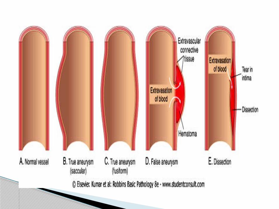
An aneurysm is a localized abnormal dilation of a blood vessel or the heart Types:1-"true" aneurysmit involves all three layers of the arterial wall (intima, media, and adventitia)
or the attenuated wall of the heart. e.g. Atherosclerotic, syphilitic, and congenital aneurysms, and ventricular
aneurysms that follow transmural myocardial infarctions. 2- “false” aneurysm (also called pseudo-aneurysm) is a breach in the vascular wall leading to an extravascular hematoma that
freely communicates with the intravascular space ("pulsating hematoma"). E.g. ventricular ruptures after MI that are contained by a pericardial
adhesion E.g. a leak at the junction of a vascular graft with a natural artery.
Aneurysm
aneurysms are classified by macroscopic shape and size (note: aspects of shape and size are not specific for any disease or clinical manifestations) :
Saccular aneurysms spherical outpouchings (involving only a portion of
the vessel wall, and often contain thrombi. Fusiform aneurysms diffuse, circumferential dilation of a long vascular
segment; they vary in diameter and length and can involve
extensive portions of the aortic arch, abdominal aorta, or even the iliacs.
The two most important causes are:1- atherosclerosis : the most common cause causes thinning and weakening of the media. The intimal plaques
compress the underlying media and also compromise nutrient and waste diffusion from the vascular lumen into the arterial wall. The media consequently undergoes degeneration and necrosis, thus allowing the dilation of the vessel
2- cystic medial degeneration of the arterial media. E.g. Marfan syndrome.
Other causes include: trauma, congenital defects (e.g., berry aneurysms), infections (mycotic aneurysms), systemic diseases, such as vasculitis.
Aortic aneurysms
Infection of a major artery that weakens its wall is called a mycotic aneurysm
possible complications: thrombosis and rupture. can originate from:(1) embolization of a septic thrombus, usually as a
complication of infective endocarditis(2) extension of an adjacent suppurative process; (3) circulating organisms directly infecting the arterial wall
Mycotic AAAs are atherosclerotic lesions infected by lodging of circulating microorganisms in the wall
- e.g. bacteremia from a primary Salmonella gastroenteritis.
Mycotic aneurysms
Atherosclerotic aneurysms occur most frequently in the abdominal aorta (abdominal aortic aneurysm, often abbreviated AAA)
the common iliac arteries, the arch, and descending parts of the thoracic aorta can also be involved
Pathogenesis AAA occurs more frequently in men and rarely develops before age
50. Atherosclerosis is a major cause of AAA other contributors include: hereditary defects in structural components of the aorta (e.g.,
defective fibrillin production in Marfan disease affects elastic tissue synthesis)
an altered balance of collagen degradation and synthesis mediated by local inflammatory infiltrates and the destructive proteolytic enzymes
Abdominal Aortic Aneurysm
Usually positioned below the renal arteries and above the bifurcation of the aorta
AAA can be saccular or fusiform as large as 15 cm in diameter, and as long as 25 cm. Microscopically: atherosclerosis with destruction and
thinning of the underlying aortic media the aneurysm frequently contains a laminated mural
thrombus
Morphology
Caused by The spirochetes T. pallidum A vanishingly rare complication in the U.S. and West thanks
to early recognition and treatment of syphilis Tertiary stage of syphilis can cause obliterative endarteritis
of the involve small vessels in any part of the body, including the vasa vasorum of the aorta
this results in ischemic medial injury, leading to aneurysmal dilation of the aorta and aortic annulus, and eventually valvular insufficiency.
valvular insufficiency and massive volume overload lead to hypertrophy of the left ventricle. The greatly enlarged hearts are sometimes called "cor bovinum" (cow's heart).
Syphilitic Aneurysm
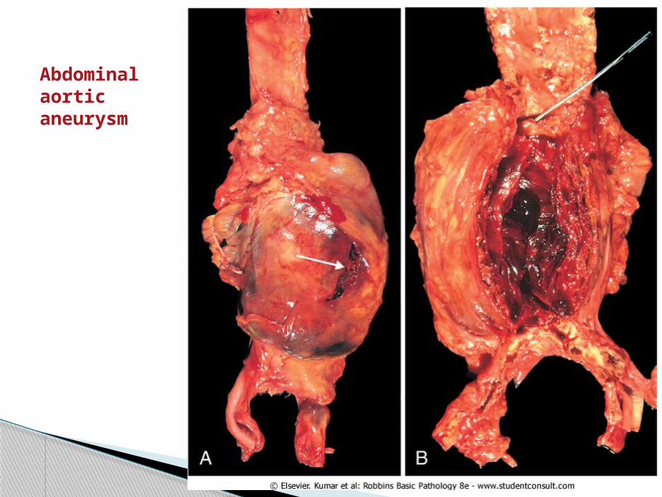
Abdominal aortic aneurysm
Rupture massive potentially fatal hemorrhage- risk of rupture is directly related to the size of the aneurysm (of 5 cm or more)- operative mortality for unruptured aneurysms 5%, after rupture the mortality rate is more than 50%
Obstruction of downstream vessel tissue ischemic injury((e.g. iliac (leg), renal (kidney), mesenteric (gastrointestinal [GI] tract), or vertebral (spinal cord) arteries))
Embolism from atheroma or mural thrombus Impingement and compression on an adjacent structure (e.g.ureter
or vertebrae) Presentation as an abdominal mass (often palpably pulsating) that
simulates a tumor
The clinical consequences of AAA:
arises when blood enters the wall of the artery, as a hematoma dissecting between its layers.
Dissections are often but not always aneurysmal. Both true and false aneurysms as well as dissections can
rupture, often with catastrophic consequences Aortic dissection : is a catastrophic event whereby blood dissects apart the media
to form a blood-filled channel within the aortic wall Complications are :
- massive hemorrhage - cardiac tamponade (hemorrhage into the pericardial sac).
Arterial dissection
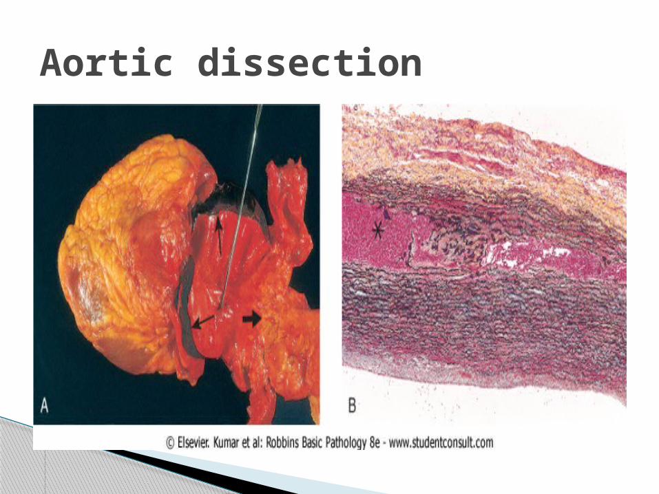
Aortic dissection
1- Hypertension is the major risk factor pressure-related mechanical injury and/or ischemic
injury. 2- inherited or acquired connective tissue disorders
causing abnormal vascular ECM (e.g., Marfan syndrome, Ehlers-Danlos syndrome,
vitamin C deficiency, copper metabolic defects)
Pathogenesis of Aortic dissection
The most common cause among the inherited or acquired connective tissue disorders associated with Aortic dissection
it is an autosomal dominant disease of fibrillin, an ECM scaffolding protein required for normal elastic tissue synthesis.
Patients have skeletal abnormalities (elongated axial bones) and ocular findings (lens subluxation) in addition to the cardiovascular manifestations
Marfan syndrome
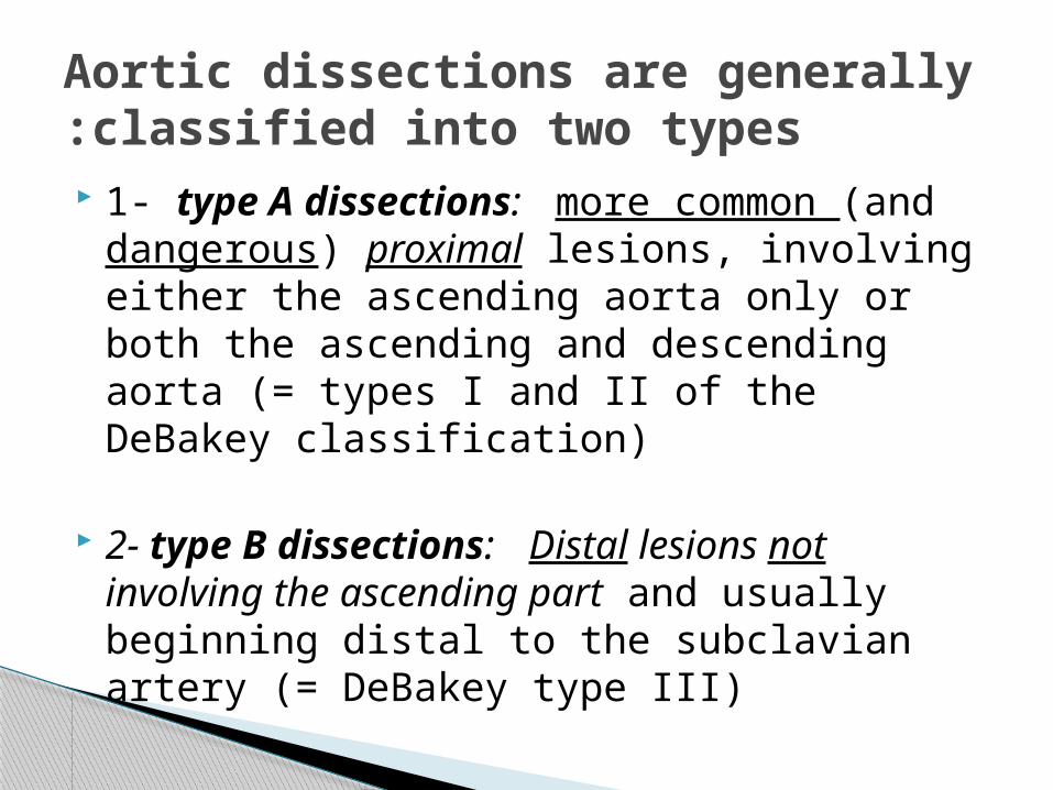
1- type A dissections: more common (and dangerous) proximal lesions, involving either the ascending aorta only or both the ascending and descending aorta (= types I and II of the DeBakey classification)
2- type B dissections: Distal lesions not involving the ascending part and usually beginning distal to the subclavian artery (= DeBakey type III)
Aortic dissections are generally classified into two types:
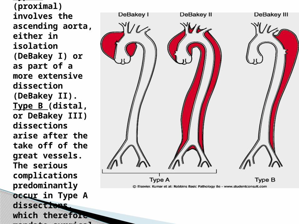
Classification of dissections. Type A (proximal) involves the ascending aorta, either in isolation (DeBakey I) or as part of a more extensive dissection (DeBakey II). Type B (distal, or DeBakey III) dissections arise after the take off of the great vessels. The serious complications predominantly occur in Type A dissections, which therefore mandate surgical intervention
Previously, aortic dissection was typically fatal, but the prognosis has markedly improved.
Rapid diagnosis and institution of intensive antihypertensive therapy, coupled with surgical procedures involving plication of the aorta permits survival of 65% to 75% of patients
Clinical course