Embed Size (px)
Citation preview

Docking Studies and Model Development of Tea PolyphenolProteasome Inhibitors: Applications toRational Drug DesignDavid M. Smith,1 Kenyon G. Daniel,1 Zhigang Wang,2 Wayne C. Guida,1,3 Tak-Hang Chan,2 and Q. Ping Dou1*1Drug Discovery Program, H. Lee Moffitt Cancer Center and Research Institute, and Departments of Biochemistry andMolecular Biology and Interdisciplinary Oncology, College of Medicine, University of South Florida, Tampa, Florida2Department of Chemistry, McGill University, Montreal, Quebec, Canada3Department of Chemistry, Eckerd College, St. Petersburg, Florida
ABSTRACT Previously, we demonstrated thatnatural and synthetic ester bond-containing greentea polyphenols were potent and specific non-peptide proteasome inhibitors. However, the molecu-lar mechanism of inhibition is currently unknown.Here, we report that inhibition of the chymotrypsinactivity of the 20S proteasome by (�)-epigallocat-echin-3-gallate (EGCG) is time-dependent and irre-versible, implicating acylation of the �5-subunit’scatalytic N-terminal threonine (Thr 1). This knowl-edge is used, along with in silico docking experi-ments, to aid in the understanding of binding andinhibition. On the basis of these docking experi-ments, we propose that (�)-EGCG binds the chymo-trypsin site in an orientation and conformation thatis suitable for a nucleophilic attack by Thr 1. Consis-tently, the distance from the electrophilic carbonylcarbon of (�)-EGCG to the hydroxyl group of Thr 1was measured as 3.18 Å. Furthermore, the A ring of(�)-EGCG acts as a tyrosine mimic, binding to thehydrophobic S1 pocket of the �5-subunit. In theprocess, the (�)-EGCG scissile bond may becomestrained, which could lower the activation energyfor attack by the hydroxyl group of Thr 1. Thismodel is validated by comparison of predicted andactual activities of several EGCG analogs, eithernaturally occurring, previously synthesized, or ratio-nally synthesized. Proteins 2004;54:58–70.© 2003 Wiley-Liss, Inc.
Key words: 20S proteasome; �5-subunit; chymotryp-sin-like activity; EGCG; green tea; or-ganic synthesis; computation; drug dis-covery
INTRODUCTION
The proteasome is a massive multicatalytic proteasecomplex that is responsible for degrading most of thecellular proteins.1,2 The 20S-core particle of the 26S protea-some is barrel-shaped, and the sites of proteolytic activityreside on the interior. The eukaryotic proteasome containsthree known activities, which are associated with its�-subunits. These are the chymotrypsin-like (cleavageafter hydrophobic residues, �5-subunit), trypsin-like (cleav-age after basic residues, �2-subunit), and caspase-like
(cleavage after acidic residues, �1-subunit) activities.3
These three activities depend on the presence of anN-terminal Thr (Thr 1) residue.2 The hydroxyl group onthe side chain of Thr 1 is responsible for catalyzingcleavage of peptides through nucleophilic attack (addition-elimination mechanism). Near this N-terminal threonine,binding pockets recognize the side-chains of peptides andgive each catalytic site its specificity. The S1 pocket of the�5-subunit is defined by the hydrophobic residues, Ala 20,Val 31, Ile 35, Met 45, Ala 49, and Gln 53,2 and thisbinding pocket has been shown to be important for sub-strate specificity and binding of several types of protea-some inhibitors.4,5
The ubiquitin/proteasome-dependent degradation path-way plays an essential role in up-regulation of cell prolifera-tion, down-regulation of cell death, and development ofdrug resistance in human tumor cells, suggesting the useof proteasome inhibitors as potential novel anticancerdrugs.6,7 This hypothesis has been supported by resultsusing various cell cultures, animal models, and clinicaltrials. In a broad range of cell culture models, proteasomeinhibitors rapidly induce tumor cell apoptosis, selectivelytrigger programmed cell death in the oncogene-trans-formed, but not normal or untransformed cells, and areable to activate the death program in human cancer cellsthat are resistant to various anticancer agents.6–9 Inhibi-tion of the chymotrypsin-like, but not trypsin-like, activityhas been found to be associated with induction of tumorcell apoptosis.8,9 In different animal studies, proteasomeinhibitors suppress tumor growth via induction of apopto-sis and inhibition of angiogenesis.10,11 MLN-341 (formerlyPS-341) is a potent and selective dipeptidyl boronic acid
Grant sponsor: National Cancer Institute-National Institutes ofHealth; Grant sponsor: the United States Army Medical Research andMaterial Command; Grant sponsor: H. Lee Moffitt Cancer Center andResearch Institute; Grant sponsor: Natural Science and EngineeringResearch Council of Canada; Grant sponsor: Molecular Imaging CoreFacility at Moffitt Cancer Center and Research Institute.
*Correspondence to: Q. Ping Dou, Barbara Ann Karmanos CancerInstitute, and Department of Pathology, Wayne State UniversitySchool of Medicine, 508 & 516 HWCRC, 110 E. Warren Ave., Detroit,MI 48201. E-mail: [email protected]
Received 19 December 2002; Accepted 15 May 2003
PROTEINS: Structure, Function, and Bioinformatics 54:58–70 (2004)
© 2003 WILEY-LISS, INC.

compound that inhibits the chymotrypsin-like activity ofthe 20S proteasome.12 This proteasome inhibitor is cur-rently being developed for the potential treatment ofhuman hematological malignant neoplasms and solid tu-mors.13,14 Preliminary data from phase I and II clinicaltrials confirm the antitumor activity of MLN-341, al-though some associated side effects were observed.13,14
The mechanism by which MLN-341 inhibits the protea-some has not yet been determined by X-ray diffractionexperiments. However, the proteasome-inhibition mecha-nism of another peptide inhibitor LLnL2 and non-peptideinhibitors lactacystin2 and the macrocyclic compound TMC-9515 have been confirmed by X-ray diffraction. Understand-ing how these inhibitors function at the molecular levelwill give insight into the structural studies of otherproteasome inhibitors where X-ray crystal structures arenot available. These studies thereby show that the protea-some is an excellent target for developing pharmacologicalanticancer drugs.
Tea, the most popular beverage in the world, is con-sumed by more than two thirds of the world’s population.Several epidemiological studies have provided evidence forthe cancer-preventive properties of green tea.16–19 Further-more, animal studies have also suggested that green teapolyphenols could suppress the formation and growth ofvarious tumors.20–24 Although numerous cancer-relatedproteins are affected by tea polyphenols,25–27 the molecu-lar basis for tea-mediated cancer prevention remainsunknown. We recently reported that the naturally occur-ring ester bond-containing green tea polyphenols (GTPs),such as (�)-epigallocatechin-3-gallate [(�)-EGCG, Fig.1(A)], possess the ability to inhibit proteasome activityboth in vitro and in vivo.28 Moreover, we demonstratedthat in a cellular extract, (�)-EGCG inhibits the �5-mediated chymotrypsin-like and �1-mediated caspase-likeactivities, but not �2-mediated trypsin-like activity, of theproteasome.28 In addition, we discovered that syntheticGTPs with an ester bond, such as (�)-EGCG [Fig. 1(A)],were also able to potently and selectively inhibit thechymotrypsin-like activity of the proteasome.29 It appearsthat a center of nucleophilic susceptibility resides at theester bond-carbon in these polyphenols.28,29 On the basisof these results, we proposed that this electrophilic car-bonyl carbon might play a role in proteasomal inhibitionby acylating Thr1 in the active site. This proposed mecha-nism of ester bond-based nucleophilic attack is similar tothat of lactacystin-based inhibition.4 However, until now,the precise mechanism of proteasome inhibition by GTPshas not been explored.
The purpose of the present study was to build a mecha-nistic model to describe how EGCG binds the proteasomebefore attack and cleavage of its ester bond.28 Because thechemistry of ester bond cleavage would in all likelihood notallow for an EGCG-proteasome complex or subsequenttetrahedral intermediate to be captured by X-ray diffrac-tion, another available tool for investigating the putativeinteraction between EGCG and the proteasome for ratio-nal drug design is computational docking. In the presentstudy, for the first time, we demonstrate that (�)-EGCG is
an irreversible mechanism-based inhibitor of the chymo-trypsin-like activity of 20S proteasome. On the basis ofthis finding, we establish a docking model for how (�)-EGCG interacts with the �5-subunit of the 20S protea-some. This model is verified by application of several othernatural and synthetic EGCG analogs, because their dock-ing free energy could be used to predict their actualproteasome-inhibitory activity. Finally, the proteasomeinhibition model of (�)-EGCG is further validated byrationally designing and synthesizing two EGCG-amidecompounds, followed by comparing their predicted andactual proteasome-inhibitory activities.
MATERIALS AND METHODSMaterials
Highly purified tea polyphenols (�)-EGCG (�95%), (�)-GCG (�98%), (�)-ECG (�98%) and (�)-CG (�98%) werepurchased from Sigma (St. Louis, MO). (�)-EGCG, benzyl-protected-(�)-EGCG, (�)-GCG, (�)-EGCG-amide, and (�)-EGCG-amide were prepared by enantioselective synthesis(see below). Purified 20S eukaryotic proteasome fromrabbit was purchased from Boston Biochem (Cambridge,MA). Purified 20S prokaryotic proteasome (Methanosar-cina thermophile, recombinant, E. coli) was purchasedfrom Calbiochem (La Jolla, CA). Fluorogenic peptide sub-strates Suc-Leu-Leu-Val-Tyr-AMC (for the proteasomalchymotrypsin-like activity) was obtained from Calbiochem(La Jolla, CA).
Enantioselective Synthesis of GTPs
Synthesis of (�)-EGCG, benzyl-protected-(�)-EGCG, and(�)-GCG were as described previously.29 The chemicalsynthesis of (�)-EGCG-amide and (�)-EGCG-amide start-ing from trans-5,7-bis-benzyloxy-2-[3,4,5-tris (benzyloxy)-phenyl] chroman-3-ol29,30 will be published elsewhereseparately.
Inhibition of Purified 20S Proteasome Activity byNatural or Synthetic GTPs
The chymotrypsin-like activity of purified 20S protea-some was measured as previously described.28 Briefly,purified prokaryotic (0.5 �g) or eukaryotic (0.02 �g) 20Sproteasome was incubated with 20 �M fluorogenic peptidesubstrate, Suc-Leu-Leu-Val-Tyr-AMC for 30 min at 37°Cin 100 �L of assay buffer (50 mM Tris-HCl, pH 7.5), with orwithout a natural or synthetic tea polyphenol. After incu-bation, production of hydrolyzed 7-amido-4-methyl-couma-rin (AMC) groups was measured by using a multiwell plateVersaFluor™ Fluorometer with an excitation filter of 380nm and an emission filter of 460 nm (Bio-Rad).
Assays for Irreversible Inhibition
To measure the effect of dialysis on (�)-EGCG-mediated proteasome inhibition, 20S prokaryotic protea-some (2 �g) was incubated with 10 �M (�)-EGCG or thecontrol solvent (H2O) in 50 mM Tris-HCl, pH 7.5 for 1 h.This was then incubated at 4°C either without or withdialysis overnight using a 10,000 MWCO Pierce Slide-A-Lyzer Dialysis Cassette (Rockford, IL) in a rotating bath
DOCKING OF POLYPHENOL PROTEASOME INHIBITORS 59

of 50 mM Tris-HCl, pH 7.5. The proteasomal chymotryp-sin-like activity was then assayed as previously de-scribed.28 As a control, an EGCG solution (without
purified 20S proteasome) was dialyzed overnight, fol-lowed by measurement of the effects on inhibition of theproteasome activity.
Fig. 1. Structures of GTPs and irreversible inhibition of 20S proteasome activity by (�)-EGCG. A: Structures of the natural and synthetic GTPs. Thenames of the synthetic GTPs are underlined to distinguish them from the natural compounds. The ring nomenclature was defined as A, C, B, or G (forgallate) rings41 and used throughout the text. B: Dialysis does not affect (�)-EGCG-mediated proteasome inhibition. The purified 20S prokaryoticproteasome (2 �g) was incubated overnight at 4°C in the presence or absence of 10 �M (�)-EGCG, with or without dialysis. Percentage of chymotrypsin(chymo) activity was then determined. The prokaryotic proteasome was used because it showed more stable kinetics after overnight dialysis, althoughsimilar result could be obtained with eukaryotic proteasome (data not shown). C: Kinetics of (�)-EGCG-mediated proteasome inhibition. (�)-EGCG at 1�M was incubated with eukaryotic 20S proteasome (0.02 �g) and suc-LLVY-AMC (20 �M) for the indicated times, followed by measurement of thechymotrypsin activity. Values are means from four independent experiments, and error bars represent standard deviations.
60 D.M. SMITH ET AL.

Molecular Modeling and Docking Studies
The crystal structure of the eukaryotic yeast 20S protea-some was obtained from the Protein Database31 (Ref.number 1JD2) and used for all the docking studies pre-sented here. The yeast 20S proteasome is structurally verysimilar to the mammalian 20S proteasome, and the chymo-trypsin active site between the two species is highlyconserved.2,4 The AutoDock 3.0 suite of programs, whichwas used for the docking calculations, uses an automateddocking approach that allows ligand flexibility as de-scribed to a full extent elsewhere.32 AutoDock has beencompared with various docking programs in several stud-ies and has been found to be able to locate docking modesthat are consistent with X-ray crystal structures.33,34
Default parameters (including a distance-dependent dielec-tric “constant”) were used as described in the AutoDockmanual except for those changes mentioned below. Thedockings were run on an i386 architecture computerrunning Redhat Linux 6.0. The crystal structure of the 20Sproteasome and the ligands were prepared for docking byfollowing the default protocols except where noted. Theenergy-scoring grid was prepared as a 20 � 20 � 20 Å boxcentered around the �5-catalytic N-terminal threonine,and the ligand was limited to this search space duringdocking. Atomic solvation parameters were assigned to theproteasome using default parameters. The default param-eters for the Lamarckian genetic algorithm32 were used asthe search protocol except for the maximum number ofenergy evaluations, which were changed to 5 million (thepopulation size was retained at 50). AutoDock relies on anempirical scoring function, which provides approximatebinding free energies. AutoDock reports a docked energythat we have referred to in this article as a “docked freeenergy” because it includes a solvation free energy term.The docked energy also includes the ligand internal energyor the intramolecular interaction energy of the ligand.AutoDock also reports a binding free energy that excludesthe ligand internal energy but includes a torsional freeenergy term for the ligand based on the number ofrotatable bonds.
In the present study, we chose to use the docked freeenergies because the number of rotatable bonds in ourinhibitors is relatively constant and because we believedthat the internal energy of the ligand should not beneglected for our compounds. Its neglect is tantamount toassuming that the intramolecular interaction energy ofthe ligand is the same in the complex as in solution. It isworth noting that the docked free energies (or binding freeenergies) that one obtains may vary, depending on theprecise force field parameters in use (e.g., charges, electro-static treatment, etc.). For the GA algorithm, the defaultparameters were kept for mutation, crossover, and elitism.The pseudo-Solis and Wets local search method wasincluded by using default parameters as well. Dockingmodes were selected on the basis of two criteria: theirproximity to the N-terminal threonine and placement ofthe A-C ring system of the molecule within the S1 hydro-phobic pocket. Of the orientations/conformations that fitthese two criteria, the docked structure of lowest dockedfree energy was chosen. Each molecule was docked with upto 30 genetic algorithm runs of 5 million energy evalua-tions for each run. AutoDock reports the best dockingsolution (lowest docked free energy) for each GA run andalso performs a cluster analysis in which the total numberof clusters and the rank of each docking mode (clusterrank) is reported. So, for a 30 GA run, for example, therewould be up to 30 total docking modes from which we thenselected the lowest energy-docking mode that met the twocriteria. In Table I, we indicate the number of clusters foreach compound docked, the cluster rank of the dockingmode selected, the range of docked free energies, and thedocked free energy of the docking mode selected. It shouldbe noted that for most of the GTPs docked, only dockingmodes from a single cluster met both of the two presetcriteria, and the docking mode with the lowest docked freeenergy was selected from this cluster. The output fromAutoDock and all modeling studies as well as images wererendered with PyMOL.35 PyMOL was used to calculate thedistances of hydrogen bonds as measured between thehydrogen and its assumed binding partner.
TABLE I. Relative Cluster Rank and Docked Free Energies of Selected Docking Modes
CompoundNumber of AutoDock
clustersaCluster rankb of selected
docked structurecDocked free energy range
of docked structuresdDocked free energy of selected
docked structured
(�)-EGCG 8 (10) 1 �10.82 to �8.75 �10.82(�)-EGCG 4 (5) 2 �10.62 to �9.83 �10.52(�)-GCG 3 (5) 3 �12.39 to �10.33 �10.33(�)-ECG 15 (30) 2 �10.81 to �8.34 �10.56(�)-EGCG-A 23 (30) 8 �11.38 to �8.23 �9.63(�)-EGCG-A 5 (5) 4 �11.70 to �9.28 �9.52(�)-CG 19 (30) 7 �10.46 to �7.93 �9.30(�)-GCG 22 (30) 13 �10.27 to �7.24 �9.10genistein 12 (100) 4 �5.40 to �4.23 �5.15aNumber of GA runs are shown in parenthesesbThe cluster rank is the absolute ranking as determined by the docked free energy defined by AutoDock.cThe criteria used for selection were as follows: (1) distance between the ester carbonyl carbon of the compound and hydroxyl oxygen of Thr1 and2) the A–C ring system located in the S1 pocket.dkcal/mol.
DOCKING OF POLYPHENOL PROTEASOME INHIBITORS 61

Determination of Nucleophilic Susceptibility
The electron density surface colored by nucleophilicsusceptibility was created by performing Extended Huckelmolecular orbital calculations with use of CaChe Worksys-tem version 3.2 (Oxford Molecular Ltd., now Accelrys), asdescribed previously.28 A colored “bull’s-eye” with a whitecenter denotes atoms that are highly susceptible to nucleo-philic attack.
RESULTS AND DISCUSSION(�)-EGCG Irreversibly Inhibits the Chymotrypsin-Like Activity of Purified 20S Proteasome
To investigate the nature of (�)-EGCG-mediated protea-some inhibition,28 we performed a dialysis experiment. Apurified prokaryotic 20S proteasome (Methanosarcina ther-mophile, recombinant E. coli) was preincubated for 1 h at37° C with either 10 �M (�)-EGCG or its control solvent(H2O), followed by overnight coincubation at 4°C with orwithout dialysis. Figure 1(B) shows that in the absence ofdialysis, (�)-EGCG was able to inhibit the chymotrypsin-like activity of the prokaryotic 20S proteasome by 85%.More importantly, overnight dialysis of the EGCG-proteasome mixture did not change the outcome: (�)-EGCG still caused 81% inhibition of the proteasomalchymotrypsin-like activity. As a control, when an aliquotof (�)-EGCG was first dialyzed overnight and then addedto the purified 20S proteasome, no inhibition was observed[Fig. 1(B), EGCG pre]. This result shows that (�)-EGCG iseither an irreversible or a tight-binding inhibitor of thechymotrypsin-like activity of the proteasome.
We also performed a kinetics experiment. (�)-EGCG at1 �M potently inhibited the chymotrypsin-like activity of apurified eukaryotic (rabbit) 20S proteasome in a time-dependent manner: 35% at 5 min, 62% at 30 min, and70–80% after 1–3 h [Fig. 1(C)], which is characteristic fora mechanism-based inhibitor.36 This result further sup-ports the argument that the mode of (�)-EGCG action isirreversible inhibition.
Previously, we hypothesized that (�)-EGCG’s suscepti-bility to a nucleophilic attack would be essential forinhibiting the proteasome.28 This hypothesis was furthersupported by our HPLC results that showed that theproteasome could attack and degrade (�)-EGCG.28 Kineticanalysis37 and X-ray diffraction2 studies using the specificproteasome inhibitor lactacystin have shown that theester bond of this inhibitor covalently modifies the N-terminal threonine of the �5-subunit, which is critical forproteasome inhibition.4 Because (�)-EGCG contains anester bond28 [Fig. 1(A)] and inhibits the proteasome irre-versibly in a time-dependent manner [Fig. 1(B) and (C)], itis probable that a lactacystin-like reaction occurs with(�)-EGCG.
Automated Docking of (�)-EGCG to the �5-Subunitof 20S Proteasome
To build a model for how (�)-EGCG binds to theproteasome, which will allow for nucleophilic attack, weperformed automated docking studies. Knowledge of theenzyme kinetics discussed above can help direct docking
solutions that would allow covalent modification and inhi-bition of the proteasome. Before acylation of Thr 1’shydroxyl by (�)-EGCG can occur, (�)-EGCG must bind tothe �5-active site in a conformation that would allow areaction to occur between the two atoms involved. There-fore, binding of (�)-EGCG in an appropriate orientationand conformation is necessary38 for ester bond scissionbecause the presence of an ester bond alone is insufficientto inhibit the proteasome. This finding is shown by benzylprotected-EGCG, which has an ester-bond, but cannotinhibit the proteasome.29 In addition, several small molecu-lar weight molecules that contain ester bonds, includingmethyl acetate, benzyl hydroxybenzoate, and methyl gal-late, cannot inhibit the proteasome (unpublished observa-tions).
Therefore, docking modes were chosen on the basis ofthe following two predefined criteria. First, the distancebetween the carbonyl carbon of (�)-EGCG and the hy-droxyl oxygen of Thr 1 must be between 3 and 4 Å.39
Second, the A-C ring system must be located within the S1pocket (for details, see Materials and Methods). On thebasis of these two criteria, the lowest docked free energy(negative �G) was chosen for such a bound conformation.40
After docking (�)-EGCG to the �5-chymotrypsin activesite using five separate genetic algorithm runs, the esterbond of (�)-EGCG in one of the two lowest docked freeenergy structures could be easily found oriented directlyover the Thr 1 side-chain and the ester bond-carbon waslocated 3.18 Å away from the hydroxyl of Thr 1 [Figs. 2(A)and (B)]. Such an orientation/conformation of the inhibitoris well suited for nucleophilic attack and is structurallyachievable, which satisfies the first predefined criterion. Inaddition, the fairly hydrophobic A-C rings of (�)-EGCG[see Fig. 1(A)] were oriented in the S1 pocket of the�5-subunit, the B ring projected up into solvent, bridgingthe two walls of the binding cleft, and the gallate (G) groupsat above Ser 131 [Figs. 2(A) and 3(A)]. (�)-EGCG filledmost of the binding cleft, which was seen by drawing awater-accessible mesh surface around (�)-EGCG whendocked into the binding site as depicted by a ribbonstructure of the �5-subunit [Figs. 2(B) and (C)]. Theoccupancy of the S1 pocket satisfied the second criterion,and the docking mode chosen possessed a docked freeenergy of �10.52 kcal/mol with a range of docked free
Fig. 2. Docking solution of (�)-EGCG. (�)-EGCG was docked to thechymotrypsin active site (�5) of the yeast 20S proteasome allowing ligandflexibility (see Materials and Methods for details). This docking mode waschosen on the basis of the two criteria set in Results and Discussion. A: Astick figure of (�)-EGCG with a transparent surface is used to show theproximity between (�)-EGCG and the N-terminal Threonine (Thr 1,represented by a space-filling model). The dotted yellow line represents adistance of 3.18 Å from the hydroxyl of Thr 1 to the carbonyl carbon of(�)-EGCG. B: Another view is given of this interaction with a meshsurface drawn around (�)-EGCG and the Thr 1 represented in sticks. Redis used for oxygen, blue for nitrogen, gray for carbon, and white forhydrogen. C: An overview of (�)-EGCG’s binding mode to the �5-subunitis also shown. (�)-EGCG is represented by stick model with a meshsurface, bound to the �5-binding cleft (ribbon representation). D: Nonco-valent interaction between the �5-subunit and (�)-EGCG. The S1hydrophobic pocket is layered with a transparent surface, and residuesthat interact hydrophobically are given along with distances from theresidue to the A-C rings in (�)-EGCG.
62 D.M. SMITH ET AL.
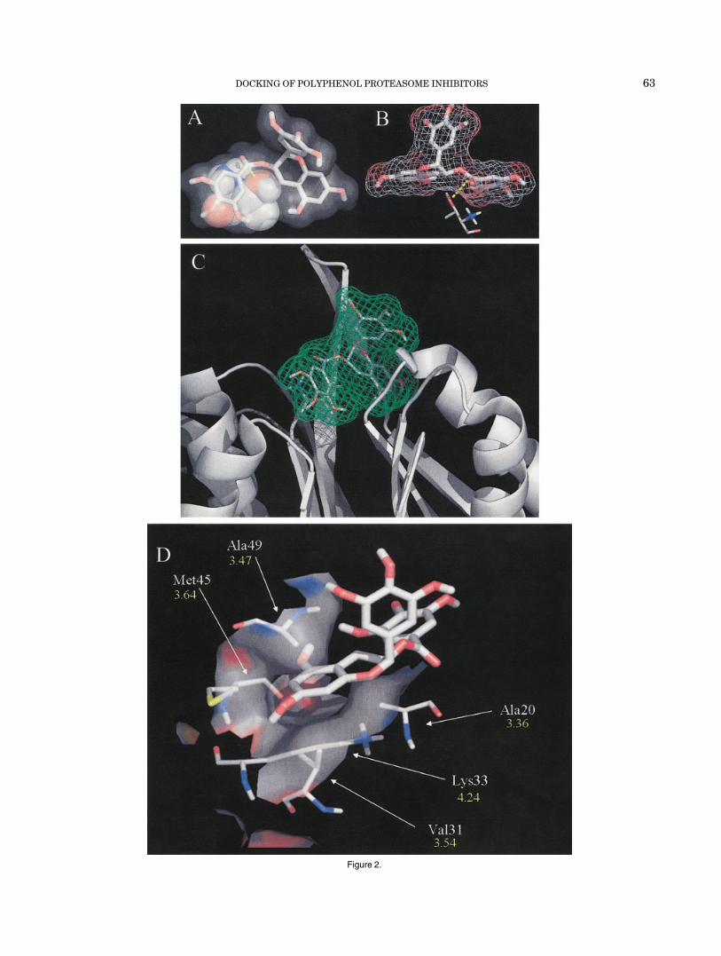
Figure 2.
DOCKING OF POLYPHENOL PROTEASOME INHIBITORS 63

Fig. 3. Binding modes of EGCG analogs. Each EGCG analog is represented by stick structure (see alsoFig. 2 for description) with their names in top left corner (underlined names are synthetic analogs). The IC50
values (in nM) to the chymotrypsin-like activity of purified rabbit 20S proteasome are listed in the top rightcorner, along with the docked free energy value (e � kcal/mol, used to score each docking mode) calculatedfrom the final docked conformations of each respective analog. Similar results for IC50 values were obtained insix or more independent experiments. For (�)-EGCG inset (B), an overlap of (�)-EGCG and (�)-EGCG(green) binding modes is shown. The �5-subunit is represented with a water accessible surface and colored byatom type (O-red, N-blue, C-gray, and H-gray). A two-dimensional scheme for (�)-GCG and (�)-GCG is given(G,H). The dotted lines represent potential hydrogen bond formations and the S1 pocket designationrepresents hydrophobic interactions (G, H).
64 D.M. SMITH ET AL.

energies between �9.83 and �10.62 kcal/mol (see Table I).Moreover, the second lowest docked free energy structuremet all the criteria described above. This resultant modelsupports the hypothesis that (�)-EGCG first binds to the�5-active site and then is attacked by the N-terminalthreonine, rendering the proteasome inactive by acylation.
To look further into the favorable binding mode of(�)-EGCG to the proteasomal chymotrypsin active site, weanalyzed hydrogen-bond (H-bond) formation. There areeight polar hydrogens and one carbonyl-oxygen on (�)-EGCG that are available for H-bonding [see Fig. 1(A)]. Itappears that all but two of these sites are capable ofactively participating in H-bonding (docked structure notshown). It should be noted that we used a relatively loosecriterion to establish the presence of a hydrogen bond.Consistent with this prediction, the fully benzyl protected-EGCG without free OH groups, which should not formH-bonds, fails to inhibit the proteasome29 and could not befound docked in an orientation/conformation that metcriteria 1 and 2 (docked structure not shown).
We then analyzed the hydrophobic interactions between(�)-EGCG and the �5-subunit. The chymotrypsin-likeactivity of the proteasome cleaves peptides after hydropho-bic residues, such as the Tyr in the model fluorogenicsubstrate Suc-Leu-Leu-Val-Tyr-AMC. This Tyr would bindto the S1 hydrophobic pocket of the �5-subunit to allow forspecific chymotrypsin-like cleavage of the AMC group. Itseems that the A ring of (�)-EGCG mimics the Tyr residueof the proteasome peptide substrate: the hydrophobicportion of this aromatic ring is oriented in the middle of theS1 pocket between the side-chains of Ala 49, Ala 20, andLys 33 [with distances of 3.47, 3.36, and 4.24 Å, respec-tively; Fig. 2(D)]. This conformation would allow thehydrophilic hydroxyls of the A ring to project out of the twosides of the S1 hydrophobic pocket and participate inH-bonding. In addition, the sidewalls of the S1 pocket thatinteract with (�)-EGCG are created by Met 45 and Val 31[3.64 and 3.54 Å; Fig. 2(D)]. Each of these hydrophobic orpartially hydrophobic residues are 4.5 Å from (�)-EGCG[see Fig 2(D)], suggesting that entropically driven hydro-phobic interactions might indeed occur between the (�)-EGCG A-C rings and the S1 pocket. Therefore, inhibitionkinetics, along with docking studies of (�)-EGCG bound tothe proteasome �5-subunit, suggests a mechanistic modelfor how (�)-EGCG inhibits the proteasomal chymotrypsin-like activity.
Docking of Other Natural and Synthetic EGCGAnalogs
We next investigated whether this established model of(�)-EGCG inhibition could also be used to interpret theproteasome-inhibitory properties of other EGCG analogs.Three natural, (�)-GCG, (�)-ECG, and (�)-CG, and twosynthetic, (�)-EGCG and (�)-GCG, polyphenols were cho-sen, all of which contain an ester bond [Fig. 1(A)]. Wefound that, similar to (�)-EGCG, all of these five polyphe-nols potently inhibited the chymotrypsin-like activity ofthe rabbit 20S proteasome, with IC50 values similar to
those obtained by using prokaryotic 20S proteasome (TableII and Fig. 3 vs Refs. 28 and 29).
We then docked each of these five polyphenols to the 20Sproteasome �5-subunit by using (�)-EGCG as a compari-son (Fig. 3). For each compound (plus the two amideanalogs, see below and Fig. 4), a single docking mode withthe lowest docked free energy was selected after applyingthe two preset criteria (Table I). For each of these com-pounds, it was possible to locate a viable docking mode(based on the two criteria previously described) with asfew as five GA runs. In many instances, the selecteddocking mode was a member of a cluster of high rank (i.e.,possessed a docked free energy that was low relative to theglobal minimum docked free energy structure).
In our docking studies, (�)-EGCG was found to beslightly more potent than (�)-EGCG with regard to puri-fied 20S proteasome [IC50 170 nM vs 205 nM; Figs. 3(A)and (B)]. (�)-EGCG was oriented in the proteasome �5-subunit with a seemingly similar mode compared to (�)-EGCG, with the A-C rings in the S1 pocket and the B ringin solvent, bridging the binding cleft [Fig. 3(B) vs (A)]. Theester bond-carbon (and gallate group) was shifted only0.38 Å away from Thr 1 but still resided over Thr 1 in asuitable position for a nucleophilic attack [see Fig. 3(B),inset, for overlap of (�)-EGCG and (�)-EGCG]. The shift ofthis gallate group placed the carbonyl oxygen into thebinding cavity created by Arg 19 and Thr 21, allowing foran increased van der Waals interaction and a slightly morefavorable docked free energy [�10.82 kcal/mol vs �10.52kcal/mol; Figs. 3(A) and (B)], explaining the increasedactivity of this compound. A closer inspection revealed that(�)-EGCG had to flip �180° (in relation to the plane of theA-C rings) to attain a similar orientation/conformation. Itis known that (�)-EGCG and (�)-EGCG have (2R, 3R) and(2S, 3S) stereochemistry, respectively [Fig. 1(A)]. Thisfinding suggests that if the B ring and the gallate group of(�)-EGCG were to bind in the same position in three-dimensional space as (�)-EGCG, the A-C rings of (�)-EGCG would then have to rotate 180° to compensate [seeinset, Fig. 3(B)]. The model suggests that proteasome does
TABLE II. Predicted Versus ObservedDocked Free Energies
CompoundPredicted �G°
(kcal/mol) IC50 �RT ln(1/IC50)a
(�)-EGCG �10.82 170 nM �9.60(�)-EGCG �10.52 205 nM �9.49(�)-GCG �10.33 270 nM �9.32(�)-ECG �10.56 710 nM �8.72(�)-EGCG-A �9.63 320 nM �9.21(�)-EGCG-A �9.52 405 nM �9.07(�)-CG �9.30 505 nM �8.93(�)-GCG �9.10 610 nM �8.81genistein �5.15 26 �M �6.50aNote that IC50 is proportional to Ki. Because Ki is the equilibriumconstant for the dissociation of the enzyme-inhibitor complex, and thefree energy (�G°) is related to the equilibrium constant for theassociation of enzyme with inhibitor, �G° is proportional to�RT ln(l/IC50), which is identical to �RT ln(IC50).
DOCKING OF POLYPHENOL PROTEASOME INHIBITORS 65

not exhibit significant enantioselectivity for EGCG29 (Fig.3) due to the partial symmetry of the A-C rings.
(�)-GCG is a non-“epi” compound that has trans stereo-chemistry about the C ring, unlike (�)-EGCG which has
cis stereochemistry [Fig. 1(A)]. The IC50 value of (�)-GCGindicates that it is nearly 3 times less potent than (�)-EGCG (610 nM vs 205 nM; Figs. 3(A) and (C), suggestingthat the trans stereochemistry may not be as beneficial for
Figure 4. (Continued on next page.)
66 D.M. SMITH ET AL.

binding to the proteasome’s active site. In agreement withthe experimental IC50 values, the calculated docked freeenergy of (�)-GCG was �9.10 kcal/mol [Fig. 3(C)] com-pared with �10.52 kcal/mol for (�)-EGCG [Fig. 3(A)]. Forclarity, a two-dimensional scheme of the binding mode for(�)-GCG is also represented [Fig. 3(G)].
The synthetic (�)-GCG was more potent than the natu-ral (�)-GCG [270 nM vs 610 nM; Figs. 3(C) and (D)].Consistent with their IC50 values, a lower docked freeenergy is obtained for the binding of (�)-GCG to �5-subunit compared to (�)-GCG (�10.33 kcal/mol vs �9.10kcal/mol; Figs. 3(C) and (D). (�)-GCG binds in a slightlydifferent conformation compared with the rest of the othercompounds [Fig. 3(H)]. The unique (�)-trans stereochemis-try of (�)-GCG allows for its B ring to potentially formthree H-bonds instead of two as with (�)-EGCG. It alsohydrophobically interacts with Tyr 170 [see Fig. 3(H)],
which has stronger affinity than the binding cleft bridgingconformation. The gallate group of (�)-GCG also extendsfarther out of the pocket and is available to form threepotential H-bonds [Figs. 3(D) and (H), instead of the twopotential H-bonds as with (�)-EGCG. However, althoughthis conformation may increase binding affinities at the Bring and gallate moieties, the A-C rings are pulled slightlyout of the S1 pocket, reducing the total number of potentialinteractions that could take place. As a net result, a slightoverall increase in docked free energy and a slight reduc-tion in in vitro proteasome-inhibitory activity occurs,compared to (�)-EGCG [Fig. 3(D) vs (A)].
The natural GTP (�)-ECG lacks one hydroxyl group onits B ring [Fig. 1(A)], which significantly reduces itssolubility in water and also decreases its potency against20S proteasome by more than threefold, compared to(�)-EGCG [710 nM vs 205 nM; Figs. 3(A) and (E)].(�)-ECG is also found to bind the �5-binding cleft withalmost exactly the same binding mode as (�)-EGCG [Fig.3(E) vs (A)]. However, this did not increase the calculateddocked free energy (�10.56 vs �10.52 kcal/mol). Becausethe B ring is protruding into solvent and, as mentionedpreviously, the loss of this hydroxyl significantly decreasesthe solubility of (�)-ECG, binding of this GTP to theproteasome might be affected in a manner that is not wellaccounted for by the solvation model used in the dockingalgorithms.
The natural GTP (�)-CG is another non-epi compoundwith a trans stereochemistry [Fig. 1(A)] and is less potentthan (�)-EGCG [Fig. 3(F) vs (A)]. Consistent with thisfinding, a significantly increased ligand internal energy[�0.42 kcal/mol for (�)-CG vs �0.38 kcal/mol for (�)-EGCG] is calculated for binding of (�)-CG to the protea-some’s active site, thereby giving a net increase in dockedfree energy [�9.30 kcal/mol vs �10.52 kcal/mol; and seethe discussion about (�)-GCG].
To test whether the developed computational model canbe applied to a range of compounds with different chemicalstructures, we selected genistein, the predominant isofla-vone found in soy products. Like (�)-EGCG, genistein alsoconsists of a ring system similar to the A, C, and B rings ofthe GTPs [see Fig. 1(A), suggesting that genistein might bea proteasome inhibitor. But different from (�)-EGCG,genistein lacks the gallate group [see Fig. 1(A)], whichsuggests that genistein would be less potent than (�)-EGCG. To test this hypothesis, we first docked genistein toyeast 20S proteasome. We found that in 60 of 100 runswith 5 million energy evaluations, genistein docks primar-ily in the S1 pocket of the active site of the proteasome�5-subunit (Table I). The B ring hydroxyl group of genisteinlies in close proximity to Thr 1, and up to four potentialhydrogen bonds could be formed within the complex ofgenistein and the proteasomal �5-subunit (docking modesnot shown). However, the docked free energy of genisteinto �5-subunit was found to be �5.15 kcal/mol (Table I),much higher than that of (�)-EGCG [�10.52 kcal/mol; Fig.3(A)]. Consistent with its higher docked free energy,genistein weakly inhibits the chymotrypsin-like activity ofpurified 20S proteasome with an IC50 value of 26 �M [also
Fig. 4. (Continued) Decreased nucleophilic susceptibility and ester bondflexibility in EGCG amides are associated with decreased proteasome-inhibitory activity. A,B: The nucleophilic susceptibility of (�)-EGCG and(�)-EGCG amides. Molecular orbital energy analysis is shown by drawing anelectron density isosurface and coloring by nucleophilic susceptibly.28 Thewhite center signifies the highest area of susceptibility. C,D: Binding modesof (�)-EGCG-amide and (�)-EGCG-amide. Also see Figure 3. E: The boundconformation of EGCG and their amides are given along with arrows to tracethe saddle conformation formed with the ester bonds in (�)- and (�)-EGCGand the linear amide bonds in (�)- and (�)-EGCG-amide compounds. F: Anoverlap of all the eight bound ligands (Figs. 3 and 4) are given along with aview of the bottom of the saddle-shaped binding pocket (occluding residueshave been removed) with surface colored by atom type. The S1 pocket andThr 1 are designated. G: The calculated docked free energy predicts theactual proteasome-inhibitory activity. A plot of predicted docked free energy(in kcal/mol) against the actual proteasome-inhibitory activity (converted fromIC50 values to kcal/mol; see below). A regression analysis R2 value for abest-fit line was 0.9893 [without (�)-ECG; see text]. IC50 values of eachcompound were used in the formula, -RT[ln(1/IC50)]. Here the value used forR (gas constant) was 0.0019872 kcal/K*mol and T was 310 K (thetemperature of the experiment was 37°C). These values were then plottedagainst the �G values computed by AutoDock in the docking process. Alsosee Table II.
DOCKING OF POLYPHENOL PROTEASOME INHIBITORS 67

see Table II and Fig. 4(G)], in contrast to an IC50 of 205 nMfor (�)-EGCG [Fig. 3(A)]. These data further support theconclusion that our established computational model couldsatisfactorily describe the EGCG-�5-interaction that isresponsible for its proteasome-inhibitory activity.
Rational Design, Synthesis, and Validation ofEGCG-Amides as Proteasome Inhibitors
To further test the model of proteasome inhibition byEGCG analogs, we decided to rationally design relatedcompounds and examine whether they possess the ex-pected properties as predicted by the in silico dockingstudies. We hypothesized that an EGCG analog withaltered nucleophilic susceptibility to the ester bond-carbonwould have altered proteasome-inhibitory potency. Forexample, replacement of the ester bond-oxygen of EGCGwith nitrogen (EGCG-amide) would render the carbonylfunctional group less susceptible to a nucleophilic attackdue to the presence of the amide nitrogen. Indeed, asexpected, molecular orbital calculations confirmed thatthe ester bond-carbon of (�)-EGCG produced an arbitraryvalue of 0.69 for nucleophilic susceptibility, whereas thesame carbon in (�)-EGCG-amide had a value of 0.55 [Figs.4(A) and (B)].28
To determine whether the reduction in nucleophilicsusceptibility would result in a decreased potency toinhibit the proteasome activity, we synthesized (�)-EGCG-amide and (�)-EGCG-amide [Fig. 1(A)]. The IC50 valuesagainst 20S eukaryotic proteasome were determined to be320 and 405 nM for both (�)-EGCG-amide and (�)-EGCG-amide, respectively [Figs. 4(C) and (D)]. Compared to (�)-and (�)-EGCG, both amide compounds have decreasedproteasome-inhibitory potencies, respectively [Figs. 4(C)and (D) vs Figs. 3(A) and (B)], although their stereochemi-cal structures were not changed [Fig. 1(A)]. This findingsuggests that reduction in nucleophilic susceptibility isindeed associated with decreased potency to inhibit 20Sproteasome activity, as predicted by the model.
It is of interest that the molecular modeling studies withthe amide compounds brought another property of (�)-EGCG inhibition to light. When (�)-EGCG binds theproteasome, a saddle shape is formed between the A-Crings extending past the ester bond and back down to thegallate moiety [Fig. 4(E)]. The more flexible nature of theester bond allows this conformation to occur so that(�)-EGCG might fit the saddle shape formed by the bottomof the binding pocket [Fig. 4(F), top/right). In fact, whenthe docked conformations of all the EGCG analogs areoverlapped into one image, this saddle shape can be easilyobserved [Fig. 4(F)]. It can be assumed that this saddle-shaped conformation of EGCG would also place additionalstrain on the scissile bond, further lowering the activationenergy for nucleophilic attack. However, introduction of anitrogen atom into EGCG, as in EGCG-amide, reducesbond flexibility. It is well known that such an amide bond(or peptide bond) is less flexible than the ester bond andprefers the trans conformation. Therefore, because of thedecreased flexibility of the amide bond, the amide polyphe-nols cannot adopt a saddle-shaped conformation that is
energetically favorable for binding. This causes a straight-ening out of the arch conformation [Fig. 4(E)], which doesnot allow the A-C rings to bind as deeply in the S1 pocketas (�)-EGCG, thus pulling the compound farther out of thebinding cleft. This consequently raises the docked freeenergy of both (�)-EGCG-amide and (�)-EGCG-amide[�9.63 and �9.52 kcal/mol, respectively; Figs. 4(C) and(D)]. This binding mode with increased docked free energyagrees with the increase in the IC50 values of both amidecompounds (Fig. 4) and may (along with their reducednucleophilic susceptibility) explain their decreased po-tency relative to the corresponding esters. Consistent withthe prediction that EGCG-amides are also irreversible ortight-binding proteasome inhibitors, we found that theamide analogs were able to accumulate levels of theproteasome target protein p27 in breast cancer MCF-7cells, with potency comparable to that of (�)-EGCG (unpub-lished data).
Finally, to compare the selected binding modes to theactual proteasome-inhibitory activities of each of the eightEGCG analogs and genistein, we plotted the predictedactivity (docked-free energy) against the actual inhibitoryactivity (IC50 values; converted to kcal/mol) [Table II andFig. 4(G)]. A decrease in the docking free energy for eight ofthe nine compounds was correlated with an increase in theactual activity of each of these compounds. Only onecompound, (�)-ECG, did not fit the linear relationshipbetween the predicted and actual activity [Fig. 4(G)]. Thissignificant loss in actual activity of (�)-ECG, which is notin congruence with the calculated docked free energy, maybe due to the orientation and solvation issues mentionedpreviously. A regression analysis R2 value of 0.9893 wasdetermined for a best-fit line, not including the valuesgenerated for (�)-ECG. These data support our model ofproteasome inhibition by EGCG analogs.
CONCLUSION
There are two aspects of proteasome inhibition by(�)-EGCG. First, it was shown that (�)-EGCG irreversiblyinhibits the chymotrypsin-like activity of the proteasomein a time-dependent manner (Figs. 1(B) and (C)], so it isplausible that a nucleophilic attack of the ester bond-carbon of (�)-EGCG occurs. Second, for the above event tooccur, (�)-EGCG must bind to the active site in such amode that allows for attack of the carbonyl carbon of(�)-EGCG to take place [Fig. 2(A)]. Our established modelsuggests that any analogs of (�)-EGCG that bind (withreasonable affinity) to the active site in an orientation/conformation more conducive to nucleophilic attack shouldproduce a higher rate of attack and thus greater inhibition.In contrast, those analogs that bind to the active site in anorientation/conformation less favorable for nucleophilicattack should produce a lower rate of attack and thus lessinhibition. Of course, it is possible that one could have amolecule that binds tightly to the enzyme but is not in anorientation/conformation favorable for nucleophilic attack.This molecule may very well inhibit the enzyme, but notirreversibly through acylation.
68 D.M. SMITH ET AL.

(�)-EGCG can bind the proteasome’s chymotrypsin ac-tive site in an orientation and conformation that is wellsuited for nucleophilic attack as described by the followingmodel. First, favorable hydrophobic surface interactionsexist (tyrosine-like mimic in S1 pocket) [Fig. 2(D)]. Second,there is a large potential van der Walls contact surfacearea [Fig. 2(C)]. Third, the calculated docked free energyvalues are favorable for binding of (�)-EGCG to theproteasome [Fig. 3(A)]. Fourth, it is likely that the scissilebond of (�)-EGCG is strained, suggesting lowering of theactivation energy for the formation of the tetrahedralintermediate in the proposed acylation reaction (Fig. 4).Finally, it was observed that one of the two dockedstructures of lowest docked free energy for (�)-EGCG hadits electrophilic carbonyl carbon 3.18 Å from the hydroxylgroup of Thr 1 [Figs. 2(A) and (B)]. All these propertiesshown by this reported docking model have supplied anattractive, empirically directed, analog-supported model ofproteasome inhibition by the green tea polyphenol (�)-EGCG.
Our immediate future studies will focus on rationallydesigning and synthesizing new EGCG analogs based onthe docking information and examining their proteasome-inhibitory activities in vitro and in vivo. We also plan toperform docking studies with (�)-EGCG to both the �1-and �2-subunits of the yeast 20S proteasome. In prelimi-nary studies (unpublished observations), we have foundthat (�)-EGCG can dock to the �1-subunit in a dockingmode that is very similar to that described in this articlefor (�)-EGCG docked to the �5-subunit. Moreover, thedocked free energy for (�)-EGCG docked to the �1-subunitis almost identical to the docked free energy observed forits docking to the �5-subunit. There were no dockingmodes found for (�)-EGCG docked to the �2-subunit inwhich the distance between the Thr-1 hydroxyl group andthe ester carbonyl carbon atom was 4.0 Å. A few struc-tures were observed in which (�)-EGCG was bound to thesurface of the protein and the distances of Thr-1 hydroxyland ester carbonyl carbon were very close to 4.0 Å. But(�)-EGCG was not actually docked to the catalytic site;therefore, nucleophilic attack of the carbonyl group byThr1 could not occur in this conformation. These resultsare consistent with our experimental observations.28 Wealso plan to further demonstrate acylation of the �5s Thr 1by (�)-EGCG via X-ray diffraction studies. It is our hopethat a better mechanistic understanding of proteasomeinhibition by (�)-EGCG will allow for the rational designof more potent and stable, but less toxic, GTP analogs forcancer prevention.
ACKNOWLEDGMENT
We thank Mr. Layne Norton (Eckerd College) for hisassistance with some of the docking studies reportedherein.
REFERENCES1. Goldberg AL. Functions of the proteasome: the lysis at the end of
the tunnel. Science 1995;268:522–523.2. Groll M, Ditzel L, Lowe J, Stock D, Bochtler M, Bartunik HD,
Huber R. Structure of 20S proteasome from yeast at 2.4 Aresolution. Nature 1997;386:463–471.
3. Seemuller E, Lupas A, Stock D, Lowe J, Huber R, Baumeister W.Proteasome from Thermoplasma acidophilum: a threonine pro-tease. Science 1995;268:579–582.
4. Kisselev AF, Goldberg AL. Proteasome inhibitors: from researchtools to drug candidates. Chem Biol 2001;8:739–758.
5. Dou QP, Nam S. Pharmacological proteasome inhibitors and theirtherapeutic potential. Exp Opin Ther Patents 2000;10:1263–1272.
6. Dou QP, Li B. Proteasome inhibitors as potential novel anticanceragents. Drug Resist Updat 1999;2:215–223.
7. Almond JB, Cohen GM. The proteasome: a novel target for cancerchemotherapy. Leukemia 2002;16:433–443.
8. An B, Goldfarb RH, Siman R, Dou QP. Novel dipeptidyl protea-some inhibitors overcome Bcl-2 protective function and selectivelyaccumulate the cyclin-dependent kinase inhibitor p27 and induceapoptosis in transformed, but not normal, human fibroblasts. CellDeath Differ 1998;5:1062–1075.
9. Lopes UG, Erhardt P, Yao R, Cooper GM. p53-dependent induc-tion of apoptosis by proteasome inhibitors. J Biol Chem 1997;272:12893–12896.
10. Adams J, Palombella VJ, Sausville EA, Johnson J, Destree A,Lazarus DD, Maas J, Pien CS, Prakash S, Elliott PJ. Proteasomeinhibitors: a novel class of potent and effective antitumor agents.Cancer Res 1999;59:2615–2622.
11. Sun J, Nam S, Lee CS, Li B, Coppola D, Hamilton AD, Dou QP,Sebti SM. CEP1612, a dipeptidyl proteasome inhibitor, inducesp21WAF1 and p27KIP1 expression and apoptosis and inhibits thegrowth of the human lung adenocarcinoma A-549 in nude mice.Cancer Res 2001;61:1280–1284.
12. Adams J, Behnke M, Chen S, Cruickshank AA, Dick LR, GrenierL, Klunder JM, Ma YT, Plamondon L, Stein RL. Potent andselective inhibitors of the proteasome: dipeptidyl boronic acids.Bioorg Med Chem Lett 1998;8:333–338.
13. Adams J. Development of the proteasome inhibitor PS-341. Oncolo-gist 2002;7:9–16.
14. Dou QP, Goldfarb RH. Evaluation of the proteasome inhibitorMLN-341 (PS-341). IDrugs 2002;5:828–834.
15. Groll M, Koguchi Y, Huber R, Kohno J. Crystal structure of the 20S proteasome:TMC-95A complex: a non-covalent proteasome inhib-itor. J Mol Biol 2001;311:543–548.
16. Ohno Y, Wakai K, Genka K, Ohmine K, Kawamura T, TamakoshiA, Aoki R, Senda M, Hayashi Y, Nagao K, et al. Tea consumptionand lung cancer risk: a case-control study in Okinawa, Japan. JpnJ Cancer Res 1995;86:1027–1034.
17. Zhong L, Goldberg MS, Gao YT, Hanley JA, Parent ME, Jin F. Apopulation based case-control study of lung cancer and green teaconsumption among women living in Shanghai, China. Epidemiol-ogy 2001;12:695–700.
18. Ji BT, Chow WH, Hsing AW, McLaughlin JK, Dai Q, Gao YT, BlotWJ, Fraumeni JF Jr. Green tea consumption and the risk ofpancreatic and colorectal cancers. Int J Cancer 1997;70:255–258.
19. Inoue M, Tajima K, Mizutani M, Iwata H, Iwase T, Miura S,Hirose K, Hamajima N, Tominaga S. Regular consumption ofgreen tea and the risk of breast cancer recurrence: follow-up studyfrom the Hospital-based Epidemiologic Research Program at AichiCancer Center (HERPACC), Japan. Cancer Lett 2001;167:175–182.
20. Taniguchi S, Fujiki H, Kobayashi H, Go H, Miyado K, Sadano H,Shimokawa R. Effect of (�)-epigallocatechin gallate, the mainconstituent of green tea, on lung metastasis with mouse B16melanoma cell lines. Cancer Lett 1992;65:51–54.
21. Wang ZY, Huang MT, Ho CT, Chang R, Ma W, Ferraro T, ReuhlKR, Yang CS, Conney AH. Inhibitory effect of green tea on thegrowth of established skin papillomas in mice. Cancer Res 1992;52:6657–6665.
22. Liao S, Umekita Y, Guo J, Kokontis JM, Hiipakka RA. Growthinhibition and regression of human prostate and breast tumors inathymic mice by tea epigallocatechin gallate. Cancer Lett 1995;96:239–243.
23. Gupta S, Ahmad N, Mohan RR, Husain MM, Mukhtar H. Prostatecancer chemoprevention by green tea: in vitro and in vivo inhibi-tion of testosterone-mediated induction of ornithine decarboxyl-ase. Cancer Res 1999;59:2115–2120.
24. Muto S, Yokoi T, Gondo Y, Katsuki M, Shioyama Y, Fujita K,Kamataki T. Inhibition of benzo[a]pyrene-induced mutagenesisby (�)-epigallocatechin gallate in the lung of rpsL transgenicmice. Carcinogenesis 1999;20:421–424.
DOCKING OF POLYPHENOL PROTEASOME INHIBITORS 69

25. Fujiki H. Two stages of cancer prevention with green tea. J CancerRes Clin Oncol 1999;125:589–597.
26. Yang CS. Tea and health. Nutrition 1999;15:946–949.27. Ahmad N, Mukhtar H. Green tea polyphenols and cancer: biologic
mechanisms and practical implications. Nutr Rev 1999;57:78–83.28. Nam S, Smith DM, Dou QP. Ester bond-containing tea polyphe-
nols potently inhibit proteasome activity in vitro and in vivo.J Biol Chem 2001;276:13322–13330.
29. Smith DM, Wang Z, Kazi A, Li LH, Chan TH, Dou QP. Syntheticanalogs of green tea polyphenols as proteasome inhibitors. MolMed 2002;8:382–392.
30. Li L, Chan TH. Enantioselective synthesis of epigallocatechin-3-gallate (EGCG), the active polyphenol component from green tea.Org Lett 2001;3:739–741.
31. Berman HM, Westbrook J, Feng Z, Gilliland G, Bhat TN, WeissigH, Shindyalov IN, Bourne PE. The Protein Data Bank. NucleicAcids Res 2000;28:235–242.
32. Morris GM, Goodsell DS, Halliday RS, R H, Hart WE, Belew RK,Olson AJ. Automated docking using a Lamarckian genetic algo-rithm and an empirical binding free energy function. J ComputChem 1998;19:1639–1662.
33. Dym O, Xenarios I, Ke H, Colicelli J. Molecular docking ofcompetitive phosphodiesterase inhibitors. Mol Pharmacol 2002;61:20–25.
34. Rao MS, Olson AJ. Modelling of factor Xa-inhibitor complexes: acomputational flexible docking approach. Proteins 1999;34:173–183.
35. DeLano WL. The PyMOL Molecular Graphics System 2002. SanCarlos, CA: DeLano Scientific; 2002.
36. Vicentini CB, Guarneri M, Andrisano V, Guccione S, Langer T,Marschhofer R, Chabin R, Edison AM, Huang X, Knight WB, GioriP. Potential of pyrazolooxadiazinone derivatives as serine pro-tease inhibitors. J Enzyme Inhib 2001;16:15–34.
37. Dick LR, Cruikshank AA, Grenier L, Melandri FD, Nunes SL,Stein RL. Mechanistic studies on the inactivation of the protea-some by lactacystin: a central role for clasto-lactacystin beta-lactone. J Biol Chem 1996;271:7273–7276.
38. Antonov VK, Ivanina TV, Ivanova AG, Berezin IV, Levashov AV,Martinek K. Binding-catalysis relationship in alpha-chymotryp-sin action as revealed from reversible inhibition study of phenylal-kylboronic acids. FEBS Lett 1972;20:37–40.
39. da Graca Thrige D, Buur JR, Jorgensen FS. Substrate binding andcatalytic mechanism in phospholipase C from Bacillus cereus: amolecular mechanics and molecular dynamics study. Biopolymers1997;42:319–336.
40. Binda C, Angelini R, Federico R, Ascenzi P, Mattevi A. Structuralbases for inhibitor binding and catalysis in polyamine oxidase.Biochemistry 2001;40:2766–2776.
41. Bravo L. Polyphenols: chemistry, dietary sources, metabolism,and nutritional significance. Nutr Rev 1998;56:317–333.
70 D.M. SMITH ET AL.


![Catalog polyphenol np_final[1]](https://img.pdfslide.us/doc/110x75/5a672d187f8b9a0c518b489f/catalog-polyphenol-npfinal1.jpg)