Embed Size (px)
Citation preview

DnaJ (Hsp40 Protein) Binding to Folded Substrate ImpactsKplE1 Prophage Excision Efficiency*□S
Received for publication, December 7, 2011, and in revised form, February 23, 2012 Published, JBC Papers in Press, February 28, 2012, DOI 10.1074/jbc.M111.331462
Tania M. Puvirajesinghe‡1, Latifa Elantak§, Sabrina Lignon¶, Nathalie Franche‡, Marianne Ilbert�,and Mireille Ansaldi‡2
From the ‡Laboratoire de Chimie Bactérienne, CNRS UMR7283, §Laboratoire d’Ingénierie des Systèmes MacromoléculairesCNRS UMR7255, the ¶Proteomic Facility (MaP Marseille Protéomique IBiSA), and �Bioénergétique et Ingénierie des ProtéinesCNRS UMR7281, Institut de Microbiologie de la Méditerranée, Aix-Marseille University, 31 Chemin Joseph Aiguier,13402 Marseille Cedex 20, France
Background: DnaJ positively modulates KplE1 prophage excision and is involved in lysogeny escape.Results: The recombination directionality factor TorI, from KplE1 prophage, interacts with the DnaJ chaperone, and theyprotect each other from limited trypsin digestion.Conclusion: DnaJ stabilizes TorI recombination directionality factor conformation.Significance: DnaJ cochaperone can bind folded substrates and induces the conformational stabilization of the TorI protein.
Temperate phages mediate gene transfer and can modify theproperties of their host organisms through the acquisition ofnovel genes, a process called lysogeny. The KplE1 prophage isone of the 10 prophage regions in Escherichia coliK12MG1655.KplE1 is defective for lysis but fully competent for site-specificrecombination. The TorI recombination directionality factor isstrictly required for prophage excision from the host genome.We have previously shown that DnaJ promotes KplE1 excisionby increasing the affinity of TorI for its site-specific recombina-tion DNA target. Here, we provide evidence of a direct associa-tion between TorI and DnaJ using in vitro cross-linking assaysand limited proteolysis experiments that show that this interac-tion allows both proteins to be transiently protected from tryp-sin digestion. Interestingly, NMR titration experiments showedthat binding of DnaJ involves specific regions of the TorI struc-ture. These regions, mainly composed of �-helices, are locatedon a surface opposite the DNA-binding site. Taken together, wepropose that DnaJ, without the aid of DnaK/GrpE, is capable ofincreasing the efficiency of KplE1 excision by causing a confor-mational stabilization that allows TorI to adopt a more favor-able conformation for binding to its specific DNA target.
Site-specific recombination is used by temperate phages tointegrate and excise from their host chromosome (1). As a con-sequence, temperate phages mediate gene transfer and canmodify the properties of their host organisms through theacquisition of novel genes, a process called lysogeny (2, 3). The
ancient prophage KplE1 (or CPS-53) is one such example,which contains 16 open reading frames and belongs to one ofthe 10 prophage regions present in the Escherichia coli K12MG1655 (4, 5). Although fully competent for site-specificrecombination, KplE1 is noninfectious and defective for virionformation and host cell lysis (6). Therefore, experimental stud-ies of excisive recombination are made easier by the defectivenature of the KplE1 prophage, because the host cells do not lyseupon prophage induction. The KplE1 integrase protein, IntS,belongs to the tyrosine recombinase family of integrases, whichare exemplified by the prototypic member phage �. In additionto the integrase protein, prophage excision from the chromo-some requires additional proteins, which are classed as recom-bination directionality factors (RDFs)3 (7). TorI is the RDF ofKplE1 and is essential for prophage excision (8). It is encoded bythe torI gene, which is located upstream of the 3� core sequencethatmarks the end of the prophage (6). SomeRDFs, such asCoxproteins of phages P2 andHP1, have additional regulatory func-tions outside recombination (9). TorI itself was originally iden-tified as a negative response regulator of the TorR protein,which activates transcription of the torCAD operon, encodingthe trimethylamine-N-oxide reductase respiratory system (10).Molecular chaperones arewidely distributed in all organisms
and are involved in protein folding, maturation, and disaggre-gation (11). Remodeling of protein complexes is also part of themolecular chaperone functions and involves the interactionwith native substrates (12). DnaJ (Hsp40) is a cochaperone thatregulates the protein folding function of DnaK (Hsp70) chap-erone by binding and delivering substrates to DnaK as well asactivating ATP hydrolysis (13). DnaJ is universally conserved inbiological kingdoms (14). Classical functions of the DnaK/DnaJ/GrpE machinery under low stress conditions includefolding of newly synthesized proteins, protein assembly anddisassembly, and translocation of proteins into organelles (15,
* This work was supported by CNRS, the French Research Ministry (Ministèrede l’Éducation Nationale, de la Recherche et de la Technologie), andNational Research Agency Grant ANR-08-BLAN-0122-01.
□S This article contains supplemental Figs. S1–S3 and Table S1.1 Present address: Polarité Cellulaire Signalisation et Cancer, Centre de
Recherche en Cancérologie de Marseille, Inserm U1068, CRCM, 27, Bd LeiRoure, 13009 Marseille, France.
2 To whom correspondence should be addressed: Laboratoire de Chimie Bac-térienne CNRS UMR7283, Institut de Microbiologie de la Méditerranée, Aix-Marseille University, 31 Chemin Joseph Aiguier, Marseille 13009, France.Tel.: 33-491164585; Fax: 33-491718914; E-mail: [email protected].
3 The abbreviations used are: RDF, recombination directionality factor;BMH, bismaleimidohexane; IHF, integration host factor; Tricine,N-[2-hydroxy-1,1-bis(hydroxymethyl)ethyl]glycine.
THE JOURNAL OF BIOLOGICAL CHEMISTRY VOL. 287, NO. 17, pp. 14169 –14177, April 20, 2012© 2012 by The American Society for Biochemistry and Molecular Biology, Inc. Published in the U.S.A.
APRIL 20, 2012 • VOLUME 287 • NUMBER 17 JOURNAL OF BIOLOGICAL CHEMISTRY 14169
by guest on April 4, 2019
http://ww
w.jbc.org/
Dow
nloaded from

16). Under conditions of cellular stress, DnaJ, together withDnaK andGrpE, play a role in the protection of proteins againststress-induced unfolding as well as refolding of unfolded pro-teins (17). The initial discovery of DnaJ is exquisitely inter-twined with initial research into phage � DNA replication (18).DnaJ was found to be involved in recruiting DnaK into the pre-initiation complex at the phage origin of replication (19). In thisprocess, DnaJ binds first to the replication initiation protein �Pand the host primase DnaB in the preprimosomal complex.This allows recruitment ofDnaK and subsequent binding to�P,which in turn results in the release of the preinitiation complexthus initiating DNA replication (19, 20). Besides its cochaper-one function, DnaJ is involved in several functions. DnaJ fromE. coli possesses an active dithiol/disulfide group, which hasbeen suggested to allowDnaJ to control the redox state of cyto-plasmic, membrane, or exported proteins (21). DnaJ is alsoknown to act as a holdase chaperone that does not activelyrefold protein clients but can bind hydrophobic exposed sur-faces of unfolded substrates and prevent their aggregation (22).Additionally, DnaJ-like proteins have been implicated in viralinfection of plants. ADnaJ-like protein has been identified fromNicotiana tabacum, designated MP-interacting protein 1(NtMPIP1), which has been shown to interactwith the essentialmovement protein of tobaccomosaic virus, thereby implicatingDnaJ as a host-encoded factor involved in the cell-to-cell spreadof tobacco mosaic virus (23).As mentioned above, in E. coli the DnaK/DnaJ/GrpE
machinery has been discovered through its role in � phage rep-lication. Recently, we have shown that DnaJ is also involved inprophage excision of a class of (pro)phages that share very sim-ilar recombination modules (24). As this role appeared to be abona fide chaperone activity of DnaJ, whichwas independent ofDnaK, we have now further characterized the structural fea-tures underlying the TorI/DnaJ interaction.
EXPERIMENTAL PROCEDURES
Overproduction and Purification of TorI—TorI was pro-duced from E. coli BL21 (DE3) harboring plasmid pETsI, asdescribed previously (6).Overproduction and Purification of DnaJ—DnaJ was pro-
duced using pGPJ-His (25) in the �5 strain (MC4100dnaK-dnaJ42::kan, grpE::tet cbpA::kan, djlA::spc, laboratorystrain fromP.Genevaux). Fresh overnight cultureswere diluted1:100 in 500 ml of LB broth supplemented with 100 �g/ml�1
ampicillin and grown with vigorous shaking at 30 °C. Cultureswere induced with L-arabinose 0.2% (w/v) and incubated for afurther 4 h. Cells were harvested and resuspended in 30 ml ofbuffer 1 (40 mM sodium phosphate buffer, pH 7.4, 20 mM imid-azole, 0.6% Brij 58P, 0.5% Triton, 1 M NaCl, 20 mM �-mercap-toethanol). Cells were lysed using a French press, and lysateswere centrifuged at 40,000 rpm for 1 h in a Beckman 70Ti rotor.The supernatant was isolated and filtered using a 0.2-�m filter.The sample was loaded onto a nickel Hi-Trap FF 1-ml column(GEHealthcare). The columnwas washed with buffer 2 (50mM
sodium phosphate, 20 mM imidazole, 1 M NaCl, 20 mM �-mer-captoethanol). Proteins were then eluted using buffer 2 con-taining 250 mM imidazole. The protein buffer was thenexchanged into storage buffer (40 mM sodium phosphate, pH
7.6, 100mMKCl, 10% glycerol, 1mMDTT), and the protein wasstored at �80 °C.Immunoblot Analysis—Proteins were prepared using 2�
SDS-Tricine sample buffer andheated to 95 °C for 10min. Sam-ples were separated according to their molecular weight bySDS-Tricine gel electrophoresis and transferred to nitrocellu-lose membranes for immunoblotting. Membranes wereblocked at 25 °C for 1 h with phosphate-buffered saline (PBS)containing 5% skimmed milk powder. Blots were incubatedovernight with a primary antibody, either rabbit polyclonal pri-mary antibodies recognizing DnaJ (diluted 1:1,000) or rabbitpolyclonal primary antibodies recognizing TorI (diluted1:10,000). Blots were washed three times with PBS. Anti-rabbitHRP-conjugated antibody (1:10,000, Amersham Biosciences)was used as a secondary antibody and incubated for 1 h at roomtemperature. After washing three times with PBS, blots weredeveloped with ECL Supersignal West Pico chemiluminescentsubstrate (Thermo Scientific).In Vitro Chemical Cross-linking—Cross-linking experiments
were carried out using bismaleimidohexane (BMH) cross-link-ing reagent (Pierce). Proteins (2–4 �g) were incubated for 30min at room temperature in PBS buffer containing 300mMKCland 1mMBMH. Samples were analyzed using 12% SDS-Tricinegels followed by PageBlue (Fermentas) protein stain and immu-noblotting analysis.Far-UV Circular Dichroism—Far-UV CD spectra were
recorded on a Jasco 815 CD spectrometer using 2-mm thickquartz cells (10 mm path length) in 40 mM sodium phosphate,pH 7.4, and 100 mM KCl at 20 °C. CD spectra were measuredfrom 190 to 260 nm, with a scanning speed of 10 nm/min, andresults were averaged from five scans. All spectra were buffer-corrected and normalized for any variation in protein concen-trations. Protein concentrations used were as follows: TorI, 25�M; DnaJ, 0.8 �M or 0.25 �M.Limited Trypsin Digestion and Mass Spectrometry Analysis—
DnaJ and TorI proteins were incubated alone and together, inthe presence of trypsin, which was used at a 1:100 (trypsin/totalprotein, w/w). Enzymatic digestion was started by addition oftrypsin, and the reaction was carried out at room temperature.Aliquots were taken at different time points of the enzymedigestion. Samples were spotted with a saturated solution ofsinapinic acid matrix (40% acetonitrile in water, 0.1% trifluoro-acetic acid (v/v)) directly onto a MALDI plate. Mass spectro-metric analysis was carried out using aMicroflexMALDI-TOFmass spectrometer (Bruker Daltonics). Mass determinationwas performed in positive linear mode to analyze monomericproteins forms and in positive reflectron mode for tryptic pep-tides. Peptidemass fingerprint analysis was processed by Bioto-ols software using internal calibration (26).Alternatively, trypsin digestion of samples was stopped by
heating samples to 99 °C for 5 min and analyzed on a 12% SDS-Tricine gel or by using an automated Experion separation sys-tem (supplemental Fig. S2). Samples were loaded onto a Pro260chip run following the manufacturer’s instructions. Data anal-ysis used Experion Software (version 3.0).NMR Titration Experiments—TorI was labeled by growing
cells in M9 minimal medium supplemented with 1 g/liter[15N]NH4Cl (Cambridge Isotopes), supplemented with ampi-
DnaJ Stabilizes TorI Recombination Protein Conformation
14170 JOURNAL OF BIOLOGICAL CHEMISTRY VOLUME 287 • NUMBER 17 • APRIL 20, 2012
by guest on April 4, 2019
http://ww
w.jbc.org/
Dow
nloaded from
cillin (100 �g/ml), thiamine (2 pg/ml), MgSO4 (1 mM), CaCl2(0.1mM). TorI proteinwas purified as described previously (16).Titration experiments were carried out in 20 mM HEPES, pH7.2, 50 mM KCl, 5% glycerol, 10% D2O as follows: 0.05, 0.1, and0.2 mM final concentration of 15N-labeled TorI (0.5, 1, and2 molecular equivalents) were added to 0.1 mM unlabeledDnaJ. Chemical shift perturbations in TorI were monitored intwo-dimensional 1H-15N HSQC experiments. Weighted aver-age chemical shift variations were calculated according to theformula, �ppm � ((��HN)2 � (��N � 0.17)2)1/2 (27). TheNMR experiments have been recorded at 290 K on a 600 MHzspectrometer (Avance III 600 MHz Bruker) equipped with acryoprobe.Site-specific Mutagenesis of TorI �3 Helix—To construct
plasmid pETsI-R63C-A64S, the codons corresponding to resi-dues Arg63 and Ala64 were replaced by Cys and Ser codons,respectively, by a one-step PCR method (28) using plasmidpETsI as a template and overlapping divergent primersIr-61–63-64 (5�-CTTATTTTTGAATTCACAATGGTCAC-GATATAACC) and Im-R63C-A64S (5�-TGTGAATTCAAA-AATAAGCTCTTAAGCTGCTCCAATGGGTAACTC). Thesequence accuracy of the construction was checked by directsequencing. The mutant protein (TorI_R63C_A64S) was pro-duced in a C41 E. coli strain and purified as the wild type.In Vitro Excision Assay—Linear att sites were amplified by
PCR with primer pairs attLSpeI/attL-KpnI for attL and attR-XbaI/attR-IHF2 for attR and then purified using QIAQuickPCR purification kit protocol (Qiagen). Reaction mixtures (50�l) included linearattDNAsites (28 nM) in buffer containing 33mM Tris-HCl, pH 7.6, 33 mM KCl, 9 mM spermidine, 4 mM
EDTA, 0.9 mg/ml�1 BSA, and 7% glycerol, IHF (0.3 �M), andIntS (0.6 �M). TorI and DnaJ were added at the same molarity:TorIWT (0.7�M), DnaJ (0.7�M), or TorI*63–64 (2.1�M), DnaJ(2.1 �M). TorI and DnaJ were initially preincubated at 37 °C for30 min, and then IntS, IHF, and linear attL and attRDNAweresimultaneously added. The reactions were carried out in opti-mized conditions at 30 °C for 2 h at an IHF/IntS/TorI/DnaJprotein ratio of 1:2:2.3:2.3 for TorI WT and 1:2:7:7 forTorI_R63C_A64S. Reaction products were purified (QIAQuickkit, Qiagen) and analyzed on a 2% agarose gel electrophoresis.
RESULTS AND DISCUSSION
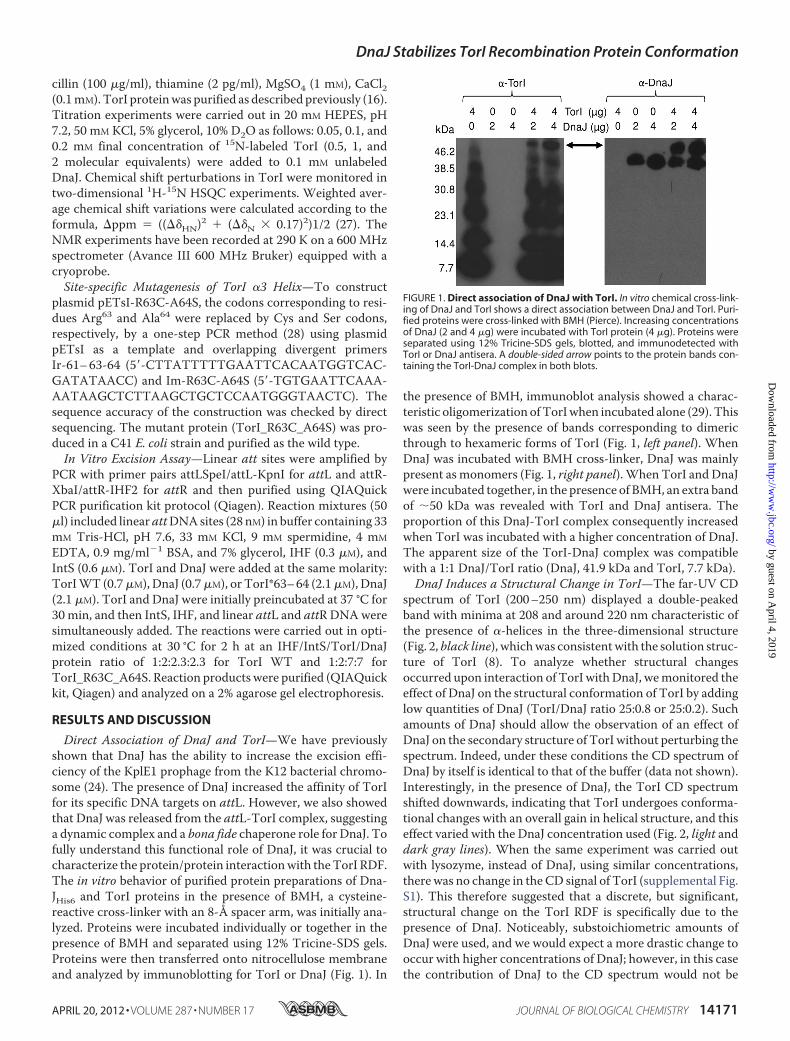
Direct Association of DnaJ and TorI—We have previouslyshown that DnaJ has the ability to increase the excision effi-ciency of the KplE1 prophage from the K12 bacterial chromo-some (24). The presence of DnaJ increased the affinity of TorIfor its specific DNA targets on attL. However, we also showedthat DnaJ was released from the attL-TorI complex, suggestinga dynamic complex and a bona fide chaperone role for DnaJ. Tofully understand this functional role of DnaJ, it was crucial tocharacterize the protein/protein interactionwith theTorI RDF.The in vitro behavior of purified protein preparations of Dna-JHis6 and TorI proteins in the presence of BMH, a cysteine-reactive cross-linker with an 8-Å spacer arm, was initially ana-lyzed. Proteins were incubated individually or together in thepresence of BMH and separated using 12% Tricine-SDS gels.Proteins were then transferred onto nitrocellulose membraneand analyzed by immunoblotting for TorI or DnaJ (Fig. 1). In
the presence of BMH, immunoblot analysis showed a charac-teristic oligomerization ofTorIwhen incubated alone (29). Thiswas seen by the presence of bands corresponding to dimericthrough to hexameric forms of TorI (Fig. 1, left panel). WhenDnaJ was incubated with BMH cross-linker, DnaJ was mainlypresent as monomers (Fig. 1, right panel).When TorI and DnaJwere incubated together, in the presence of BMH, an extra bandof �50 kDa was revealed with TorI and DnaJ antisera. Theproportion of this DnaJ-TorI complex consequently increasedwhen TorI was incubated with a higher concentration of DnaJ.The apparent size of the TorI-DnaJ complex was compatiblewith a 1:1 DnaJ/TorI ratio (DnaJ, 41.9 kDa and TorI, 7.7 kDa).DnaJ Induces a Structural Change in TorI—The far-UV CD
spectrum of TorI (200–250 nm) displayed a double-peakedband with minima at 208 and around 220 nm characteristic ofthe presence of �-helices in the three-dimensional structure(Fig. 2, black line), whichwas consistentwith the solution struc-ture of TorI (8). To analyze whether structural changesoccurred upon interaction of TorI withDnaJ, wemonitored theeffect of DnaJ on the structural conformation of TorI by addinglow quantities of DnaJ (TorI/DnaJ ratio 25:0.8 or 25:0.2). Suchamounts of DnaJ should allow the observation of an effect ofDnaJ on the secondary structure of TorI without perturbing thespectrum. Indeed, under these conditions the CD spectrum ofDnaJ by itself is identical to that of the buffer (data not shown).Interestingly, in the presence of DnaJ, the TorI CD spectrumshifted downwards, indicating that TorI undergoes conforma-tional changes with an overall gain in helical structure, and thiseffect varied with the DnaJ concentration used (Fig. 2, light anddark gray lines). When the same experiment was carried outwith lysozyme, instead of DnaJ, using similar concentrations,there was no change in the CD signal of TorI (supplemental Fig.S1). This therefore suggested that a discrete, but significant,structural change on the TorI RDF is specifically due to thepresence of DnaJ. Noticeably, substoichiometric amounts ofDnaJ were used, and we would expect a more drastic change tooccur with higher concentrations of DnaJ; however, in this casethe contribution of DnaJ to the CD spectrum would not be
FIGURE 1. Direct association of DnaJ with TorI. In vitro chemical cross-link-ing of DnaJ and TorI shows a direct association between DnaJ and TorI. Puri-fied proteins were cross-linked with BMH (Pierce). Increasing concentrationsof DnaJ (2 and 4 �g) were incubated with TorI protein (4 �g). Proteins wereseparated using 12% Tricine-SDS gels, blotted, and immunodetected withTorI or DnaJ antisera. A double-sided arrow points to the protein bands con-taining the TorI-DnaJ complex in both blots.
DnaJ Stabilizes TorI Recombination Protein Conformation
APRIL 20, 2012 • VOLUME 287 • NUMBER 17 JOURNAL OF BIOLOGICAL CHEMISTRY 14171
by guest on April 4, 2019
http://ww
w.jbc.org/
Dow
nloaded from
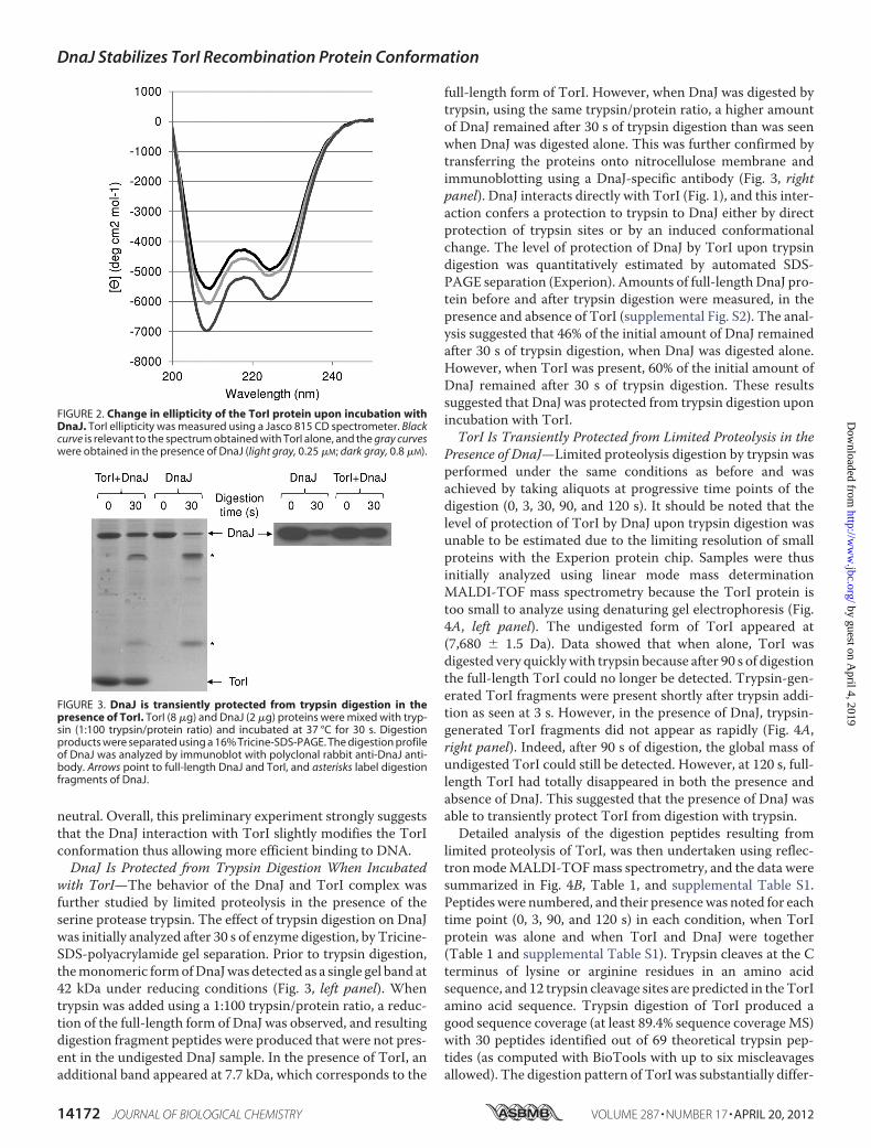
neutral. Overall, this preliminary experiment strongly suggeststhat the DnaJ interaction with TorI slightly modifies the TorIconformation thus allowing more efficient binding to DNA.DnaJ Is Protected from Trypsin Digestion When Incubated
with TorI—The behavior of the DnaJ and TorI complex wasfurther studied by limited proteolysis in the presence of theserine protease trypsin. The effect of trypsin digestion on DnaJwas initially analyzed after 30 s of enzyme digestion, by Tricine-SDS-polyacrylamide gel separation. Prior to trypsin digestion,themonomeric formofDnaJwas detected as a single gel band at42 kDa under reducing conditions (Fig. 3, left panel). Whentrypsin was added using a 1:100 trypsin/protein ratio, a reduc-tion of the full-length form of DnaJ was observed, and resultingdigestion fragment peptides were produced that were not pres-ent in the undigested DnaJ sample. In the presence of TorI, anadditional band appeared at 7.7 kDa, which corresponds to the
full-length form of TorI. However, when DnaJ was digested bytrypsin, using the same trypsin/protein ratio, a higher amountof DnaJ remained after 30 s of trypsin digestion than was seenwhen DnaJ was digested alone. This was further confirmed bytransferring the proteins onto nitrocellulose membrane andimmunoblotting using a DnaJ-specific antibody (Fig. 3, rightpanel). DnaJ interacts directly with TorI (Fig. 1), and this inter-action confers a protection to trypsin to DnaJ either by directprotection of trypsin sites or by an induced conformationalchange. The level of protection of DnaJ by TorI upon trypsindigestion was quantitatively estimated by automated SDS-PAGE separation (Experion). Amounts of full-length DnaJ pro-tein before and after trypsin digestion were measured, in thepresence and absence of TorI (supplemental Fig. S2). The anal-ysis suggested that 46% of the initial amount of DnaJ remainedafter 30 s of trypsin digestion, when DnaJ was digested alone.However, when TorI was present, 60% of the initial amount ofDnaJ remained after 30 s of trypsin digestion. These resultssuggested that DnaJ was protected from trypsin digestion uponincubation with TorI.TorI Is Transiently Protected from Limited Proteolysis in the
Presence of DnaJ—Limited proteolysis digestion by trypsin wasperformed under the same conditions as before and wasachieved by taking aliquots at progressive time points of thedigestion (0, 3, 30, 90, and 120 s). It should be noted that thelevel of protection of TorI by DnaJ upon trypsin digestion wasunable to be estimated due to the limiting resolution of smallproteins with the Experion protein chip. Samples were thusinitially analyzed using linear mode mass determinationMALDI-TOF mass spectrometry because the TorI protein istoo small to analyze using denaturing gel electrophoresis (Fig.4A, left panel). The undigested form of TorI appeared at(7,680 � 1.5 Da). Data showed that when alone, TorI wasdigested very quicklywith trypsin because after 90 s of digestionthe full-length TorI could no longer be detected. Trypsin-gen-erated TorI fragments were present shortly after trypsin addi-tion as seen at 3 s. However, in the presence of DnaJ, trypsin-generated TorI fragments did not appear as rapidly (Fig. 4A,right panel). Indeed, after 90 s of digestion, the global mass ofundigested TorI could still be detected. However, at 120 s, full-length TorI had totally disappeared in both the presence andabsence of DnaJ. This suggested that the presence of DnaJ wasable to transiently protect TorI from digestion with trypsin.Detailed analysis of the digestion peptides resulting from
limited proteolysis of TorI, was then undertaken using reflec-tronmodeMALDI-TOFmass spectrometry, and the data weresummarized in Fig. 4B, Table 1, and supplemental Table S1.Peptideswere numbered, and their presencewas noted for eachtime point (0, 3, 90, and 120 s) in each condition, when TorIprotein was alone and when TorI and DnaJ were together(Table 1 and supplemental Table S1). Trypsin cleaves at the Cterminus of lysine or arginine residues in an amino acidsequence, and 12 trypsin cleavage sites are predicted in theTorIamino acid sequence. Trypsin digestion of TorI produced agood sequence coverage (at least 89.4% sequence coverageMS)with 30 peptides identified out of 69 theoretical trypsin pep-tides (as computed with BioTools with up to six miscleavagesallowed). The digestion pattern of TorI was substantially differ-
FIGURE 2. Change in ellipticity of the TorI protein upon incubation withDnaJ. TorI ellipticity was measured using a Jasco 815 CD spectrometer. Blackcurve is relevant to the spectrum obtained with TorI alone, and the gray curveswere obtained in the presence of DnaJ (light gray, 0.25 �M; dark gray, 0.8 �M).
FIGURE 3. DnaJ is transiently protected from trypsin digestion in thepresence of TorI. TorI (8 �g) and DnaJ (2 �g) proteins were mixed with tryp-sin (1:100 trypsin/protein ratio) and incubated at 37 °C for 30 s. Digestionproducts were separated using a 16% Tricine-SDS-PAGE. The digestion profileof DnaJ was analyzed by immunoblot with polyclonal rabbit anti-DnaJ anti-body. Arrows point to full-length DnaJ and TorI, and asterisks label digestionfragments of DnaJ.
DnaJ Stabilizes TorI Recombination Protein Conformation
14172 JOURNAL OF BIOLOGICAL CHEMISTRY VOLUME 287 • NUMBER 17 • APRIL 20, 2012
by guest on April 4, 2019
http://ww
w.jbc.org/
Dow
nloaded from

ent in the presence of DnaJ. Certain regions of TorI were tran-siently protected from trypsin digestion in the presence ofDnaJ, and cleavage in these regions only appeared at later timepoints of digestion. After 3 s of trypsin digestion, peptidesappearing from cleavage at amino acids 51, 57, 59, and 63 onlyappeared when TorI was alone and not when DnaJ was presentwith TorI.Together, limited trypsin digestion experiments indicated a
transient protection of TorI and DnaJ on each other, whichconfirmed the formation of a transient complex of the two pro-teins, and this complex involves the C-terminal region of TorI
either directly or indirectly through discrete conformationalchanges of this region.Mapping of Amino Acid Residues Implicated in the Forma-
tion of TorI-DnaJ Complex by Heteronuclear NMR—An NMRtitration experiment was carried out to locate the amino acidresidues at the surface of the TorI structure, which wereinvolved in binding to DnaJ. This was achieved by titrating iso-topically labeled 15N-TorI with unlabeled DnaJ, and the reac-tion was carried out at 25 °C. Chemical shift variations weremonitored using two-dimensional 1H-15NHSQC spectra (sup-plemental Fig. S3). Significant chemical shift perturbations
FIGURE 4. Monomeric TorI is protected from trypsin digestion in the presence of DnaJ. Limited proteolysis with trypsin was carried out for the TorI andDnaJ proteins together and alone. Samples were analyzed using MALDI-TOF MS using two different modes of peptide determination. A, linear mode MALDI-TOF MS analysis showed monomeric TorI remained in excess in the digestion fragment peaks until 90 s. However, in the absence of DnaJ, TorI was more quicklydigested, with the digestion fragment peaks appearing after only 3 s of trypsin digestion. B, cleavage sites of trypsin in TorI are indicated by arrows, and missingcleavage sites in the presence of DnaJ are indicated in red (amino acids 51, 57, 59, and 63). These amino acids mapped onto the C-terminal �-helix of the TorIstructure.
DnaJ Stabilizes TorI Recombination Protein Conformation
APRIL 20, 2012 • VOLUME 287 • NUMBER 17 JOURNAL OF BIOLOGICAL CHEMISTRY 14173
by guest on April 4, 2019
http://ww
w.jbc.org/
Dow
nloaded from

were observed upon the addition of increasing amounts of 15N-labeled TorI to unlabeled DnaJ, indicative of equilibriumbetween the bound and unbound states in the fast exchangeregime. The weighted average chemical shift displacements areshown in Fig. 5A. The largest chemical shift variations betweenthe free and the bound forms of TorI are mainly observed forbackbone amides of the first �-strand (Gln6, Ser9, Leu10, andVal11), the helix �1 (Ile16, Asp19, and Gly21), the �3-strandinvolved in anti-parallel �-sheet flexible loop region (Arg47),and the C-terminal helix 3 (Glu55, Lys57, Lys59, Leu61, Arg63,andAla64). These latter perturbations are in agreementwith themass spectrometry data showing the protection of the TorIC-terminal part in the presence of DnaJ. The perturbed resi-dues congregate onto one face of theTorI protein surface oppo-site the TorI DNA-binding site, which we have previouslymapped (Fig. 5, B and C) (8). Indeed, none of the residuesinvolved in the interaction with DNA were perturbed uponDnaJ binding. However, some residues (Leu10, Val11, and Ile16)are very close to the wing and the helix �1 contacting DNA.This close proximity of the DnaJ and DNA-binding sites onTorI structure may have a direct influence on the observedincreased affinity of TorI for DNA (24).To validate whether the residues identified by NMR were
involved in a direct interactionwithDnaJ, we constructed threedifferent TorI mutants, TorI_L10Q_V11S, TorI_L61S, andTorI_R63C_A64S, that behaved differently in terms of theirprotein production and stability. TorI_L10Q_V11S proved to
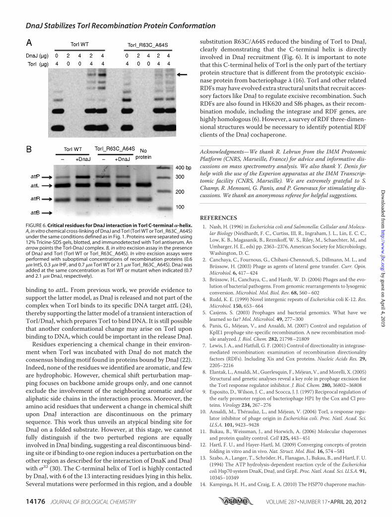
be very unstable and was barely produced. TorI_L61S was pro-duced and stable, but this single substitution was insufficient todisrupt the TorI/DnaJ interaction (data not shown). Finally, thedouble mutant TorI_R63C_A64S was stably produced andshowed the same overall structure as the wild-type protein byCD (data not shown). This latter mutant was affected in DnaJbinding as shown in Fig. 6A, thus validating that the residuesidentified and mapped through NMR titration assay wereindeed involved in DnaJ binding. This double-substituted pro-tein was assayed in vitro for its propensity to recruit DnaJ in arecombination assay. As seen in Fig. 6B, the TorI_R63C_A64S,although active in an in vitro excision assay, failed to recruitDnaJ. As a result, no increased efficiency in the presence ofDnaJ was observed in contrast to what was observed when thewild-type protein was used. Together, these results confirm theresults obtained by NMR titration indicating that the C-termi-nal �-helix in TorI was important for DnaJ recruitment.Conclusions—Wehave previously identified a functional role
of DnaJ in assisting prophage excision (24). Initial experimentaldata showed that DnaJ did not bind to the attL DNA itself butsomehow increased the affinity of TorI for this site. Here, wehave shown that DnaJ binds directly to the TorI RDF inducing astabilization of its conformation. Taken together, the datashown here demonstrate that DnaJ interacts with a folded sub-strate, TorI. This interaction results in a transient and recipro-cal protection of these two proteins, either due to direct bindingor to induced conformational change. Conclusions drawn from
TABLE 1TorI peptides detected upon trypsin hydrolysis in the presence or absence of DnaJTransient protection of specific trypsin cleavage sites in the TorI amino acid sequence was analyzed. TorI cleavage peptides were identified using reflectron modeMALDI-TOFMS analysis and listed at each time point of trypsin digestion (0, 3, 30, 90 and 120 s). 1 indicates the presence of a given peptide in the sample; 0 indicates itsabsence.
Numericalassignmenta
Peptide size(amino acid)
Digestion time 3 s Digestion time 30 s Digestion time 90 s Digestion time 120 sTorI DnaJ � TorI TorI DnaJ � TorI TorI DnaJ � TorI TorI DnaJ � TorI
1 66 1 1 1 1 0 0 0 02 45 1 1 1 1 0 0 0 03 40 1 1 1 1 0 0 0 04 38 1 1 1 1 0 0 0 05 32 1 1 1 1 1 1 1 16 30 1 1 1 1 1 1 1 17 24 1 1 1 0 1 1 1 18 14 1 0 1 0 1 1 1 19 31 1 1 1 1 1 1 0 010 26 1 1 1 0 0 0 0 011 24 0 0 1 1 0 0 0 012 18 1 1 1 1 1 1 1 013 16 1 1 1 1 1 1 1 114 10 1 0 1 0 1 1 1 115 21 1 0 0 0 0 0 0 016 18 1 0 1 0 0 0 0 017 14 1 1 1 1 1 1 1 018 12 1 0 1 0 1 1 1 019 19 1 1 0 0 0 0 0 020 16 1 0 0 0 0 0 0 021 12 1 0 1 1 1 1 1 122 10 1 0 1 0 1 1 1 123 31 0 0 1 0 0 0 0 024 15 0 0 1 0 0 0 0 025 27 0 0 0 0 0 0 1 026 8 0 0 0 0 1 0 0 027 6 1 0 1 0 1 1 1 128 8 1 0 1 0 1 1 1 129 6 0 0 0 0 0 0 1 030 8 0 0 0 0 0 0 1 031 6 0 0 1 1 0 0 0 0Total 23 13 24 13 15 14 16 12
a Numerical assignment refers to supplemental Table S1.
DnaJ Stabilizes TorI Recombination Protein Conformation
14174 JOURNAL OF BIOLOGICAL CHEMISTRY VOLUME 287 • NUMBER 17 • APRIL 20, 2012
by guest on April 4, 2019
http://ww
w.jbc.org/
Dow
nloaded from

bothNMR andCD experiments led us to propose that DnaJ hasan atypical effect on a native protein that induces conforma-tional �-helix stabilizations. The NMR data collected providesinformation on the residues that are or are not trypsin cleavagesites. If we look at residues that undergo a chemical shift andthat are also transiently protected from trypsin, we can identifyresidues Arg47, Lys57, Lys59, and Arg63, many of which lie in theC-terminal helix of TorI.The 1H-15N HSQC spectrum of TorI upon binding of DnaJ
does not showdramaticmodifications that could correspond toimportant conformational changes of the protein. However,CD experiments suggest that slight structural modifications
occur in TorI when in the presence of DnaJ (Fig. 2). The resi-dues perturbed upon binding of DnaJ on TorI in the NMRexperiments are mostly located within �-helices of TorI. Bind-ing of DnaJ might then have the effect of stabilizing the helicesas illustrated by the downward shift in the CD signal of TorI.This could provide a stabilizing effect, which probably helps toposition TorI to obtain optimal binding to its target DNA.How and when TorI is released from DnaJ still remains
unknown.We show that the segment of TorI that interactswithattL is not altered upon binding to the DnaJ protein. Thismeans that there are two possible models, either DnaJ acts atthe same time asTorIwith attLor preparesTorI beforehand for
FIGURE 5. Mapping the binding site of DnaJ in the TorI structure by heteronuclear NMR. A, plot of the weighted average chemical shift variations of15N-TorI backbone upon binding of DnaJ. Secondary structures of TorI are shown above the plot. The red bars correspond to residues showing the largestchemical shift variations. B, chemical shift mapping onto ribbon (right) and surface (left) representations of TorI. Residues undergoing the largest chemical shiftchanges upon DnaJ binding are colored in red. C, ribbon (right) and surface (left) representations of the data presented in B, after 180° rotation according to they axis, with the residues involved in DNA binding colored blue.
DnaJ Stabilizes TorI Recombination Protein Conformation
APRIL 20, 2012 • VOLUME 287 • NUMBER 17 JOURNAL OF BIOLOGICAL CHEMISTRY 14175
by guest on April 4, 2019
http://ww
w.jbc.org/
Dow
nloaded from
binding to attL. From previous work, we provide evidence tosupport the latter model, as DnaJ is released and not part of thecomplex when TorI binds to its specific DNA target attL (24),thereby supporting the lattermodel of a transient interaction ofTorI/DnaJ, which prepares TorI to bind DNA. It is still possiblethat another conformational change may arise on TorI uponbinding to DNA, which could be important in the release DnaJ.Residues experiencing a chemical change in their environ-
ment when TorI was incubated with DnaJ do not match theconsensus binding motif found in proteins bound by DnaJ (22).Indeed, none of the residueswe identified are aromatic, and feware hydrophobic. However, chemical shift perturbation map-ping focuses on backbone amide groups only, and one cannotexclude the involvement of the neighboring aromatic and/oraliphatic side chains in the interaction process. Moreover, theamino acid residues that underwent a change in chemical shiftupon DnaJ interaction are discontinuous on the primarysequence. This work thus unveils an atypical binding site forDnaJ on a folded substrate. However, at this stage, we cannotfully distinguish if the two perturbed regions are equallyinvolved in DnaJ binding, suggesting a real discontinuous bind-ing site or if binding to one region induces a perturbation on theother region as described for the interaction of DnaK and DnaJwith �32 (30). The C-terminal helix of TorI is highly contactedby DnaJ, with 6 of the 13 interacting residues lying in this helix.Several mutations were performed in this region, and a double
substitution R63C/A64S reduced the binding of TorI to DnaJ,clearly demonstrating that the C-terminal helix is directlyinvolved in DnaJ recruitment (Fig. 6). It is important to notethat this C-terminal helix of TorI is the only part of the tertiaryprotein structure that is different from the prototypic excisio-nase protein from bacteriophage � (16). TorI and other relatedRDFsmayhave evolved extra structural units that recruit acces-sory factors like DnaJ to regulate excisive recombination. SuchRDFs are also found in HK620 and Sf6 phages, as their recom-bination module, including the integrase and RDF genes, arehighly homologous (6). However, a survey of RDF three-dimen-sional structures would be necessary to identify potential RDFclients of the DnaJ cochaperone.
Acknowledgments—We thank R. Lebrun from the IMM ProteomicPlatform (CNRS, Marseille, France) for advice and informative dis-cussions on mass spectrometry analysis. We also thank Y. Denis forhelp with the use of the Experion apparatus at the IMM Transcrip-tomic facility (CNRS, Marseille). We are extremely grateful to S.Champ, R. Menouni, G. Panis, and P. Genevaux for stimulating dis-cussions. We thank an anonymous referee for helpful suggestions.
REFERENCES1. Nash, H. (1996) in Escherichia coli and Salmonella: Cellular and Molecu-
lar Biology (Neidhardt, F. C., Curtiss, III, R., Ingraham, J. L., Lin, E. C. C.,Low, K. B., Magasanik, B., Reznikoff, W. S., Riley, M., Schaechter, M., andUmbarger, H. E., eds) pp. 2363–2376, American Society forMicrobiology,Washington, D. C.
2. Canchaya, C., Fournous, G., Chibani-Chennoufi, S., Dillmann, M. L., andBrüssow, H. (2003) Phage as agents of lateral gene transfer. Curr. Opin.Microbiol. 6, 417–424
3. Brüssow, H., Canchaya, C., and Hardt, W. D. (2004) Phages and the evo-lution of bacterial pathogens. From genomic rearrangements to lysogenicconversion.Microbiol. Mol. Biol. Rev. 68, 560–602
4. Rudd, K. E. (1999) Novel intergenic repeats of Escherichia coli K-12. Res.Microbiol. 150, 653–664
5. Casjens, S. (2003) Prophages and bacterial genomics. What have welearned so far?Mol. Microbiol. 49, 277–300
6. Panis, G., Méjean, V., and Ansaldi, M. (2007) Control and regulation ofKplE1 prophage site-specific recombination. A new recombination mod-ule analyzed. J. Biol. Chem. 282, 21798–21809
7. Lewis, J. A., andHatfull, G. F. (2001) Control of directionality in integrase-mediated recombination: examination of recombination directionalityfactors (RDFs). Including Xis and Cox proteins. Nucleic Acids Res. 29,2205–2216
8. Elantak, L., Ansaldi,M., Guerlesquin, F.,Méjean, V., andMorelli, X. (2005)Structural and genetic analyses reveal a key role in prophage excision forthe TorI response regulator inhibitor. J. Biol. Chem. 280, 36802–36808
9. Esposito, D., Wilson, J. C., and Scocca, J. J. (1997) Reciprocal regulation ofthe early promoter region of bacteriophage HP1 by the Cox and Cl pro-teins. Virology 234, 267–276
10. Ansaldi, M., Théraulaz, L., and Méjean, V. (2004) TorI, a response regu-lator inhibitor of phage origin in Escherichia coli. Proc. Natl. Acad. Sci.U.S.A. 101, 9423–9428
11. Bukau, B., Weissman, J., and Horwich, A. (2006) Molecular chaperonesand protein quality control. Cell 125, 443–451
12. Hartl, F. U., and Hayer-Hartl, M. (2009) Converging concepts of proteinfolding in vitro and in vivo. Nat. Struct. Mol. Biol. 16, 574–581
13. Szabo, A., Langer, T., Schröder, H., Flanagan, J., Bukau, B., and Hartl, F. U.(1994) The ATP hydrolysis-dependent reaction cycle of the EscherichiacoliHsp70 systemDnaK, DnaJ, and GrpE. Proc. Natl. Acad. Sci. U.S.A. 91,10345–10349
14. Kampinga, H. H., and Craig, E. A. (2010) The HSP70 chaperone machin-
FIGURE 6. Critical residues for DnaJ interaction in TorI C-terminal �-helix.A, in vitro chemical cross-linking of DnaJ and TorI (TorI WT or TorI_R63C_A64S)under the same conditions defined as in Fig. 1. Proteins were separated using12% Tricine-SDS gels, blotted, and immunodetected with TorI antiserum. Anarrow points the TorI-DnaJ complex. B, in vitro excision assay in the presenceof DnaJ and TorI (TorI WT or TorI_R63C_A64S). In vitro excision assays wereperformed with suboptimal concentrations of recombination proteins (0.6�M IntS, 0.3 �M IHF, and 0.7 �M TorI WT or 2.1 �M TorI_R63C_A64S). DnaJ wasadded at the same concentration as TorI WT or mutant when indicated (0.7and 2.1 �M DnaJ, respectively).
DnaJ Stabilizes TorI Recombination Protein Conformation
14176 JOURNAL OF BIOLOGICAL CHEMISTRY VOLUME 287 • NUMBER 17 • APRIL 20, 2012
by guest on April 4, 2019
http://ww
w.jbc.org/
Dow
nloaded from
ery. J proteins as drivers of functional specificity. Nat. Rev. Mol. Cell Biol.11, 579–592
15. Genevaux, P., Georgopoulos, C., and Kelley, W. L. (2007) The Hsp70chaperone machines of Escherichia coli. A paradigm for the repartition ofchaperone functions.Mol. Microbiol. 66, 840–857
16. Genevaux, P., Keppel, F., Schwager, F., Langendijk-Genevaux, P. S., Hartl,F. U., and Georgopoulos, C. (2004) In vivo analysis of the overlappingfunctions of DnaK and trigger factor. EMBO Rep. 5, 195–200
17. Langer, T., Lu, C., Echols, H., Flanagan, J., Hayer, M. K., and Hartl, F. U.(1992) Successive action of DnaK, DnaJ, and GroEL along the pathway ofchaperone-mediated protein folding. Nature 356, 683–689
18. Georgopoulos, C. (2006) Toothpicks, serendipity, and the emergence ofthe Escherichia coli DnaK (Hsp70) and GroEL (Hsp60) chaperone ma-chines. Genetics 174, 1699–1707
19. Zylicz, M., Ang, D., Liberek, K., and Georgopoulos, C. (1989) Initiation of�DNA replication with purified host- and bacteriophage-encoded pro-teins. The role of the dnaK, dnaJ, and grpE heat shock proteins. EMBO J. 8,1601–1608
20. Zylicz, M. (1993) The Escherichia coli chaperones involved in DNA repli-cation. Philos. Trans. R. Soc. Lond. B Biol. Sci. 339, 271–278
21. de Crouy-Chanel, A., Kohiyama, M., and Richarme, G. (1995) A novelfunction of Escherichia coli chaperone DnaJ. Protein-disulfide isomerase.J. Biol. Chem. 270, 22669–22672
22. Rüdiger, S., Schneider-Mergener, J., and Bukau, B. (2001) Its substratespecificity characterizes the DnaJ co-chaperone as a scanning factor forthe DnaK chaperone. EMBO J. 20, 1042–1050
23. Shimizu, T., Yoshii, A., Sakurai, K., Hamada, K., Yamaji, Y., Suzuki, M.,Namba, S., and Hibi, T. (2009) Identification of a novel tobacco DnaJ-like
protein that interacts with themovement protein of tobaccomosaic virus.Arch. Virol. 154, 959–967
24. Champ, S., Puvirajesinghe, T. M., Perrody, E., Menouni, R., Genevaux, P.,and Ansaldi, M. (2011) Chaperone-assisted excisive recombination, a sol-itary role for DnaJ (Hsp40) chaperone in lysogeny escape. J. Biol. Chem.286, 38876–38885
25. Cajo,G. C., Horne, B. E., Kelley,W. L., Schwager, F., Georgopoulos, C., andGenevaux, P. (2006) The role of the DIF motif of the DnaJ (Hsp40) co-chaperone in the regulation of the DnaK (Hsp70) chaperone cycle. J. Biol.Chem. 281, 12436–12444
26. Erales, J., Lignon, S., and Gontero, B. (2009) CP12 from Chlamydomonasreinhardtii, a permanent specific “chaperone-like” protein of glyceralde-hyde-3-phosphate dehydrogenase. J. Biol. Chem. 284, 12735–12744
27. Grzesiek, S., Bax, A., Clore, G. M., Gronenborn, A. M., Hu, J. S., Kaufman,J., Palmer, I., Stahl, S. J., andWingfield, P. T. (1996) The solution structureof HIV-1 Nef reveals an unexpected fold and permits delineation of thebinding surface for the SH3 domain of Hck tyrosine protein kinase. Nat.Struct. Biol. 3, 340–345
28. Ansaldi, M., Lepelletier, M., andMéjean, V. (1996) Site-specific mutagen-esis by using an accurate recombinant polymerase chain reactionmethod.Anal. Biochem. 234, 110–111
29. Panis, G., Duverger, Y., Champ, S., andAnsaldi,M. (2010) Protein-bindingsites involved in the assembly of the KplE1 prophage intasome. Virology404, 41–50
30. Rodriguez, F., Arsène-Ploetze, F., Rist, W., Rüdiger, S., Schneider-Mer-gener, J., Mayer,M. P., and Bukau, B. (2008)Molecular basis for regulationof the heat shock transcription factor �32 by the DnaK and DnaJ chaper-ones.Mol. Cell 32, 347–358
DnaJ Stabilizes TorI Recombination Protein Conformation
APRIL 20, 2012 • VOLUME 287 • NUMBER 17 JOURNAL OF BIOLOGICAL CHEMISTRY 14177
by guest on April 4, 2019
http://ww
w.jbc.org/
Dow
nloaded from
Ilbert and Mireille AnsaldiTania M. Puvirajesinghe, Latifa Elantak, Sabrina Lignon, Nathalie Franche, Marianne
Excision EfficiencyDnaJ (Hsp40 Protein) Binding to Folded Substrate Impacts KplE1 Prophage
doi: 10.1074/jbc.M111.331462 originally published online February 28, 20122012, 287:14169-14177.J. Biol. Chem.
10.1074/jbc.M111.331462Access the most updated version of this article at doi:
Alerts:
When a correction for this article is posted•
When this article is cited•
to choose from all of JBC's e-mail alertsClick here
Supplemental material:
http://www.jbc.org/content/suppl/2012/02/28/M111.331462.DC1
http://www.jbc.org/content/287/17/14169.full.html#ref-list-1
This article cites 29 references, 10 of which can be accessed free at
by guest on April 4, 2019
http://ww
w.jbc.org/
Dow
nloaded from


![Research Paper E3 Ubiquitin Ligase Siah- 1 is Down ...free-journal.umm.ac.id/files/file/v07p0418.pdf · 40 (Hsp40) [10], HBV core proteins [11], tumor sup-pressor p53 [12], and transcription](https://img.pdfslide.us/doc/110x75/5f773a28edc69301e35444e5/research-paper-e3-ubiquitin-ligase-siah-1-is-down-free-40-hsp40-10-hbv.jpg)