Embed Size (px)
Citation preview
Divine Intervention Episode 51Comprehensive Step 1
Biochemistry Review (Session 2)Some PGY1

Absorbing Fe/Breaking Down Heme-Fe is absorbed (also only carries O2) in the 2+ form only. Vit C encourages this
process (what are 2 other HY functions of Vit C that have been discussed?).
-HFE regulates this process. A HFE mutation can cause too much Fe reabsorption
(hemochromatosis, tx w/phlebotomy). What should your first step in diagnosis be?
-I’d encourage you to also try recalling the relationship b/w Fe2+/Fe3+ w/pathologies.
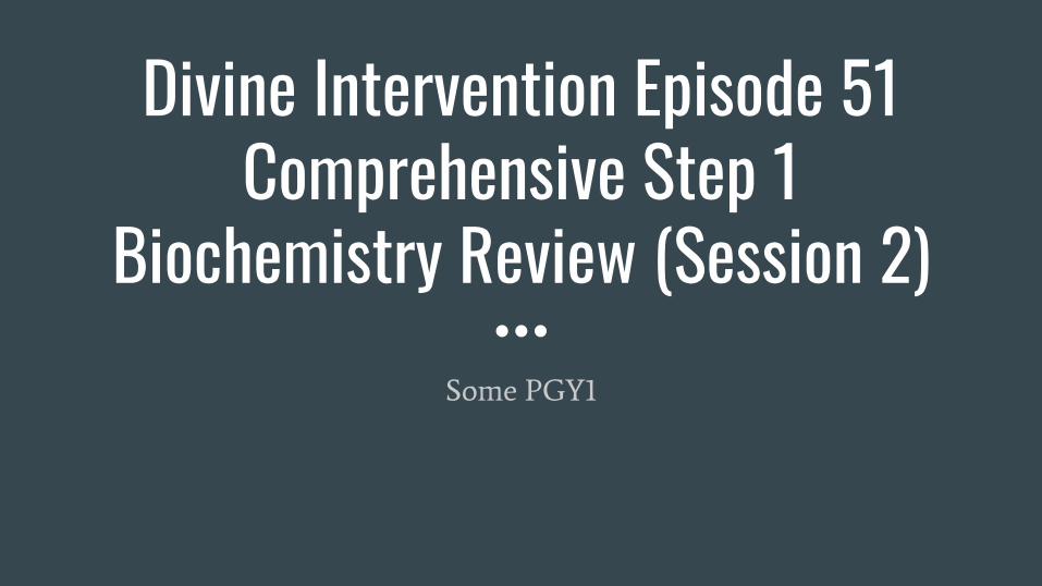
-It is HY to know the breakdown pathway for heme and the different diseases that
could arise from issues along that pathway (as well as the associated kind of
hyperbilirubinemia)-> Hemolytic anemia, Newborn jaundice, TMP-SMX toxicity,
Crigler Najjar (T1 and 2), Gilbert’s, Dubin Johnson, Rotor, Obstructive process, etc.
-Remember that Fe is absorbed in the duodenum, folate is absorbed in
duodenum/jejunum, B12 is absorbed in the terminal ileum (re-Crohn’s association).
Heme Breakdown

Another Step 1 Worthy Question/Thought Can you explain these lesions?
Option A-Increased urine bilirubin, decreased urine
urobilinogen, increased direct bilirubin, dark/tea
colored urine, acholic stools.
Option B-Increased urine urobilinogen, no urine
bilirubin, increased indirect bilirubin, normal
colored urine, dark colored stools.
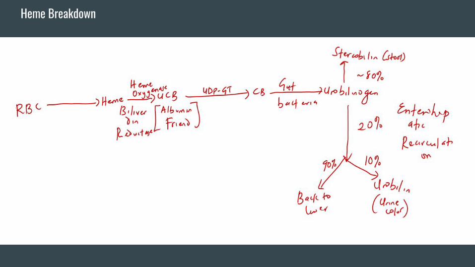
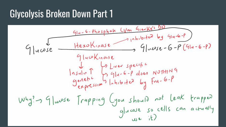
Some General Principles (make thy life super easy!)-Insulin works through tyrosine kinase receptors. Insulin is a dephosphorylator.
-Glucagon works through G protein coupled receptors which activate PKA. Glucagon
is a phosphorylator.
-If you know this, you can easily reason that if an enzyme is activated by insulin, the
activated form must be a “dephosphorylated form” of the enzyme (and vice versa for
glucagon).
-Carboxylase enzymes are ABC enzymes (they use ATP and Biotin, hence the AB). C
stands for carboxylase (and CO2).
-Kinase enzymes as a rule add phosphate groups to stuff.
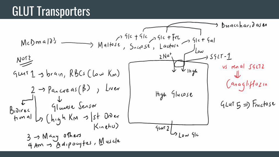
GLUT Transporters
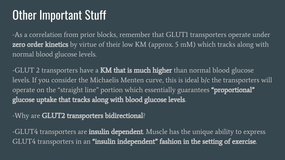
Other Important Stuff-As a correlation from prior blocks, remember that GLUT1 transporters operate under
zero order kinetics by virtue of their low KM (approx. 5 mM) which tracks along with
normal blood glucose levels.
-GLUT 2 transporters have a KM that is much higher than normal blood glucose
levels. If you consider the Michaelis Menten curve, this is ideal b/c the transporters will
operate on the “straight line” portion which essentially guarantees “proportional”
glucose uptake that tracks along with blood glucose levels.
-Why are GLUT2 transporters bidirectional?
-GLUT4 transporters are insulin dependent. Muscle has the unique ability to express
GLUT4 transporters in an “insulin independent” fashion in the setting of exercise.
Glycolysis Broken Down Part 1
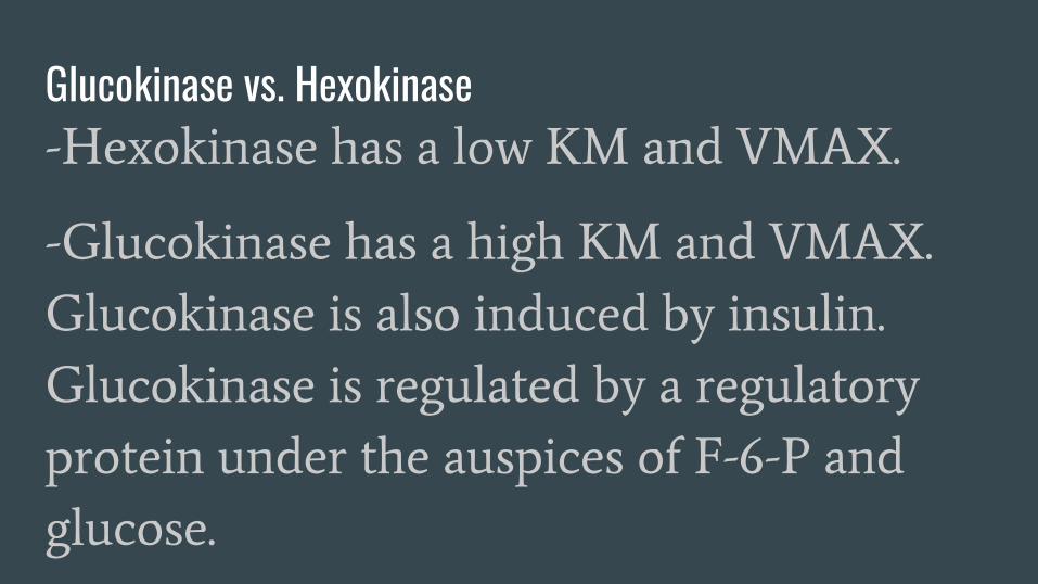
Glucokinase vs. Hexokinase-Hexokinase has a low KM and VMAX.
-Glucokinase has a high KM and VMAX.
Glucokinase is also induced by insulin.
Glucokinase is regulated by a regulatory
protein under the auspices of F-6-P and
glucose.

Glucokinase Regulatory Protein-Is an inhibitor of glucokinase (GK).
-Binds GK and sends it to the nucleus (where it is inactive).
-GKRP has the ability to bind both F6P and glucose.
-When bound by F6P, GKRP has a higher affinity for GK (which sequesters GK by
taking it to the nucleus).
-When bound by glucose, GKRP has a much lower affinity for GK (which brings it
back to the cytoplasm for reaction).
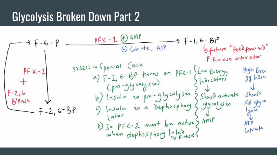
Glycolysis Broken Down Part 2

Glycolysis Broken Down Part 3

Glycolysis Broken Down Part 4

Some Other Important Stuff-Overall, glycolysis gives rise to the rule of 2s (2 ATPs, 2 NADH, and 2 Pyruvates).
Pyruvate has multiple fates;
-It can form lactate under the action of lactate DH. This step regenerates NAD to keep
the Glyceraldehyde-3-P DH step working.
-Pyruvate can go into mitochondria to receive special attention from the PDH complex
ultimately leading to Acetyl-coA formation.
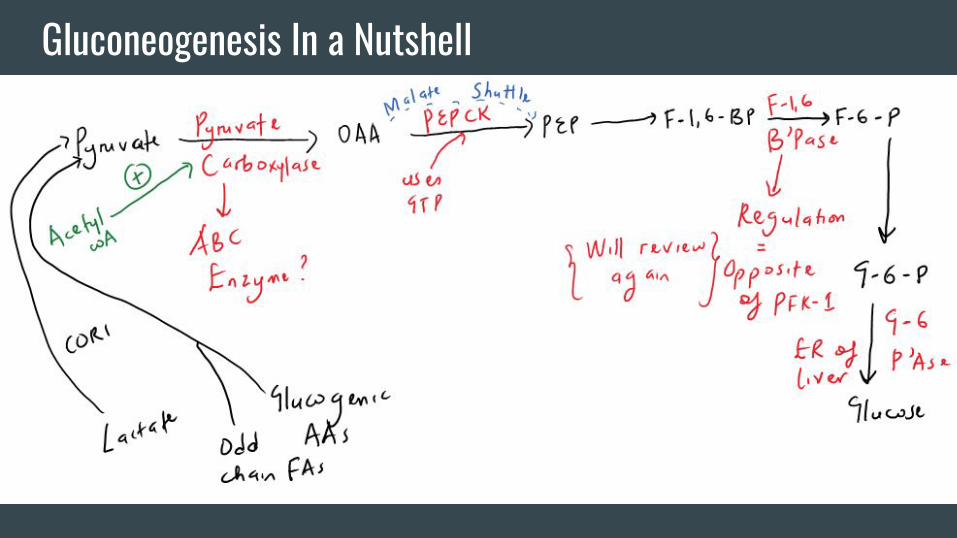
-Pyruvate can receive special attention from Pyruvate carboxylase (what is a HY
cofactor utilized by this enzyme???) to form OAA that can reverse course in
gluconeogenesis (through subsequent PEPCK action).

Galactose Metabolism

Fructose Metabolism
Other Important Stuff-You should by now notice that the main job of a kinase is to
phosphorylate stuff. “Phosphorylated stuff” cannot leave cells.
They also tend to be osmotically active. This explains the many
problems associated with the “2nd enzyme” in the 2 metabolic
pathways we just discussed.
-Note the temporal association of the diseases presented to
breastfeeding and introduction of fruits.
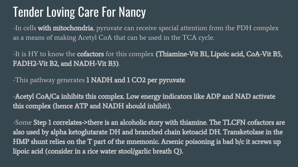
Tender Loving Care For Nancy-In cells with mitochondria, pyruvate can receive special attention from the PDH complex
as a means of making Acetyl CoA that can be used in the TCA cycle.
-It is HY to know the cofactors for this complex (Thiamine-Vit B1, Lipoic acid, CoA-Vit B5,
FADH2-Vit B2, and NADH-Vit B3).
-This pathway generates 1 NADH and 1 CO2 per pyruvate.
-Acetyl CoA/Ca inhibits this complex. Low energy indicators like ADP and NAD activate
this complex (hence ATP and NADH should inhibit).
-Some Step 1 correlates->there is an alcoholic story with thiamine. The TLCFN cofactors are
also used by alpha ketoglutarate DH and branched chain ketoacid DH. Transketolase in the
HMP shunt relies on the T part of the mnemonic. Arsenic poisoning is bad b/c it screws up
lipoic acid (consider in a rice water stool/garlic breath Q).

The TCA Cycle

TCA Cycle Takeaways-Makes (per pyruvate) CO2, NADH, FADH2, ATP (2311).
-The VOMIT pathway feeds into succinyl-coA (Valine, Odd Chain FAs, Methionine,
Isoleucine, Threonine).
-It is also HY to know certain keto acid, AA relationships;
OAA is the keto acid to aspartate. Pyruvate is the keto acid to alanine. Alpha
ketoglutarate is the keto acid to glutamate.
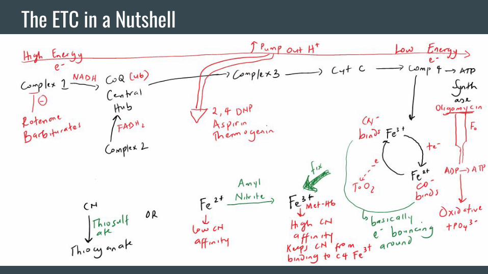
The ETC in a Nutshell

Glycogen Formation and Breakdown In a Nutshell
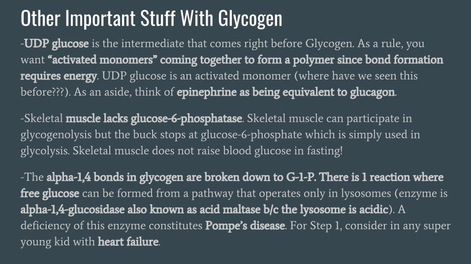
Other Important Stuff With Glycogen-UDP glucose is the intermediate that comes right before Glycogen. As a rule, you
want “activated monomers” coming together to form a polymer since bond formation
requires energy. UDP glucose is an activated monomer (where have we seen this
before???). As an aside, think of epinephrine as being equivalent to glucagon.
-Skeletal muscle lacks glucose-6-phosphatase. Skeletal muscle can participate in
glycogenolysis but the buck stops at glucose-6-phosphate which is simply used in
glycolysis. Skeletal muscle does not raise blood glucose in fasting!
-The alpha-1,4 bonds in glycogen are broken down to G-1-P. There is 1 reaction where
free glucose can be formed from a pathway that operates only in lysosomes (enzyme is
alpha-1,4-glucosidase also known as acid maltase b/c the lysosome is acidic). A
deficiency of this enzyme constitutes Pompe’s disease. For Step 1, consider in any super
young kid with heart failure.
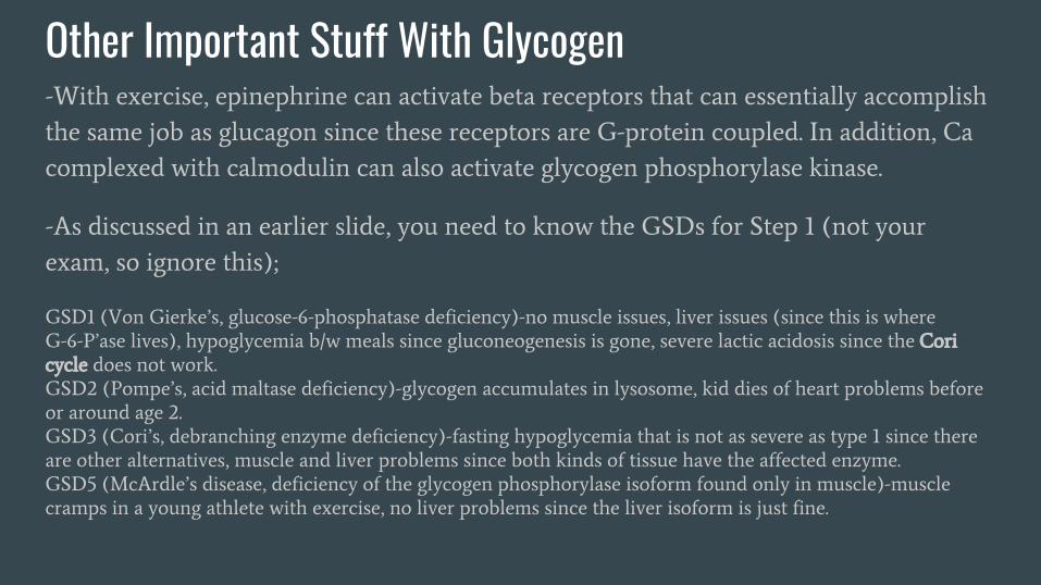
Other Important Stuff With Glycogen-With exercise, epinephrine can activate beta receptors that can essentially accomplish
the same job as glucagon since these receptors are G-protein coupled. In addition, Ca
complexed with calmodulin can also activate glycogen phosphorylase kinase.
-As discussed in an earlier slide, you need to know the GSDs for Step 1 (not your
exam, so ignore this);
GSD1 (Von Gierke’s, glucose-6-phosphatase deficiency)-no muscle issues, liver issues (since this is where
G-6-P’ase lives), hypoglycemia b/w meals since gluconeogenesis is gone, severe lactic acidosis since the Cori
cycle does not work.
GSD2 (Pompe’s, acid maltase deficiency)-glycogen accumulates in lysosome, kid dies of heart problems before
or around age 2.
GSD3 (Cori’s, debranching enzyme deficiency)-fasting hypoglycemia that is not as severe as type 1 since there
are other alternatives, muscle and liver problems since both kinds of tissue have the affected enzyme.
GSD5 (McArdle’s disease, deficiency of the glycogen phosphorylase isoform found only in muscle)-muscle
cramps in a young athlete with exercise, no liver problems since the liver isoform is just fine.
Gluconeogenesis In a Nutshell

Other Important Stuff With Gluconeogenesis
-For Step 1, remember the association of
serious egg white consumption (too much
avidin) with a deficiency of Vitamin B7
which is needed for pyruvate carboxylase
function.
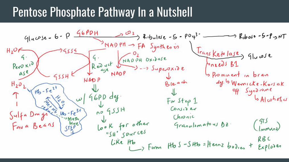
Pentose Phosphate Pathway In a Nutshell
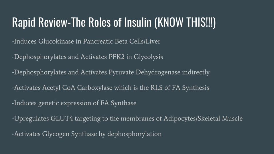
Rapid Review-The Roles of Insulin (KNOW THIS!!!)-Induces Glucokinase in Pancreatic Beta Cells/Liver
-Dephosphorylates and Activates PFK2 in Glycolysis
-Dephosphorylates and Activates Pyruvate Dehydrogenase indirectly
-Activates Acetyl CoA Carboxylase which is the RLS of FA Synthesis
-Induces genetic expression of FA Synthase
-Upregulates GLUT4 targeting to the membranes of Adipocytes/Skeletal Muscle
-Activates Glycogen Synthase by dephosphorylation

Intro To Fatty Acid Metabolism/FA Synthesis-Any reason why cis unsaturated fats are better than trans unsaturated/saturated fats?
-In a patient with steatorrhea, how would you differentiate between a pancreatic
enzyme deficiency/bile non-delivery as a cause in comparison with malabsorption?
-To cook FAs (in the cytoplasm), you need a few ingredients -> Acetyl coA (from the
mitochondria through citrate shuttle), ATP (from glycolysis, TCA, ETC), CO2
(everywhere), NADPH (from the HMP shunt and from the Malic enzyme).
-When the cell has a lot of energy from the TCA cycle and ETC -> ATP and NADH
build up -> inhibit Isocitrate dehydrogenase -> Citrate builds up -> to cytoplasm.
-RLE here is Acetyl coA carboxylase (induced by insulin, also activated by
dephosphorylation, activated by citrate as well ->what is the feedforward activating
mechanism in glycolysis?).ACCASE deactivation is via glucagon med. phosphorylation.

FA Synthesis
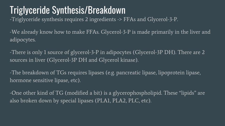
Triglyceride Synthesis/Breakdown-Triglyceride synthesis requires 2 ingredients -> FFAs and Glycerol-3-P.
-We already know how to make FFAs. Glycerol-3-P is made primarily in the liver and
adipocytes.
-There is only 1 source of glycerol-3-P in adipocytes (Glycerol-3P DH). There are 2
sources in liver (Glycerol-3P DH and Glycerol kinase).
-The breakdown of TGs requires lipases (e.g. pancreatic lipase, lipoprotein lipase,
hormone sensitive lipase, etc).
-One other kind of TG (modified a bit) is a glycerophospholipid. These “lipids” are
also broken down by special lipases (PLA1, PLA2, PLC, etc).
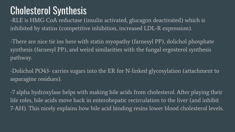
Cholesterol Synthesis-RLE is HMG CoA reductase (insulin activated, glucagon deactivated) which is
inhibited by statins (competitive inhibition, increased LDL-R expression).
-There are nice tie ins here with statin myopathy (farnesyl PP), dolichol phosphate
synthesis (farnesyl PP), and weird similarities with the fungal ergosterol synthesis
pathway.
-Dolichol PO43- carries sugars into the ER for N-linked glycosylation (attachment to
asparagine residues).
-7 alpha hydroxylase helps with making bile acids from cholesterol. After playing their
life roles, bile acids move back in enterohepatic recirculation to the liver (and inhibit
7-AH). This nicely explains how bile acid binding resins lower blood cholesterol levels.
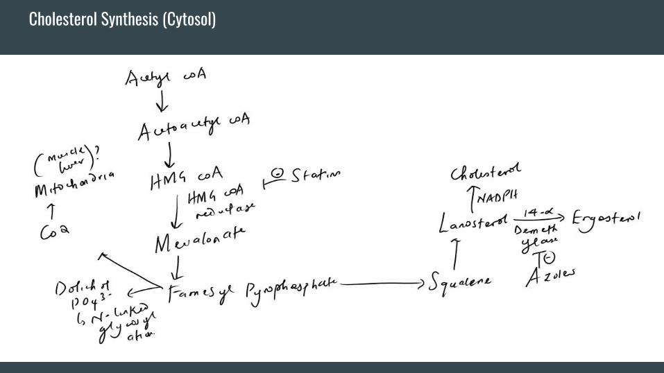
Cholesterol Synthesis (Cytosol)
FA Breakdown (Beta Oxidation)-Occurs in the mitochondria (unlike synthesis in the cytosol).
We don’t want both processes to be operating at the same time
so malonyl coA from FA synthesis inhibits the transport
mechanism of FAs (CAT1) into the mitochondria.
-Step 1 of BOX involves activating the FA by losing ATP (kinda
like glycolysis).
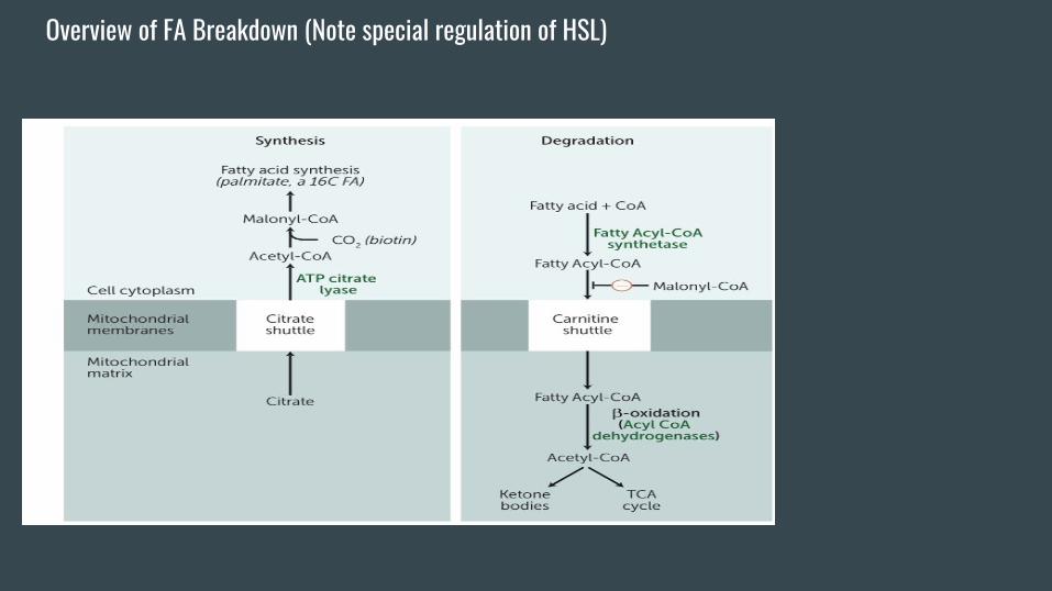
Overview of FA Breakdown (Note special regulation of HSL)

A Step 1 Worthy Series of Questions-CAT deficiency presents with muscle weakness. Why is that?
-It so happens that GSD5 (McArdle’s disease) also presents with muscle weakness.
-How would you differentiate these two?
-MCAD deficiency is classically said to present with hypoketotic hypoglycemia. What
is the mechanism behind the “hypoketosis”? What is the mechanism behind the
“hypoglycemia”? What is one characteristic “acid” that rises in the blood?
-Is there some way to differentiate MCAD deficiency from CAT deficiency? Your
friends at the NBME love this!
This is a SUPER HY slide to understand and explain based on your understanding of
the biochemistry.
Some Thoughts on Ketone Body Synthesis-Takes place in the mitochondria. Don’t get dinged on Step 1 for not recognizing the
difference between the cytosolic HMG CoA Synthase (cholesterol synthesis) and the
mitosolic HMG CoA synthase (KB synthesis)
-The liver has the ability to make ketone bodies. It however, lacks the ability to use
ketone bodies (thiophorase).
-This is a relatively mute point, but in fasting states, the liver does not use AcCoA
generated from BOX in the TCA cycle. It uses it almost exclusively for KB synthesis.
Why might this be?
-However, extrahepatic tissues have the ability to use KBs to generate energy (Acetyl
coA) which can be fed into the TCA cycle. Why can they do this?

Ketone Body Synthesis and Utilization
References-First Aid for The USMLE Step 1 2017


![HMG Capability Statement[1]](https://img.pdfslide.us/doc/110x75/58ee1b161a28abae778b45b9/hmg-capability-statement1.jpg)