Embed Size (px)
Citation preview
RESEARCH Open Access
Divergent differentiation and variantmorphology in invasive urothelialcarcinomas – association with muscle-invasive diseaseSuelen Cunha Santana1, Maiara Ferreira de Souza2, Maria Estela Pompeu Amaral2 and Daniel Abensur Athanazio1,2*
Abstract
Introduction: To evaluate the frequency of divergent differentiations / variant morphology in urothelial carcinoma,and their association with muscle-invasive disease at diagnosis.
Methods: All consecutive cases of invasive urothelial carcinoma from a busy pathology laboratory were reviewed.Clinical and pathological data were recorded including data on divergent and variant morphologies and theirpercentage within the invasive component.
Results: Among 91 cases, 46 (51%) showed some form of divergent/variant morphology. The most commondivergent morphology was squamous which was present in 18/46 (39% of cases with some divergent or variantmorphology) followed by micropapillary (28%), plasmacytoid (20%) and poorly differentiated (17%). Only squamousdifferentiation was associated with higher rate with muscularis propria invasion.
Conclusions: Although common, squamous differentiation should be still recognized as a feature of aggressivedisease.
Keywords: Urologic neoplasms, Bladder Cancer, Pathology
IntroductionInvasive urothelial carcinoma commonly shows diver-gent differentiation and variant morphology. CurrentWHO classification of urothelial carcinoma requiressubclassification based on presence or absence of diver-gent differentiation and variant morphology. Urothelialcarcinoma may show divergent differentiation (such assquamous, glandular, trophoblastic) and variant morph-ologies include nested, micropapillary, microcystic,lymphoepithelioma-like, plasmacytoid, sarcomatoid,giant cell, lipid-rich, clear cell (glycogen-rich) and poorlydifferentiated. The WHO recognizes micropapillary,
plasmacytoid, sarcomatoid and poorly differentiatedmorphologies as associated with more aggressive behav-ior. Due to some reports that correlate percentage ofmicropapillary and plasmacytoid variants with prognosis,international guidelines recommend that some or all di-vergent differentiation or variant morphology should bequantified (Moch et al. 2016; College of American Pa-thologists 2018; International Collaboration on CancerReporting (ICCR), n.d.).Data from institutions with mandatory central path-
ology review suggest that up to 44% of all divergent/vari-ant morphologies may be under-recognized and themost commonly overlooked variants are lymphoepithe-lial, plasmacytoid, nested, micropapillary and - not avariant but a concomitant tumor - small cell neuroendo-crine carcinoma (Shah et al. 2013).
© The Author(s). 2020 Open Access This article is licensed under a Creative Commons Attribution 4.0 International License,which permits use, sharing, adaptation, distribution and reproduction in any medium or format, as long as you giveappropriate credit to the original author(s) and the source, provide a link to the Creative Commons licence, and indicate ifchanges were made. The images or other third party material in this article are included in the article's Creative Commonslicence, unless indicated otherwise in a credit line to the material. If material is not included in the article's Creative Commonslicence and your intended use is not permitted by statutory regulation or exceeds the permitted use, you will need to obtainpermission directly from the copyright holder. To view a copy of this licence, visit http://creativecommons.org/licenses/by/4.0/.
* Correspondence: [email protected] Universitário Professor Edgard Santos, R. Augusto Viana, s/n –Canela, Salvador 40301-155, Brazil2Imagepat Laboratory, Rua Lucaia, 209 - Edf. EventusEmpresarial, RioVermelho, Salvador 41940-660, Brazil
Surgical and ExperimentalPathology
Santana et al. Surgical and Experimental Pathology (2020) 3:14 https://doi.org/10.1186/s42047-020-00066-z
The NCCN and some institutional guidelines (e.g. MDAnderson) suggest early cystectomy for T1 tumors withmicropapillary, plasmacytoid and sarcomatoid morph-ologies (National Comprehensive Cancer Network Clin-ical (NCCN), n.d.; MD Anderson Cancer Center, n.d.).The European Association of Urology states that micro-papillary, plasmacytoid, nested, sarcomatoid, microcysticvariants; and squamous and glandular (adeno) divergentdifferentiations are associated with poorer prognosis(Babjuk et al. 2017).We evaluated the frequency of divergent differenti-
ation / variant morphology in urothelial carcinoma, andtheir association with muscle-invasive disease at diagno-sis in a busy Pathology Laboratory in the light of thecurrently adopted WHO classification (2016).
MethodsAll consecutive cases of invasive urothelial carcinomafrom a private Laboratory of Pathology - Imagepat La-boratory (Salvador Bahia) - were included. All casesfrom January 2017 to October 2019 were reviewed. Allcases were signed or reviewed by a senior Urologic path-ologist. Clinical and pathological data were recorded in-cluding data on divergent differentiation / variantmorphology and their percentage within the invasivecomponent. Small biopsies were defined as casesresected without intention to remove the entire tumor –that did not include muscularis propria and, therefore,were not analyzed for the association with muscle-invasive disease. All cases defined as TURB had adequatesampling of muscularis propria (in the same sample orin a re-biopsy within few weeks). Qualitative variableswere analyzed using Fisher’s exact test. This study wasapproved by the Human Research Ethics Committee ofthe Faculty of Medicine, Federal University of Bahia (ap-proval number: 3.709.215).
ResultsA total of 91 cases of invasive urothelial carcinoma wereidentified. Frequency of specimens obtained by trans-urethral resection of the bladder (TURB) and cystectomywere 87 and 3 cases, respectively. Among TURB speci-mens, maximum extension of tumor was lamina propriain 29 and at least muscularis propria in 41.Overall, 45 cases had no divergent differentiation or
variant morphology while 46 showed some form ofthem. The most common divergent differentiation wassquamous which was present in 18/46 (39% of caseswith some divergent/variant morphology) ranging from5 to 95% of tumor area. The second most common wasmicropapillary – seen in 13/46 (28%), range < 5–80%.Third, plasmacytoid was observed in 9/46 (20%) (range:30–90%). These were followed by poorly differentiated8/46 (17%) (range: 20–100%), glandular 5/46 (11%)
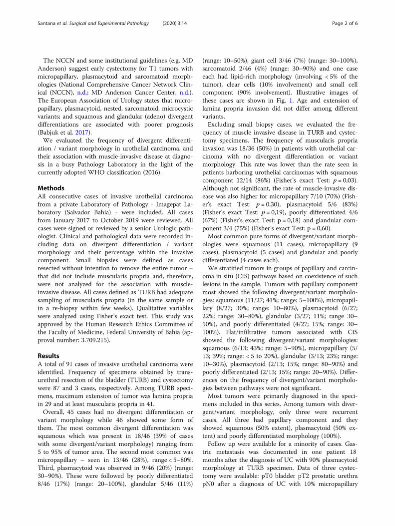
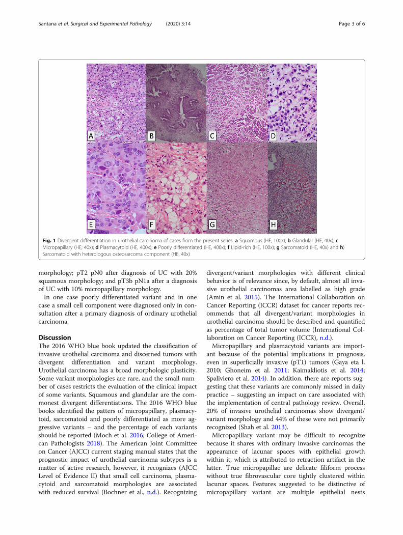
(range: 10–50%), giant cell 3/46 (7%) (range: 30–100%),sarcomatoid 2/46 (4%) (range: 30–90%) and one caseeach had lipid-rich morphology (involving < 5% of thetumor), clear cells (10% involvement) and small cellcomponent (90% involvement). Illustrative images ofthese cases are shown in Fig. 1. Age and extension oflamina propria invasion did not differ among differentvariants.Excluding small biopsy cases, we evaluated the fre-
quency of muscle invasive disease in TURB and cystec-tomy specimens. The frequency of muscularis propriainvasion was 18/36 (50%) in patients with urothelial car-cinoma with no divergent differentiation or variantmorphology. This rate was lower than the rate seen inpatients harboring urothelial carcinomas with squamouscomponent 12/14 (86%) (Fisher’s exact Test: p = 0,03).Although not significant, the rate of muscle-invasive dis-ease was also higher for micropapillary 7/10 (70%) (Fish-er’s exact Test: p = 0,30), plasmacytoid 5/6 (83%)(Fisher’s exact Test: p = 0,19), poorly differentiated 4/6(67%) (Fisher’s exact Test: p = 0,18) and glandular com-ponent 3/4 (75%) (Fisher’s exact Test: p = 0,60).Most common pure forms of divergent/variant morph-
ologies were squamous (11 cases), micropapillary (9cases), plasmacytoid (5 cases) and glandular and poorlydifferentiated (4 cases each).We stratified tumors in groups of papillary and carcin-
oma in situ (CIS) pathways based on coexistence of suchlesions in the sample. Tumors with papillary componentmost showed the following divergent/variant morpholo-gies: squamous (11/27; 41%; range: 5–100%), micropapil-lary (8/27; 30%; range: 10–80%), plasmacytoid (6/27;22%; range: 30–80%), glandular (3/27; 11%; range 30–50%), and poorly differentiated (4/27; 15%; range: 30–100%). Flat/infiltrative tumors associated with CISshowed the following divergent/variant morphologies:squamous (6/13; 43%; range: 5–90%), micropapillary (5/13; 39%; range: < 5 to 20%), glandular (3/13; 23%; range:10–30%), plasmacytoid (2/13; 15%; range: 80–90%) andpoorly differentiated (2/13; 15%; range: 20–90%). Differ-ences on the frequency of divergent/variant morpholo-gies between pathways were not significant.Most tumors were primarily diagnosed in the speci-
mens included in this series. Among tumors with diver-gent/variant morphology, only three were recurrentcases. All three had papillary component and theyshowed squamous (50% extent), plasmacytoid (50% ex-tent) and poorly differentiated morphology (100%).Follow up were available for a minority of cases. Gas-
tric metastasis was documented in one patient 18months after the diagnosis of UC with 90% plasmacytoidmorphology at TURB specimen. Data of three cystec-tomy were available: pT0 bladder pT2 prostatic urethrapN0 after a diagnosis of UC with 10% micropapillary
Santana et al. Surgical and Experimental Pathology (2020) 3:14 Page 2 of 6
morphology; pT2 pN0 after diagnosis of UC with 20%squamous morphology; and pT3b pN1a after a diagnosisof UC with 10% micropapillary morphology.In one case poorly differentiated variant and in one
case a small cell component were diagnosed only in con-sultation after a primary diagnosis of ordinary urothelialcarcinoma.
DiscussionThe 2016 WHO blue book updated the classification ofinvasive urothelial carcinoma and discerned tumors withdivergent differentiation and variant morphology.Urothelial carcinoma has a broad morphologic plasticity.Some variant morphologies are rare, and the small num-ber of cases restricts the evaluation of the clinical impactof some variants. Squamous and glandular are the com-monest divergent differentiations. The 2016 WHO bluebooks identified the patters of micropapillary, plasmacy-toid, sarcomatoid and poorly differentiated as more ag-gressive variants – and the percentage of each variantsshould be reported (Moch et al. 2016; College of Ameri-can Pathologists 2018). The American Joint Committeeon Cancer (AJCC) current staging manual states that theprognostic impact of urothelial carcinoma subtypes is amatter of active research, however, it recognizes (AJCCLevel of Evidence II) that small cell carcinoma, plasma-cytoid and sarcomatoid morphologies are associatedwith reduced survival (Bochner et al., n.d.). Recognizing
divergent/variant morphologies with different clinicalbehavior is of relevance since, by default, almost all inva-sive urothelial carcinomas area labelled as high grade(Amin et al. 2015). The International Collaboration onCancer Reporting (ICCR) dataset for cancer reports rec-ommends that all divergent/variant morphologies inurothelial carcinoma should be described and quantifiedas percentage of total tumor volume (International Col-laboration on Cancer Reporting (ICCR), n.d.).Micropapillary and plasmacytoid variants are import-
ant because of the potential implications in prognosis,even in superficially invasive (pT1) tumors (Gaya eta l.2010; Ghoneim et al. 2011; Kaimakliotis et al. 2014;Spaliviero et al. 2014). In addition, there are reports sug-gesting that these variants are commonly missed in dailypractice – suggesting an impact on care associated withthe implementation of central pathology review. Overall,20% of invasive urothelial carcinomas show divergent/variant morphology and 44% of these were not primarilyrecognized (Shah et al. 2013).Micropapillary variant may be difficult to recognize
because it shares with ordinary invasive carcinomas theappearance of lacunar spaces with epithelial growthwithin it, which is attributed to retraction artifact in thelatter. True micropapillae are delicate filiform processwithout true fibrovascular core tightly clustered withinlacunar spaces. Features suggested to be distinctive ofmicropapillary variant are multiple epithelial nests
Fig. 1 Divergent differentiation in urothelial carcinoma of cases from the present series. a Squamous (HE, 100x); b Glandular (HE; 40x); cMicropapillary (HE; 40x); d Plasmacytoid (HE, 400x); e Poorly differentiated (HE, 400x); f Lipid-rich (HE, 100x); g Sarcomatoid (HE, 40x) and h)Sarcomatoid with heterologous osteosarcoma component (HE, 40x)
Santana et al. Surgical and Experimental Pathology (2020) 3:14 Page 3 of 6
within a single lacunaor epithelial ring formation (Aminet al. 2015).Micropapillary morphology has been associated with
poor prognosis and the percentage of micropapillarymorphology in transurethral resections was reported topredict higher stage (Gaya et al. 2010; Samaratunga andKhoo 2004) and cancer specific death (Samaratunga andKhoo 2004; Comperat et al. 2010). Those findings arethe main reason to report the presence and the percent-age of micropapillary morphology in pathology reports.The association with poorer outcome led some authorsto suggest early cystectomy even in pT1 tumors (Kamatet al. 2006). Since some cases treated with early cystec-tomy still show advanced stage and nodal metastasis,consideration of neoadjuvant chemotherapy has beensuggested (Ghoneim et al. 2011), while a recent studysuggested benefit of prior chemotherapy only in muscleinvasive disease (Fernández et al. 2017). A recent meta-analysis suggests that although associated with more ad-vanced disease at diagnosis, micropapillary urothelialcarcinoma is not associated with poorer outcomes aftersurgical treatment (Abufaraj et al., 2019).Plasmacytoid variant is the current term of choice for
obsolete terminology of signet ring or diffuse variants ofurothelial carcinoma (Moch et al. 2016). It is usually as-sociated with advanced stage at diagnosis and poor sur-vival (Fox et al. 2017). In a trial of muscle-invasiveurothelial carcinoma, treated by radical cystectomy andadjuvant cisplatin-based chemotherapy, it was shownthat plasmacytoid morphology was an independent pre-dictor of poor survival when compared to ordinaryurothelial carcinoma and micropapillary variant (Kecket al. 2013). On the other hand, a recent series did notshow the impact of plasmacytoid morphology on out-come (Li et al. 2017). A distinctive clinical feature is thehigh rate of recurrence with peritoneal spread (Dayyaniet al. 2013; Ricardo-Gonzalez et al. 2012). A potentialpitfall in cystoscopy is that the tumor may invade mus-cularis propria without grossly identifiable mucosaltumor (Fritsche et al. 2008). Mirroring the discussion onearly cystectomy for the micropapillary variant, some au-thors also suggest aggressive therapy in pT1 disease withplasmacytoid morphology (Kaimakliotis et al. 2014).Both micropapillary and plasmacytoid variants com-
monly show HER2 oncogene alterations including amp-lification and mutation and, therefore, may be prone totarget therapy in the future (Ching et al. 2011; Ross et al.2014; Schneider et al. 2014; Kim et al. 2016).The direct role of divergent/variant morphology on
treatment decision is controversial. Some authorspropose a treatment algorithm with early cystectomy innon-muscle invasive bladder cancer (T1) with micropa-pillary, plasmacytoid and sarcomatoid morphologies(Willis and Kamat 2015). Although an early surgical
treatment is not explicitly recommended in the MD An-derson practice algorithm, patients with micropapillaryand sarcomatoid variants may be followed as T2 tumors.Early cystectomy should be considered in variantmorphologies demonstrating concurrent carcinoma insitu (MD Anderson Cancer Center, n.d.). The NationalComprehensive Cancer Network (NCCN) guideline forbladder cancer states that non-muscle invasive bladdercancer with micropapillary, plasmacytoid and sarcoma-toid morphologies are at higher risk of progression andmore aggressive approach should be considered (Na-tional Comprehensive Cancer Network Clinical (NCCN),n.d.). The American Urological Association (AUA)/ So-ciety of Urologic Oncology (SUO) Guideline mentionsthat an experienced genitourinary pathologist should re-view the pathology of a patient with any doubt inregards to variant or suspected variant histology (e.g.,micropapillary, nested, plasmacytoid, neuroendocrine,sarcomatoid), extensive squamous or glandular differen-tiation, or the presence/absence of angiolymphatic inva-sion. (Moderate Recommendation; Evidence Strength:Grade C). Presence of variant histology requires re-staging transurethral resection within 4–6 weeks when abladder sparing approach is considered, or considerationof early cystectomy due to high risk of upstaging (ExpertOpinion) (Chang et al. 2016). The European Associationof Urology (EAU) guidelines on Non-muscle-invasiveBladder Cancer mentions micropapillary, nested, plas-macytoid, sarcomatoid, microcystic, squamous andadeno divergent/variant morphologies as associated withpoor prognosis (Babjuk et al. 2017). For muscle-invasivedisease, the EAU acknowledges that neoadjuvant chemo-therapy may be beneficial for patients with micropapil-lary, plasmacytoid, sarcomatoid, and mixed variants, andespecially for patients with neuroendocrine tumours(Veskimäe et al. 2019).In our experience, only squamous morphology was as-
sociated with a higher rate of advanced (muscle-invasive)disease. Squamous morphology is the most common di-vergent differentiation observed in urothelial carcinomaand occurs in 20–40% of all cases (Lopez-Beltran et al.2019). Several reports have associated squamous differ-entiation with more advanced stage disease at presenta-tion (Krasnow et al. 2017; Gofrit et al. 2016; Liu et al.2017). However, it is controversial if such morphology isan independent predictor of poor prognosis (Moch et al.2016).Awareness of proper variant histology classification
will probably gain additional importance since recentdata suggest that squamous and lymphoepithelioma -likemorphologies may predict pathological response of neo-adjuvant anti-PD-1 immunotherapy (Necchi et al. 2020).In our study, the frequency of divergent/variant
morphologies did not differ between tumors from the
Santana et al. Surgical and Experimental Pathology (2020) 3:14 Page 4 of 6
papillary or CIS (flat/infiltrative) pathways. Few caseswere diagnosed as recurrent disease precluding analysisof the relationship between primary vs recurrent diagno-sis (length of disease) with frequency of divergent/vari-ant morphology. A major limitation of this study wasthe lack of clinical follow-up.
ConclusionDivergent/variant morphologies in invasive urothelialcarcinomas is common in routine practice of a busyPathology Laboratory. They have clinical implicationsand should be cautiously searched. Central pathology re-view should be considered in large institutions dedicatedto treat urothelial carcinomas. In our almost three-yearexperience after new WHO classification of urologic tu-mors, only squamous differentiation is associated withhigher rate of muscle invasive disease. Although com-mon, squamous differentiation should be still recognizedas a feature of aggressive disease.
AbbreviationsAJCC: American Joint Committee on Cancer; AUA: American UrologicalAssociation; CAP: College of American Pathologists; EAU: EuropeanAssociation of Urology; ICCR: International Collaboration on CancerReporting; NCCN: National Comprehensive Cancer Network;TURB: Transurethral resection of the bladder; WHO: World HealthOrganization
AcknowledgementsNone.
Adherence to national and international regulationsNot applicable.
Authors’ contributionsDAA conceived the idea. DAA was the major contributor to the writing ofthe manuscript. MFS, MEPA, DAA diagnosed all cases. MFS, MEPA, SCC weremajor contributors for critically revising the manuscript for importantintellectual content. All authors read and approved the final manuscript.
FundingThis study had no funding resources.
Availability of data and materialsAll data generated or analyzed during this study are included in thispublished article.
Ethics approval and consent to participateThis study was approved by the Human Research Ethics Committee of theFaculty of Medicine, Federal University of Bahia (approval number: 3.709.215).
Consent for publicationThe authors declare that they have no competing interests.
Competing interestsThe authors declare that they have no competing interests.
Received: 6 April 2020 Accepted: 3 June 2020
ReferencesAbufaraj M, Foerster B, Schernhammer E, Moschini M, Kimura S, Hassler MR et al
(2019) Micropapillary Urothelial carcinoma of the bladder: a systematicreview and meta-analysis of disease characteristics and treatment outcomes.Eur Urol 75(4):649–658
Amin MB, Smith SC, Reuter VE, Epstein JI, Grignon DJ, Hansel DE et al (2015)Update for the practicing pathologist: the international consultation onurologic disease-European association of urology consultation on bladdercancer. Mod Pathol 28(5):612–630
Babjuk M, Bohle A, Burger M, Capoun O, Cohen D, Comperat EM et al (2017) EAUguidelines on non-muscle-invasive urothelial carcinoma of the bladder:update 2016. Eur Urol 71(3):447–461
Bochner BH, Hansel DE, Efstathiou JA, Konety B, Lee CT, McKiernan JM et al (n.d.)AJCC Cancer staging manual, 8th edn. Springer, Chicago, pp 757–765
Chang SS, Boorjian SA, Chou R, Clark PE, Daneshmand S, Konety BR et al (2016)Diagnosis and treatment of non-muscle invasive bladder Cancer: AUA/SUOguideline. J Urol 196(4):1021–1029
Ching CB, Amin MB, Tubbs RR, Elson P, Platt E, Dreicer R et al (2011) HER2 geneamplification occurs frequently in the micropapillary variant of urothelialcarcinoma: analysis by dual-color in situ hybridization. Mod Pathol 24(8):1111–1119
College of American Pathologists. Protocol for the examination of specimensfrom patients with carcinoma of the urinary bladder, 2018
Comperat E, Roupret M, Yaxley J, Reynolds J, Varinot J, Ouzaid I et al (2010)Micropapillary urothelial carcinoma of the urinary bladder: aclinicopathological analysis of 72 cases. Pathology 42(7):650–654
Dayyani F, Czerniak BA, Sircar K, Munsell MF, Millikan RE, Dinney CP et al (2013)Plasmacytoid urothelial carcinoma, a chemosensitive cancer with poorprognosis, and peritoneal carcinomatosis. J Urol 189(5):1656–1661
Fernández MI, Williams SB, Willis DL, Slack RS, Dickstein RJ, Parikh S et al (2017)Clinical risk stratification in patients with surgically resectable micropapillarybladder cancer. BJU Int 119(5):684–691
Fox MD, Xiao L, Zhang M, Kamat AM, Siefker-Radtke A, Zhang L et al (2017)Plasmacytoid urothelial carcinoma of the urinary bladder: a Clinicopathologicand Immunohistochemical analysis of 49 cases. Am J Clin Pathol 147(5):500–506
Fritsche HM, Burger M, Denzinger S, Legal W, Goebell PJ, Hartmann A (2008)Plasmacytoid urothelial carcinoma of the bladder: histological and clinicalfeatures of 5 cases. J Urol 180(5):1923–1927
Gaya JM, Palou J, Algaba F, Arce J, Rodriguez-Faba O, Villavicencio H (2010) Thecase for conservative management in the treatment of patients with non-muscle-invasive micropapillary bladder carcinoma without carcinoma in situ.Can J Urol 17(5):5370–5376
Ghoneim IA, Miocinovic R, Stephenson AJ, Garcia JA, Gong MC, Campbell SCet al (2011) Neoadjuvant systemic therapy or early cystectomy? Single-centeranalysis of outcomes after therapy for patients with clinically localizedmicropapillary urothelial carcinoma of the bladder. Urology 77(4):867–870
Gofrit ON, Yutkin V, Shapiro A, Pizov G, Zorn KC, Hidas G et al (2016) Theresponse of variant histology bladder Cancer to Intravesical immunotherapycompared to conventional Cancer. Front Oncol 6:43
International Collaboration on Cancer Reporting (ICCR) (n.d.) - Carcinoma of theBladder Histopathology Reporting Guide Cystectomy, Cystoprostatectomyand Diverticulectomy Specimen. http://www.iccr-cancer.org/getattachment/Datasets/Published-Datasets/Urinary-Male-Genital/Carcinoma-of-the-Bladder-Cystectomy-Cystoprostatec/ICCR-Urinary-Tract-Bladder-bookmarked-guide.pdfAccessed 1 Apr 2020
Kaimakliotis HZ, Monn MF, Cary KC, Pedrosa JA, Rice K, Masterson TA et al (2014)Plasmacytoid variant urothelial bladder cancer: is it time to update thetreatment paradigm? Urol Oncol 32(6):833–838
Kamat AM, Gee JR, Dinney CP, Grossman HB, Swanson DA, Millikan RE et al(2006) The case for early cystectomy in the treatment of nonmuscle invasivemicropapillary bladder carcinoma. J Urol 175(3 Pt 1):881–885
Keck B, Wach S, Stoehr R, Kunath F, Bertz S, Lehmann J et al (2013) Plasmacytoidvariant of bladder cancer defines patients with poor prognosis if treated withcystectomy and adjuvant cisplatin-based chemotherapy. BMC Cancer 13:71
Kim B, Kim G, Song B, Lee C, Park JH, Moon KC (2016) HER2 Protein Overexpressionand Gene Amplification in Plasmacytoid Urothelial Carcinoma of the UrinaryBladder. Dis Markers. 2016:8463731. https://doi.org/10.1155/2016/8463731
Krasnow RE, Drumm M, Roberts HJ, Niemierko A, Wu CL, Wu S et al (2017)Clinical outcomes of patients with histologic variants of Urothelial Cancertreated with Trimodality bladder-sparing therapy. Eur Urol 72(1):54–60
Li Q, Assel M, Benfante NE, Pietzak EJ, Herr HW, Donat M et al (2017) The impactof Plasmacytoid variant histology on the survival of patients with urothelialcarcinoma of bladder after radical cystectomy. Eur Urol Focus 5(1):104–108
Liu Y, Bui MM, Xu B (2017) Urothelial carcinoma with squamous differentiation isassociated with high tumor stage and pelvic lymph-node metastasis. CancerControl 24(1):78–82
Santana et al. Surgical and Experimental Pathology (2020) 3:14 Page 5 of 6
Lopez-Beltran A, Henriques V, Montironi R, Cimadamore A, Raspollini MR, ChengL (2019) Variants and new entities of bladder cancer. Histopathology 74(1):77–96
MD Anderson Cancer Center (n.d.) Urothelial Carcinoma of Bladder and UpperTract [https://www.mdanderson.org/documents/for-physicians/algorithms/cancer-treatment/ca-treatment-bladder-web-algorithm.pdf]. Accessed 28 Dec2019
Moch H, Humphrey PA, Ulbright TM, Reuter VE (2016) Chapter 2 Tumours of theurinary tract. In: WHO classification of Tumours of the urinary system andmale genital organs, 4th edn. IARC, Lyon, pp 77–133
National Comprehensive Cancer Network Clinical (NCCN) (n.d.) PracticeGuidelines in Oncology - Bladder Cancer [https://www.nccn.org/professionals/physician_gls/default.aspx]. Accessed 28 Feb 2018
Necchi A, Raggi D, Gallina A, Madison R, Colecchia M, Lucianò R et al (2020)Updated results of PURE-01 with preliminary activity of NeoadjuvantPembrolizumab in patients with muscle-invasive bladder carcinoma withvariant Histologies. Eur Urol 77(4):439–446
Ricardo-Gonzalez RR, Nguyen M, Gokden N, Sangoi AR, Presti JC Jr, McKenney JK(2012) Plasmacytoid carcinoma of the bladder: a urothelial carcinoma variantwith a predilection for intraperitoneal spread. J Urol 187(3):852–855
Ross JS, Wang K, Gay LM, Al-Rohil RN, Nazeer T, Sheehan CE et al (2014) A highfrequency of activating extracellular domain ERBB2 (HER2) mutation inmicropapillary urothelial carcinoma. Clin Cancer Res 20(1):68–75
Samaratunga H, Khoo K (2004) Micropapillary variant of urothelial carcinoma ofthe urinary bladder; a clinicopathological and immunohistochemical study.Histopathology 45(1):55–64
Schneider SA, Sukov WR, Frank I, Boorjian SA, Costello BA, Tarrell RF et al (2014)Outcome of patients with micropapillary urothelial carcinoma followingradical cystectomy: ERBB2 (HER2) amplification identifies patients with pooroutcome. Mod Pathol 27(5):758–764
Shah RB, Montgomery JS, Montie JE, Kunju LP (2013) Variant (divergent)histologic differentiation in urothelial carcinoma is under-recognized incommunity practice: impact of mandatory central pathology review at alarge referral hospital. Urol Oncol 31(8):1650–1655
Spaliviero M, Dalbagni G, Bochner BH, Poon BY, Huang H, Al-Ahmadie HA et al(2014) Clinical outcome of patients with T1 micropapillary urothelialcarcinoma of the bladder. J Urol 192(3):702–707
Veskimäe E, Espinos EL, Bruins HM, Yuan Y, Sylvester R, Kamat AM et al (2019)What is the prognostic and clinical importance of Urothelial andNonurothelial histological variants of bladder Cancer in predictingoncological outcomes in patients with muscle-invasive and metastaticbladder Cancer? A European Association of Urology muscle invasive andmetastatic bladder Cancer guidelines panel systematic review. Eur Urol Oncol2(6):625–642
Willis D, Kamat AM (2015) Nonurothelial bladder cancer and rare varianthistologies. Hematol Oncol Clin North Am 29(2):237–252 viii
Publisher’s NoteSpringer Nature remains neutral with regard to jurisdictional claims inpublished maps and institutional affiliations.
Santana et al. Surgical and Experimental Pathology (2020) 3:14 Page 6 of 6


![complex, and related syndromes Author Manuscript NIH ...kincsesz/seminar/downloads...glandular differentiation[27, 89, 117, 118] (Figure 2h,i). Another rare neurofibroma variant that](https://img.pdfslide.us/doc/110x75/60f7b75799a3976448468c78/complex-and-related-syndromes-author-manuscript-nih-kincseszseminardownloads.jpg)