Embed Size (px)
Citation preview
J. Hirnforsch. 28 (1987) 5, 529-544
Zoological Institute, University of RegensburgD-8400 Regensburg, Federal Republic of Germany
Distribution of Monoamine-containing Neurons in the Brain of a Teleost, Carassius auratus(Cyprinidae)
Udo BONN
With 9 Figures and 1 Table
(Received August 14, 1986)
Summary: The occurrence and distribution of monoamine-(MA) containing neurons and fibres in the brain ofCarassius was investigated by formaldehyde-induced fluorescence (FIF) histochemistry (Falck-Hillarp techni-que) . Many brightly green-fluorescent nerve cell perikarya were found in the nucleus dorsolateralis and ventro-medialis, in the nucleus posterioris periventricularis, in the nucleus recessus lateralis and posterioris. They alsooccurred in the mesencephalic nucleus lateralis valvulae, in the metencephalic nucleus gustatorius secundus andnear the ventricular borders of the facial and vagal lobes in the myelencephalon.Many fluorescent fibres and nerve terminals were localized in the frontal and medio-lateral parts of the telence-phalon, showing fluorescent connections to the caudal parts. In the diencephalon, MA-fibres branched in ahorizontal and ventral tract, leading to the medulla oblongata and the hypothalamic nuclei, respectively. Therewere laterally situated fibres connecting the hypothalamic nuclei with the medulla and the nucleus gustatoriussecundus. Many fluorescent fibres were found in the middle layers of the tectum opticum, in the torus semicircu-laris, in the lobus inferior and in the medulla oblongata. Considerably fewer fibres occurred in the corpus cerebelliand in the dorsal parts of the hindbrain lobes.These results are compared with the MA-system in the brains of other fish.
Key words: Teleostean brain — Catecholamines — Fluorescence microscopy — Carassius auratus (Teleostei)
Introduction
Many catecholamine- and serotonin-containing neu-rons have been found in various tissues since FALCK
and HILLARP (FALCK et al. 1962) introduced theirfluoroscopic method for the localization of biogenicamines. There are early biochemical findings on thepresence of monoamines (MA) in the brains of fishes(BOGDANSKI et al. 1963; JUORIO 1973). However,since then, most work has focussed on the MA-systemin the mammalian CNS, particularly in the rat brain.Much less is known about the distribution of mono-aminergic neurons in non-mammalian brains (forliterature see SANTER, 1977; PARENT et al. 1984).
BERTLER et al. (1963) were the first to describeMA-specific fluorescence in the fish brain, followedby several studies on the MA-distribution in thewhole brain of fishes (LEFRANC et al. 1969; PARENT
et al. 1978; WATSON 1980; KOTRSCHAL and ADAM
1983; and PARENT and NORTHCUTT 1982). Other stu-dies investigated the hypothalamic area only (HON-MA and HONMA 1970; WILSON and DODD 1973; EKEN-
GREN 1975; SWANSON et al. 1975; FREMBERG et al.
1977; TERLOU et al. 1978; BATTEN et al. 1979; EK-
STROM and VAN VEEN 1982). Two of these, BAUM-
GARTEN and BRAAK (1967) and BRAAK (1967), studiedthe goldfish.
The aim of the present study is to show the distri-bution of the MA-system in the whole brain of Caras-sius auratus.
Material and Methods
The distribution of monoaminergic neurons in the brain ofthe goldfish was studied by means of the FALCK-HILLARPhistofluorescence method (FALCK and OWMAN 1965). A totalof 94 goldfish (5 —10 cm body length) were used for thepresent study. All fish were obtained from a commercialdealer.
1) FIF.
Fourty one animals were used for formaldehyde-inducedfluorescence microscopy (FIF). Twelve animals were pro-cessed for FIF according to the modification of LOREN et al.(1976) by intracardial perfusion. Eight animals were treatedwith the CA-precursor L-Dopa (Sigma, 400 mg/kg i.m.,30 and/or 6 h before sacrifice) or the MAO-inhibitor nial-amide (Roche, 100 mg/kg i.m., 31 and/or 6 h before sacrifice).These drugs were used to increase the MA-concentrations inthe tissues.
One animal was treated with reserpine (Serpasil, Ciba,10 mg/kg i.m., 18 h before sacrifice). This was one of thetests for specificity of the observed fluorescence.
The animals were killed with MS 222 (Sandoz) and thebrains were rapidly dissected out, shock frozen, freeze-dried,
530 Journal für Hirnforschung 28 (1987) 5
gassed for 1 h at 80°C with p-formaldehyde (70% rel. humi-dity) prepared according to HAMBERGER et al. (1965), andthen vacuum-embedded in liquid paraplast. Sequentialtransverse or sagittal sections (10—20 μm) were mounted on
clean slides, briefly dipped in celloidine (0.5 — 1%) and then
covered with Depex-xylene (9 : 1). Where no celloidin-cover
was used, adjacent slices were mounted and stained by
standard histological techniques for precise localization of
the fluorescent structures. Four brains similarly treated but
not exposed to the formaldehyde gas, served as another
control for MA-specific fluorescence.
2) Fluorescence microscopy.
The slices were examined with an Ortholux II fluorescence
microscope (Leitz) using Leitz filter set D (Excitation; BP
355-425; beamsplitter: RKP 455; barrier filter: LP460).
The mechanical stage of the microscope was modified with
an additional stage for fine movements which was connected
to an X-Y-plotter (HP 7035 B, Hewlett Packard) via two
potentiometer-controlled d.c.-circuits, one for the X- and
one for the Y-axis. All structures of the brain were recorded
on paper using a cross-hair microscope eyepiece.
3) Histology.
Complete series of transverse, sagittal and horizontal sections
of 30 goldfish brains, and whole decalcified heads, were stain-
ed according to Kliiver-Barrera, Masson-Goldner, Azan, or
silverimpregnated according to Bielschowsky, Bodian or
Cajal (ROMEIS 1968).
4) HPLC.
The brains of 17 Carassius auratus were homogenized and
their MA-concentrations measured electrochemically after
separation by high performance liquid chromatography
(HPLC; KISSINGER et al. 1981).
5) Microspectrofluorometry.
The emission-spectra of 80 green- and yellow-fluorescent
perikarya and of varicose fibres in 2 Carassius brains were
measured microspectrofluorometrically with an MPV-2
microscope (Leitz). This set-up was equipped with a Xenon
high pressure lamp (Osram, XBO 75), filter set D (Leitz), a
photomultiplier (EM 9558) and an S 20 cathod. The aperture
could be closed to 2 —3 μ^i. The measurements were made
from 430 to 700 nm in 2 nm steps. Calibration of the photo-
multiplier and the correction of the spectra were according
to Leitz-Mitteilungen (16/01.76).
The presence of noradrenaline was recognized by bead-
like (varicose) fluorescent fibres (FALCK 1962). The discrimi-
nation of other monoamines (and their precursors) was diffi-
cult because all show green to yellow fluorescence after
Falck-Hillarp treatment. The subjective fluorescence of CA
at high concentrations only differs from 5-HT-/5-HTP-
fluorescence by its slow fading (BJORKLUND et al. 1975). Be-
cause of a possible coexistence of CA and 5-HT/5-HTP in the
same neuron the specifically fluorescent neurons were referred
to as monoaminergic.
Results
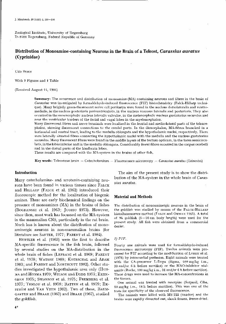
The whole brain and the cranial nerves of Carassiusauratus are shown in Fig. 1. The distribution of mono-amine-containing neurons in the CNS is schematicallyindicated in a series of transversal sections (Fig. 2).
BCDEFGHIJK L M O P QRS
NLL N V -VIII NIX
Fig. 1. Lateral view of the whole brain of Carassius auratus.
Planes of transverse sections are indicated alphabetically in
Fig. 2. Unless indicated otherwise, dorsal is up in all figures;
bar =500 μm. Abbreviations: bo bulbus olfactorius; ce — cere-
bellum; hyp hypophysis; Li lobus inferior; L X lobus vagi;
mo medulla oblongata; ms medulla spinalis; Nil nervus
opticus; N III-X cranial nerves; tel telencephalon; trol
tractus olfactorius.
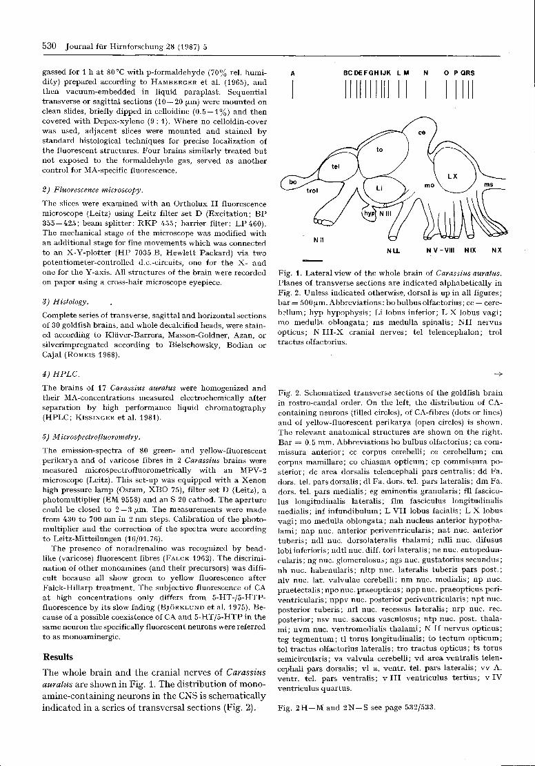
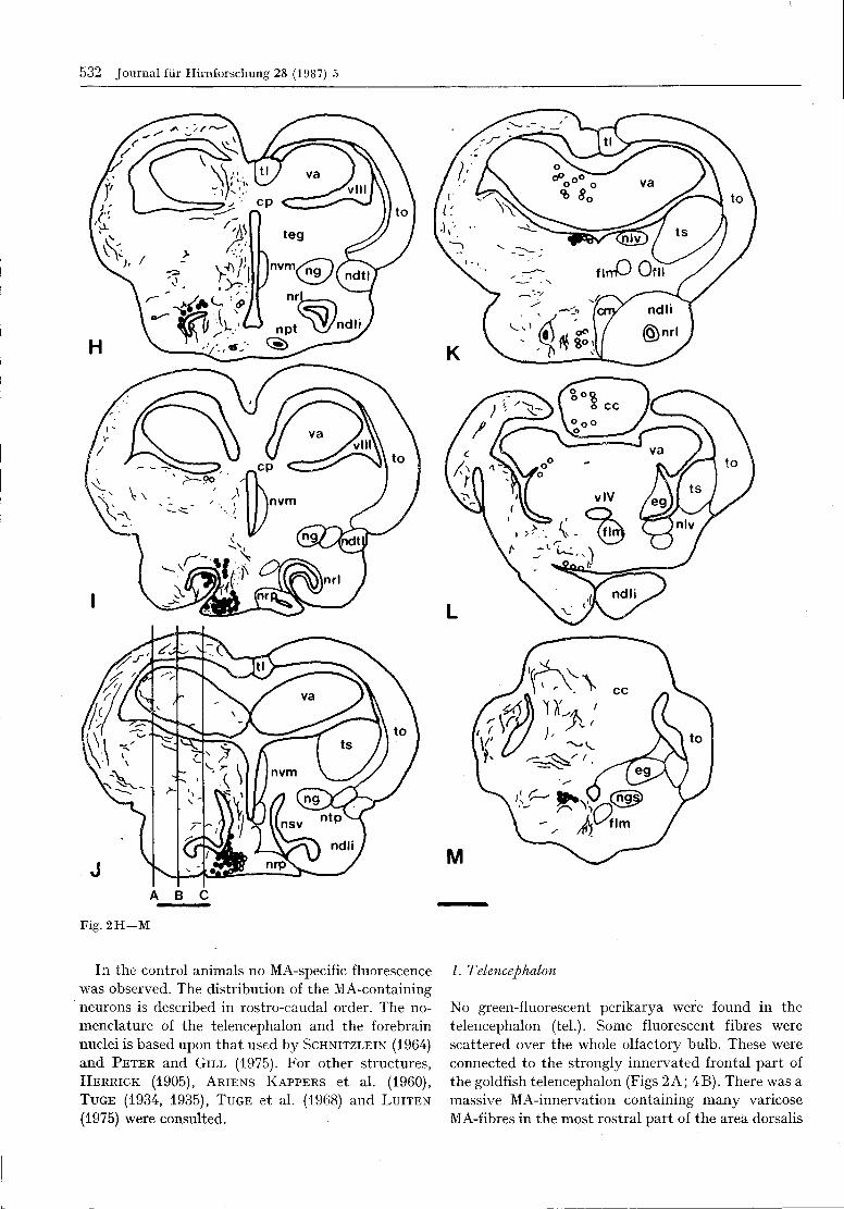
Fig. 2. Schematized transverse sections of the goldfish brain
in rostro-caudal order. On the left, the distribution of CA-
containing neurons (filled circles), of CA-fibres (dots or lines)
and of yellow-fluorescent perikarya (open circles) is shown.
The relevant anatomical structures are shown on the right.
Bar = 0 . 5 mm. Abbreviations bo bulbus olfactorius; ca com-
missura anterior; cc corpus cerebelli; ce cerebellum; cm
corpus mamillare; co chiasma opticum; cp commissura po-
sterior; dc area dorsalis telencephali pars centralis; dd Fa.
dors. tel. pars dorsalis; dl Fa. dors. tel. pars lateralis; dm Fa.
dors. tel. pars medialis; eg eminentia granulans; fll fascicu-
lus longitudinalis lateralis; flm fasciculus longitudinalis
medialis; inf infundibulum; L VII lobus facialis; L X lobus
vagi; mo medulla oblongata; nah nucleus anterior hypotha-
lami; nap nuc. anterior periventricularis; nat nuc. anterior
tuberis; ndl nuc. dorsolateralis thalami; ndli nuc. difusus
lobi inferioris; ndtl nuc. diff. tori lateralis; ne nuc. entopedun-
cularis; ng nuc. glomerulosus; ngs nuc. gustatorius secundus;
nh nuc. habenularis; nltp nuc. lateralis tuberis pars post.;
nlv nuc. lat. valvulae cerebelli; nm nuc. medialis; np nuc.
praetectalis; npo nuc. praeopticus; npp nuc. praeopticus peri-
ventricularis; nppv nuc. posterior periventricularis; npt nuc.
posterior tuberis; nrl nuc. recessus lateralis; nrp nuc. rec.
posterior; nsv nuc. saccus vasculosus; ntp nuc. post, thala-
mi; nvm nuc. ventromedialis thalami; N II nervus opticus;
teg tegmentum; tl torus longitudinalis; to tectum opticum;
tol tractus olfactorius lateralis; tro tractus opticus; ts torus
semicircularis; va valvula cerebelli; vd area ventralis telen-
cephali pars dorsalis; vl a. ventr. tel. pars lateralis; vv A.
ventr. tel. pars ventralis; v III ventriculus tertius; v IV
ventriculus quartus.
Fig. 2H —M and 2N —S see page 532/533.
BONN, U.: Monoamines in the goldfish brain 531
Fig. 2 A - G
532 Journal fur Hirnforschung 28 (1987) 5
Fig. 2 H - M
In the control animals no MA-specific fluorescencewas observed. The distribution of the MA-containingneurons is described in rostro-caudal order. The no-menclature of the telencephalon and the forebrainnuclei is based upon that used by SCHNITZLEIN (1964)and PETER and GILL (1975). For other structures,HERRICK (1905), ARIENS KAPPERS et al. (1960),TUGE (1934, 1935), TUGE et al. (1968) and LUITEN
(1975) were consulted.
1. Telencephalon
No green-fluorescent perikarya were found in thetelencephalon (tel.). Some fluorescent fibres werescattered over the whole olfactory bulb. These wereconnected to the strongly innervated frontal part ofthe goldfish telencephalon (Figs 2A; 4B). There was amassive MA-innervation containing many varicoseMA-fibres in the most rostral part of the area dorsalis

BONN, U.: Monoamines in the goldfish brain 533
Fig. 2 N - S
534 Journal fur Hirnforschung 28 (1987) 5
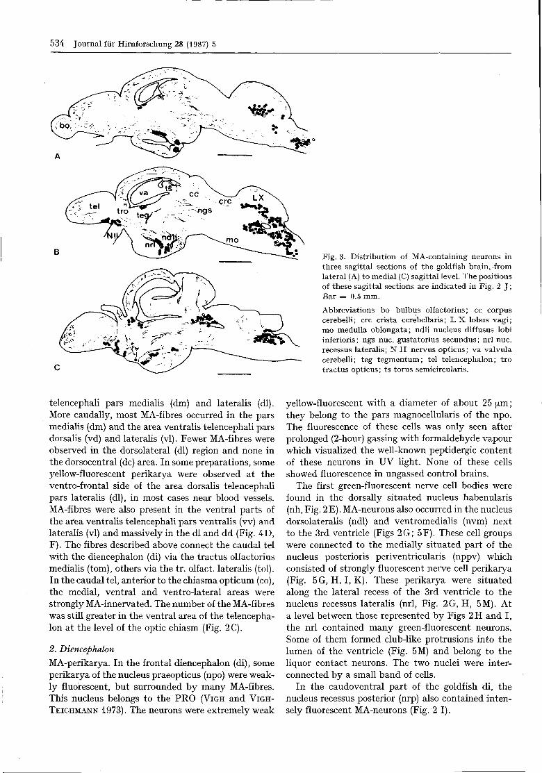
Fig. 3. Distribution of MA-containing neurons inthree sagittal sections of the goldfish brain, .fromlateral (A) to medial (C) sagittal level. The positionsof these sagittal sections are indicated in Fig. 2 J;Bar = 0 . 5 mm.
Abbreviations bo bulbus olfactorius; cc corpuscerebelli; crc crista cerebellaris; L X lobus vagi;mo medulla oblongata; ndli nucleus diffusus lobiinferioris; ngs nuc. gustatorius secundus; nrl nuc.recessus lateralis; N II nervus opticus; va valvulacerebelli; teg tegmentum; tel telencephalon; trotractus opticus; ts torus semicircularis.
telencephali pars medialis (dm) and lateralis (dl).More caudally, most MA-fibres occurred in the parsmedialis (dm) and the area ventralis telencephali parsdorsalis (vd) and lateralis (vl). Fewer MA-fibres wereobserved in the dorsolateral (dl) region and none inthe dorsocentral (dc) area. In some preparations, someyellow-fluorescent perikarya were observed at theventro-frontal side of the area dorsalis telencephalipars lateralis (dl), in most cases near blood vessels.MA-fibres were also present in the ventral parts ofthe area ventralis telencephali pars ventralis (vv) andlateralis (vl) and massively in the dl and dd (Fig. 4D,F). The fibres described above connect the caudal telwith the diencephalon (di) via the tractus olfactoriusmedialis (torn), others via the tr. olfact. lateralis (tol).In the caudal tel, anterior to the chiasma opticum (co),the medial, ventral and ventro-lateral areas werestrongly MA-innervated. The number of the MA-fibreswas still greater in the ventral area of the telencepha-lon at the level of the optic chiasm (Fig. 2C).
2. Diencephalon
MA-perikarya. In the frontal diencephalon (di), someperikarya of the nucleus praeopticus (npo) were weak-ly fluorescent, but surrounded by many MA-fibres.This nucleus belongs to the PRO (VIGH and VIGH-
TEICHMANN 1973). The neurons were extremely weak
yellow-fluorescent with a diameter of about 25 μm;they belong to the pars magnocellularis of the npo.The fluorescence of these cells was only seen afterprolonged (2-hour) gassing with formaldehyde vapourwhich visualized the well-known peptidergic contentof these neurons in UV light. None of these cellsshowed fluorescence in ungassed control brains.
The first green-fluorescent nerve cell bodies werefound in the dorsally situated nucleus habenularis(nh, Fig. 2E). MA-neurons also occurred in the nucleusdorsolateral (ndl) and ventromedialis (nvm) nextto the 3rd ventricle (Figs 2G; 5F). These cell groupswere connected to the medially situated part of thenucleus posterioris periventricularis (nppv) whichconsisted of strongly fluorescent nerve cell perikarya(Fig. 5G, H, I, K). These perikarya were situatedalong the lateral recess of the 3rd ventricle to thenucleus recessus lateralis (nrl, Fig. 2G, H, 5M). Ata level between those represented by Figs 2H and I,the nrl contained many green-fluorescent neurons.Some of them formed club-like protrusions into thelumen of the ventricle (Fig. 5M) and belong to theliquor contact neurons. The two nuclei were inter-connected by a small band of cells.
In the caudoventral part of the goldfish di, thenucleus recessus posterior (nrp) also contained inten-sely fluorescent MA-neurons (Fig. 2 I).
BONN, U.: Monoamines in the goldfish brain 535
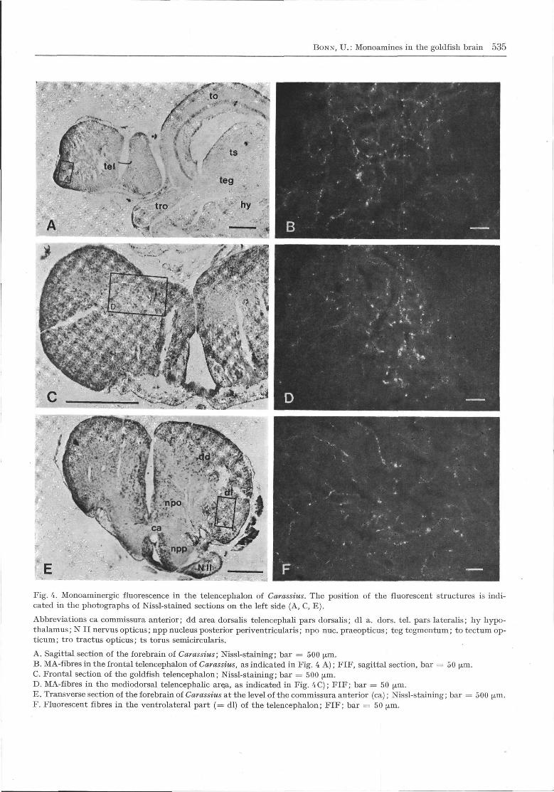
Fig. 4. Monoaminergic fluorescence in the telencephalon of Carassius. The position of the fluorescent structures is indi-cated in the photographs of Nissl-stained sections on the left side (A, C, E).
Abbreviations ca commissura anterior; dd area dorsalis telencephali pars dorsalis; dl a. dors. tel. pars lateralis; hy hypo-thalamus; N II nervus opticus; npp nucleus posterior periventricularis; npo nuc. praeopticus; teg tegmentum; to tectum op-ticum; tro tractus opticus; ts torus semicircularis.
A. Sagittal section of the forebrain of Carassius; Nissl-staining; bar = 500 μm.B. MA-fibres in the frontal telencephalon of Carassius, as indicated in Fig. 4 A); FIF, sagittal section, bar = 50 μm.C. Frontal section of the goldfish telencephalon; Nissl-staining; bar = 500 μm.D. MA-fibres in the mediodorsal telencephalic are.a, as indicated in Fig. 4C); FIF; bar = 50 μm.E. Transverse section of the forebrain of Carassius at the level of the commissura anterior (ca); Nissl-staining; bar = 500 ^m.F. Fluorescent fibres in the ventrolateral part (= dl) of the telencephalon; FIF; bar = 50 μm.
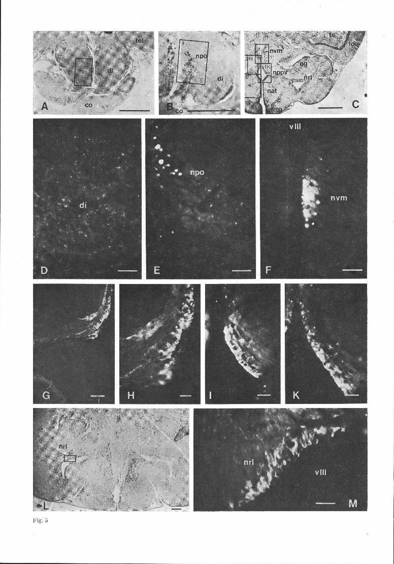
Fig. 5
BONN, U.: Monoamines in the goldfish brain 537
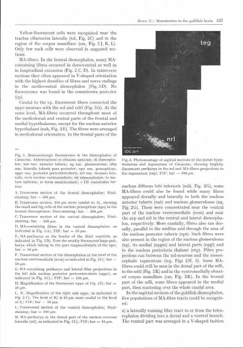
Yellow-fluorescent cells were recognized near thetractus olfactorius lateralis (tol, Fig. 2C) and in theregion of the corpus mamillare (cm, Fig. 2 J, K, L).Only few such cells were observed in ungassed sec-tions.
MA-fibres. In the frontal diencephalon, many MA-containing fibres occurred in dorsoventral as well asin longitudinal extension (Fig. 2 C, D). In transversesections they often appeared in V-shaped orientationwith the highest densities of fibres and nerve endingsin the medioventral diencephalon (Fig. 5D). Nofluorescence was found in the commissura posterior
(cp).Caudal to the cp, fluorescent fibres connected the
nppv-neurons with the nrl and ndtl (Fig. 5 G). At thesame level, MA-fibres occurred throughout most ofthe mediodorsal and ventral parts of the frontal andmedial hypothalamus, except for the nucleus anteriorhypothalami (nah, Fig. 2E). The fibres were arrangedin mediodorsal orientation. In the frontal parts of the
Fig. 5. Monoaminergic fluorescence in the diencephalon of
Carassius. Abbreviations co chiasma opticum; di diencepha-
lon; nat nuc. anterior tuberis; ng nuc. glomerulosus; nltp
nuc. lateralis tuberis pars posterior; npo nuc. praeopticus;
nppv nuc. posterior periventricularis; nrl nuc. recessus late-
ralis; nvm nucleus ventromedialis; tel telencephalon; to tec-
tum opticum; ts torus semicircularis; v III ventriculus ter-
tius.
A. Transverse section of the frontal diencephalon; Nissl-
staining; bar = 500 μm.
B. Transverse section, 340 μ^i more caudal to A), showing
the small and big cells of the nucleus praeopticus (npo) in the
frontal diencephalon; Nissl-staining; bar = 500 μm.
C. Transverse section of the ventral diencephalon; Nissl-
staining; bar = 500 μm.
D. MA-containing fibres in the ventral diencephalon, as
indicated in Fig. 5A); FIF; bar = 50 μm.
E. MA-perikarya at the border of the third ventricle, as
indicated in Fig. 5 B). Note the weakly fluorescent large peri-
karya which belong to the pars magnocellularis of the npo;
bar = 50 ^m,
F. Transversal section of the diencephalon at the level of the
nucleus ventromedialis (nvm) as indicated in Fig. 5C); bar =
50 μm.
G. MA-containing perikarya and lateral fibre projections in
the left side nucleus posterior periventricularis (nppv), as
indicated in Fig. 5C).; FIF; bar = 1 0 0 μm.
H. Magnification of the fluorescent nppv of Fig. 51); bar =
50 μ^i.
I., K. Magnification of the right side nppv, as indicated in
Fig. 5 C). The level of K) is 40 μ^i more caudal to the level
of I); FIF; bar = 50 μm.
L. Transversal section of the ventral diencephalon; Nissl-
staining; bar = 100 μ^i.
M. MA-perikarya in the dorsal part of the nucleus recessus
lateralis (nrl), as indicated in Fig. 5L); FIF; bar = 1 0 μ^i.
Fig. 6. Photomontage of sagittal sections of the dorsal hypo-
thalamus and tegmentum of Carassius, showing brightly
fluorescent perikarya in the nrl and MA-fibres projections to
the tegmentum (teg); FIF; bar = 1 0 0 μm.
nucleus diffusus lobi inferioris (ndli, Fig. 2G), someMA-fibres could also be found while many fibresappeared dorsally and laterally to both the nucleusanterior tuberis (nat) and nucleus glomerulosus (ng,Fig. 2G). These were concentrated near the ventralpart of the nucleus ventromedialis (nvm) and nearthe nrp and nrl in the ventral and lateral diencepha-lon, respectively. More caudally, fibres also ran dor-sally, parallel to the midline and through the area ofthe nucleus posterior tuberis (npt). Such fibres werealso present in the region of the nucleus glomerulosus(ng), its medial (npgm) and lateral parts (npgl) andin the nucleus posterioris thalami (ntp). Fibre pro-jections ran between the nrl-neurons and the mesen-cephalic tegmentum (teg, Figs 2H, 6). Some MA-fibres could still be seen in the dorsal part of the ndli,in the ndtl (Fig. 2 K) and in the ventromedially situat-ed corpus mamillare (cm, Fig. 2K). In the frontalpart of the ndli, some fibres appeared in the medialpart, then scattering over the whole caudal area.
In the sagittal sections of the goldfish diencephalon,five populations of MA-fibre tracts could be recogniz-ed:
a) a laterally running fibre tract to or from the telen-cephalon dividing into a dorsal and a ventral branch.The ventral part was arranged in a V-shaped fashion
538 Journal fiir Hirnforschung 28 (1987) 5
to
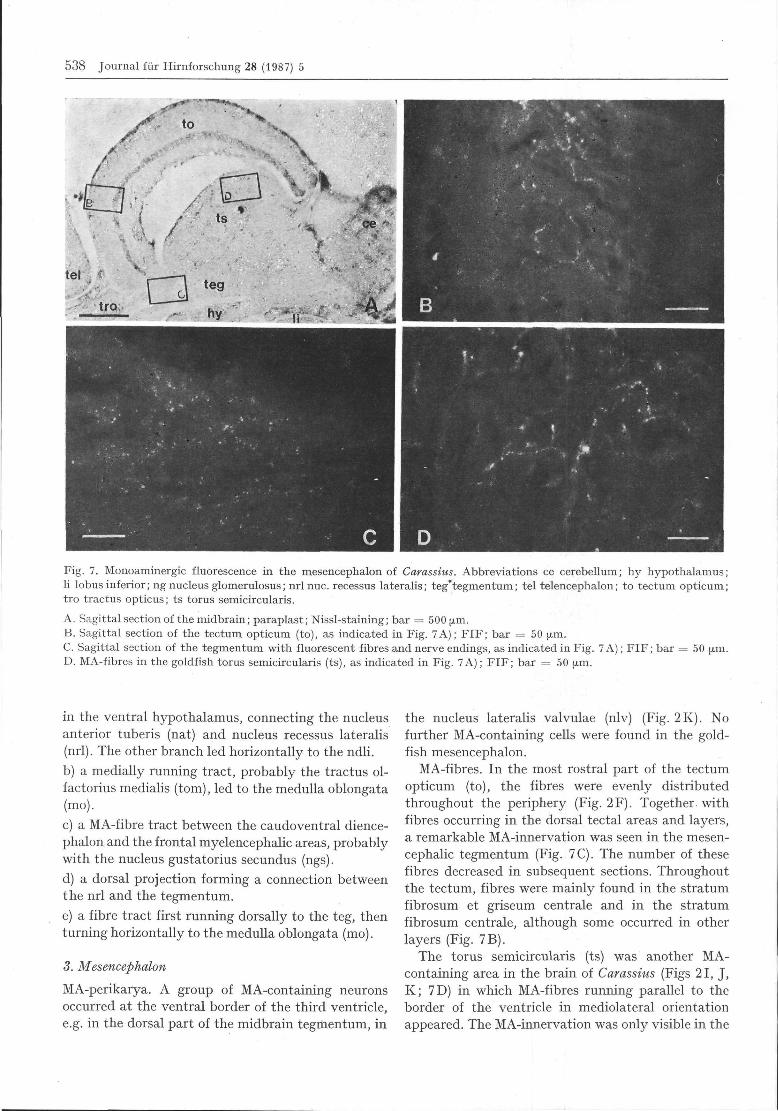
Fig. 7. Monoaminergic fluorescence in the mesencephalon of Carassius. Abbreviations ce cerebellum; hy hypothalamus;li lobus inferior; ng nucleus glomerulosus; nrl nuc. recessus lateralis; teg'tegmentum; tel telencephalon; to tectum opticum;tro tractus opticus; ts torus semicircularis. . ...
A. Sagittal section of the midbrain; paraplast; Nissl-staining; bar = 500 μ^i.B. Sagittal section of the tectum opticum (to), as indicated in Fig. 7A); FIF; bar = 5 0 μm.C. Sagittal section of the tegmentum with fluorescent fibres and nerve endings, as indicated in Fig. 7 A); FIF; bar = 50 μm.D. MA-fibres in the goldfish torus semicircularis (ts), as indicated in Fig. 7 A); FIF; bar = 50 μm.
in the ventral hypothalamus, connecting the nucleusanterior tuberis (nat) and nucleus recessus lateralis(nrl). The other branch led horizontally to the ndli.
b) a medially running tract, probably the tractus ol-factorius medialis (torn), led to the medulla oblongata(mo).
c) a MA-fibre tract between the caudoventral dience-phalon and the frontal myelencephalic areas, probablywith the nucleus gustatorius secundus (ngs).
d) a dorsal projection forming a connection betweenthe nrl and the tegmentum.
e) a fibre tract first running dorsally to the teg, thenturning horizontally to the medulla oblongata (mo).
3. Mesencephalon
MA-perikarya. A group of MA-containing neuronsoccurred at the ventral border of the third ventricle,e.g. in the dorsal part of the midbrain tegmentum, in
the nucleus lateralis valvulae (nlv) (Fig. 2K). Nofurther MA-containing cells were found in the gold-fish mesencephalon.
MA-fibres. In the most rostral part of the tectumopticum (to), the fibres were evenly distributedthroughout the periphery (Fig. 2F). Together, withfibres occurring in the dorsal tectal areas and layers,a remarkable MA-innervation was seen in the mesen-cephalic tegmentum (Fig. 7C). The number of thesefibres decreased in subsequent sections. Throughoutthe tectum, fibres were mainly found in the stratumfibrosum et griseum centrale and in the stratumfibrosum centrale, although some occurred in otherlayers (Fig. 7B).
The torus semicircularis (ts) was another MA-containing area in the brain of Carassius (Figs 21, J,K; 7D) in which MA-fibres running parallel to theborder of the ventricle in mediolateral orientationappeared. The MA-innervation was only visible in the
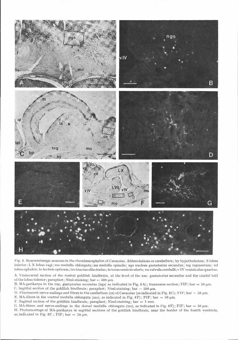
Fig. 8. Monoaminergic neurons in the rhombencephalon of Carassius. Abbreviations ce cerebellum; hy hypothalamus; li lobusinferior; LX lobus vagi; mo medulla oblongata; ms medulla spinalis; ngs nucleus gustatorius secundus; teg tegmentum; teltelencephalon; to tectum opticum; tro tractus olfactorius; ts torus semicircularis; va valvula cerebelli ;v IV ventriculus quartus.
A. Transversal section of the rostral goldfish hindbrain, at the level of the nuc. gustatorius secundus and the caudal halfof the lobus inferior; paraplast; Nissl-staining; bar = 500 μ^i.B. MA-perikarya in the nuc. gustatorius secundus (ngs) as indicated in Fig. 8 A); transverse section; FIF; bar = 50 μ^i.C. Sagittal section of the goldfish hindbrain; paraplast; Nissl-staining; bar = 500 μm.D. Fluorescent nerve-endings and fibres in the cerebellum (ce) of Carassius (as indicated in Fig. 8C); FIF; bar = 50 μm.E. MA-fibres in the ventral medulla oblongata (mo), as indicated in Fig. 8F); FIF; bar = 50 ^m.F. Sagittal section of the goldfish hindbrain; paraplast; Nissl-staining; bar = 1 mm.G. MA-fibres and nerve-endings in the dorsal medulla oblongata (mo), as indicated in Fig. 8F); FIF; bar = 50 μm.H. Photomontage of MA-perikarya in sagittal sections of the goldfish hindbrain, near the border of the fourth ventricle,as indicated in Fig. 8F.; FIF; bar = 50 μm.
540 Journal fiir Hirnforschung 28 (1987) 5
exterior part of the ts, whereas in its centre no fluo-rescent axons or nerve terminals were seen. Belowthis region, a bundle of MA-fibres was observed inlongitudinal extent (Fig. 3B).
4. Rhombencephalon
a) Metencephalon
MA-perikarya. Except for some green-fluorescentperikarya in the nucleus gustatorius secundus (ngs)(Figs 2M, N; 3C; 8B) and a few yellow-fluorescentperikarya in the mediodorsal area of the valvula cere-belli (va) and in the frontal parts of the corpus cere-belli (cc; Fig. 2L), no further fluorescent cells wereseen in the goldfish metencephalon.
MA-fibres. In the va, only few fibres occurred(Figs 2G—K). This was also true in the cc (Fig. 8D),where some MA-fibres appeared in ventral orienta-tion (Fig. 3).
b) Myelencephalon.
MA-perikarya. Fluorescent cells always appearedthroughout most of the lateral border of the 4th ven-tricle (Figs 2 O - S ; 3 A - C ; 8H). At the caudal endof the lobus facialis (L VII), the number of the green-fluorescent perikarya increased continuously, most ofthem lying in the ependymal wall of the hindbrainventricle (v IV), dorsally to the fasciculus longitudi-nalis medialis (flm) which was completely non-fluo-rescent. Green-fluorescent cells appeared at the ven-tricular border of the. vagal lobe (Figs 20—R).Among this very large area of green-fluorescent MA-cells, relatively few neuronal somata showed yellowfluorescence. The number of these perikarya wasgreatly increased after nialamide administration, butmarkedly reduced following reserpine treatment. Inuntreated brains the number of yellow-fluorescentcells was rather small.
MA-fibres. A strong MA-innervation was observedin the ventral and dorsomedial parts of the medullaoblongata (mo; Figs 2M—S; 3A; 8E—G). Thesefibres were oriented mediolaterally (Fig. 2N) as wellas longitudinally (Fig. 3A). They seem to connectthe mid- and hindbrain areas in the goldfish brain(Fig. 3). At the level of the cc and caudally, the MA-innervation of the medulla was concentrated in theventral and dorsomedial areas (Figs 3A;8G). Inmost sections, the fibres were concentrated in twobundles in the ventral medulla (Fig. 3D). There wasalso a remarkably dense MA-innervation in the vagal(LX) and facial (L VII) lobes (Fig. 2P). In caudaldirection, fascial MA-innervation increased and fluo-rescent fibres were observed running in vertical orien-tation.
5. MA-concentrations in the brain of Carassius
The MA-concentrations were determined by HPLC(Table 1). Adrenaline was below the limit of detection(about 4 ng/g wet weight).
Noradrenaline (NA) concentrations were signifi-cantly greater than dopamine (DA) concentrations inall brains and brain parts (p < 0.01; Wilcoxon match-ed-pairs signed-ranks test). The NA-concentrationsof the tel and di-mes were markedly higher than thoseof the hindbrain (0.02 < p < 0.05).
The DA-concentration in the tel was significantlylower than those of the other brain regions (p < 0.02).The DA-concentration of the combined di-mes wassignificantly higher compared with the hindbrainparts. There was no difference in the DA-concentra-tions of the two hindbrain areas. The NA- over DA-concentration ratios of the telencephalon as well asthose of the di- and mesencephalon differed signifi-cantly from the NA- over DA-concentration ratios ofthe caudal brain parts. There was no difference be-tween the NA/DA-ratios of the cerebellum (includingthe frontal medulla oblongata) and the myelencepha-lon.
Tab ell e 1. Mean values and standard deviations (SD), ofweight (W), brain weight (BW), and the concentrations ofthe catecholamines noradrenaline (NA) and dopamine (DA),and the indoleamine 5-hydroxytryptamine (5-HT). Na/DA= mean concentration ratio of NA over DA (N — 27 except* where N = 19; pi = pituitary; n.d. = not detected).
brainpart
total(SD)
W
(g)
4.000.22
BW
(mg)
63.72.4
NA
(ng/g
64025
DA
wet weight)
1309
5-HT
88*19
NA/DA
5.5
telencepha- (N == 7) 781 d= 149 40 d= 6 n.d. 20Ion
di-/mesen- (N = 7) 772 ± 60 120 ± 12 n.d. 6.7cephalon
cc/mo (N == 7) 404 ± 3 9 78 ± 6 n.d. 5.2
myelence- (N = 7) 422 ± 4 8 83 ± 9 n.d. 5.2phalonpituitary (N = 3) 53 d= 18 107 d= 13 n.d. 0.5
The mean 5-HT-concentration of the whole brainof Carassius was about 75% of the DA-concentra-tion. Because of technical difficulties the 5-HT-con-centrations was not determined for individual brainparts.
6. Microspectrofluorometry
The emission spectra of green and yellow-fluorescentperikarya and axons of the nuc. praeopticus, nuc.post, periventricularis, nuc. rec. lateralis, nuc. rec.post, lobus inferior, tectum opticum, torus semicir-
BONN, U.: Monoamines in the goldfish brain 541
1.0 n
COzw
0.0
1.0 -,
UJ»—z
0.0
B
440 480 520 560 600 640
WAVELENGTH
680 nm 440 480 520 560 600 640 680 nrr
WAU£LENGTH
1.0 ,
COzUJ
0.0
1.0 .,
D
(f>zUJ
0.0
440 480 520 560 600
WAVELENGTH
640 680 nm 440 480 520 560 600
WAVELENGTH
640 680 nm
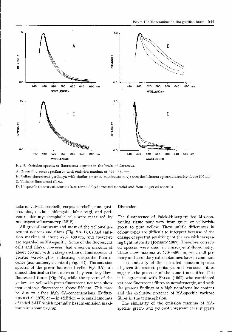
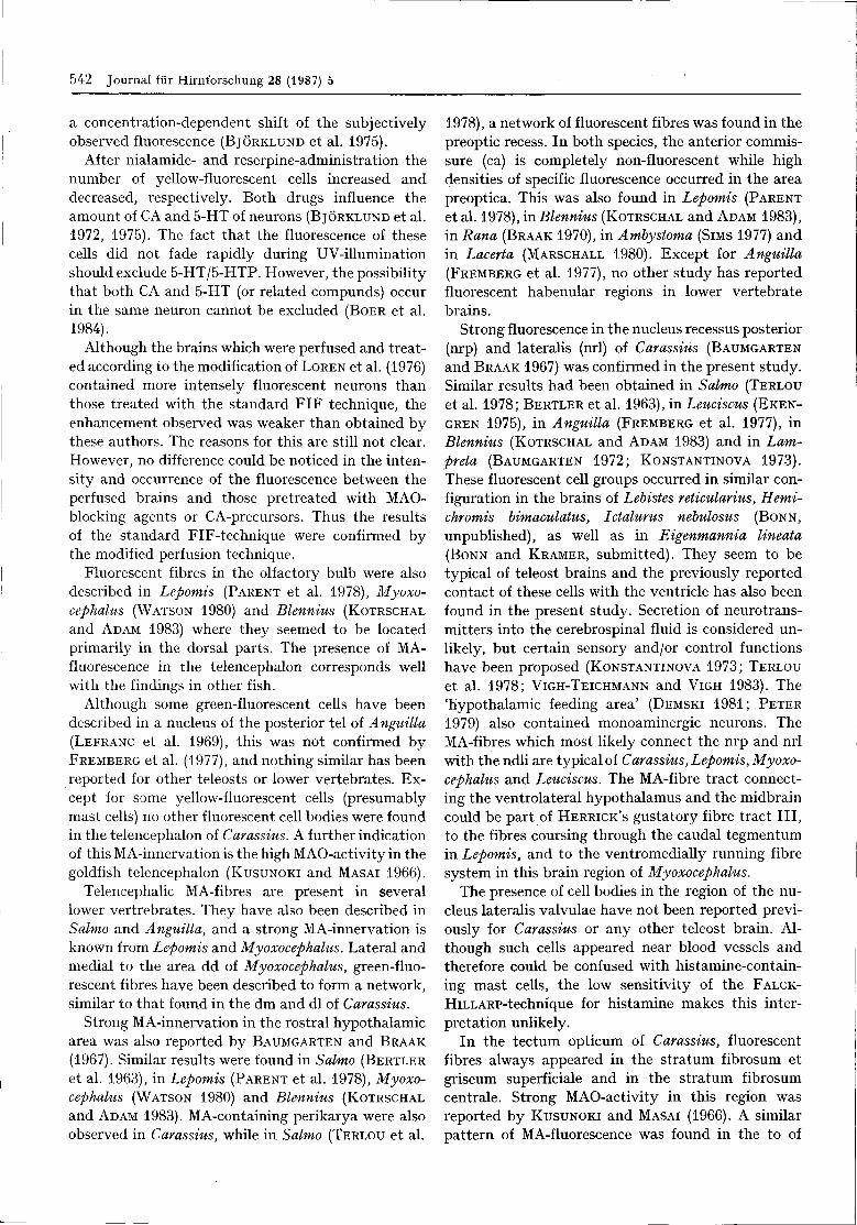
Fig. 9. Emission spectra of fluorescent neurons in the brain of Carassius.
A. Green fluorescent perikarya with emission maxima of 475 — 480 nm.B. Yellow-fluorescent perikarya with similar emission maxima as in A); note the different spectral intensity above 500 nm.C. Varicose fluorescent fibres.D. Unspecif ic fluorescent neurons from formaldehyde-treated material and from ungassed controls.
cularis, valvula cerebelli, corpus cerebelli, nuc. gust,secundus, medulla oblongata, lobus vagi, and peri-ventricular myelencephalic cells were measured bymicrospectrofluorometry (MSF).
All green-fluorescent and most of the yellow-fluo-rescent neurons and fibres (Fig. 9A, B, C) had emis-sion maxima of about 470—480 nm, and thereforeare regarded as MA-specific. Some of the fluorescentcells and fibres, however, had emission maxima ofabout 460 nm with a steep decline of fluorescence atgreater wavelengths, indicating unspecific fluores-cence (non-aminergic content; Fig. 9D). The emissionspectra of the green-fluorescent cells (Fig. 9 A) arealmost identical to the spectra of the green- to yellow-fluorescent fibres (Fig. 9C), while the spectra of theyellow- or yellowish-green-fluorescent neurons showmore intense fluorescence above 520 nm. This maybe due to either high CA-concentrations (BJORK-
LUND et al. 1975) or — in addition — to small amountsof faded 5-HT which normally has its emission maxi-mum at about 520 nm.
Discussion
The fluorescence of Falck-Hillarp-treated MA-con-taining tissue may vary from green or yellowish-green to pure yellow. These subtle differences incolour tones are difficult to interpret because of thechange of spectral sensitivity of the eye with increas-ing light intensity (JONSSON 1967). Therefore, correct-ed spectra were used in microspectrofluorometry.These show maxima at 470—480 nm, which all pri-mary and secondary catecholamines have in common.
The similarity of the corrected emission spectraof green-fluorescent perikarya and varicose fibressuggests the presence of the same transmitter. Thisis in agreement with FALCK (1962) who consideredvaricose fluorescent fibres as noradrenergic, and withthe present findings of a high noradrenaline contentand the exclusive presence of MA-specific varicosefibres in the telencephalon.
The similarity of the emission maxima of MA-specific green- and yellow-fluorescent cells suggests
542 Journal fiir Hirnforschung 28 (1987) 5
a concentration-dependent shift of the subjectivelyobserved fluorescence (BJORKLUND et al. 1975).
After nialamide- and reserpine-administration thenumber of yellow-fluorescent cells increased anddecreased, respectively. Both drugs influence theamount of CA and 5-HT of neurons (BJORKLUND et al.1972, 1975). The fact that the fluorescence of thesecells did not fade rapidly during UV-illuminationshould exclude 5-HT/5-HTP. However, the possibilitythat both CA and 5-HT (or related compunds) occurin the same neuron cannot be excluded (BOER et al.1984).
Although the brains which were perfused and treat-ed according to the modification of LOREN et al. (1976)contained more intensely fluorescent neurons thanthose treated with the standard FIF technique, theenhancement observed was weaker than obtained bythese authors. The reasons for this are still not clear.However, no difference could be noticed in the inten-sity and occurrence of the fluorescence between theperfused brains and those pretreated with MAO-blocking agents or CA-precursors. Thus the resultsof the standard FIF-technique were confirmed bythe modified perfusion technique.
Fluorescent fibres in the olfactory bulb were alsodescribed in Lepomis (PARENT et al. 1978), Myoxo-cephalus (WATSON 1980) and Blennius (KOTRSCHAL
and ADAM 1983) where they seemed to be locatedprimarily in the dorsal parts. The presence of MA-fluorescence in the telencephalon corresponds wellwith the findings in other fish.
Although some green-fluorescent cells have beendescribed in a nucleus of the posterior tel of Anguilla(LEFRANC et al. 1969), this was not confirmed byFREMBERG et al. (1977), and nothing similar has beenreported for other teleosts or lower vertebrates. Ex-cept for some yellow-fluorescent cells (presumablymast cells) no other fluorescent cell bodies were foundin the telencephalon of Carassius. A further indicationof this MA-innervation is the high MAO-activity in thegoldfish telencephalon (KUSUNOKI and MASAI 1966).
Telencephalic MA-fibres are present in severallower vertrebrates. They have also been described inSalmo and Anguilla, and a strong MA-innervation isknown from Lepomis and Myoxocephalus. Lateral andmedial to the area dd of Myoxocephalus, green-fluo-rescent fibres have been described to form a network,similar to that found in the dm and dl of Carassius.
Strong MA-innervation in the rostral hypothalamicarea was also reported by BAUMGARTEN and BRAAK
(1967). Similar results were found in Salmo (BERTLER
et al. 1963), in Lepomis (PARENT et al. 1978), Myoxo-cephalus (WATSON 1980) and Blennius (KOTRSCHAL
and ADAM 1983). MA-containing perikarya were alsoobserved in Carassius, while in Salmo (TERLOU et al.
1978), a network of fluorescent fibres was found in thepreoptic recess. In both species, the anterior commis-sure (ca) is completely non-fluorescent while highdensities of specific fluorescence occurred in the areapreoptica. This was also found in Lepomis (PARENT
et al. 1978), in Blennius (KOTRSCHAL and ADAM 1983),in Rana (BRAAK 1970), in Ambystoma (SIMS 1977) andin Lacerta (MARSCHALL 1980). Except for Anguilla(FREMBERG et al. 1977), no other study has reportedfluorescent habenular regions in lower vertebratebrains.
Strong fluorescence in the nucleus recessus posterior(nrp) and lateralis (nrl) of Carassius (BAUMGARTEN
and BRAAK 1967) was confirmed in the present study.Similar results had been obtained in Salmo (TERLOU
et al. 1978; BERTLER et al. 1963), in Leuciscus (EKEN-
GREN 1975), in Anguilla (FREMBERG et al. 1977), inBlennius (KOTRSCHAL and ADAM 1983) and in Lam-preta (BAUMGARTEN 1972; KONSTANTINOVA 1973).These fluorescent cell groups occurred in similar con-figuration in the brains of Lebistes reticularius, Hemi-chromis bimaculatus, Ictalurus nebulosus (BONN,
unpublished), as well as in Eigenmannia lineata(BONN and KRAMER, submitted). They seem to betypical of teleost brains and the previously reportedcontact of these cells with the ventricle has also beenfound in the present study. Secretion of neurotrans-mitters into the cerebrospinal fluid is considered un-likely, but certain sensory and/or control functionshave been proposed (KONSTANTINOVA 1973; TERLOU
et al. 1978; VIGH-TEICHMANN and VIGH 1983). The'hypothalamic feeding area,
(DEMSKI 1981; PETER
1979) also contained monoaminergic neurons. TheMA-fibres which most likely connect the nrp and nrlwith the ndli are typical of Carassius, Lepomis, Myoxo-cephalus and Leuciscus. The MA-fibre tract connect-ing the ventrolateral hypothalamus and the midbraincould be part of HERRICK'S gustatory fibre tract III,to the fibres coursing through the caudal tegmentumin Lepomis, and to the ventromedially running fibresystem in this brain region of Myoxocephalus.
The presence of cell bodies in the region of the nu-cleus lateralis valvulae have not been reported previ-ously for Carassius or any other teleost brain. Al-though such cells appeared near blood vessels andtherefore could be confused with histamine-contain-ing mast cells, the low sensitivity of the FALCK-
HiLLARP-technique for histamine makes this inter-pretation unlikely.
In the tectum opticum of Carassius, fluorescentfibres always appeared in the stratum fibrosum etgriseum superficiale and in the stratum fibrosumcentrale. Strong MAO-activity in this region wasreported by KUSUNOKI and MASAI (1966). A similarpattern of MA-fluorescence was found in the to of
BONN, U.: Monoamines in the goldfish brain 543
Myoxocephalus, Lepomis, Lampreta, Rana and Lacer-ta, while KOTRSCHAL and ADAM (1983) reported a uni-form MA-fibre distribution in the tectum of Blennius.The present findings of fibres in the torus semicircula-ris are similar to those in Lepomis and Myoxocephalus.
The yellow-fluorescent cells demonstrated in thepresent study have not been observed in the rhomben-cephalon before. The yellow-fluorescent cell bodiesdorsolateral to the fasciculus longitudinalis regionsof Lepomis and Myoxocephalus were not observed inCarassius.
In the valvula cerebelli (va) of the goldfish (presentstudy), as well as in Lepomis (PARENT et al. 1978),only few fibres were observed while in Myoxocephalusneither the va nor the corpus cerebelli (cc) containedfluorescent fibres. The weak fluorescence in the ccof Carassius may account for the faint MAO-activityin that region (KUSUNOKI and MASAI 1966). AlthoughCA-perikarya also occurred in the myelencephalon ofLepomis and Myoxocephalus, the number of MA-cells in the hindbrain of Carassius was much greater.
In addition to its fast fading, the absence of speci-fically fluorescent yellow neurons in the raphe-regionof the Carassius brain may be due to 1) the low sen-sitivity to 5-HT/5-HTP (about 30% of the sensitivityfor CA; BJORKLUND et al. 1975) of the FIF technique.2) the relatively low 5-HT content in the brain ofCarassius, 3) to diurnal changes in the 5-HT-concen-trations in the teleost brain (MARGOLIS-KAZAN etal. 1985).
According to EVANS (1934), the structure of thehindbrain lobes reflects the feeding habits in teleostfishes. Neither in Lepomis nor in Myoxocephalus arethe myelencephalic lobes so strongly developed asin Carassius which has well developed gustatory andolfactory systems. The gustatory and olfactory fibressystem in the brain of the carp brain (HERRICK 1905)is very similar to the MA-fibres decribed here. Be-cause of the close relationship of the carp and thegoldfish, both belonging to the Cyprininae, and theirsimilar brain structures and feeding habits, this simi-larity could be expected (for literature concerningolfaction and taste in fishes, see FINGER 1975, 1978).
Abbreviations
CA: catecholamines; DA: dopamine; FIF: formaldehydeinduced fluorescence; HPLC: high performance liquid chro-matography; MA: monoamines; MAO: monoamineoxidase;MSF: microspectrofluorometry; NA: noradrenaline; S. E.:standard error; 5-HT: 5-hydroxytryptamine (serotonin) ;5-HTP: 5-hydroxytryptophane.
Acknowledgements
I am greatly indebted to Prof. B. KRAMER for his provisionof facilities and for critically reading the manuscript. Thanksalso to Dr. F. KEES for HPLC and electrochemical detection
of the MA-concentrations in the brain tissue and for ErnstLeitz GmbH, Wetzlar, for the use of their microspectro-photometer. This work formed part of a doctoral thesis andwas supported by the Deutsche Forschungsgemeinschaft(SFB4, TeilprojektHl).
References
ARIENS KAPPERS, C. U., G. C. HUBER and E. C. CROSBY:The comparative anatomy of the nervous system of verte-brates, including man. 3 Vols. Hafner Publ. Co, New York,1960.
BATTEN, T. F. C, P. M. INGLETON and J. N. BALL: Ultra-structure and formaldehyde-fluorescence studies on thehypothalamus of Poecilia latipinna (Teleostei, Cyprini-dontiformes). Gen. comp. Endocrinol. 39, 87 — 109 (1979).
BAUMGARTEN, H. G.: Biogenic amines in the cyclostome andlower vertebrate brains. Progr. Histochem. Cytochem., 4,1-90(1972)..
BAUMGARTEN, H. G. and H. BRAAK : Catecholamine im Hypo-thalamus vom Goldfisch (Carassius auratus). Z. Zellforsch.80, 246-263 (1967).
BERTLER, A., B. FALCK and C. von MECKLENBURG: Mono-aminergic mechanisms in special ependymal areas in therainbow trout, Salmo irideus. Gen. comp. Endocrinol., 3,685-686(1963).
BJQRKLUND, A., B. FALCK and O. LINDVALL: Microspectro-fluorometric analysis of cellular monoamines after form-aldehyde or glyoxylic acid condensation. In: Methods inbrain research, Ed. by P. B. BRADLEY, Wiley & sons,London, pp. 249-294 (1975).
BJORKLUND, A., B. FALCK and Chr. OWMAN: Fluorescencemicroscopic and microspectrofluorometric techniques forthe cellular localization and characterization of biogenicamines. In: Methods of investigative and diagnostic endo-crinology, Ed. by S. A. BERSON, in: The thyroid and bio-genic amines, Ed. by J. E. RALL and I. J. KOPIN, Vol. 1,North-Holland Publ. Co., Amsterdam, pp. 318-368 (1972).
BOER, H. H., L. P. C. SCHOT, H. W. M. STEINBUSCH, C.MONTAGNE and D. REICHELT: Coexistance of immuno-reactivity to anti-dopamine, anti-serotonin and anti-vaso-tocin in the cerebral giant neuron of the pond snail Lym-naea stagnalis. Cell Tissue Res., 238, 411-412 (1984).
BoGDANSKi, D., L. BONOMI and B. BRODIE: Occurrence ofserotonin and catecholamines in brain and peripheralorgans of various vertebrate classes. Life ScL, 1, 80 — 84(1963).
BRAAK, H.: Biogene Amine im Gehirn vom Frosch (Ranaesculenta). Z. Zellforsch., 106, 269 — 308 (1970).
BRAAK, H.: Elektronenmikroskopische Untersuchungen anCatecholaminkernen im Hypothalamus vom Goldfisch(Carassius auratus). Z. Zellforsch., 83, 398 — 415 (1967).
DEMSKI, L. S.: Hypothalamic mechanisms of feeding infishes. In: Brain mechanisms of behaviour in lower verte-brates, Ed. by P. R. LAMING, Cambridge Univ. Press,Cambridge, pp. 225-237 (1981).
EKENGREN, B.: Aminergic nuclei in the hypothalamus ofthe roach, Leuciscus rutilus. Cell Tissue Res., 159, 493 — 502(1975).
EKSTROM, P. and T. VAN VEEN : The monoaminergic para-ventricular organ in the teleost Ictalurus nebulosus LeSueur, with special reference to its vascularization. ActaZool. (Stockh) 63, 45-54 (1982).
EVANS, H.: The correlation of brain pattern and feedinghabits in four species of cyprinid fishes. J. comp. Neurol.,97, 133-142 (1934).
5 J. Hirnforsch. 28 (1987) 5
544 Journal fur Hirnforschung 28 (1987) 5
FALCK, B.: Observations on the possibilities of the cellularlocalization of monoamines by a fluorescence method.Acta Physiol. Scand., Vol. 56, Suppl. 197 (1962).
FALCK, B., N. A. HILLARP, G. THIEME and A. TORP: Fluo-rescence of catecholamines and related compounds condens-ed with formaldehyde. J. Histochem. Cytochem., 10,348-354 (1962).
FALCK, B. and Chr. OWMAN: A detailed methodologicaldescription of the fluorescence method for the cellulardemonstration of biogenic monoamines. Acta Universi-tatis Lundensis (1965).
FINGER, T. E.: The distribution of the olfactory tracts inin the bullhead catfish, Ictalurus nebulosus. J. comp.Neurol., 161, 125-142 (1975).
FINGER, T. E.: Gustatory pathways in the bullhead catfish.II. Facial lobe connections. J. comp. Neurol., 180, 691 — 706(1978).
FREMBERG, M., T. VAN VEEN and H. G. HARTWIG: Form-aldheyde induced fluorescence in the telencephalon anddiencephalon of the eel (Anguilla anguilla L.). Cell TissueRes., 176, 1-22 (1977).
HAMBERGER, B., T. MALMFORS and C. SACHS: Standardiza-tion of paraformaldehyde and of certain procedures forthe histochemical demonstration of catecholamines. J.Histochem. Cytochem., 13, 147 (1965).
HERRICK, C. J.: The central gustatory paths in the brains ofbony fishes. J. comp. Neurol., 15, 375 — 456 (1905).
JONSSON, G.: Further studies on the specificity of the histo-chemical fluorescence method for the determination ofcatecholamines. Acta Histochem. (Jena), 26, 379 — 390(1967).
HONMA, S. and Y. HONMA: Histochemical demonstration ofmonoamines in the hypothalamus of lampreys and ice-goby. Bull. Jap. Soc. Sci. Fish., 36, 125-134 (1970).
JUORIO, A.: The distribution of catecholamines in the hypo-thalamus and other brain areas of some lower vertebrates.J. Neurochem., 20, 641-645 (1973).
KISSINGER, P. T., C. S. BRUNFLETT and R. E. SHOUP: Neuro-chemical applications of liquid chromatography withelectrochemical detection. Life Sci., 28, 455 — 465 (1981).
KONSTANTINOVA, M.: Monoamines in the liquor-contactingnerve cells in the hypothalamus of the lamprey, Lampvetafluviatilis. Z. Zellforsch., 144, 549-557 (1973).
KOTRSCHAL, K. and H. ADAM: The aminergic system in thebrain of Blennius incognitus (Bath 1968) (Teleostei, Perci-formes). Cell Tissue Res., 229, 403-409 (1983).
KUSUNOKI, T. and H.MASAI: Chemoarchitectonic in theCNS of the goldfish. Arch. Histol. Jap., 27, 333-371(1966).
LEFRANC, G., A. L. L'HERMITE and J. TUSQUE: Mise enevidence de neurones monoaminergiques par la techniquede fluorescence dans Tencephale d'Anguille. C. R. Soc.Biol. (Paris), 163, 1193-1196 (1969).
LOREN, I., A. BJORKLUND, B. FALCK and O. LINDVALL: Animproved histofluorescence procedure for freeze-dried andparaffin-embedded tissue based on combined formalde-hyde-glyoxylic acid perfusion with high magnesium con-tent and acid pH. Histochem. 49, 177-192 (1976).
LUITEN, P.: The central projections of the trigeminal, facialand anterior lateral line nerves in the carp (Cyprinuscarpio L.). J. comp. Neurol., 160, 399 — 418 (1975).
MARGOLIS-KAZAN, H., L. R. HALPERN-SEBOLD and M. P.SCHREIBMAN: Immunocytochemical localization of sero-tonin in the brain and pituitary gland of the platyfish,Xiphophorus maculatus. Cell Tissue Res., 240, 311 — 314(1985).
MARSCHALL, C.: Hypothalamic monoamines in lizards(Lacerta). A histofluorescence study. Cell Tissue Res., 205,95-105(1980).
PARENT, A., L. DUBE, M. BRAFORD and R. NORTHCUTT:The organization of monoamine-containing neurons in thebrain of the sunfish (Lepomisgibbosus) as revealed by fluores-cence microscopy. J. comp. Neurol., 182, 495 — 526 (1978).
PARENT, A., D. POITRAS and L. DUBE : Comparative anatomyof central monoaminergic systems. In: Handbook of che-mical neuroanatomy. Eds.: A. BJORKLUND and T. HOK-FELT. Vol. 2, Classical transmitters in the CNS, Part I.Amsterdam: Elsevier, pp. 409-439, (1984).
PARENT, A. and R. E. NORTHCUTT: The monoamine-contain-ing neurons in the brain of the garfish, Lepisosteus osteus.Brain Res. Bull. 9, 189-204 (1982).
PETER, R. E.: The brain and feeding behavior. In: Fishphysiology, Ed. by W. S. HOAR, D. H. RANDALL and J. R.BRETT, Academic Press, New York, Vol. VIII, pp. 121 bis159 (1979).
PETER, R. E. and V. E. GILL: A stereotaxic atlas and tech-nique for forebrain nuclei of the goldfish (Carassius aura-tus). J. comp. Neurol., 159, 69-102 (1975).
ROMEIS, B.: Mikroskopische Technik, 16. AufL, Oldenbourg,Miinchen (1968).
SANTER, R. M.: Monoaminergic nerves in the central andperipheral nervous system of fishes. Gen. Pharmac, 8,157-172 (1977).
SCHNITZLEIN, N.: The habenula and dorsal thalamus ofsome teleosts. J. comp. Neurol., 118, 225 — 268 (1964).
SIMS, T.: The development of monoamine-containing neuronsin the brain and spinal cord of the salamander Ambystomamexicanum. J. comp. Neurol., 173, 319 — 336 (1977).
SWANSON, D. D., R. S. NiSHiOKAandH. A. BERN : Aminergicinnervation of the cranial and caudal neurosecretorysystems in the teleost Gillichthys mirabilis. Acta zool.(Stockh) 56, 225-237 (1975).
TERLOU, M., B. EKENGREN and K. HIEMSTRA: Localizationof monoamines in the forebrain of two salmonid species,with special reference to the hypothalamo-hypophysialsystem. Cell Tissue Res., 190, 417-434 (1978).
TUGE, H.: Studies on cerebellar function in the teleost. J.comp. Neurol., 61, 347-370 (1935).
TUGE, H.: Studies on cerebellar function in the teleost. J.comp. Neurol., 60, 225-236 (1934).
TUGE, H., K. UCHIHASHI and H. SHIMAMURA: An atlas ofthe brains of the fishes of Japan. Tsukiji Shokan Publ.Co., Tokyo (1968).
VIGH, B. and I. VIGH-TEICHMANN : Comparative ultrastruc-ture of the cerebrospinal fluid contacting neurons. Int.Rev. Cytol. 35, 189-251 (1973).
VIGH-TEICHMANN, I. and B. VIGH: The system of cerebro-spinal fluid-contacting neurons. Arch, histol. jap. 46, 427 —468(1983).
WATSON, A. H. D.: The distribution of aminergic neuronsand their projections in the brains of the teleost, My oto-cephalus scorpius. Cell Tissue Res., 208, 299 — 313 (1980).
WILSON, J. and J. DODD: Distribution of monoamines in thediencephalon and pituitary of the dogfish, Scyliorhinuscanicula L. Z. Zellforsch. 137, 451-469 (1973).
A ddress:
Dr. U. BONNInstitut für AnatomieLehrstuhl Prof. Dr. E. LindnerUniversitat RegensburgD-8400 Regensburg, FRG